The Division and Cell Wall Gene Cluster of Enterococcus hirae S185
C. Duez, I. Thamm, F. Sapunaric, J. Coyette and J.M. Ghuysen
Centre d'Ingénierie des Protéines, Université de Liège, Institut de Chimie, B6, B-4000 Sart Tilman (Liège), Belgium Abstract
A chromosomal 10355-bp segment of Enterococcus hirae S185 contains nine orfs which occur in the same order as the MraW-, FtsL-, PBP3-, MraY-, MurD-, MurG-, FtsQ-, FtsA- and FtsZ-encoding genes of the division and cell wall clusters of Escherichia coli and Bacillus subtilis. The E. hirae DNA segment lacks the genes which in E. coli encode the ligases Ddl, MurC, MurE and MurF and the integral membrane protein FtsW. The encoded E. hirae and E. coli proteins share 25 % to 50 % identity except FtsL and FtsQ (s 14 % identity).
Keywords : dcw gene cluster ; Enterococcus hirae ; Escherichia coli ; Bacillus subtilis ; cell septation
The dcw (division and cell wall) cluster at the 2-min region of the chromosome of Escherichia coli contains genes the products of which are involved in the bacterial cell wall peptidoglycan synthesis and assembly (For primary references, see Yura et al., 1992 ; van Heijenoort, 1994 ; Ayala et al., 1994 ; Ghuysen et al., 1996 ; Vicente and Errington, 1996). Five ligases, Ddl, MurC, MurD, MurE and MurF, catalyse the formation of alanyl-alanine (Ddl) and the sequential addition to UDP-N-acetylmuramic acid of L-alanine (MurC), D-glutamic acid (MurD), meso-diaminopimelic acid (MurE) and the preformed D-alanyl-D-alanine (MurF), achieving the synthesis of the nucleotide precursor UDP- N-acetylmuramoyl-L-alanine-γ-D-glutamyl-(L) meso-diaminopimelyl-D-alanyl-D-alanine. The transphosphorylase MraY transfers the phospho-N-acetylmuramoyl-pentapeptide moiety of the uridylic acid carrier to the transmembrane C55-isoprenoid alcohol phosphate, and the
transglycosylase MurG transfers the acetylglucosamine residue of UDP-acetylglucosamine to the N-acetylmuramoyl moiety of the lipid precursor, achieving the synthesis of the lipid-linked disaccharide (N-acetylglucosaminyl-N-acetylmuramoyl)-pentapeρtide, or lipid II intermediate. The multimodular class B penicillin-binding protein (PBP)3 and several non-penicillin-binding proteins target the wall peptidoglycan assembly machinery to septum formation. PBP3 (the penicillin-binding module of which is an acyl serine transferase), FtsL (a protein with a putative leucine zipper motif) and FtsQ are membrane-bound with the bulk of the polypeptide chains exposed in the periplasm. FtsW is an integral membrane protein with loops exposed on both sides of the membrane. FtsA, an isologue of the DnaK-actin family of ATPases, is cytosolic when
phosphorylated and membrane-associated when unphosphorylated. MraW, a protein with a putative S-adenosyl-methionine-binding motif, and FtsZ, a GTPase that has similarity to tubulin, are cytosolic. FtsZ functions as a cytoskeletal element mediating the invagination of the septum.
The Bacillus subtilis dcw cluster is located at the 130°-135° region of the chromosome (Buchanan et al., 1994 ; Kunst et al., 1997). It contains genes encoding proteins analogous to the E. coli ligases MurD and MurE, the transphosphorylase MraY, the transglycosylase MurG, the multimodular class B PBP3 (i.e. PBP2b and SpoVD) and the cell cycle proteins MraW, FtsL, FtsW, FtsQ, FtsA and FtsZ. The B. subtilis cluster, however, lacks the ligase-encoding genes ddl, murC and murF and it contains additional genes related to sporulation.
The high-molecular-mass PBP3s of Enterococcus hirae (the suffix « s » denotes the susceptibility of this PBP to
β-lactam antibiotics in opposition to the low affinity of PBP3r) was known to be involved in cell septation
(Coyette et al., 1983). It was expected to be the counterpart of E. coli PBP3 and B. subtilis PBP2b and its encoding gene was expected to be part of a dcw cluster. To check the validity of the hypothesis, a 10355-bp DNA segment of E. hirae S185, containing the PBP3s-encoding gene, was sequenced and analysed.
The E. hirae genomic DNA was isolated (Hopwood et al., 1985) from cells grown unshaken at 37°C in Brain-Heart medium and collected at the end of the exponential phase. Restricted fragments were cloned into pUC18 or pUCBM20 and the inserts were sequenced on both strands using the T7 sequencing kit with [35S]dATP labelling, and the Autoread or ThermoSequenase sequencing kits with 5'-fluorescein or Cy5 primers, in which case the electrophoresis was performed on an ALF express DNA sequencer. The nucleotide sequences were introduced in GELASSEMBLE (Pearson et al., 1988), the ORFs were identified with CODON PREFERENCE (Devereux et al., 1984) and homology searches (SWISS-PROT, PIR, Genpept) were made by using FASTA or BLASTP
(Altschul et al., 1990).
The results of these studies are summarized in Figs 1 and 2. Fig. 1 gives the nucleotide sequence of the 10355-bp segment of E. hirae S185 and it translates the orfs into amino acid sequences. Fig. 2 compares pair-wise the proteins encoded by the dcw clusters of E. hirae, E. coli and B. subtilis.
The E. hirae dcw cluster was identified using a six-step strategy. The nucleotides and amino acid residues mentioned below refer to Fig. 1.
FIGURE 1 Nucleotide and deduced amino acid sequences of the E. hirae S185 dcw cluster. Putative
ribosome-binding sites are indicated by asterisks above the nucleotide sequence. Motifs (boxes) 1-9 of PBP3s, characteristic of the multimodular class B PBPs (Ghuysen, 1997), are indicated above the amino acid sequence. t-pbp3s : tryptic fragment-encoding sequence (Piras et al., 1990). Nucleotide sequence accession number : Y13922 (EMBL data bank)
Step1. The primers
and
(with I denoting inosine) were synthesized on the basis of the sequence of the amino terminal region (37 residues) of a 58-kD tryptic fragment of E. hirae PBP3s (Piras et al., 1990). They allowed a 119-bp DNA segment of the genomic DNA to be amplified by PCR. The reaction product generated by the Taq and
Dynazyme polymerases each encoded the polypeptide T62-(S)95, the sequence of which was that of the amino-terminal end of the tryptic fragment except that E occurred at position 63 instead of G and S occurred at position 95 instead of V.
Step 2. The primer
the 24 last nucleotides of which were complementary to nt 924 - nt 901, and the primer
the 20 last nucleotides of which corresponded to nt 925 - nt 944 were used in inverse PCRs (Silver et al., 1991) carried out on BamHI, BglII, DraI, EcoRI, HindIII and XbaI genomic libraries. The 2-kb DNA fragments amplified from the HindIII library by the Taq and Dynazyme polymerases each were digested with HindIΠ and Asp718, and with HindIΠ and SacI. The inserts were cloned and sequenced, yielding the sequence nt 1 - nt 2066. Step 3. The primer
the 24 last nucleotides of which were complementaryto nt 65 - nt 42 and the primer
the 18 last nucleotides of which corresponded to nt 1841 - nt 1858, were used in inverse PCRs carried out on BglII, EcoRI and HindIII genomic libraries using the Goldstar polymerase. A 5.5-kb DNA fragment of the EcoRI library was digested with SacI and EcoRI. Sequencing of the released 3-kb DNA fragment allowed several orfs to be identified.
orf1 (nt 1 - nt 287; truncated at the 5' end) encoded a 94 amino acid residue polypeptide similar to the carboxy terminal region of the E. coli MraW (38 % identity) and B. subtilis ORFB (72 % identity). orf2 (nt 292 - nt 603) encoded a 103 amino acid residue protein weakly related to the E. coli MraR/FtsL (14 % identity) and B. subtilis ORFA (21 % identity). orf3 (nt 689 - nt 2878) encoded the 730 amino acid residue multimodular class B PBP3s similar to the 588 amino acid residue E. coli PBP3 (27 % identity) and the 716 amino acid residue B. subtilis
PBP2b (36 % identity). Most class B PBPs, including E. coli PBP3, terminate 60-90 residues downstream from the KTGTA motif of the acyl serine transferase-penicillin-binding module. In contrast, B. subtilis PBP2b and SpoVD, and E. hirae PBP3s each bear a carboxy terminal extension, 150-200 amino acid residues long. orf4 (nt 2908 - nt 3873) encoded a 321 amino acid residue protein similar to the E. coli and B. subtilis MraY (43 % and 47 % identity). orf5 (nt 3877 - nt 4857) was truncated at the 3' end.
Step 4. In analogy with E. coli and B. subtilis, E. hirae was expected to possess a ftsW/rodA-like gene downstream from murD (orf5). Consequently the primer
the 20 last nucleotides of which corresponded to nt 4804 - nt 4823 and the primer
the 19 last nucleotides of which were complementary to the G77VIVLT82-encoding sequence of the E. hirae FtsW (unpublished data) were used in a direct PCR carried out on genomic DNA. Sequencing the 2.6-kb product generated by the Goldstar polymerase allowed the 3' portion of orf5 (nt 4858 - nt 5259) to be completed and two additional orfs to be identified.
orf5 (nt 3877 - nt 5259) encoded a 460 amino acid residue protein similar to the E. coli and B. subtilis MurD (33 % and 51 % identity). orf6 (nt 5276 - nt 6358) encoded a 360 amino acid residue protein similar to the E. coli and B. subtilis MurG (32 % and 52 % identity). orf7 (nt 6603 - nt 7350; truncated at the 3' end) encoded a polypeptide which had similarity with the amino terminal region of the B. subtilis DivIB (Harry et al., 1989) and E. coli FtsQ.
Step 5. In analogy with E. coli and B. subtilis, E. hirae was expected to possess ftsA- and ftsZ-like genes downstream from ftsQ. The FtsZ sequences of E. coli, B. subtilis and some other bacteria each possess the hexapeptide GADMVF (Margolin et al., 1996). On this basis, the primer
the 17 last nucleotides of which were complementary to the hexapeptide-encoding sequence and the primer
the 24 last nucleotides of which corresponded to nt 7252 - nt 7275 were used in a direct PCR carried out on genomic DNA with the Dynazyme polymerase. Sequencing the 2.1-kb product allowed the 3' portion of orf7 (nt 7351 - nt 7610) to be completed and two additional orfs to be identified.
orf7 (nt 6603 - nt 7610) encoded a 335 amino acid residue protein weakly related to E. coli FtsQ (14 % identity) but significantly similar to B. subtilis DivIB/FtsQ (33 % identity). orf8 (nt 7764 - nt 9092) encoded a 442 amino acid residue protein similar to the E. coli and B. subtilis FtsA (33 % and 39 % identity). orf9 (nt 9115 - nt 9416; truncated at the 3'end) encoded a 100 amino acid residue polypeptide similar to the amino-terminal region of the E. coli and B. subtilis FtsZ.
(with I denoting iosine) complementary to the sequence encoding the peptide PFFRRK(R) which occurs at the carboxy end of the protein, and the primer 5'GGACTAGGTGCAGGCTCTC AACC3' corresponding to nt 9313 - nt 9341, were used in a direct PCR carried out on genomic DNA with the Taq DNA polymerase. Sequencing of the amplified 1043-bp DNA fragment allowed orf9 to be completed. This orf (nt 9115 - nt 10355) encoded a protein at least 413 amino acid residues long, similar to the E. coli and B. subtilis FtsZ (50 % and 63 % identity). One may note that because of the degenerated primer used, the 17 nucleotide sequence at the 3' end of the 10355-bp segment may not be accurate but the encoded amino acid residues are likely to be exact.
The dcw cluster shown in Fig. 2 is that of E. hirae strain S185. All the genes are oriented in the same direction of transcription and they do not overlap. PCRs were also carried out on the genomic DNAs of E. hirae S185, E. hirae ATCC9790 and E. hirae R40 using as primers the sequences nt 252-273 and nt 1620-1597 (pair 1), nt 1278-1301 and nt 3759-3737 (pair 2), nt 3737-3759 and nt 6456-6434 (pair 3), nt 5210-5231 and nt 7364-7346 (pair 4) and nt 7255-7278 and nt 8177-8148 (pair 5). Consistent with the patterns of the reaction products, the three E. hirae strains are expected to have similar or identical dcw clusters. Likewise, the dcw cluster of E. faecalis has exactly the same organization as that of E. hirae and the encoded proteins are very homologous ; the Staphylococcus aureus dcw cluster is also very similar except that it lacks murG (Pucci et al., 1997).
In E. coli, mrdB which encodes the integral membrane protein RodA (which is very similar to FtsW) is located outside the dcw cluster at the 14-min region of the chromosome. Likewise, a ftsW/rodA-like gene is not present in the 10355-bp DNA segment of E. hirae. The gene, however, has been identified in plasmid pDML540 upstream from psr, itself located upstream from the low-affinity PBP5-encoding gene (unpublished results). dcw clusters are likely to be ubiquitous in the bacterial world but with species-specific variations. The E. coli dcw cluster (see the Introduction) contains the complete set of genes that encode the ligases Ddl, MurC, MurD, MurE and MurF involved in the conversion of UDP-N-acetylmuramic acid into UDP-N-acetylmuramoyl-pentapeptide. The B. subtilis dcw cluster lacks the Ddl- and MurC-encoding genes and the E. hirae dcw cluster lacks the Ddl-, MurC-, MurE- and MurF-encoding genes. However, the E. coli, B. subtilis and E. hirae dcw clusters, each contain the genes that encode the MraY transphosphorylase and the MurG transglycosylase involved in the synthesis of the lipid II intermediate, which is the immediate precursor used for wall
peptidoglycan assembly. They each also contain the genes that encode MraW, FtsL, PBP3 (PBP2B/SpoVD), FtsQ (DivIB), FtsA and FtsZ, which are essential components of the cell septation network.
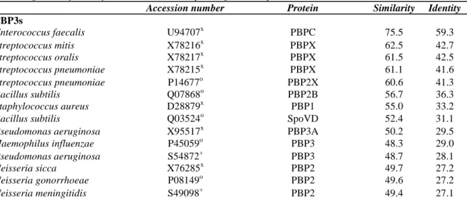
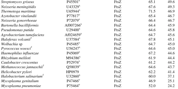
These cell division proteins are widespread in the bacterial world and most of them are much conserved (Table I). The percentages of identity relative to the E. hirae proteins are greater than 24 % except for the E. coli FtsQ (14 % identity). FtsZ which polymerizes to form a circumferential ring at the division site, is also present in mycoplasma (Wang and Lutkenhaus, 1996) which are wall-less eubacterial organisms and in archaeobacteria. Interestingly, phylogenetic trees consistently place the archaeobacterial FtsZ closer to the eukaryotic tubulins relative to the eubacterial FtsZ proteins (Margollin et al., 1996).
TABLE I Proteins isologous to the E. hirae PBP3s, MraY, MurG, FtsQ, FtsA and FtsZ. The percentages of
amino acid similarity and identity were calculated with the BESTFIT algorithm. The proteins are listed in decreasing order of identity relative to the corresponding E. hirae proteins
Accession number Protein Similarity Identity
PBP3s
Enterococcus faecalis U94707x PBPC 75.5 59.3
Streptococcus mitis X78216x PBPX 62.5 42.7 Streptococcus oralis X78217x PBPX 61.5 42.5 Streptococcus pneumoniae X78215x PBPX 61.1 41.6 Streptococcus pneumoniae P14677o PBP2X 60.6 41.3 Bacillus subtilis Q07868o PBP2B 56.7 36.3 Staphylococcus aureus D28879x PBP1 55.0 33.2
Bacillus subtilis Q03524o SpoVD 52.4 31.1
Pseudomonas aeruginosa X95517x PBP3A 50.2 29.5
Haemophilus influenzae P45059o PBP3 48.3 29.0
Pseudomonas aeruginosa S54872+ PBP3 48.7 28.1
Neisseria sicca X76285x PBP2 49.7 27.2
Neisseria gonorrhoeae P08149o PBP2 49.6 27.2
Escherichia coli P04286o PBP3 48.7 27.0
Bacillus subtilis P42971o hypothetical 74.4 kD prot. 48.7 26.5
Enterococcus hirae R40 X62280x PBP5 47.4 26.4
Escherichia coli P08150o PBP2 48.8 26.3
Helicobacter pylori HP1565● PBP2 48.0 26.3
Enterococcus hirae A36903+ PBP3r 49.6 25.9
Enterococcus faecium X92687x PBP5 49.2 25.2
Helicobacter pylori HP1556● FtsI 47.6 24.3
MraY
Enterococcus faecalis U94707x MraY 90.9 72.8
Staphylococcus aureus U94706x MraY 74.8 50.7
Bacillus subtilis Q03521o MraY 73.7 46.8
Escherichia coli P15876o MraY 68.6 43.4
Haemophilus influenzae A64185+ MraY 67.4 42.8
Borrelia burgdorferi X96432x MraY 68.8 42.8
Synechocystis sp. D64005x MraY 63.9 40.0
Staphylococcus aureus A55856+ L1m1 60.6 30.5
Escherichia coli P24235o Rfe2 58.9 28.7
Mycobacterium leprae P45830o Rfe homolog 57.3 28.3
Pseudomonas aeruginosa U17293x Rfb3033 58.6 27.7
Methanococcus jannaschii5 U67554x Diaminopimelate epimerase 57.2 27.3
Yersinia enterocolitica S51265+ TrsF4 58.6 26.7
Haemophilus influenzae A64138+ Rfe homolog 54.5 25.0
MurG
En terococcus faecalis U94707x MurG 79.3 66.8
Bacillus subtilis P37585o MurG 71.0 52.1
Haemophilus influenzae P45065o MurG 53.3 32.0
Escherichia coli P17443o MurG 57.2 31.7
FtsQ
Enterococcus faecalis U94707x DivIB 57.4 39.4
Bacillus licheniformis U01958x DivIB 54.1 33.7
Bacillus subtilis P16655o DivIB 52.5 32.8
Staphylococcus aureus U94707x DivIB 50.3 28.9
Escherichia coli K02668x FtsQ 38.1 14.3
FtsA
Enterococcus faecalis U94707x FtsA 87.8 76.2
Bacillus subtilis P28264o FtsA 63.6 39.1
Borrelia burgdorferi Z12164x FtsA 56.6 32.6
Escherichia coli P06137o FtsA 56.9 33.0
Haemophilus influenzae P45068o FtsA 57.4 30.9
Sinorhizobium meliloti Af024660x FtsA 54.8 30.2
Staphylococcus aureus U94706x FtsA 52.0 28.8
Helicobacter pylori HP0978● FtsA 49.0 27.0
FtsZ
Enterococcus faecalis U94707x FtsZ 88.5 82.2
Bacillus subtilis P17865° FtsZ 75.9 62.9
Staphylococcus aureus U94706x FtsZ 74.3 59.7
Anabaena sp JC4289+ FtsZ 68.3 55.3
Streptomyces coelicolor P45500o FtsZ 68.4 52.2
Synechocystis sp P73456o FtsZ 68.2 51.3
Borrelia burgdorferi P45483o FtsZ 70.8 50.9
Brevibaclerium lactofermentum P94337o FtsZ 68.0 50.9
Corynebacterium glutamicum Ab003132x FtsZ 68.7 50.5
Mycoplasma pulmonis Q50318o FtsZ 68.1 50.4
Pseudomonas aeruginosa P47204o FtsZ 66.4 50.3
Streptomyces griseus P45501o FtsZ 65.1 49.6
Neisseria meningitidis U43329x FtsZ 67.6 49.3
Thermotoga maritima U65944x FtsZ 71.5 46.9
Azotobacter vinelandii P77817o FtsZ 65.4 46.7
Neisseria gonorrhoeae P72079o FtsZ 66.4 46.7
Bartonella bacilliformis Af007266x FtsZ 64.4 45.9
Pseudomonas putida U29400x FtsZ 64.6 45.8
Agrobacterium tumefaciens Af024659x FtsZ 64.7 45.6
Haloferax volcanii5 U37584x FtsZ 67.8 45.1
Wolbachia sp P45485o FtsZ 64.7 45.0
Pyrococcus woesci5 U56247x FtsZ 64.6 45.0
Haemophilus influenzae P45069o FtsZ 67.2 44.6
Rhizobium meliloti M94386x FtsZ 61.9 44.4
Caulobacter crescentus P52976o FtsZ 61.2 44.2
Methanococcus jannaschii5 Q58039o FtsZ 64.6 43.7
Helicobacter pylori HP0979● FtsZ 62.2 41.4
Halobacterium salinarium5 U32860x FtsZ 60.0 37.1
Mycoplasma genitalium P47466o FtsZ 46.9 25.1
Mycoplasma pneumoniae P75464o FtsZ 52.0 24.2
1Llm: protein affecting the methicillin resistance level and the autolysis rate in S. aureus. 2Rfe: putative undecaprenyl-phosphate α-N-acetylglucosaminyl transferase.
3Rfb303: B-band lipopolysaccharide biosynthesis protein. 4TrsF: protein involved in lipopolysaccharide core biosynthesis. 5
M. jannaschii, H. volcanii, P. woesci and H. salinarium are archaeobacteria.
+: PIR bank ;o: SWISSPROT bank ;x: EMBL/GENbank/DDBJ;●: TIGR databank.
FIGURE 2 The dcw cluster of E. hirae S185, E. coli and B. subtilis. Genes are boxed. Numbers of amino acid
residues of the encoded proteins are given below and above the genes. Similarity between pairs of amino acid sequences is expressed in percent identity
Acknowledgements
This work was supported by the Belgian programme on Interuniversity Poles of Attraction initiated by the Belgian State, Prime Minister's Office, Services fédéraux des affaires scientifiques, techniques et culturelles (PAI n° 19 and P4/03) and the Fonds de la Recherche Fondamentale Collective (contract 2.4534.95). CD is Research Associate of the Fonds National de la Recherche Scientifique. FS is indebted to the Fonds pour la Formation à la Recherche dans l'Industrie et dans l'Agriculture.
References
Altschul, S.F., Gish, W., Miller, W., Myers, E.W. and Lipman, D.J. (1990). Basic local alignment search tool. Journal of Molecular Biology 215, 403-410.
Ayala, J.A., Garrido, T., De Pedro, M.A. and Vicente, M. (1994). Molecular biology of bacterial septation. In Ghuysen, J.M. and Hakenbeck, R. (eds), Bacterial Cell Wall (Elsevier, Amsterdam), pp. 73-101.
Buchanan, C.E., Henriques, A.O. and Piggot, P.J. (1994). Cell wall changes during bacterial endospore formation. In Ghuysen, J.M. and Hakenbeck, R. (eds), Bacterial cell wall (Elsevier, Amsterdam), pp. 167-183.
Coyette, J., Somzé, A., Briquet, J.J., Ghuysen, J.M. and Fontana, R. (1983). Function of penicillin-binding protein 3 in Streptococcus faecium. In Hakenbeck, R., Höltje, J.V. and Labischinski, H. (eds), The target of penicillin (de Gruyter W. and Co., Berlin), pp. 523-530. Devereux, J., Haeberli, P. and Smithies, O. (1984). A comprehensive set of sequence analysis programs for the VAX. Nucleic Acids Research 12, 387-395.
Ghuysen, J.M. (1997) Penicillin-binding proteins. Wall peptidoglycan assembly and resistance to penicillins : facts, doubts and hopes. International Journal of Antimicrobial Agents 8, 45-60.
Ghuysen, J.M., Charlier, P., Coyette, J., Duez, C., Fonzé, E., Fraipont, C., Goffin, C., Joris, B. and Nguyen-Distèche, M. (1996). Penicillin and beyond : evolution, protein fold, multimodular polypeptides, and multiprotein complexes. Microbial Drug Resistance 2, 163-175. Harry, E.J. and Wake, R.G. (1989). Cloning and expression of a Bacillus subtilis division initiation gene for which a homolog has not been identified in another organism. Journal of Bacteriology. 171, 6835-6839.
Hopwood, D.A., Bibb, M.J., Chater, K.F., Kieser, T., Bruton, C.J., Kieser, H.M., Lydiate, D.J., Smith, C.P., Ward, J.M. and Schrempf, H. (1985). Genetic Manipulation of Streptomyces. A Laboratory Manual. The John Innes Foundation, Norwich, U.K.
Kunst, F. and 150 coauthors (1997). The complete sequence of the Gram-positive bacterium Bacillus subtilis. Nature 390, 249-256. Margolin, W., Wang, R. and Kumar, M. (1996). Isolation of an ftsZ homolog from the archaebacterium Halobacterium salinarium : implications for the evolution of FtsZ and tubulin. Journal of Bacteriology 178, 1320-1327.
Pearson, W.R. and Lipman, D.J. (1988). Improved tools for biological sequence analysis. Proceedings of the National Academy of Sciences USA 85, 2444-2448.
Piras, G., El Kharroubi, A., Van Beeumen, J., Coeme, E., Coyette, J. and Ghuysen, J.M. (1990). Characterization of an Enterococcus hirae penicillin-binding protein 3 with low penicillin affinity. Journal of Bacteriology 172, 6856-6862.
Pucci, M.J., Thanassi, J.A., Discotto, L.F., Kessler, R.E. and Dougherty, T.J. (1997). Identification and characterization of cell wall-cell division gene clusters in pathogenic Gram-positive cocci. Journal of Bacteriology 179, 5632-5635.
Silver, J. (1991). Inverse polymerase chain reaction. In McPherson, M.J., Quirke, P. and Taylor, G.R. (eds), PCR a practical approach (IRL Press at Oxford University Press), pp. 137-146.
van Heijenoort, J. (1994). Biosynthesis of the bacterial peptidoglycan unit. In Ghuysen, J.M. and Hakenbeck, R. (eds), Bacterial Cell Wall (Elsevier, Amsterdam), pp. 39-54.
Vicente, M. and Errington, J. (1996). Structure, function and controls in microbial division. Molecular Microbiology 20, 1-7.
Wang, X. and Lutkenhaus, J. (1996). Characterization of the ftsZ gene from Mycoplasma pulmonis, an organism lacking a cell wall. Journal of Bacteriology 178, 2314-2319.
Yura, T., Mori, H., Nagai, T., Nagata, A. Ishihama, N., Fujita, N., Isono, K., Mizobuchi, K. and Nakata, A. (1992). Systematic sequencing of the Escherichia coli genome : analysis of the 0 - 2.4 min region. Nucleic Acids Research 20, 3305-3308.