Université de Montréal
EVALUATION OF VALOSIN CONTAINING PROTEIN
(P97) AS A CANCER BIOMARKER IN CANINE
LYMPHOMAS
parSABIN DRAGOS FILIMON
Département de Biomédecine Vétérinaire Faculté de médecine vétérinaire
Mémoire présenté à la Faculté de médecine vétérinaire en vue de l’obtention du grade de maître ès science (M.Sc)
en Sciences Vétérinaires option Biomédecine
Août, 2014
Université de Montréal
Faculté des études supérieures et postdoctorales
Ce mémoire intitulé
EVALUATION OF VALOSIN CONTAINING PROTEIN (P97) AS A CANCER BIOMARKER IN CANINE LYMPHOMAS
Présenté par :
SABIN DRAGOS FILIMON
a été évalué par un jury composé des personnes suivantes :
Carolyn Grimes, présidente-rapporteuse Derek Boerboom, directeur Marie-Ève Nadeau, codirectrice Marie-Claude Blais, membre du jury
Résumé
Le lymphome est l'une des tumeurs les plus communes tant chez le chien que l’humain. Chaque année, un nombre important de chiens développe ce cancer agressif. La majorité décédant un an suivant le diagnostic. Le lymphome canin est maintenant identifié comme un excellent modèle de recherche pour la tumeur chez l'homme, particulièrement en ce qui concerne la biologie moléculaire de la maladie. En conséquence, la recherche sur le lymphome canin sera bénéfique non seulement pour les chiens mais aussi pour l’oncologie humaine. Parmi les méthodes diagnostiques de choix pour dépister de façon hâtive le lymphome se trouve la mesure de marqueurs tumoraux. Ceci a l’avantage d’être peu invasive, simple et peu dispendieuse. Ainsi, dans le but d’évaluer la protéine VCP (valosin containing protein) comme biomarqueur tumoral dans les lymphomes canins à cellules B et T, nous avons évalué la protéine VCP par immunobuvardage sur sérums et tissus tumoraux de chiens atteints et par immunohistochimie sur des tumeurs de haut grade, grade intermédiaire et bas grade. Pour mieux définir l’expression de VCP dans les cellules cancéreuses, nous avons également examiné par immunobuvardage les niveaux de VCP dans 3 lignées cellulaires: CLBL-1, CL-1, et 17-71. Il s’avère que les lymphomes à cellules B de haut grade avaient une élévation significative du taux de VCP comparé aux tumeurs de bas grade (P < 0,05). De même, une accumulation importante de VCP a également été détectée dans les lignées tumorales comparées aux cellules mononucléaires du sang périphérique (P < 0,05). D’autre part, le taux sérique de VCP est resté similaire à ceux des chiens normaux. Ces résultats suggèrent une corrélation entre le taux de VCP et le degré de malignité des lymphomes à cellules B. En conclusion, la protéine VCP doit faire l’objet d’une évaluation approfondie pour déterminer son utilité comme marqueur pronostique.
Mots-clés : la protéine VCP, marqueurs tumoraux, lymphome canine, lymphome à cellules B, oncologie vétérinaire, chien, tumeur maligne
Abstract
Lymphoma is one of the common malignancies in both dogs and humans. Annually, an important number of canine patients develop this aggressive cancer and a majority succumbs to the disease within one year. In recent years, canine lymphoma has been increasingly recognized as an excellent model for the disease in humans, especially with regards to the molecular biology of the disease. Consequently, research targeted at canine lymphoma benefits not only dogs but the field of human oncology as well. Among the most desirable diagnostic and screening tests for lymphoma is the measurement of cancer biomarkers. They have the advantage of being minimally invasive, simple, and inexpensive. Thus, with the aim of evaluating valosin containing protein (VCP) as a cancer biomarker in canine B and T-cell lymphomas, we first performed western blots on sera and tumor tissue of dogs with lymphoma and then immunohistochemical analysis on low, intermediate and high-grade tumors. To further determine VCP expression in cancer cells, we also examined VCP levels by immunoblotting in 3 tumor cell lines: CLBL-1, CL-1, and 17-71. High-grade B-cell lymphomas had significantly increased levels of VCP compared to low-grade tumors (P < 0.05). Additionally, we detected a corresponding accumulation of VCP in tumor cells lines compared to peripheral blood mononuclear cells (PBMCs) (P < 0.05). In contrast, VCP levels were not elevated in sera of dogs with lymphoma compared to healthy controls. These results suggest that VCP positively correlates with malignancy in canine B-cell lymphomas. We conclude that VCP merits further investigation to determine its potential as a clinically useful prognosis biomarker for canine B-cell lymphoma.
Keywords : Valosin containing protein (p97), Cancer biomarker, Canine Lymphoma, B-cell lymphoma, Veterinary oncology, Dog, Malignancy
Table of contents
Résumé ……….i
Abstract ………..iii
List of tables ……….vii
List of figures ………...…viii
List of acronyms and abbreviations ………...ix
Acknowledgments ………xii
Introduction ……….1
Literature review ……….5
Chapter 1: Cancer in dogs ………...5
1.1. Lymphoma, a type of cancer most prevalent in dogs ………...6
1.1.1. Incidence and prevalence ………..6
1.1.2. Causes ………8 1.1.3. Clinical findings ………9 1.1.4. Classification ………...10 1.1.4.1. Anatomical classification ……….10 1.1.4.2. Histopathological classification ………12 1.1.4.3. Immunophenotypical classification ………..14 1.1.5. Diagnosis ……….15 1.1.6. Treatment ………16 1.1.6.1. Chemotherapy ………...17
1.1.6.3. Radiation therapy ………..19
1.1.6.4. Surgery ……….19
1.1.7. Prognosis ………...……..20
Chapter 2: Development of cancer biomarkers as tools for improving disease detection and treatment ………...21
2.1. Cancer biomarkers ……….21
2.2. Cancer biomarkers in human medicine ………. 25
2.2.1. Serum markers in human medicine ……… 25
2.2.1.1. Specific serum markers for lymphoma ……….28
2.2.2. Tumor markers in human medicine ………...…………..29
2.2.2.1. Specific tumor markers for lymphoma ……….31
Chapter 3: Cancer biomarkers in veterinary medicine ……….33
3.1. Serum markers in veterinary medicine ………..35
3.1.1. Detection and monitoring of lymphoma using serum makers …………37
3.2. Tumor markers in veterinary medicine ………..38
3.2.1. Detection and monitoring of lymphoma using tumor markers ………...39
Chapter 4: Valosin containing protein (VCP) as a cancer biomarker ………...40
4.1. Structure of VCP ………40
4.2. Role of VCP in the cell ………..41
4.3. VCP as a cancer biomarker in human medicine ……….46
4.3.1. VCP as a tumor marker ………...47
4.3.2. VCP as a serum marker ………...48
Hypothesis and objectives ……….50
Publication as first author ……….51
General discussion ………87
Conclusion ………98
List of tables
Table 1. The updated Kiel classification of canine lymphomas ………...12
Table 2. The WF classification of Non Hodgkin’s Canine Lymphomas ………..13
Table 3. WHO clinical staging for domestic animals with lymphoma ……….16
Table 4. Use of cancer biomarkers in patient care ………23
Table 5. FDA approved serum biomarkers ………...26
Table 6. Lymphoma serum biomarkers identified in current literature ………29
Table 7. FDA approved tumor biomarkers ………...30
Table 8. Tumor biomarkers for lymphoma identified in the current literature ……….32
Table 9. Serum markers in veterinary medicine ………...36
Table 10. Lymphoma serum biomarkers detectable in dogs ………37
List of Figures
Figure 1. Structure of the hexameric AAA+ protein VCP/p97 ………41
Figure 2. VCP and its three core adaptors regulate diverse cellular functions ……….42
Figure 3. Multiple functions of VCP ………43
Figure 4. Protein-induced chromatin stress (PICHROS) ………..45
Article
Figure 1. VCP protein expression in canine lymphoma tumors ………...82Figure 2. Western blotting analysis of VCP in PBMCs and lymphoma cell lines (17-71, CL-1, and CLBL-1) ……….83
Figure 3. Immunohistochemical analysis of VCP expression in lymphatic nodule compared to B-cell and T-cell lymphomas ………84
Figure 4. K-48 Polyubiquitinated protein expression in canine lymphoma tumors ………….85
List of acronyms and abbreviations
AAA+: ATPases-Associated with diverse cellular Activities ACTB: β-ActinALS: amyotrphic lateral sclerosis AUC: area under curve
CCOGC: Canine Comparative Oncology and Genomics Consortium CGH: comparative genomic-hybridization
CHOP: cyclophosphamide (C), hydroxydaunorubicin or doxorubicin (H), oncovin or vincristine (O) and prednisone (P)
CID: inflammatory chronic disease DNA: deoxyribonucleic acid Eey 1 : Eeyarestatin 1
ELISA: enzyme-linked immunosorbent assay ER: endoplasmatic reticulum
ERAD: endoplasmatic reticulum associated degradation FDA: US Food and Drug Administration
FFPE: formalin fixed paraffin embedded tissue FISH: florescence in situ Hybridization
GCT: granulosa cell tumor GEP: gene expression profiling HDAC6: histone deacetylase 6
IBMPFD: Inclusion Body Myopathy and Paget’s disease of the bone and Frontotemporal Dementia
kDa : kilo Daltons
MALT: mucosa-associated lymphoid tissue MDR: multiple drug resistance
NHL: Non-Hodgkin’s Lymphoma
NPL4-UFD1: ubiquitin fusion degradation 1 – nuclear protein localization 4 NSCLC: non-small cell lung carcinoma
NSFL1C: N-ethylmaleimide-sensitive factor L 1 cofactor PBMC: peripheral blood mononuclear cells
PCR: polymerase chain reaction
PICHROS: protein induced chromatin stress RIPA: radioimmunoprecipitation assay RNA: ribonucleic acid
ROC: receiver operating characteristic RPMI: Roswell Park Memorial Institute SEM: standard error of the mean
TK1: serum thymidine kinase 1 UBD: ubiquitin-fold domain UBX: Ubiquitin regulatory X
UBXD1: ubiquitin regulatory X domain 1 UFD: ubiquitin-fusion domain
VEGF: Vascular Endothelial Growth Factor WB: western blot
WF: Working Formulation
Acknowledgements
I would like to thank my research director, Dr. Derek Boerboom, for his guidance and advice all throughout these years. Dr. Boerboom provided me an opportunity to work in the interesting area of canine lymphoma and gave me full support in accomplishing my postgraduate studies. I will never be able to forget him, nor his absolute amazing commentaries on my writing. Special thanks to Dr. Marie-Éve Nadeau, my co-director, who helped me enormously with the collection of samples and clinical advice. To Dr. Marilène Paquet, thank for you excellent analyses. I would also like to thank the members of the Boerboom laboratory: Charlène Rico, Meggie Girard, Mayra Tsoi, Atefeh Abedini, and Alexandre Boyer. I could not have accomplished this work without their help. Thank you to the members of my advisory committee and to the members of the jury who graciously agreed to assist me in this endeavour. Last but not least, I would like to thank my girlfriend and my friends for their felt support.
Introduction
As presented in a plethora of studies, particularly in the last decade, cancer has become one of the major causes of death in humans and companion animals alike. The increasing incidence of diagnosed cases has pushed the research community to study and develop new screening and therapeutic methods for specific types of cancers. Among these, lymphoma, a common canine tumor, represents a continuing interest of research (1).
Lymphomas are a type of cancer characterized by the proliferation of malignant lymphoreticular cells, or cells pertaining to both lymphoid and reticuloendothelial systems, within solid organs (2). The primary lymphoid organs, represented by the bone marrow and thymus, along with the secondary lymphoid structures, such as lymph nodes and spleen, are the preferred sites of neoplastic growth (3). However, due to the facilitated access of lymphocytes to the bloodstream and lymph, the tumor can originate virtually in any organ (4).
In veterinary literature, lymphoma is presented as the most common hematopoietic canine neoplasm (4). Despite its ubiquity, the precise etiology of the disease has not yet been identified. Causes vary from genetic to environmental, with several breeds of dogs presenting a higher incidence for the disease (5). The tumor can manifest a wide spectrum of clinical and biological behaviours depending on its anatomical location and its stage of development (6). Furthermore, the symptomology of the disease can vary accordingly to the neoplastic transformation of either B or T-lymphocytes (7). To complicate matters even further, each individual tumor can present an innate heterogeneity, leaving early detection and complete curative therapies elusive for most veterinary patients.
proven to be the major limitation on the efficacy of available lymphoma treatments. Many patients seem to undergo intensive chemotherapy sessions with little apparent benefit in metastasis control or life expectancy (8). Along the years it was understood that early tumor detection has a strong correlation with net gains in quality of life and prolonged survival (4). As such, veterinary scientists are faced with the growing clinical demand to develop and validate new therapeutical targets that will assist oncologists in early cancer screening.
Among the most desirable tools for the diagnosis and prognosis of lymphomas are cancer biomarkers. They are defined as measurable molecules, in clinical or laboratory settings that appear in a modified state, quantitatively or qualitatively, in the presence of tumoral processes (9). Historically, the research for biomarkers was targeted at bodily fluids (blood, plasma, urine) and/or tumor tissue (10). Therefore, two distinctive categories of cancer biomarkers emerged: serum and tumor markers. Although, both operate on the same principle, their detection, measurement and utility are quite different.
Serum biomarkers are usually measured in bodily fluids, having considerable value in cancer screening, diagnosis, prognosis and follow-up of treatment efficacy (11, 12). As molecules, serum markers can be hormones, glycoproteins, or other proteins overexpressed by cancer cells. Although they are routinely used for screening and diagnosis of several human cancers, few serum markers have been identified in dogs, and none are currently used in clinical settings (13-15).
The ideal serum marker should have high sensitivity and/or high specificity. More specifically, the test needs to discriminate cancer patients form healthy subjects and/or patients with benign tumors or non-cancerous conditions. Furthermore, the markers need to show
targeted proteins, done by enzyme-linked immunosorbent essay (ELISA) or western blot, has the advantage of being minimally invasive, simple, and inexpensive (10).
Tumor biomarkers, on the other hand, are measurable biochemical molecules associated with malignancy. They can be produced by tumors cells or by the body in response to the neoplastic process (9). Although measurement of tumor markers is only possible once the cancer has been identified and a biopsy has been obtained, making them ineffective for screening, they have an important role to play during the development of the disease (9). These markers are highly desirable in cancer staging, prognosis, monitoring treatment efficacy, and/or in evaluating the cancer’s recurrence (9). The markers can be detected in tissue by several immunohistochemistry and immunofluorescence protocols, immunoblotting and mass spectrometry (10).
Despite their tremendous potential value, the use of serum and tumor biomarkers in veterinary oncology remains in its infancy. Recently, a new protein has been identified as a promising serum marker for human ovarian, breast, and colon cancer (17, 18). Based on our laboratory’s preliminary results, valosin-containing protein, or VCP, also seems to have a higher sensibility for certain types of canine tumors, indicative of superior potential for early diagnosis (17). Lymphomas are found among the specific cancers with higher expression of VCP during their development.
As such, this thesis will present the evaluation of VCP expression as a cancer biomarker in canine lymphomas. First off, a review of the cancers in dogs and canine lymphoma will be presented, followed by current knowledge of the development of cancer biomarkers as a diagnostic tool in human and veterinary medicine, focusing on lymphoma. The emphasis is put on the use of serum and tissue markers as early detection and monitoring
tools. Next, the thesis will describe the cellular functions of VCP and will explain its potential usefulness as a diagnosis and prognosis biomarker for human and canine malignancies. Finally, the findings from this study will be presented and discussed.
Literature Review
Chapter 1: Cancer in dogs
Recent studies identify cancer as one of the most common causes of death among dogs, with values as high as 30% of registered cases (19). The total incidence of neoplasia in the United States was estimated at 1134 cases per 100,000 dogs per year (20). In 2000, a Canadian study conducted by Reid-Smith and colleagues reported 4817 cases per 100,000 dogs per year, almost four times more patients than the incidence rates reported in the States (20). Across the ocean, studies carried out in the United Kingdom present 2671 tumors per 100,000 dogs per year (20). Finally, an Italian study conducted in 2008 revealed an incidence of 1070 cases per 100,000 dogs per year (21). The discrepancy can be a direct consequence of the differences in the population of dogs found in the distinct geographical territories, since some breeds of dogs are reported to have higher genetic predispositions for lymphoma, and/or the method used in tumor classification and diagnosis (20). Whatever the variables influencing the data might be, the overall message remains the same: a relatively high incidence of canine neoplasia is encountered around the world.
The studies also presented estimated figures for the frequency of different types of canine tumors. Consequently, with small variations, certain types of cancers have been identified as having higher incidence and frequency. For female dogs, cancers of the mammary gland are the most significant, followed by Non-Hodgkin’s Lymphoma (NHL) and other connective and soft tissue tumors (21). For males, lymphoma represents the most important neoplasm, before skin (excluding melanoma) and soft tissue cancers (21).
The major limitation in presenting specific canine cancers as more prevalent revolves around diagnosis and detection methods. Mammary, genital and skin tumors are easily recognizable during a general physical exam, whereas the detection of internal cancers is less obvious. The latter might require specific methods of diagnosis such as imagery, exploratory surgery and/or histopathological consult (21). Often, the client’s decision to halt further clinical investigation represents a serious limitation in correctly estimating the incidence of different types of canine cancers such as lymphoma.
1.1. Lymphoma, a type of cancer most prevalent in dogs
Lymphomas, malignant or lymphosarcomas, are a diverse group of cancers (3). They present a high genetic variability with diverse, poorly understood causality (3). Clinical data combined with a comprehensive series of metanalyses, were able to thoroughly describe the biological behaviour of these cancers. In general, lymphomas have a high incidence with a rather aggressive development and a low life expectancy in the absence of treatment (3).
1.1.1. Incidence and prevalence
The annual incidence, or the number of new lymphoma cases per year, varies anywhere between 24 to 107 new cases per 100 00 dog, however there is a significant number of lymphoma case not recorded limiting our ability to detected the true incidence which seems to be much higher (3, 22, 23). In fact lymphoma occurs about 2 to 5 times in dogs as in people and it estimated that approximately 1 of every 15 dogs born today will get lymphoma at some point in his life. Incidence of lymphoma was found to increase with age, peaking at 10 years
less than one year, and as high as 84 cases per 100,000 for dogs older than 10 years (3). The maximal incidence can be found in the mid-age population, with dogs aged between 6 and 9 years old (25).
Although gender it is not an influential factor in lymphoma frequency, certain breeds appear to have a higher incidence for the disease (25). These include Golden Retrievers, Boxers, Bull Mastiffs, Basset Hounds, Saint Bernards, Scottish Terriers, Airedales and Bulldogs (26). Moreover, particular breeds present higher tendencies to develop specific types of lymphoma (25). Hence, Boxers and Shar-peis tend to develop alimentary lymphoma, Cocker Spaniels, Basset Hounds and Doberman Pinchers are more prone to develop B-cell lymphoma, and Boxers, Irish Wolfhounds, Siberian Huskies and Shih Tzus present more often than not T-cell lymphoma (27). Finally, Golden Retrievers can indiscriminately develop B or T-cell lymphomas (3).
Historically, lymphomas have a lower risk of appearance in intact females and in certain breeds, namely, Dachshunds and Pomeranians (3). Furthermore, because of closed gene pools and repeated inbreeding, purebred dogs present slightly higher tumor rates than crossbred dogs (28).
Lymphoma prevalence, also known as the cumulative incidence, is estimated at 7% to 24% of all canine neoplasia, with an average of 20% (3, 27). Among the hematopoietic malignancies, lymphoma occupies a staggering 83% of all registered cases (3). The most prevalent are the solid forms, such as lymphosarcomas and malign lymphomas (25). Within the solid forms, the most common type is diffuse large B-cell lymphoma, a similarity canis familiaris shares with humans (27).
1.1.2. Causes
The causes of lymphoma, despite extensive research, are still not completely understood and likely multifactorial. Among the etiological factors that trigger the cells’ aberrant multiplication we can distinguish: genetics and molecular causes, infectious and environmental factors, and immunosuppression (3).
The recent advances in coding the canine genome have allowed detection of chromosomal aberrations in dogs affected by lymphoma. As such, studies have reported genetic material gain on canine chromosomes 13 and 31 and loss of it on chromosome 14 (3). Among other molecular events leading to tumorigenesis, deoxyribonucleic acid hypomethylation, and mutation of genes p53 and N-ras are common features of cancerous lymphocytes (3). The cellular pathways of the Bcl-2 family also seem to be involved in the process, however, more research is needed in order to fully understand their complete implication (3).
Besides the genetic causes, several studies have made a link between lymphomas and infectious and/or environmental factors. A weak association has been proposed between Helicobacter pylori infections and the development of gastric lymphomas (3). Viral infections appear to trigger the formation of mucosa-associated lymphoid tissue (MALT) lymphoma (3). Environmentally, the tumor appears to be related to the use of phenoxyacetic acid herbicides, particularly 2,4-dicholorophenoxyacetic acid (2,4-D) (3). In households were the substance was regularly used, dogs developed malignant lymphoma twice as frequently as in households where environmentally friendly or no herbicides were employed (3). In recent literature, there is a debate concerning the correlation between strong magnetic fields and the increased risk of
households with very high current codes were 6.8 times more inclined to develop the disease (28).
Finally, immunosuppression is considered an important risk factor in the development of lymphomas. Immune system alterations, such as immune-mediated thrombocytopenia and immunosuppressive therapies (e.g., cyclosporine) are reported to help in the development of the neoplastic process (3).
1.1.3. Clinical findings
The prevalent ontogenic site of lymphomas is the lymphoid tissue (lymph nodes, spleen and bone marrow), however, almost any tissue can be affected (3). Due to this variability in anatomical locations, lymphoma presents with variously different clinical signs which are most often site dependent (29).
Multicentric lymphoma, the most common form encountered, presents a painless generalized lymphadenopathy, often accompanied by hepatosplenomegaly and bone marrow infiltration (3). The attack on the bone marrow provokes blood dyscrasias, a condition associated with anemia, thrombocytopenia and neutropenia (3). Due to the incapacity of the myeloid stem cells to replicate, the disorder can also lead to fever, sepsis and/or hemorrhage (3). Occasionally, the lesions will lead to nonspecific signs such as anorexia, weight loss, vomiting, diarrhea, emaciation, ascites, dyspnea, polydipsia, polyuria, and fever (3). T-cell lymphoma, a subtype of multicentric lymphoma, can add hypercalcemia to the long list of symptoms (25).
In the case of gastric or alimentary lymphoma, lesions to the mucosa and lamina propria will cause vomiting, diarrhea, weight loss, panhypoproteinemia, and malabsorption
(3). As secondary lesions, the tumor could metastasize to the mesenteric and pelvic lymph nodes, spleen and liver (3).
Canine lymphomas can also be encountered in the mediastinal cavity, where lesions create their own set of clinical symptoms. These types of tumors are characterized by the enlargement of the craniomediastinal structures and/or the thymus (3). Depending the extent of the disease, the animal might present with one or more of the following signs: respiratory distress, pleural effusion, exercise intolerance, regurgitation, polydipsia, polyuria, and possible hypercalcemia (3). If the mass compresses or invades the cranial vena cava, a pitting edema of the head, neck, and forelimbs might be observed (3).
The lesions for extranodal lymphomas are organ specific and, although important, their description exceeds the scope of this thesis. It is worth mentioning the involvement of tumors in the central nervous system (CNS) because of their serious consequences such as: paraparesis, ataxia, hyperesthesia, blindness, lethargy, and seizures (3).
1.1.4. Classification
Over the years, several classification systems have been used to differentiate between the various types of lymphomas. As such, the tumors can be classified on the basis of anatomical location, histopathological description and immunophenotypic analysis (3).
1.1.4.1. Anatomical classification
Multicentric lymphoma is encountered in approximately 80% of cases (29). It has the aspect of a ganglionary, rubbery, painless, mobile, smooth, and generally symmetrical tumor
however, if left untreated, it will spread to other organs and parts of the lymphoreticular system (25).
The second most encountered form, representing 7% of cases, is gastrointestinal or alimentary lymphoma (29). The digestives lesions, mostly ulcers, can have different stages of infiltration throughout the submucosa and lamina propria (25). Mesenteric, gastric and retrocaecal adenopathies can be observed in the last stages of the disease (25). Historically, it was believed that alimentary lymphoma originates form B-cell lymphocytes, however, recent findings place the origin of the tumor as T-cell derived neoplasia (3).
Six percent of dogs affected by lymphoma develop the cutaneous or mucocutaneous form (29). The tumor is characterized by non-specific skin changes, progressing from scaly alopecia to thickened erythematous ulcerations to plaque-like lesions (29). Recent classifications have distinguished between two forms: epitheliotropic cutaneous and non-epitheliotropic cutaneous lymphomas (25). Epitheliotropic cutaneous lymphoma is encountered more often and is characterized by multicentricity and slow development, originating most often from CD8+ T-cells (3, 25). The latter type, although rarely encountered in clinical settings, develops in the deep dermis, being more invasive and difficult to treat (25). Extranodal forms of lymphoma are rarely encountered, with a prevalence of only 3% of cases (29). The tumors appear randomly with predilections for eyes, central nervous system, bones, testis and nasal cavity (29). The form specific to the central nervous system is known as intravascular or angiotrophic lymphoma (3). It originates from either B or T-cell lymphocytes, and is characterized by diffuse infiltration of the wall and lumen of blood vessels (3).
1.1.4.2. Histopathological classification
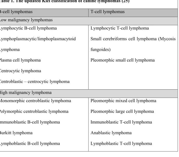
Due to numerous cellular analogies between human and canine malignancies, the histological classification systems have been adapted from human oncology (25). The current systems used in veterinary medicine include the Kiel classification, Working Formulation (WF) and World Health Organization (WHO) (3).
The Kiel classification is useful in distinguishing between B or T-cell tumors and in differentiating between high grade or low grade malignancies (25). We present the identifiable types of lymphoma using this system in table 1.
Table 1. The updated Kiel classification of canine lymphomas (25)
B-cell lymphomas T-cell lymphomas
Low malignancy lymphomas Lymphocytic B-cell lymphoma
Lymphoplasmacytic/limphoplasmacytoid Lymphoma
Plasma cell lymphoma Centrocytic lymphoma
Centroblastic – centrocytic lymphoma
Lymphocytic T-cell lymphoma
Small cerebriforms cell lymphoma (Mycosis fungoides)
Pleomorphic small cell lymphoma
High malignancy lymphoma
Monomorphic centroblastic lymphoma Polymorphic centroblastic lymphoma Immunoblastic B-cell lymphoma Burkitt lymphoma
Lymphoblastic B-cell lymphoma
Pleomorphic mixed cell lymphoma Pleomorphic large cell lymphoma Immunoblastic T-cell lymphoma Anablastic lymphoma
The WF, developed by the National Cancer Institute, categorizes lymphomas as low, intermediate, and high grade, with no distinction between B or T-cell origin (3, 25). The tumoral classification mainly uses cytomorphological characteristics such as size, type (small cleaved cells, large cells, and immunoblastic cells), shape of the nucleus, and the architecture of the neoplastic tissue (follicular or diffuse) (25). The WF classification (table 2) is used to analyze the biology of the tumor and is better at predicting patient survival (3).
Table 2. The WF classification of Non Hodgkin’s Canine Lymphomas (25)
Low malignancy lymphomas
Small cell lymphocytic lymphoma:
- Diffuse small cell lymphocytic lymphoma - Plasmocytoid lymphoma
Small cleaved cell follicular lymphoma Mixed small cleaved cell
Large cell follicular lymphoma
Intermediate malignancy lymphomas
Large cell follicular lymphoma Diffuse small cleaved cell lymphoma
Diffuse mixed small and large cell lymphoma Diffuse large cell lymphoma
Diffuse large non-cleaved cell lymphoma
High malignancy lymphomas
Large cell immunoblastic lymphoma Convoluted cell lymphoblastic lymphoma Non-convoluted cell lymphoblastic lymphoma Burkitt lymphoma
Other types of lymphomas
Mixed lymphoma Mycosis fungoides
Extramedullary plasmocytoma Histiocytic lymphoma
The Veterinary Lymphoma Study has recently adapted the latest WHO classification of neoplastic diseases of lymphoid tissue in humans (30). The classification was developed to incorporate both morphological, genotypic, and immunohistologic criteria, thereby providing a system for categorizing lymphoid neoplasms according to the level of cellular maturation (3). The strength of this system resides in the capacity to categorize canine lymphomas within the B or T-cell subtypes as well as offering a clinical prognosis (31). The WHO classification combines with success the previous two systems, insuring a better diagnostical uniformity among veterinary pathologists.
1.1.4.3. Immunophenotypical classification
The development of monoclonal antibodies has made possible the detection of specific markers on the surface of lymphocytes, giving more insight into the classification of canine lymphomas. Based on this method, lymphomas are further differentiated into B, T or null-cell (neither B or T immunoreactive) tumors (29). The immunophenotypic classification has lead to the realization that most canine lymphomas have a B-cell immonophenotype (25). Thus, as presented by the veterinary literature, B-cell lymphoma accounts for 60% to 80% of cases, T-cell lymphomas for 10% to 38%, mixed B and T-T-cells for 22%, and null-T-cell for fewer than 5% (3). Furthermore, classifying lymphomas with the help of monoclonal antibodies led pathologists to the conclusion that T-cell lymphomas have a higher incidence than previously recorded (25). Clinically, making a clear distinction between a T-cell or a B-cell derived tumor has major importance since the latter can be more aggressive and present a lower response rate
1.1.5. Diagnosis
When suspecting lymphoma, the veterinary clinical oncologist has to perform a thorough clinical evaluation which includes a complete physical examination, blood work, serum biochemistry, and urinalysis (3). Advanced imaging modalities like computer tomography, magnetic resonance imaging, and positron emission/computed tomography are becoming more accessible in veterinary medicine (3). The final diagnosis is largely given by the veterinary pathologist who will employ histologic and cytologic tissue evaluation, molecular analysis, immunophenotyping, and clonality assay as diagnostic tools (3).
Histological and cytological evaluations can be done on biopsied tissue using histochemical/immunohistochemical and cytochemical/immunocytochemical staining and/or flow cytometry (3). Although all protocols are clinically available, for the moment immunophenotyping seems to be the most reliable (3). The technique uses specific cellular membrane markers to improve the precision of diagnosis and provide the clinician with a certain prognosis (3). Antibodies against CD3+, CD4+ and CD8+ for T-cells and CD79a+ and CD21+ for B-cells are applied against tissue sections (immunohistochemistry), cytologic specimens (immunocytochemistry), or single cells (flow cytometry) (3). When the characterization of lymphomas proves to be challenging utilizing standard histology, PCR techniques are used to amplify the genes of the immunoglobulin receptor for either B or T-cell lymphocytes (3).
Once the precise diagnosis is reported by the pathologist, the WHO classification system (Table 3) is used to stage the tumor (3). Stages I and II are more favorable if index “b” is not present, however, the other higher stages carry with them a poor prognosis (25). Stage V
leads for the most part to the death of the animal in a matter of days to weeks (25). Because lower stages are asymptomatic and regular medical examination is not standard veterinary practice, most dogs present with advanced stages, thus reducing drastically the treatment’s efficacy (3).
Table 3. WHO clinical staging for domestic animals with lymphoma
Stage Criteria
I
Single lymph node
a. Without clinical sign of disease b. With clinical signs of disease
II
Multiple lymph nodes in a regional area a. Without clinical signs of disease b. With clinical signs of disease
III
Generalized lymphadenopathy a. Without clinical signs of disease b. With clinical signs of disease
IV
Liver and/or spleen involvement (with or without stage III) a. Without clinical signs of disease
b. With clinical signs of disease
V
Bone marrow or blood involvement and/or non-lymphoid organ (with or without stage I-IV)
a. Without clinical signs of disease b. With clinical signs of disease
1.1.6. Treatment
In clinical practice treatment options and the order of their administration differ from patient to patient, depending on the type of lymphoma and the extent of the disease. The
current golden standard in veterinary oncology concerning lymphoma treatment includes: systemic chemotherapy and immunotherapy, radiation therapy, and surgery (29).
1.1.6.1. Chemotherapy
Veterinary oncologists have at their disposal a variety of chemotherapeutic protocols that can be adapted according to the stage and substance (the presence or absence of clinical signs) (12), the presence of paraneoplastic syndrome, and the dog’s overall physical condition (3, 29). Moreover, the owners’ financial situation and time constraints, along with possible apprehension vis-a-vis the side effects of treatment, may also play a deciding factor (3, 29).
Multidrug chemotherapy protocols were first introduced over 20 years ago and have developed considerably since (3, 29). The most used treatment is a modification of the CHOP protocol, used for the first time in human oncology (3). This first line treatment is composed of cyclophosphamide (C), hydroxydaunorubicin or doxorubicin (H), oncovin or vincristine (O) and prednisone (P) (32). For this protocol, remission rates can reach 80% to 90% and can present an average survival time of 8 to 12 months (29). In 20 to 25% of cases, survival times might be as high as two years, albeit complete healing can rarely be achieved (25).
When the CHOP-based protocol does not represent a desirable option, the oncologist can implement a single agent chemotherapy protocol. Several pharmaceutical agents are available: doxorubicin, L-asparaginase, polyethylene glycol-L-asparaginase, vincristine, cyclophosphamide, and prednisone (3). This type of protocol is often avoided as single agent chemotherapy induction does not lead to long-lasting remission, with the exception of doxorubicin (3). Compared to a complex chemotherapy treatment, a non-doxorubicin
treatment protocol has more often than not resulted in short-lived remission and survival times, and higher chances of developing multi drug resistance (3).
Despite the existence of several induction chemotherapy protocols, the majority of dogs will relapse (29). Several theories are trying to account for this regression, although the most accepted in the scientific literature is the emergence of multiple drug resistant cancer cells (25). Treating a patient with chemotherapeutic regimens exerts strong selective pressure on tumor cells: those that have activated genes that reduce their susceptibility to drugs will survive treatment and continue proliferating while those that have not will die. Chemoresistance comes in many forms, from molecular pumps that remove drugs from cells to impaired DNA damage response mechanisms that allow damaged cells to continue dividing In the eventuality the tumor will develop such cells, a new chemotherapy protocol has to be initiated: the reinduction protocol. The treatment is started with the previously used protocol and changed if remission delays appear (3). Favourable results are obtained in only 50% of cases with better outcomes if the patient fully completed the initial induction protocol (25).
The last resort for treating a lymphoma with the help of chemotherapy is the rescue protocol (29). The drugs used in a single agent or combination protocol may include, but not exclusive to, actinomycin D, mitoxantrone, lomustine etc. (33-35). The overall response rates rarely exceed 40% to 50% and the remission is short lived, with an average remission time of one and a half to 3 months (25).
Although chemotherapy is the treatment of choice in veterinary hospitals and clinics, the effectiveness of this method seems to have reached its limits. For the last couple of years, no important changes have been recorded in response and remission rates. New generations of
agents such as immunoconjugate therapies (an antibody conjugated to a second molecule, usually a toxin or radioisotope) (36).
1.1.6.2. Immunotherapy
In recent years, several specific (i.e. bacterial vaccines) and non-specific (analogous lymphatic cells, monoclonal antibodies, anti-tumoral cell) immunotherapy trials have been developed (13). Unfortunately, these new treatment options have not yet passed phase two of clinical trials, leaving veterinary oncology without a viable chemotherapy replacement.
1.1.6.3. Radiation therapy
Used frequently in human oncology, radiation therapy has been used in veterinary medicine only for selected cases (3). Trained oncologists have the option of performing focal, whole body or half-body radiation. Focal radiotherapy is usually reserved only for localized stage I and II tumors (3). Whole body irradiation becomes a viable option when bone marrow transplant can be performed, and half-body irradiation only when the animal is in remission (3). Present investigations are in place to determine if radiation therapy could be used between chemotherapy sessions to reduce treatment length and induce faster remission (3).
1.1.6.4. Surgery
Curative surgical resection can only be used for ganglionary stage I that is localized and solitary extranodal lymphomas (25). Alimentary and cutaneous forms are the best candidates for this form of treatment (25). Dogs that present with massive splenomegaly can only benefit from splenectomy only when the cancer is not responsive to chemotherapy (37).
1.1.7. Prognosis
Historically, survival times for canine lymphoma have been associated with a number of factors, namely: the location of the disease, clinical stage, presence of clinical signs, histological grade, immonophenotype, previous treatment, development of multi-drug resistance, tumour’s apoptosis and proliferation rate, presence of previous medical conditions, and paraneoplastic syndrome (3). According to the WHO, stage I and II carry a more favourable prognosis especially when associated with high and medium histopathological grades (3). Although high-grade tumors are normally associated with reduced survival times, in low stages they respond better to treatment. This is explained by their high mitotic rate which is directly targeted by the chemotherapeutic agents (3). For the same stage and grade a comparison between B-cell and T-cell lymphomas has shown for the latter shorter remission and survival times, especially when associated with hypercalcemia and reduced renal function (38, 39).
The multifactorial aspect of the disease makes it difficult to provide the client with a precise prognosis. Once the diagnosis is established, untreated dogs are expected to live 4 to 6 weeks, with some exceptions in the case of low-grade tumors (29). The prognosis improves to 8 to 12 months when the appropriate treatment is provided, however, the disease remains aggressive by nature and full recovery is rarely attained.
Chapter 2: Development of cancer biomarkers as tools for improving disease detection and treatment
2.1. Cancer biomarkers
Clinically, most cancers could be curable by conventional therapies if detected prematurely. Early, non-invasive cancer screening has been a long time endeavour for the scientific community. The realization that cancers could overexpress distinct biomolecules, in addition to the development of the field of proteomics, made scientists’ efforts easier in identifying molecules overproduced in malignant processes. In turn, this has led to the possibility of early cancer detection and the development of individualized target therapies. As such, in the last decade, much attention was given to cancer biomarkers as a non-invasive and economically affordable method of diagnosis and treatment.
Cancer biomarkers can be defined as measurable molecules found in biological tissues or fluids that act as a surrogate indication for the presence of a tumor (40). They may be produced by the tumor itself or by the host in response to the presence of the tumor (40). The molecules may be measured qualitatively or quantitatively by several methods such as proteomic, genomic or chemical to determine their presence (40). The ideal tumor marker should be both specific and sensitive for the detection of a certain type of tumor early in its progression. Currently, most tumor markers are not sensitive or specific enough to be used in routine population screening and are used in monitoring patients after therapy (40).
Cancer biomarkers can take many different forms such as: DNA, RNA, proteins, gene-expression and proteomic signature, methylation patterns, metabolites, carbohydrates and lipids (40). The nucleic acid analysis has revealed that cancer cells can be detected due to
DNA based mutations, single nucleotide polymorphisms, altered expression of certain genes and epigenetic modifications that alter gene expression (41). Moreover, scientists realized that changes in peptide and protein levels could also represent a reliable way to detect malignant tumors (41). The small molecule metabolites resulting from the metabolic pathways of cancer cells, together with the concentration of certain micronutrients by some cancers represent yet another opportunity to identify tumors in their early stage of growth (41).
The opportunity to identify candidate cancer biomarkers has materialized in the past decade with the completion of the human genome sequence and the introduction of high throughput sequencing technologies, microarrays and mass spectrometric approaches for the identification of proteins. As a result, there have been large efforts in developing methods to identify novel cancer biomarkers that show clinical utility for diagnosis, prognosis or therapeutic monitoring. Two main approaches, genomic and proteomics are applied in today laboratories to the search for cancer biomarkers.
Historically, cancer biomarkers have been divided into serum and tumor markers, each with its own specific clinical application (16). Besides their biological classification, in human oncology three major utilitarian definitions have emerged for biomarkers: diagnostic biomarker, prognostic biomarker, and stratification or predictive biomarker (42). Diagnostic biomarkers are defined as molecules used to detect a specific type of cancer (42). On the other hand, prognostic biomarkers are used once the patient has a definite diagnosis and the clinician needs to predict the probable course of the disease including its recurrence (42). Finally, the measurement of a stratification biomarker is made when there is a need to predicting the response to a drug before treatment is started (42). In this case, the marker is
used to look for specific characteristics of the tumor that will inform the oncologist if the patient is likely to be responsive to treatment (42).
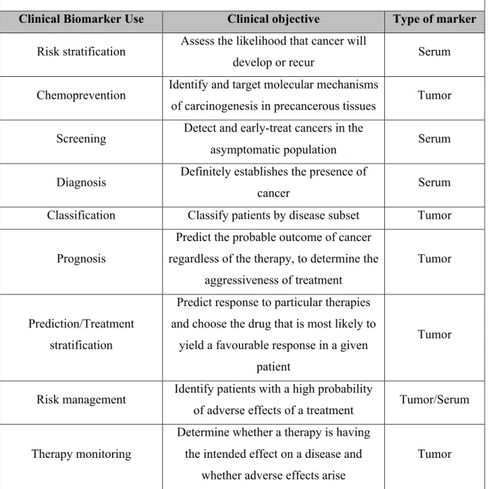
Besides the well-established clinical applications, cancer biomarkers have extended in the last decade into all the major segments of the treatment continuum. Table 4 depicts the full extent of the possible clinical utility of biomarkers according to their type.
Table 4. Use of cancer biomarkers in patient care (40)
Clinical Biomarker Use Clinical objective Type of marker Risk stratification Assess the likelihood that cancer will
develop or recur Serum
Chemoprevention Identify and target molecular mechanisms
of carcinogenesis in precancerous tissues Tumor Screening Detect and early-treat cancers in the
asymptomatic population Serum Diagnosis Definitely establishes the presence of
cancer Serum
Classification Classify patients by disease subset Tumor
Prognosis
Predict the probable outcome of cancer regardless of the therapy, to determine the
aggressiveness of treatment
Tumor
Prediction/Treatment stratification
Predict response to particular therapies and choose the drug that is most likely to
yield a favourable response in a given patient
Tumor
Risk management Identify patients with a high probability
of adverse effects of a treatment Tumor/Serum
Therapy monitoring
Determine whether a therapy is having the intended effect on a disease and
whether adverse effects arise
Post-treatment surveillance Early detection and treatment of recurrent
disease Serum
Target validation Demonstrate that a potential drug target
plays a key role in the disease process Tumor Early compound screening Identify compounds with the most
promise for efficacy and safety Serum Pharmacodynamic assays Determine drug activity; select dose and
schedule Tumor
Patient selection
In clinical trials, patient selection by disease subset or probability or
response//adverse events
Serum
Surrogate end-point
Use of a short-term outcome measure in place of the long-term primary endpoint to determine more quickly whether the treatment is efficacious and safe in drug
regulatory approval
Tumor
Clinical efficacy and analytical robustness are two of the main criteria used in selecting cancer biomarkers for use (16). A biomarker should be specific for its application, be it screening, prediction or therapeutic response monitoring. Overall it should exhibit high sensitivity and specificity with a high area under curve (AUC) in a receiver operating characteristic curve (ROC) (16). It should also be well-defined biochemically with the possibility of detection by reagents such as antibodies (16). The analytical method for measuring the marker should be accurate and precise with no cross-reactivity and should be automated for efficient execution (16, 40). In the following sections, this thesis will summarize the clinical efficacy of cancer biomarkers both in human and veterinary medicine.
2.2. Cancer biomarkers in human medicine
The emergence of evidence-based medicine in the late 19th century compelled human oncology to develop early ideas about premature cancer detection and personalized therapies. Consequently, human medicine already benefits from several FDA (Food and Drug Administration) approved cancer biomarker tests. The tests use quantifiable molecules to diagnose, prognose, monitor cancerous processes and evaluate treatment response (16). It is worth mentioning that, despite obvious progress, biomedical research has not yet been able to develop any reliable biomarker-based tests for early cancer screening.
The published literature reveals that in human medicine biomarkers of interest can be measured in serum or plasma, tumor, in vivo, urine, proximal fluids (pancreatic juices, ascites), exhaled breath, etc. (16). Due to the minor importance of the other sampling sources, this thesis will focus mainly on the utility of serum and tumor markers.
2.2.1. Serum markers in human medicine
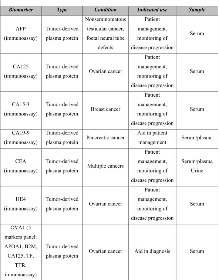
Several serum biomarkers, mostly tumor derived soluble proteins, have been identified and are presently used in clinical settings (16). As aforementioned, their main clinical benefit is the ability to diagnose cancer and monitor its recurrence (16). The molecular biomarkers summarized in the table 5 are approved for clinical practice, thus used in oncological settings. Despite their large-scale usage none is clinically used in veterinary medicine.
Table 5. FDA approved serum biomarkers (16, 43)
Biomarker Type Condition Indicated use Sample
AFP (immunoassay) Tumor-derived plasma protein Nonseminomatous testicular cancer, foetal neural tube
defects Patient management, monitoring of disease progression Serum CA125 (immunoassay) Tumor-derived
plasma protein Ovarian cancer
Patient management, monitoring of disease progression Serum CA15-3 (immunoassay) Tumor-derived
plasma protein Breast cancer
Patient management, monitoring of disease progression Serum CA19-9 (immunoassay) Tumor-derived
plasma protein Pancreatic cancer
Aid in patient
management Serum/plasma
CEA (immunoassay)
Tumor-derived
plasma protein Multiple cancers
Patient management, monitoring of disease progression Serum/plasma Urine HE4 (immunoassay) Tumor-derived
plasma protein Ovarian cancer
Patient management, monitoring of disease progression Serum OVA1 (5 markers panel: APOA1, B2M, CA125, TF, TTR, Tumor-derived
PSA (immunoassay)
Tumor-derived
plasma protein Prostate cancer
Diagnosis, patient management, monitoring of disease progression Serum ROMA (2-Marker panel: CA125, HE4, immunoassay) Tumor-derived
plasma protein Ovarian cancer Diagnosis Serum
Alpha-Fetoprotein
(AFP)
Glycoprotein Nonseminomatous
testicular cancer Staging Serum Human
chorionic
gonadotropin-beta
Glycoprotein Testicular cancer Staging Serum
Thyroglobulin
(Tg) Protein Thyroid cancer Monitoring Serum
Progesterone receptor HER2/NEU
Protein Breast cancer
Monitoring, prognosis, therapy
selection
Serum/Tumor
Increasing research in the field of proteomic cancer profiling will most likely allow the discovery of new serum biomarkers (41). All the same, special attention has to be given to the improvement of present tests. Today’s cancer biomarkers tend to have high sensitivity but low specificity since they do not detect cancer per se but rather a general cellular process intensified during pathological dysplasia (44). In this sense, early cancer detection proves to be a difficult task as numerous pathologies can have as a side effect an increased level of the molecule (41). Furthermore, not all cancer patients express high serum marker levels and
when they do, the cancer is too advanced, making the tests obsolete (41). To complicate matters even more, the majority of the biochemical and biocellular pathways involved in cancer growth are permanently active as part of the normal homeostatic process. Therefore, almost everyone has a trace amount of the interest molecule in their blood, thereby increasing the number of false positive tests (41). As such, it is imperative to remember that although an aggressive investigative approach may be warranted on the basis of raised serum marker values, treatment cannot be initiated without a confirmatory pathological report (44). For the moment, the development of a marker that would prove to be such a precise diagnostic tool is still in its infancy.
2.2.1.1. Specific serum markers for Lymphoma
Generally, the diagnosis of haematological cancers represents a difficult task. The malignant cells have the capacity of arising from different stages of hematopoietic differentiation giving birth to heterogeneous tumors (40). In recent years, several scientific advances have led to a better understanding of the biochemical processes underlying haematological malignancies. As such, potential biomolecular targets, with possible applications in detection, prognosis, and treatment monitoring, have been identified (40). Lymphoma was not deprived of the scientific advancement; several serum biomarkers have been acknowledged in human oncology. Table 6 presents the biomarkers that are of most interest, with the mention that only a small number are presently used in clinical settings mainly due to their low specificity or sensitivity (40).
2.2.2. Tumor markers in human medicine
Cancer is a heterogeneous disease. During the tumorigenic process, different genetic or epigenetic lesions may occur that can result in distinct transcriptome, which is associated with a distinct tumor phenotype (51). Gene expression profiling (GEP) using microarray platforms represent a powerful tool to explore the expression of thousands of gene simultaneously (51, Table 6. Lymphoma serum biomarkers identified in current literature (45-50)
Biomarker Type Condition Indicated use Sample
Serum Deoxythymidine (immunoassay) Protein (enzyme) Non-Hodgkin’s Lymphoma Prognostic and monitor of patients Serum
Serum Ferritin Protein Lymphomas Patient
follow-up Serum Interleukin-2 Receptors Protein Non-Hodgkin’s Lymphoma Prognostic significance for survival Serum Beta-2 Microglobulin Protein Non-Hodgkin’s Lymphoma Prognostic significance for survival Serum CA125 Tumor-derived plasma protein Non-Hodgkin’s Lymphoma Staging, monitoring and follow-up Serum TNF-R1 and CD27 (theoretical stage) Proteins Non-Hodgkin’s Lymphoma Cancer screening (increased future risk of NHL) Serum C-reactive Protein Protein Non-Hodgkin’s
52). In the context of cancer, GEP has been used to accurately classify tumors or define tumor subtypes. The molecular signatures derived from gene expression profiling might have an impact on diagnosis, prognosis, and therapy selection (52).
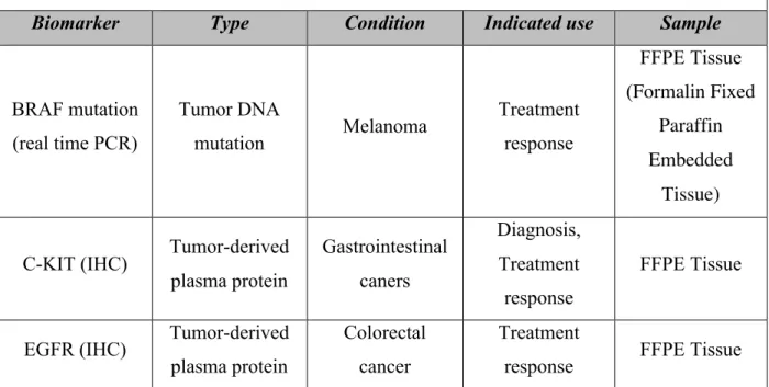
Recently, in human cancer studies, the application of a range of different microarray-based technologies such as, array-microarray-based comparative genomic-hybridization (CGH), cDNA microarray, and methylation array, has increased the understanding of cancer development, and, more importantly, has generated a large number of candidate molecular markers with potential clinical value (40). While several tumor biomarkers have made the object of clinical trials in the recent decade, due to inconclusive results, side effects of personalized treatments, or low efficacy compared to the golden standard, only a small number are present in clinical settings (40). Table 7 presents the FDA approved markers used in human medicine. Similar to serum markers, none of these molecules have applications in veterinary medicine.
Table 7. FDA approved tumor biomarkers (16)
Biomarker Type Condition Indicated use Sample
BRAF mutation (real time PCR) Tumor DNA mutation Melanoma Treatment response FFPE Tissue (Formalin Fixed Paraffin Embedded Tissue)
C-KIT (IHC) Tumor-derived plasma protein Gastrointestinal caners Diagnosis, Treatment response FFPE Tissue
EGFR (IHC) Tumor-derived plasma protein
Colorectal cancer
Treatment
Mammaprint DNA microarray
Gene expression
signature Breast cancer Prognosis Fresh tissue TOP2A (FISH) Chromosomal
aberration Breast cancer Prognosis FFPE Tissue
2.2.2.1. Specific tumor markers for lymphoma
Non-Hodgkin lymphoma (NHL), or more commonly known as the solid tumor of lymphocytic origin, has the highest incidence among hematopoietic cancers in humans (53, 54). It is the fifth most common cancer in North America and its incidence has been increasing over the last three decades (54). NHL comprises a group of clinically and biological diverse diseases, which range from indolent to aggressive clinical course. Although, immunochemotherapy has significantly increased complete remission rates, leading to improved survival, cure rates reach only around 60% (55). Patients that develop resistance to the primary drug regiments have a poor survival, even with subsequent high-dose chemotherapy (55). Moreover, primary refractory patients have a dismal outcome (55). Many new treatment strategies are being explored, some involving targeted molecules (55).
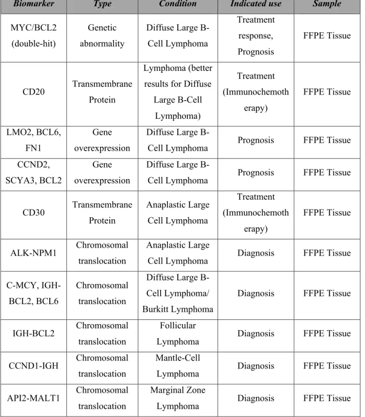
A myriad of tumor markers have been described to help significantly increase survival rates. However, the field is constantly becoming more complex, and few markers have been validated in independent studies and have moved to controlled clinical trials. A PubMed search was performed using the terms “tumor biomarkers”, “lymphoma”, “prognosis”, “outcome”, and “survival”. Priority was given to studies analyzing the biomarkers already in clinical use. Table 8 summarizes the tumor biomarkers identified in the current literature.
Table 8. Tumor biomarkers for lymphoma identified in the current literature (55-59)
Biomarker Type Condition Indicated use Sample
MYC/BCL2 (double-hit) Genetic abnormality Diffuse Large B-Cell Lymphoma Treatment response, Prognosis FFPE Tissue CD20 Transmembrane Protein Lymphoma (better results for Diffuse
Large B-Cell Lymphoma) Treatment (Immunochemoth erapy) FFPE Tissue LMO2, BCL6, FN1 Gene overexpression Diffuse Large
B-Cell Lymphoma Prognosis FFPE Tissue CCND2,
SCYA3, BCL2
Gene overexpression
Diffuse Large
B-Cell Lymphoma Prognosis FFPE Tissue
CD30 Transmembrane Protein Anaplastic Large Cell Lymphoma Treatment (Immunochemoth erapy) FFPE Tissue ALK-NPM1 Chromosomal translocation Anaplastic Large
Cell Lymphoma Diagnosis FFPE Tissue C-MCY, IGH-BCL2, BCL6 Chromosomal translocation Diffuse Large B-Cell Lymphoma/ Burkitt Lymphoma
Diagnosis FFPE Tissue
IGH-BCL2 Chromosomal translocation
Follicular
Lymphoma Diagnosis FFPE Tissue
CCND1-IGH Chromosomal translocation
Mantle-Cell
Lymphoma Diagnosis FFPE Tissue
API2-MALT1 Chromosomal translocation
Marginal Zone
Chapter 3: Cancers biomarkers in veterinary medicine
Although routinely used for screening, diagnosis and prognosis of several human cancers, few cancer biomarkers have been identified in dogs, and none are currently used in veterinary clinical settings (60). Moreover, the use of cancer biomarkers in veterinary medicine has been recently challenged on the basis that the identified biological molecules do not reach the specificity, sensitivity, and accuracy required for routine clinical practice (61). According to Mukaratirwa (61), despite the advances in human oncology, human markers cannot be simply used as multiple species, or “bridging biomarkers” since the molecular etiology of most types of cancers varies from humans to dogs.
Despite this criticism, other authors consider the pet dog population is an ideal model group for many diseases in humans, especially cancers (1, 62). Their large body size and similar metabolic rate make for easy translation of drugs and surgical techniques to the human population (62). Furthermore, they share a living environment with humans and are more closely genetically related to people than are mice (62). Compared to humans, dogs are a relatively inbred population; this may lend itself to successful identification of disease-causing genetic factors that are otherwise obscured in the human population (62). Finally, unlike mouse models, diseases in dogs are spontaneous and more likely to reflect similar human disease (1).
Just like in human medicine, there are several possible uses for cancer biomarkers in helping veterinary clinical management of patients with cancer. The properties of the biomarker and the eventual test used to measure it will dictate which application the marker is suitable for. These include cancer screening, diagnosis, prognosis, prediction of therapeutic response and monitoring effectiveness of therapy, determining tumor recurrence or remission,
or even targeted therapies.
In order to be considered as screening tests in human medicine, biomarkers need to meet two essential requirements: (1) they must be able to detect cancers at a sub-clinical state, and (2) early detection must contribute to the improvement of clinical outcome (63). By these standards, cancer screening in veterinary medicine is far from being a clinical reality. However, the obvious criticism could be opposed by the development of promising biomarker discovery tools such as transcriptomics (the study of RNA transcript expression), metabolomics (the study of metabolites), and proteomics (the study of protein expression) (63). With the help of these large-scale comprehensive studies, veterinary scientists were able to develop new candidate molecules that are presently being evaluated (17). According to preliminary results, the future development of cancer screening tests in veterinary oncology is likely to enhance the prevention of cancer-related deaths and to reduce the morbidity associated with advanced tumors.
A tumour marker used in diagnosis of cancer involves the same considerations as a biomarker for screening. Most markers identified to date in veterinary research, not surprisingly, have either low diagnostic sensitivity or specificity (61). Notwithstanding these limitations, for patients in high-risk groups where prevalence is relatively high, such as dog with lymphoma (27), using a diagnostic marker would aid in deciding whether more invasive tests are required.
Determining the prognosis of a tumour is essential in deciding the course of therapy to be taken. Most tumour markers used in human medicine are correlated with cancer prognosis and other prognostic indications such as tumour grade and staging (16). However, due to the
of action cannot be taken based on the levels of the marker alone. However, the availability of such a biomarker in veterinary clinical practice would obviously help the oncologists to predict and monitor therapeutic response. This determines if the therapy was effective and also determines if there is tumour recurrence, as the biomarker level would be seen to increase again.
Finally, the advantage of actively researching for biomarkers in veterinary oncology, besides the obvious possible improvement in the remission rates and survival of the man’s best friend, resides in the idea that the dog might be a possible animal model for human NHL. In dogs, the tumors seem to progress more rapidly, thus permitting clinical trials to be completed quickly, thereby acting as a bridge to human clinical applications (1). Furthermore, the short remission interval followed by resistance to treatment offers the possibility to test novel antitumoral drugs (1). Many interesting markers currently under investigation have proved efficacious in canine lymphoma (64-66).
The following sections of this thesis will focus on presenting the cancer biomarkers currently identified in the veterinary literature. Although, several publications address the need of advancing the study of cancer biomarkers, for now no ideal maker has been discovered for any canine tumor. As the progress in molecular techniques in oncology advances, so does the understanding of tumor biology, and more cancer biomarkers with better sensitivity and specificity will be found and used in clinical assay.
3.1. Serum markers in veterinary medicine
As in human medicine, measuring cancer biomarkers in bodily fluids has several advantaged such as easier sampling and handling, pain reduction in patients, and non-invasive
detection (17). These serum biomarkers have the potential to be beneficial not only for diagnostic of cancer, but also for monitoring the recurrence of tumors after surgical resection and the effect of anticancer drug therapies (67). Progressively, and with increasing rapidity, these prominent advantages have pushed the scientific community to the search for novel serum markers that could reliable be used in clinical settings. However, due to overlapping results, the data in not compelling enough to warrant routine use of these markers. The potential molecules are still not standardized and expensive for veterinary practice, thereby limited to preclinical exploratory research.
The following table (Table 9) summaries the serum markers mentioned in the veterinary literature. Most of the molecules present elevated serum levels in several cancers, however, their low specificity remains the dispute subject of several articles.
Table 9. Serum markers in veterinary medicine (68-72)
Biomarker Type Condition Potential use Sample
Serum Ferritin Protein Histiocytic
Sarcoma Diagnosis Serum
Alpha1-acid
glycoprotein Protein
Carcinoma, Sarcoma and Round cell tumors
Diagnosis Serum
CK19, ERBB2, CLDN7, ELF3
Gene
overexpression Mammary tumors
Diagnosis, Prognosis,
Treatment response Serum
VEGF Protein Several Aggressive canine tumors Diagnosis Serum Thymidine