HAL Id: dumas-02099993
https://dumas.ccsd.cnrs.fr/dumas-02099993
Submitted on 15 Apr 2019
HAL is a multi-disciplinary open access
archive for the deposit and dissemination of sci-entific research documents, whether they are pub-lished or not. The documents may come from teaching and research institutions in France or abroad, or from public or private research centers.
L’archive ouverte pluridisciplinaire HAL, est destinée au dépôt et à la diffusion de documents scientifiques de niveau recherche, publiés ou non, émanant des établissements d’enseignement et de recherche français ou étrangers, des laboratoires publics ou privés.
Safety and long term efficacy of radiofrequency ablation
for lung tumors
Jean-Baptiste Sautiere
To cite this version:
Jean-Baptiste Sautiere. Safety and long term efficacy of radiofrequency ablation for lung tumors. Human health and pathology. 2018. �dumas-02099993�
UNIVERSITÉ de CAEN NORMANDIE ---
FACULTÉ de MÉDECINE
Année 2017/2018
THÈSE POUR L’OBTENTION
DU GRADE DE DOCTEUR EN MÉDECINE
Présentée et soutenue publiquement le : 18 octobre 2018
par
M SAUTIERE Jean-Baptiste Né le 5 mai 1989 à Croix (Nord)
SAFETY AND LONG TERM EFFICACY OF RADIOFREQUENCY
ABLATION FOR LUNG TUMORS
Président : Monsieur le Professeur PELAGE Jean-Pierre Membres : Monsieur le Professeur BERGOT Emmanuel
Monsieur le Professeur DAO Manh Thông Monsieur Docteur OULKHOUIR Youssef
UNIVER S ITÉ DE CA EN · NOR MA NDIE
UF R S AN TÉ
Année Universitaire 2017 / 2018 Doyen
Professeur Emmanuel TOUZÉ
Assesseurs
Professeur Paul MILLIEZ (pédagogie) Professeur Guy LAUNOY
Professeur Sonia DOLLFUS & Professeur Evelyne Emery (3ème cycle)
Directrice administrative
Madame Sarah CHEMTOB
PROFESSEURS DES UNIVERSITÉS - PRATICIENS HOSPITALIERS
M. AOUBA Achille Médecine interne
M. AGOSTINI Denis Biophysique et médecine nucléaire M. AIDE Nicolas Biophysique et médecine nucléaire M. ALLOUCHE Stéphane Biochimie et biologie moléculaire
M. ALVES Arnaud Chirurgie digestive
M. BABIN Emmanuel Oto-Rhino-Laryngologie
M. BÉNATEAU Hervé Chirurgie maxillo-faciale et stomatologie M. BENOIST Guillaume Gynécologie - Obstétrique
M. BERGER Ludovic Chirurgie vasculaire
M. BERGOT Emmanuel Pneumologie
M. BIBEAU Frédéric Anatomie et cytologie pathologique Mme BRAZO Perrine Psychiatrie d’adultes
M. BROUARD Jacques Pédiatrie
M. BUSTANY Pierre Pharmacologie
Mme CHAPON Françoise Histologie, Embryologie Mme CLIN-GODARD Bénédicte Médecine et santé au travail
M. COQUEREL Antoine Pharmacologie
M. DAO Manh Thông Hépatologie-Gastro-Entérologie M. DAMAJ Ghandi Laurent Hématologie
M. DEFER Gilles Neurologie
M. DELAMILLIEURE Pascal Psychiatrie d’adultes
M. DENISE Pierre Physiologie
M. DERLON Jean-Michel Éméritat jusqu’au 31/08/2018 Neurochirurgie
Mme DOLLFUS Sonia Psychiatrie d'adultes M. DREYFUS Michel Gynécologie - Obstétrique
M. DU CHEYRON Damien Réanimation médicale M. DUHAMEL Jean-François Éméritat jusqu’au 31/08/2018 Pédiatrie
Mme ÉMERY Evelyne Neurochirurgie
M. ESMAIL-BEYGUI Farzin Cardiologie
Mme FAUVET Raffaèle Gynécologie – Obstétrique M. FISCHER Marc-Olivier Anesthésiologie et réanimation M. GÉRARD Jean-Louis Anesthésiologie et réanimation
M. GUILLOIS Bernard Pédiatrie
Mme GUITTET-BAUD Lydia Epidémiologie, économie de la santé et prévention M. HABRAND Jean-Louis Cancérologie option Radiothérapie
M. HAMON Martial Cardiologie
Mme HAMON Michèle Radiologie et imagerie médicale M. HANOUZ Jean-Luc Anesthésiologie et réanimation M. HÉRON Jean-François Éméritat jusqu’au 31/08/2018 Cancérologie
M. HULET Christophe Chirurgie orthopédique et traumatologique M. HURAULT de LIGNY Bruno Éméritat jusqu’au 31/01/2020 Néphrologie
M. ICARD Philippe Chirurgie thoracique et cardio-vasculaire M. JOIN-LAMBERT Olivier Bactériologie - Virologie
Mme JOLY-LOBBEDEZ Florence Cancérologie
Mme KOTTLER Marie-Laure Biochimie et biologie moléculaire M. LAUNOY Guy Epidémiologie, économie de la santé et prévention
M. LE COUTOUR Xavier Epidémiologie, économie de la santé et prévention
Mme LE MAUFF Brigitte Immunologie
M. LEPORRIER Michel Éméritat jusqu’au 31/08/2020 Hématologie
M. LEROY François Rééducation fonctionnelle
M. LOBBEDEZ Thierry Néphrologie
M. MANRIQUE Alain Biophysique et médecine nucléaire
M. MARCÉLLI Christian Rhumatologie
M. MARTINAUD Olivier Neurologie
M. MAUREL Jean Chirurgie générale
M. MILLIEZ Paul Cardiologie
M. MOREAU Sylvain Anatomie/Oto-Rhino-Laryngologie M. MOUTEL Grégoire Médecine légale et droit de la santé
M. NORMAND Hervé Physiologie
M. PELAGE Jean-Pierre Radiologie et imagerie médicale Mme PIQUET Marie-Astrid Nutrition
M. RAVASSE Philippe Chirurgie infantile
M. REZNIK Yves Endocrinologie
M. ROUPIE Eric Thérapeutique
Mme THARIAT Juliette Radiothérapie
M. TILLOU Xavier Urologie
M. TOUZÉ Emmanuel Neurologie
M. TROUSSARD Xavier Hématologie
Mme VABRET Astrid Bactériologie - Virologie
M. VERDON Renaud Maladies infectieuses
Mme VERNEUIL Laurence Dermatologie
M. VIADER Fausto Neurologie
M. VIVIEN Denis Biologie cellulaire
Mme ZALCMAN Emmanuèle Anatomie et cytologie pathologique PROFESSEUR DES UNIVERSITÉS
M. LUET Jacques Éméritat jusqu’au 31/08/2018 Médecine générale
PROFESSEUR ASSOCIÉ DES UNIVERSITÉS A TEMPS PLEIN
M. VABRET François Addictologie
PROFESSEURS ASSOCIÉS DES UNIVERSITÉS A MI-TEMPS
M. de la SAYETTE Vincent Neurologie Mme DOMPMARTIN-BLANCHÈRE Anne Dermatologie
Mme LESCURE Pascale Gériatrie et biologie du vieillissement
M. SABATIER Rémi Cardiologie
PRCE
Année Universitaire 2017 / 2018 Doyen
Professeur Emmanuel TOUZÉ
Assesseurs
Professeur Paul MILLIEZ (pédagogie) Professeur Guy LAUNOY
Professeur Sonia DOLLFUS & Professeur Evelyne Emery (3ème cycle)
Directrice administrative
Madame Sarah CHEMTOB
MAITRES DE CONFERENCES DES UNIVERSITÉS - PRATICIENS HOSPITALIERS M. ALEXANDRE Joachim Pharmacologie clinique
Mme BENHAÏM Annie Biologie cellulaire
M. BESNARD Stéphane Physiologie
Mme BONHOMME Julie Parasitologie et mycologie
M. BOUVIER Nicolas Néphrologie
M. COULBAULT Laurent Biochimie et Biologie moléculaire
M. CREVEUIL Christian Biostatistiques, info. médicale et tech. de communication Mme DEBRUYNE Danièle Éméritat jusqu’au 31/08/2019 Pharmacologie fondamentale
Mme DERLON-BOREL Annie Éméritat jusqu’au 31/08/2020 Hématologie
Mme DINA Julia Bactériologie - Virologie
Mme DUPONT Claire Pédiatrie
M. ÉTARD Olivier Physiologie
M. GABEREL Thomas Neurochirurgie
M. GRUCHY Nicolas Génétique
M. GUÉNOLÉ Fabian sera en MAD à Nice jusqu’au 31/08/18 Pédopsychiatrie
M. HITIER Martin Anatomie - ORL Chirurgie Cervico-faciale M. LANDEMORE Gérard sera en retraite à partir du 01/01/18 Histologie, embryologie, cytogénétique
M. LEGALLOIS Damien Cardiologie
Mme LELONG-BOULOUARD Véronique Pharmacologie fondamentale Mme LEPORRIER Nathalie Éméritat jusqu’au 31/10/2017 Génétique
Mme LEVALLET Guénaëlle Cytologie et Histologie
M. LUBRANO Jean Chirurgie générale
M. MITTRE Hervé Biologie cellulaire
M. REPESSÉ Yohann Hématologie
M. SESBOÜÉ Bruno Physiologie
MAITRES DE CONFERENCES ASSOCIÉS DES UNIVERSITÉS A MI-TEMPS
Mme ABBATE-LERAY Pascale Médecine générale M. COUETTE Pierre-André Médecine générale
M. GRUJARD Philippe Médecine générale
M. LE BAS François Médecine générale
Remerciements
J’adresse mes remerciements aux membres du jury pour avoir accepté d’avoir évalué mon travail.
A mon Président de jury et directeur de thèse, Monsieur le Professeur Jean-Pierre PELAGE. Je vous remercie pour votre aide et votre disponibilité tout au long de ce travail ainsi pour vote bienveillance tout au long des cinq années de mon internat.
A mon juge, Monsieur le Professeur Emmanuel BERGOT. Pour m’avoir fait l’honneur de participer au jury, je vous adresse Monsieur le Professeur mes sincères remerciements. A mon juge, Monsieur le Professeur DAO Manh Thông. Pour m’avoir fait l’honneur de participer au jury, je vous adresse Monsieur le Professeur mes sincères remerciements. A mon juge, Monsieur le Docteur Youssef OULKHOUIR. Pour m’avoir fait l’honneur de participer au jury, je vous adresse Monsieur le Professeur mes sincères remerciements
A Marine, pour ton soutien ces cinq dernières années, ton calme, ton objectivité, tes encouragements, et ta confiance ces six derniers mois et pour les années à venir. Merci. A mes Parents, ma sœur Héloïse mon beau-frère Marco, mon frère Théo pour vos
encouragements et votre soutien tout au long de ces années. Merci.
A ma famille et plus particulièrement à Bruno, Michael et mon grand-père qui m’ont supportés, accompagnés et rassurés tout au long de ces années. Merci.
A la famille de Marine, pour votre gentillesse et votre soutien.
A Axel, pour ta bonne humeur, ton soutien, ta gentillesse, ta disponibilité et ta pédagogie. A Vincent, pour ton soutien, tes conseils, ta sérénité et ta franchise.
A mes amis et particulièrement à Maxime et Adrien, pour votre soutien, votre bonne humeur et votre générosité.
Au Dr Fohlen et à Yassine pour leur aide et leurs conseils avisés.
Aux équipes de manipulateurs et aux secrétaires du service de radiologie du CHU, pour votre gentillesse.
Abréviations
RFA : Radiofrequency ablation MWA : Microwave ablation
SABR: stereotatic ablative radiotherapy SBRT: stereotatic body radiotherapy CT: Computed tomography US: Ultrasonography MR: Magnetic resonance G: Gauge kHz: kilo hertz W: Watt mm: millimeter
Tableaux et figures Figures
Figure 1 : Histological and immune pathological changes in RFA Figure 2: Cockade phenomenon
Figure 3: RFA of lung metastasis of colorectal carcinoma Figure 4: RFA of the longest lesion
Figure 5: Flow diagram showing course of treatment Figure 6: Overall survival after RFA
Figure 7: Overall without recurrence after RFA
Tables:
Table 1: Demographics Table 2: Adverse events Table 3: Imaging follow up
Summary I. Introduction
A. Lung radiofrequency ablation 1
1. Physical principle 1
2. Available technologies 2
3. Ablation procedure 3
4. Histological and immune pathological changes after RFA 3 5. Imaging feature of post ablation zone 4
a. Early phase 4
b. Intermediate phase 6
c. Late phase 6
6. Complication 7
7.Indications 8
B. Others ablation techniques 9
1. Thermoablation technologies 9
a. Microwave ablation 9
b. Cryoablation 9
c. laser induced thermotherapy 10
2. Stereotatic body radiotherapy 10
II. Materials and methods 11
1 Study design 11 2. RFA technique 12 3. Follow up 13 4. Statistical analysis 13 III. Results 14 1. Demographics 14
2. Initial imaging evaluation 14
3. Thermal ablation 15
4. Complications 16
5. Imaging follow up 16
6. Progression 17
a. Pulmonary progression 17
b. Extra pulmonary progression 17
c. Progression summary 18
d. Survival rates 18
IV Discussion 19
1. Complications 19
2. Clinical results 21
3. Follow up and future directions 25
V. Conclusion 27
Tables and figures 28
Bibliography 39
I/ Introduction
Surgical resection is the treatment of choice for early non-small cell lung cancer and for the treatment of lung metastases (1). Depending on the localization of tumor, the number of lesions and the patient comorbidities surgery may not be a valid therapeutic option. Since last decade, thermoablation techniques have been developed in the liver, the kidney and the lungs to treat small and unresectable tumors (2). The purpose of this single center study was to evaluate safety and long-term efficacy of radiofrequency ablation using small 17-Gauge (17-G) needles in the management of primary and secondary lung cancer.
A/ Lung radiofrequency ablation 1/ Physical principle
Radiofrequency is a physical technique used to destroy tumor cells by thermo-ablation (3). An oscillating electric current (in the radio waves frequency, 400-500 kHz) passes between an active electrode (applicator) and a reference electrode (grounding pad). Ionic agitation caused by electron collisions in the tissues adjacent to the electrode tip leads to local frictional tissue heating (Joule effect). The aim of this technique is to produce a temperature exceeding 60°C that will induce irreversible damages on cells. As a consequence, an area of tissue necrosis encompassing both the tumor and a margin of normal surrounding lung parenchyma will be obtained.
The characteristics of lung parenchyma that make thermal ablation so effective are low electrical conductivity and heat insulation (4).
However, the presence of vessels within the ablation zone induces a cooling by convection known as heat-sink effect (5). Gilliams AR and Lees WR reported that presence of vessels greater than 3 mm within the ablation zone was associated with a higher rate of tumor recurrence (6).
2/ Available technologies
As previously described, radiofrequency generator (150-200W), produces an oscillating current (400-500 kHz), which passes between an active electrode and a reference electrode-grounding pad most frequently put on lower limbs of the patient. Actives electrodes are divided in two groups: monopolar electrodes and bipolar electrodes.
In monopolar electrodes energy, scatters centrifugally from the active electrode put to the center of tumor.
Several technical improvements allow an increase in the size of radiofrequency ablation treatment area:
-Cool tip electrode (Covidien, Mansfield, Massachusetts) is an internally cooled electrode. It is a dual-lumen electrode with a non-insulated active tip in which is deliver a continuous perfusion of chilled fluid. This prevents heat accumulation and thereby charring of tissue adjacent to the tip. This cooling of the electrode allows a progressive increase of temperature and thus a size increase of radiofrequency ablation treatment zone.
-Expandable electrodes are electrodes with fine metallic tines that are deployed through a coaxial access needle. This allows an increase of radiofrequency treatment zone.
Several sizes and shapes of electrodes are proposed by different manufactures:
The superslim electrode (Leveen, Boston Scientific) is a small 17-G multitined expandable electrode, a diameter comparable to biopsy needles. The diameter once electrodes are expanded varies between 2 and 3 cm and should then be used to destroy tumors measuring 3-cm or less. Beyond this diameter, there are larger multitined expandable electrode with size ranging from 15-G to 13-G (Leveen CoAcces needle, Boston Scientific), a diameter larger than for biopsy needles. Inside the tumor electrodes tines are deployed in an umbrella like fashion. StarBurst XL and StarBurst Talon (Angiodynamics, Latham, NY) are also expandable devices with Christmas tree like shape once electrode tines are deployed.
With bipolar electrodes, oscillating current does not cross the body but circulates between two parts of the same needle or between two needles inserted in the tumor. This allow not to use a reference electrode (grounding pad) and to increase the size of treatment zone by positioning these two electrodes inside a same tumor
3/ Ablation procedure
CT is the preferred imaging modality for safe guidance even if ultrasonography (US) may be used for superficial pleural lesion (7). New angiography rooms include 3D-CT reconstructions and fusion tool allowing users to perform thermal ablation of large and peripheral nodules without CT (8).
Most centers prefer to use general anaesthesia for thermal ablation but radiofrequency ablation (RFA) can be performed with conscious sedation. General anesthesia is useful in the treatment of lower lobe lesions and diaphragmatic lesions that can be mobile. It is also preferred when a long time of ablation is needed to treat multiple lesions or in case multiple needles need to be inserted to treat a large lesion.
Grounding pad is placed on lower limbs of patient and tumor lesion is targeted with active electrode using fluoro-CT control. If an expandable electrode is used metallic tines are deployed within the lesion after confirmation of the optimal position.
The generator is then activated. Most generators allow evaluation of post-ablation local temperature, time of treatment and impedance. Every manufacturer offers a specific algorithm and instruction for use depending on the type of needle and the size of lesion. During ablation, it is also possible to monitor the temperature inside the needle and inside the ablation zone.
When tissue get charred, impedance of external loads increases. This prevents radiofrequency generator to deliver energy in the tumor. This increase of impedance and decrease of radiofrequency power is called “roll-off”.
At the end of treatment, chest CT is performed to assess the adequacy of the ablation zone and to identify early complications including pneumothorax, alveolar hemorrhage and pleural effusion that may require immediate treatment.
4/ Histologic and immune-pathological changes after RFA
Inside the treated lesion, three areas are classically described (Figure 1) (9).
A central area where the ablation temperature is greater than or equal to 50°C. In this area coagulation necrosis occurs defined by cell membrane collapse, protein denaturation, mitochondrial dysfunction, failure of enzymatic function especially of polymerase function that lead to an interruption of DNA replication.
A peripheral zone surrounds this central area. In this area temperature of treatment is between 41°C and 45°C. This temperature induces sub-lethal and reversible damages in enzymatic and metabolic cell functions. These enzymatic and metabolic cell dysfunctions are not associated with cell death but induce a vulnerability to further injury. In this zone, inflammatory phenomena occur like an increase of the blood flow that may increase the production of free radical and the circulation of liposomal agents (good vector of chemotherapeutic agents). Exposition of damaged tissue markers such as markers of endothelial injury and hyaluronic acid, associated with chemokines and cytokines, first promotes a recruitment of innate immunity (dendritic cells, neutrophilia, antigen-presenting cells, natural killer cells) followed by the recruitment of humoral cells (B and T lymphocytes).
An area of normal tissue surrounds the peripheral zone. In this area “heat sink effect” occurs that causes tissue cooling and induces a decrease of ablation efficiency. Lymphatic drainage of tumor antigens promotes stimulation of dendritic cells and naïve T cells.
5/ Imaging features of the post ablation zone
There is no well-defined criterion to follow the ablation zone. Immediate post treatment CT allows identification of modifications in the ablation area that will change and disappear in the CT follow-up. RECIST 1.1 criteria cannot be used initially to follow the treated lesion. CT performed at one month will be used as the baseline CT since size will increase immediately after ablation (10).
a) Early phase
In the early phase (less than one week after ablation), a cone-shaped or a rim of ground glass opacity may completely or partially surround the targeted lesion. Anderson et al have observed that a thickness of ground glass opacity margin less than 3mm was associated with a higher risk of local recurrence (11). Ground glass opacity margin at least 5 mm around the complete tumor volume is suggested to be the ideal margin after RFA. Intra-lesional bubbles are frequently seen. On the needle tract, increased attenuation, and a lightbulb-shaped opacity may also be seen between the ablated tumor and the skin entry. Thus, immediately after RFA the ablated lesion is larger than the initial tumor (10).
After radiofrequency, the ablated lesion shows a central hypo-attenuating area with reduction of enhancement (12).
Benign peri-ablation rim of enhancement with a size less than 5 mm may be seen. This rim is thick, has a concentric aspect and probably represents inflammatory reaction to radiofrequency. This benign rim must be differentiated from irregular and nodular peripheral enhancement, which represents incompletely ablated tumor or residual tumor.
On immediate post treatment CT, some patients may exhibit a particular pattern of post ablation imaging. It is called the cockade phenomenon and is defined by the presence of multiple concentric rings of varying attenuation surrounding the ablated lesion (Figure 2) (14). Six areas are commonly described:
-In A area, we can find a thin area of charred tissue corresponding to needle tract.
-In B area, we can find a partial emptying due to the vaporization of lesion and coagulative necrotic area with destroyed capillaries and ghost phenomenon (seemingly intact tissue after sudden thermal coagulation (13)).
-In C area, coagulation necrosis is found surrounding the tumor with collapsed alveoli, entrapped air and ghost phenomenon.
-In D area: enzymatic necrosis is observed, associated with blood capillaries destruction, micro thrombosis and lysosomal enzymatic activation.
-In D1 area, micro hemorrhages are found.
-In E area, edema, inflammatory reaction and vascular congestion are found.
These different concentric rings of attenuation seem to correspond to the different areas describe by Miao et al in the MR follow-up and histologic analysis of mice lung tumors ablated by RFA (13).
b) Intermediate phase
In the intermediate phase (between one week and two months after ablation), involution of the ground glass opacity occurs and therefore a nodular aspect of the scar may be present. Cavitation is a common feature found in 24%-31% of treated lesions (15). The ablated lesion is larger than the initial lesion, but its size decreases compare to the diameter found on CT obtained at one month. This decrease can be explained by a regression of parenchymal edema, inflammation and hemorrhage (involution of ground glass opacity). Linear opacities nearby the scar may be found as a result of segmental volume loss. Pleural thickening on the tract of needle is also commonly found. No enhancement of the scar is normally found. An irregular or nodular enhancement is suggestive of residual disease or progression. Nevertheless, like on early CT evaluation, a thin peripheral regular enhancement may be found.
c) Late phase
In the late phase, observed more than two months after treatment, the intra-lesional bubbles have disappeared. Wall thickness and size of cavities tend to decrease. Cavitation may completely disappear and be replaced by an architectural distortion of lung parenchyma. Residual pleural effusion and pleural thickening of tract needle have to totally disappear. Satellite nodules, nodules along needle tract and an irregular or nodular aspect of the scar are indicators of recurrence (10). An increase in size of the ablated zone three months after RFA is to be considered as a local tumor recurrence. In the late phase, RFA scar may have an inferior enhancement compare to the initial tumor, a central enhancement more than 10 mm or more than 15 Hounsfield units (HU) suggests local disease progression or incompletely ablated tumor (12)
6/ Complications
It is well known that the size of the needle used for lung biopsy has an effect on risk of complication like pneumothorax. Kuban et al found that the use of a 19-G coaxial guide needle significantly decreased the risk of pneumothorax and chest tube placement compared with an 18-G coaxial guide needle after computed tomography-guided lung biopsy (16). Not surprisingly, the most common complication reported after radiofrequency ablation is pneumothorax (17). In a retrospective study of de Baere et al 67% of pneumothorax were found (18). No treatment was needed in 28%, single aspiration was made in 14% of pneumothorax cases but 58% of pneumothorax required chest tube placement. Gillams et al defined that the number of tumors, the electrode position and the length of electrode trajectory through aerated lung, impact on the likelihood of pneumothorax (19). Pre-existing emphysema is also an independent risk factor for pneumothorax requiring pleurodesis (20).
Unlike pneumothorax after lung biopsy, pneumothorax after lung radiofrequency treatment may occur or recur late after ablation procedure (12) These are called delayed or recurrent pneumothorax. Yoshimatsu et al describe that contact of ground glass opacity to the pleura was associated with a higher risk of delayed or recurrent pneumothorax (21). The absence of pneumothorax should aways be checked one day after procedure on X rays chest control.
Pleural effusion is a common side effect after radiofrequency procedure. Large pleural effusions needing drain are relatively rare and can be reactive. Kashima et al (20) describe two significant risk factors of aseptic pleuritis: number of pleural punctures and previous systemic chemotherapy. Fortunately, haemothorax is a rare complication and should be suspected in case of circulatory failure and massive pleural effusion.
In a retrospective study of 1,000 lung RFA procedures, an incidence of pneumonia of 1.8% and an incidence of 1.6% of lung abscess were reported (20). Care should be taken to diagnose these infectious complications, indeed abscess like appearance and cavitation might be seen on CT controls several months after RFA procedure.
Hemoptysis is rare even if large parenchymal hemorrhage are found in 7-8% after RFA procedure but are most of the time asymptomatic (22)(23). Pseudo-aneurysm caused by vascular injury are rare events, in a retrospective series of 583 RFA procedures Sakurai J et
al found an incidence of 0.2% of pseudoaneurysm (24). Rupture and massive hemorrhage require urgent catheter angiography and embolization (25).
Other complications including rib fractures, superficial skin burns, intercostal nerve injury and damage to the diaphragm have also been described (26).
7/ Indications
Decision for radiofrequency ablation is always taken during a multidisciplinary tumor board. Gold standard treatment for lung primitive tumors and metastasis remains surgery. Thus, RFA is discussed in case of contraindications to surgery or if patient refused surgery. Radiofrequency is a second intention and curative treatment.
Indications for radiofrequency have been well established: - Lesion less than 3 cm in diameter (at to 4 cm).
- Less than 5 metastasis in the two lungs. Contraindications for radiofrequency include: - Contraindication for general anesthesia. - Low platelet level (< 50,000/mm3).
- Low prothrombin time (<50%) and anticlotting medicines. - Pacemaker.
B/ Other ablation techniques. 1/ Thermo-ablation technologies a/ Microwave ablation
Microwave ablation uses electromagnetic waves in the microwaves spectrum (300MHz to 300GHz). Electromagnetic microwave produce friction between water molecules. The spinning between water molecules interacts with surrounding tissue and causes tissue heating by transfer of kinetic energy (5).
This allows a raise of temperature to 60°C-150°C in a short time leading to coagulation necrosis of the cell. In microwave ablation energy is not transmitted by electrical current thus grounding pad is not necessary.
Microwave enables simultaneous delivery with multiple probe so the ablation zone is larger than RFA. Microwave ablation procedure is made under general anesthesia (4). Microwave electrode size is 13G or 14G.
Lower convective cooling (heat sink effect), because of high work temperature in MWA, has been demonstrated in animal studies (27).
b/ Cryo-ablation
Cryo-ablation uses pressurized argon gas and distributes it to area of lower pressure. This allows the gas to expand and to reach ultracold temperature leading to the formation of iceball (5).
With this iceball, probe is stuck inside the tumor (like with expandable electrode). When temperature is less than -40°C, protein denaturation, cell rupture and tissue ischemia occur, leading to cryogenic cell destruction (5).
Cryoablation allows simultaneous delivery of energy though several probes which can be activated at the time provided a synergistic effect.
Ground glass opacity is not created during cryoablation. Indeed, an ice ball formation at the extremity of cryoablation tip and edematous changes in the surrounding lung parenchyma are used to estimate the ablated margins (4).
c/ Laser-induced thermotherapy
Laser ablation is less developed than the three other techniques.
The physical mechanism of laser induced thermotherapy is temperature elevation within the tumor by laser fiber to induce coagulation necrosis of tumor cells (28). Laser ablation is performed by using neodymium-doped yttrium aluminum garnet laser with the wavelength of 1064nm. Optical fiber is introduce within a 21 gauge needle so there are less complications. Ablation is short because of instant release of laser energy.
2/ Stereotatic body radiotherapy
Stereotatic body radiotherapy (SBRT) also known as stereotatic ablative radiotherapy (SABR) uses a three dimensional coordinate system with internal fiducial or image guidance for tumor tracking (29). Precise adjustment of the focus of the radiation beam to compensate respiratory and unintentional motions of patient or targeted lesion is achieved. The delivery of SBRT generally uses multiple non coplanar arcing fields directed at the radiation target. Thus, the dose gradient is deeper than conventional radiotherapy treatment. Stereotatic body radiation fractionation uses doses ranging from 5 to 33Gy per fraction (29).
Since, radiofrequency ablation is not the treatment of choice for centrally-based tumors and it could be difficult to place needles in small apical tumor, in posteriorly positioned tumor, and in tumor close to scapula, SBRT may be considered as an alternative treatment to treat small solitary lung tumors (30)
II/ Materials and methods
1/ Study design
Retrospective analysis of a prospectively acquired database. This study was approved by our institutional review board and informed consent was obtained from patients.
All patients with primary or secondary lung tumors treated using RFA using 17-G needles between June 2005 to October 2008 at University Hospital Ambroise-Paré Paris, were included to obtain a potential follow-up of more than 10 years.
Indication for RFA was confirmed by a pluridisciplinary tumor board with interventional radiologists, medical oncologist, pulmonologists, surgeons and pathologists involved.
Inclusion criteria for ablation were: proven diagnosis of lung tumor (biopsy or previous history of neoplasm), age greater than 18 years, unilateral or bilateral unresectable primary or secondary lung tumors, less than three tumors in the two lungs, Eastern Cooperative Oncology Group (ECOG) performance status of 0,1 or 2, platelet count greater than 100x109/L and
international normalized ratio of 1.5 or less. Patient with minimal extra-pulmonary metastasis were not excluded.
Exclusion criteria for ablation were: patient considered high risk for ablation because of major comorbidities, more than three tumors in the two lungs, ECOG performance status of more than 2, platelet count less than or equal to 100x109/L, international normalized ratio more than
1.5, pregnant or breast-feeding patient. In addition, all patients treated during the study period using microwave or RFA with needles larger than 17-G were also excluded from analysis.
2/ Radiofrequency ablation technique
All procedures were performed under general anesthesia and intubation.
CT planning was obtained to assess the size of nodules and to select the optimal access route. The electrodes were percutaneously introduced into tumor using CT fluoroscopic guidance. Two types of small 17-G electrodes were used for ablation: a multitined expandable electrode (LeVeen Superslim; Boston Scientific, Nattick MA) and a single internally cooled electrode (Cool Tip; Covidien, Mansfield, Massachusetts). Prophylactic antibiotics were given before and after ablation procedure.
Recommended algorithms provided by the manufactor were used and ablation time and power were recorded.
If tumor ablation was incomplete or others nodules remained untreated a second procedure of RFA was planned.
Fluoro-CT was used to monitor the procedure and identify complications such as: pneumothorax, pleural effusion hemorrhage, every early complication was reported.
Per procedural and post procedural complications were carefully recorded.
Complications were defined using the Common Terminology Criteria for Adverse Event version 5.0.
Any death 30 days after the procedures was considered as a grade 5 adverse event. The grade 3 or 4 adverse event (severe symptoms or life threatening) were considered as major complication. The grade 2 or 1 adverse event (moderate symptom or asymptomatic was considered as minor complication.
Pneumothorax were divided in three groups: minimal which not requiring any treatment, mild treated with simple aspiration and large or clinically poorly-tolerated requiring chest tube placement.
Chest CT was performed at the end of procedure to assess the ablation zone and diagnose complication.
3/ Follow up
Clinical and imaging follow up using whole-body CT were obtained at 3, 6, 12, 15 months after ablation and every 6 months thereafter.
The assessment target tumor response was based on the CT analysis of lesion size and shape. In CT follow-up during first months after RFA, a high density area representing the ablation zone usually larger than the initial tumor is found.
Local tumor progression was defined as enlargement in the overall size of the ablation zone after the first CT post-ablation used as a baseline examination.
When a nodule appeared outside the ablation area it was defined as new pulmonary metastasis corresponding to lung progression. Distant progression in the liver, brain and other organs (except lung) corresponded to disease progression outside lung.
Time of last office visit and death were used to define overall survival (OS). Lung-free survival (at the treated site or distant lung progression) and extra-pulmonary progression-free survival were defined based on CT findings.
4/ Statistical analysis
Continuous variables were expressed as mean ± standard deviation (SD). Median and range were provided when appropriate. Categorial variables were expressed as frequency and percent. Time to progression was measure from date of treatment (radiofrequency ablation) to date of progression, death or last follow-up at the site of ablation, in the lungs or outside the lungs (progression in other organs). Comparisons of means were performed using the Wilcoxon signed-rank test. Overall survival and overall survival without progression were calculated by Kaplan-Meir method. Differences were considered statistically significant if the p value was less than 0.05. Statistical analysis was performed using the SAS 9.4 software (SAS Institute, Cary, NC, USA).
III/ Results
1/ Demographics
Forty-two consecutive patients composed of 17 women (41%) and 25 men (59%) aged 70.7 ± 9.4 (39-77) were included in this study. Thirty-eight (90%) patients were Caucasian and 4 patients (10%) belonged to other ethnic groups.
Nineteen additional patients treated with ablation technique during the study period were excluded: 13 patients were treated with radiofrequency needle with a diameter greater than 17-G and 6 additional patients were treated using microwave ablation.
Five patients (12%) had primary lung carcinoma including adenocarcinoma (n=2) and epidermoid (n=3). Thirty-seven patients (88%) presented with lung metastases. The location of the primary tumor was colorectal in 19 patients (45%), breast in 5 (12%), kidney in 4 (9.5%), head and neck in 4 (9.5%), lung in 2 (5%), miscellaneous in 3 (7%). (Table 1).
Thirteen patients (31%) had previous lung surgery and 26 patients (62%) had prior chemotherapy (Table 1).
Nine patients (21%) had extra-pulmonary metastases. Liver metastases were treated by surgery and RFA whereas brain and bone metastases were treated using radiation therapy.
2/ Initial imaging evaluation
Twenty-nine (69%) patients had only one nodule, 11 (26%) patients had two nodules and 2 (5%) patients had 3 nodules. The diameters of treated nodules were 22.7 ± 6.1 mm (median 24.4 mm; range 11-60 mm) for the first nodule and 17.6 ± 7.2 mm (median 16.0mm; range 8.5-35 mm) for the second nodule respectively.
3/ Thermal ablation
Fifty-one procedures were performed in 42 patients. Fifty-one nodules were ablated during 56 thermal-ablations. Thirty-three patients had one procedure and 9 patients had two procedures for retreatment or presence of a second nodule. LeVeen electrodes were used in 19 procedures (37%) and Cooltip electrodes were used in 32 procedures (63%). The choice of equipment was left at the operator discretion.
The delay between diagnosis and first RFA was 10.2 ± 17.5 months. During the first RFA, LeVeen electrode (2.5-3.5 cm diameter when electrodes are expanded) was used in 16 patients (38%) and Cooltip electrode (with a theorical ablation zone of 3 cm) was used in 26 patients (62%). In the cooltip electrode group, switching system (two electrodes used simultaneously) was used in 4 patients.
The mean duration of the first RFA was 18.6 ± 4.8 min (range 10-38 min) with a maximum power output of 105.3 ± 15.4 W (range 60-80 W).
A second RFA procedure was performed in 9 patients (21%). In 5 patients for incomplete ablation after the first RFA and in four patients to treat additional nodules after the first RFA. Delay between first and second ablation was 7.2 ± 5.6 months. LeVeen electrodes (2.5-3.5 cm) were used in 3 patients and Cooltip electrodes (3 cm) were used in 6 RFA sessions, respectively. In the Cooltip electrode group, switching system was used in 1 patient and three electrodes were inserted to treat a single lesion in one patient. Figure 4.
The mean duration of the second RFA was 25.0 ± 15.4 min (range 10-60 min) with a maximum power output of 112.2 ± 51.1 W (range 40-200 W).
4/ Complications
Pneumothorax was found in 14/42 (33%) patients and in 14/51 (27%) procedures. One (2%) patient had a large volume pneumothorax, rated as CTCAE grade 3, that required chest tube placement and one (2%) patient had mild pneumothorax, rated as CTCAE grade 2, which only required aspiration. A total of 12 (29%) other patients had minor pneumothorax diagnosed using CT but not requiring any treatment (CTCAE grade 1).
Hemoptysis which occurred in 2 (5%) patients was rated CTCAE grade 3 and treated conservatively. One patient had pleural effusion not requiring treatment (CTCAE grade 1). Ground glass opacity and alveolar hemorrhage at the ablation site and along the needle track were found in 16 (38%) patients and in 16 (31%) procedures. These minor complications were rated CTCAE grade 1. Major complications occurred in 3 patients (7%). (Table 2)
5/ Imaging follow-up
The mean and median long diameter of nodules at baseline were respectively 22.7 ± 6.1 mm and 24.4 mm (range 11-60 mm). On the first CT evaluation obtained 2.2 ± 1.8 months after RFA in all patients, the mean long diameter of the lesions showed a significant size increase compare to pretreatment: 31.5 ± 8.3 mm corresponding to an increase of 44% ± 36 (p=0,0001). On the second CT evaluation obtained 6.8 ± 4.4 months after RFA on 41 patients, a significant size increase still noticeable compare to pretreatment values 27.6 ± 11,8 mm (+ 23% ± 30, p=0,0001) was found. On the third CT evaluation obtained 11.9 ± 5.3 months after RFA on 37 patients, no significant difference in size was found compared to pretreatment mean long diameter 22.7 ± 11,9 mm (-2% ± 37, NS). On the fourth CT evaluation obtained 17.0 ± 6.2 months after RFA on 23 patients, a significant decrease in size was found with a mean long diameter of 17.5 ± 8.4 mm (-19 ± 33%, p=0.002).
6/ Progression
a/ Pulmonary progression
After the first RFA pulmonary progression of ablated nodules was found in eight patients (19%), 4 of these patients was previously treated by using LeVeen electrodes (4/16, 25%) and 4 of these patients was previously treated by using Cooltip electrodes (4/26, 15%). The difference between both needles was not significant.
Long diameter of 27.0 ± 7.5 mm vs 21.5 ± 5.1 mm was a predictive factor of progression in univariate analysis (p=0.02).
Second RFA was performed in 5 patients, systemic chemotherapy was performed in 2 patients and 1 patient refuse to have additional treatment.
Figure 5 shows treatments performed after the first RFA.
At the end of follow up, 3 patients had excusive local tumor progression (progression in ablation site) (7.1%), mean time of exclusive local tumor progression was 7.9 ± 2.1 months, 16 patients had pulmonary progression outside site of treatment (38.1%), mean time of pulmonary progression was 21.2 ± 15.6 months. Six patients had both local tumor progression and progression outside RFA treatment site (14.3%), mean time of progression was 32.6 ± 18.8 months.
b/ Extra-pulmonary progression
After 16.1 ± 8.7 months extra pulmonary progression was detected in 8 patients (19%). Six patients had bone progression, including one tract needle seeding, 3 patients presented liver metastasis, 1 patient presented adrenal gland metastasis and one patient had muscle metastasis. In univariate analysis, no predictive factor was identified.
At the end of follow up 25 patients had extrapulmonary progression (59.5%), mean time of extra pulmonary progression was 32.6 ± 26.3 months.
c/ Progression summary
After 16.1 ± 8.7 months, pulmonary progression was found in 22 patients (52%), 8 patients had local tumor progression (19%) and 14 patients had new nodules (33%).
At the end of follow-up, 9 patients had local tumor progression, rate of local tumor progression per patient was 21.4%. Twelve patients did not have progressive disease, thus 30 patients had overall progressive disease (71.4%).
Thirty-two patients (76%) required additional chemotherapy.
d/ Survival rates
After 16.1 ± 8.7 months lung-free and progression free survival was 46% and 41%, respectively.
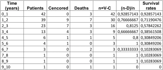
Median and mean OS was 29.8 and 39.5 months (1.8 -154.4), respectively and 1-, 2-, 3-, 4-, 5 and 9- years OS rate were 93%, 71%, 58%, 39%, 31% and 10% respectively (figure 6). Table 5 (appendix) shows evaluation of OS.
Median and mean OS without progression were 25.6 months and 34.9 months (SE= 6.1), respectively. (figure 7)
IV/ Discussion
1/ Complications
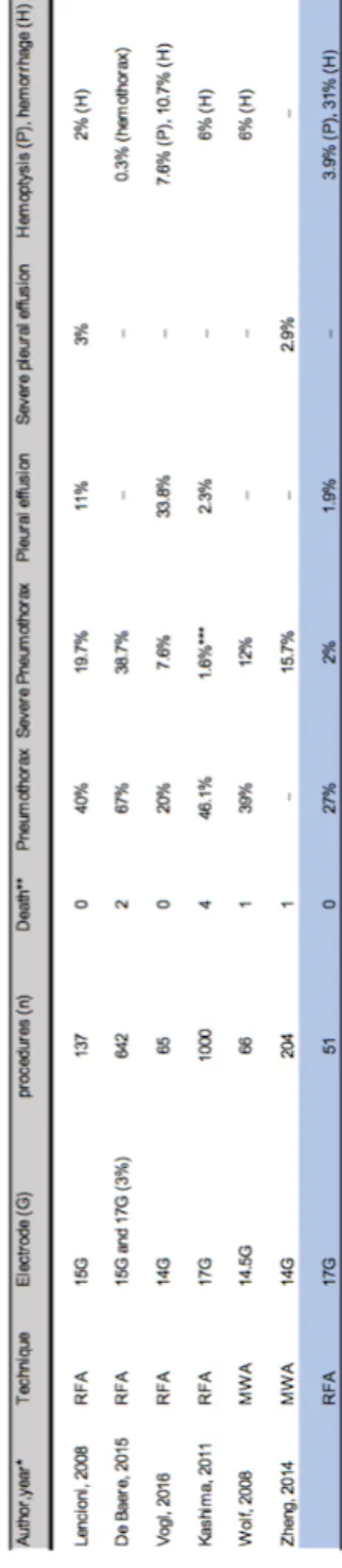
In this study, only used 17-G electrodes were used whereas in the literature most authors do not specify the electrode size. This information seems clinically relevant since the rate of pneumothorax varies between 11% and 67% (31)(18). Moreover, rate of pneumothorax is sometimes expressed in rate per lesion or rate per patient which makes the frequency of this complication difficult to understand.
The rate of pneumothorax per procedure in this study was 27% with 2% of severe pneumothorax requiring treatment. In a prospective study made in 137 RFA procedures using 15-G electrode, Lencioni et al found a rate pneumothorax and severe pneumothorax of 40% and 19.7%, respectively (32). de Baère et al in a retrospective study of 642 RFA procedures using mainly 15-G electrodes (3% of 17-G electrodes) reported a rate of pneumothorax of 67% with 38.7% of severe pneumothorax, much higher than in our series (18). Vogl et al reported their experience using large 14-G electrodes in 65 procedures and found a rate of pneumothorax of 20% with 7.6% of severe pneumothorax (28).
Finally, in a retrospective study on 1,000 RFA procedures using 17-G internally cooled electrode Kashima et al found a pneumothorax rate of 46.1% and a rate of severe pneumothorax (treated with pleurodesis) of 1.6% (20).
As expected, these results tend to prove that the size of electrode have an impact on pneumothorax occurrence during RFA procedure.
The significant difference regarding the rate of pneumothorax between the study by de Baère and our own experience should be taken with care indeed as it may also be explained by the difference of population. In de Baere study there was a high proportion of patients (127/566 corresponding to 22%) with 3 lesions or more whereas in our study only 2/42 patients had 3 lesions (18).
In a retrospective study made on 66 MWA procedures using large 14.5-G electrode in patient with metastases and primary tumors, Wolf et al reported 39% of pneumothorax and 12% of severe pneumothorax (33). In the MWA literature, the rate of pneumothorax varies widely, ranging from 8.5% to 63% (34). These results seem comparable to those reported using RFA. SABR is not associated with a risk of pneumothorax but it requires the placement of fiducials in most cases. In a retrospective study, Trumm et al reported 33.3% of pneumothorax
placement in 105 patients with lung tumors (35). These results are also closely similar to those reported after RFA.
In our study we found 1.9% of pleural effusion similar to Kashima et al who reported a rate of pleural effusion of 2.3% (20). In our study, alveolar hemorrhage occurred in 31% of cases but hemoptysis occurred only in 2 patients (1.9% of procedure). In a meta-analysis based on 32 studies and 8133 transthoracic lung core biopsy, Heerink et al found an occurrence of 18% of lung hemorrhage and 4.1% of hemoptysis after lung biopsy (36). In a retrospective study of 249 CT-guided transthoracic biopsies with 17-G and 18-G needle core, Chassagnon et al found that the concordance between occurrence of significant alveolar hemorrhage and hemoptysis was poor (k= 0.28) (37).
(Table 4)
In our study, one case of tumor seeding (rib metastasis) along the needle tract occurred. This is a serious delayed complication after RFA. Hiraki et al reported two cases out of 661 procedures (0.3%) (38). It is a poorly understood phenomenon because of the paucity of reported cases. The main hypothesis is that the removing of electrode without cauterizing the needle tract may be a risk factor of tumor seeding (38).
2/ Clinical results
For patients with early-stage lung cancer (NSCLC), surgical resection remains the standard treatment and provides the best curative option, with 5 year survival rates of 60 to 80% for stage I NSCLC and 30 to 50% for stage II (39).
Pulmonary metastasectomy has also been described as standard treatment for pulmonary metastases. Criteria for metastasectomy in curative intent has been describeb by Thomford et al in 1965 and with few changes, they remain relevant today (40). Nowadays, the criteria for surgery are as follow: controlled or controllable primary malignancy, absence or controlled or controllable extrathoracic metastasis, all lesions are resectable, adequate remaining respiratory reserve, absence of alternative treatment options with lower morbidity. Only 15 to 25% of patients with pulmonary metastases will be appropriate candidates for surgery (41). In an important publication in 1997 reported from the internal registry of lung metastasectomy of 5,206 patients from Europe and North America, Pastorino et al found that the overall survival after complete metastasectomy was 36% at 5 years (42). Better 5-years survival rate was found for patients with a single metastasis (43%) when compared to those with 2 to 3 metastases (34%) or those with 3 or more metastases (27%) (42).
There is also a wide difference in 5-year survival, depending on the type of cancer. In a systematic review and meta-analysis of 25 studies, including 2,925 patient treated with pulmonary metastasectomy for colorectal cancer, Gonzalez et al, found an overall 5-year survival after complete resection ranging from 27% to 68% (43).
In our study, progression free survival and lung-free survival after 16.1 ± 8.7 months were receptively 41% and 46%. Median and mean OS was 29.8 and 39.5 months, respectively and 1-, 2-, 3-, 4-, 5- and 9 years OS rate were 93%, 71%, 58%, 39%, 31% and 10% respectively. Our results are a little bit different from the largest report on 566 patients made by de Baère et al: median OS was 62 months and OS rates was 92.4%, 79.4%, 67.7%, 58.9% and 51.5% at 1-, 2-, 3-, 4-, and 5 years respectively, rate of local tumor progression per patient were 10.4%, 15.5%, 17.5% and 18.1% at 1, 2, 3 and 4 years respectively (18). Moreover, local tumor progression at the ablation site was associated with poor overall survival with local tumor progression being a time dependent variable.
Several hypotheses may account for our results.
First, patients were carefully selected regarding the number of metastases, most patients presenting with a single lesion.
de Baère reported results obtained in a population with 22% of patients presenting with 3 or more metastases (18). In our study patients, only 2/42 patients had 3 lesions. Even if we had very strict criteria regarding the overall control of metastatic disease at the time of inclusion 31% of our patient had previous lung surgery and 21% had extrapulmonary metastases. In de Baère study 22% of patients had extrapulmonary active diseases amenable to local therapy and rate of patient with previous lung surgery was not indicated (18).
Secondly, we reported a high proportion (45%) of colorectal metastases in our population. Our OS rates are quite similar to those found in the RAPTURE study, Lencioni et al reported an OS of 70% and 48% at 1 and 2 years after RFA in the subgroup of patients with NSCLC, and OS of 89% and 66% at 1 and 2 years after RFA in the subgroup of patients with colorectal metastases (32).
In our study, 62% of patient had prior chemotherapy before RFA and at 16.1 ± 8.7 months, 76% of patients had additional chemotherapy but we did not evaluated the effect of chemotherapy on overall survival after RFA. Chua et al in a retrospective study have demonstrated that the use of adjunctive chemotherapy was an independent predictor for survival after thermo-ablation of colorectal metastases (44).
We found that tumor diameter was predictive of local progression, with significantly larger diameter in patients with local progression (27.0 ± 7.5mm) compared to patients without local progression (vs 21.5 ± 5.1mm; p=0.02). In a prospective study evaluating factors influencing success after RFA in 37 patients with lung metastases, Gilliams and Lee demonstrated that recurrence was significantly more common in tumors larger than 3.5 cm (100%) than in tumors smaller than 3.5 (28%) (6).
In another monocentric retrospective study of 153 patients treated with RFA for pulmonary malignancy, tumor size was a statistically significant predicator of local tumor progression in patients with tumors larger than 3 cm presenting with a median time to progression of 12 months vs patients with size lower than to 3 cm with a median time to progression of 45 months (45).
Microwave ablation in treatment of lung metastases and NSCLC is a thermal ablation technique with a relatively poor literature. There are a high variability of results and methods in MWA studies. Vogl et al reported a 1 and 2 year OS rates of 91.3% and 75% respectively
in a series of 130 ablated metastases (46). Lu et al found a 1-, 2 and 3 year OS rates of 47%, 21% and 14% after the ablation of 37 metastases (47).
Even if MWA technology allows ablation of larger volumes faster and has demonstrated less susceptibility to heat sink effect in animal study, the relatively few and heterogenous results do not provide sufficient data to indicate any superiority of MWA compare to RFA in local control and survival (48).
Treatment of lung oligo-metastatic disease by SABR is based on few phase I/II studies and relatively small retrospective studies (49). One of the largest study analyzed outcome and complications after SABR over 13 years in 321 patients with 587 metastases (50). The overall survival at 1, 3, 5 and 7.5 years were respectively 80%, 39%, 23% and 12%. WHO performance PS (0-1) (HR=0.49; p<0.001), solitary metastasis (HR=0.75; p=0.049), metastasis with size inferior to 30 mm (HR=0.053; p<0.001), metachronous metastases (HR=0.71; p=0.02) and pre SABR chemotherapy (HR=0.59; p<0.001) were independently related to favorable OS. In this study 201/587 lesions were colorectal metastases. A meta-analysis of SABR used in lung oligometastases, based on 18 studies and 1,920 patients found that the local rate of control in patients with pulmonary oligo metastases from colorectal cancer was significantly lower than that in patients with pulmonary oligo metastases from other cancers (OR= 3.10, p<0.00001) (51). Laarhoven et al suggested that the large amount of hypoxic cells in liver metastases from colorectal cancer provides radio resistance compare to metastases from other cancer (52).
The effectiveness of SABR in NSCLC was evaluated by Chang et al in a study that pooled, two independent randomized phase 3 trials which failed to complete accrual (STAR and ROSEL registered with Clinicaltrial.gov), eligible patients were those with clinical T1-2a (<4cm), N0M0, operable NSCLC (49). These patients were randomly assigned in a 1:1 ratio to SABR or lobectomy with lymph node dissection or sampling. A total of 31 patients were assigned to SABR and 27 patients were assigned to surgery. OS at 3 years was 95% in SABR group and 79% in surgery group (HR=0.14, 95% CI: 0.017-1.190; p=0.037), recurrence free survival at 3 years was 86% in SABR group and 80% in surgery group (HR=0.69, 95% CI: 0.21-2.29; p=0.54).
These pooled analysis underwent many criticisms but SABR should be considered has an alternative to surgery in stage I NSCLC for patient with clinically significant morbidities.
Even if SABR is considered as a non-invasive technique some complications such as post radiation toxicity complications may occur. That is why the main limitation of SABR is to treat
several metastases in the same region because of the risk of overlapping of radiation fields and the difficulty to re-treat a recurrent lesion after previous SABR treatment (53).
In NSCLC, surgery remain the standard treatment especially because of lymph node dissection, which provides a survival benefit (39). A possible advantage but not demonstrated of SABR treatment of lung metastases is low dose lymph node irradiation.
3/ Follow up and future direction
One of the major issue in the post ablation follow up is the early detection of recurrence. In the study of de Baere, the follow up CT evaluation only detected 50% of case of local progression during the first year; the rates of local progression per tumor were 5.9%, 8.5%, 10.2% and 11% at 1, 2, 3 and 4 years respectively (18).
As we previously described, there are several CT patterns in the area ablation that vary with time. This induce a real challenge for radiologists to detect the local recurrence.
Apart from the size of the ablation zone, some authors suggest that the evaluation of dynamic enhancement should be used to detect recurrence. To obtain dynamic contrast-enhanced images, CT acquisition is repeated at 45, 90, 180 and 300 seconds after intravenous injection of 100ml of iodinated contrast material at a rate of 2-3mL/second (10). The identification of a nodular or a central enhancement in the ablation area may be an added value to measurements but perfusion CT is associated with increased radiation dose, and thus is not routinely performed in most centers.
Even if no standard imaging protocol for the follow-up of patients treated with ablation for lung tumors has been validated, some authors suggest that PET/CT should be incorporated into the imaging follow-up.
FDG-PET allows a whole-body survey, evaluating metabolic activity of malignant tissues. In a retrospective study Suzawa et al evaluated the diagnostic performance of detection of local tumor progression of FDG PET and CT in 143 patient with 231 tumors treated RFA, local progression was identified in 47 tumors (20.4%): the area under the receiver operating channel curve of PET diagnostic performance was significantly higher than the ROC curve of CT performance at 4 time points; (0.71 vs 0.55 at 3 months, 0.82 vs 0.60 at 6 months, 0.84 vs 0.66 at 9 months and 0.92 vs 0.68 at 12 months) (54).
However, few days after RFA, the peripheral area of treated nodules shows an increase in FDG uptake when PET is obtained because of inflammatory and necrotic changes. Because of inflammation, false-positive results mimicking active tumor and false negative masking tumor foci may be found (55) .In a prospective study evaluating the effectiveness of RFA in 20 patients (24 lesions) with thoracic malignancy, Higuchi et al evaluated tumor response using FDG-PET and CT (56). The FDG-PET results 7-14 days after RFA did not correlate with local recurrence whereas positive findings 3-6 months after RFA were significantly correlated with local recurrence (p=0.0016). PET-CT seems to be an effective tool for assessing treatment response and recurrence and should be included in the strategy of post RFA follow up after 3
months. However, because of the cost and availability, PET-CT is used only in selected patients.
Personalized strategy of therapy using histology, immunohistochemistry and molecular biology is actually a subject of increased interest. Wan et al found that incomplete RFA promoted expression of HSP70/HIF-a in experimental nude mice model with xenograft of cell tumor (57). Combination of RFA and treatment targeting HSP70/HIF-a was associated with a synergistic reduction of tumor growth compare to RFA alone (57). The authors concluded that accelerated proliferation and angiogenesis of lung carcinoma following RFA treatment was induced by HSP70/HIF-a. This study shows theoretical basis for the use of combined therapies for lung cancer treatment.
V/ Conclusion
The use of 17-G needles for radiofrequency ablation of lung nodules is safe with a lower rate of complications than in studies using larger needles. The threshold for complete ablation was 27 mm slightly lower than previously reported. Median OS was 29.8 months, overall survival rates at 1, 2, 3, 4 and 5 years were 93%, 71%, 58%, 39% and 31% respectively. Local recurrence of ablated nodule was higher than expected 19% at 16.1 ± 8.7 months and 21.4%. at the end of follow up. The majority of our patients had extra pulmonary progression 59.5%. These results tend to demonstrate the usefulness of combined treatment strategies. Systemic chemotherapy and ablation are needed to improve overall survival and allow therapeutic windows free of chemotherapy.
Tables and figures
Figure 1
Figure 2
Figure 3
Radiofrequency of a lung metastasis of colorectal carcinoma. Typical evolution of the ablation zone after successful ablation. A Pre interventional aspect of the tumor. B Image obtained just after the ablation. C Image obtained 1 month after ablation showing a size increase of the ablation zone and a regression of ground glass opacity. D image obtained at 3 months, E image obtained at 6 months and F one year after ablation showing a continuous contraction of the ablation zone.
Figure 4
Radiofrequency of the longest lesion. A Pre-interventional CT demonstrates a large tumor (6-cm in long axis) adjacent to the pleura. B CT control during RFA, demonstrates the satisfactory position of the three electrodes inserted inside the lesion. C Successful CT control during the follow up with cavitation of the treated lesion.
Figure 6 Overall survival curve of patients with primary or secondary pulmonary malignancies who received RFA.
Figure 7 Overall survival without progression curve of patient with primary or secondary pulmonary malignancies who received RFA.
Bibliography
1.Hoffman PC, Mauer AM, Vokes EE. Lung cancer. Lancet 2000; 355:479-85
2. Dupuy DE, Zagoria RJ, Akerley W, Mayo-Smith WW, Kavanagh PV, Safran H.
Percutaneous radiofrequency ablation of malignancies in the lung. AJR 2000;174:57-59. 3. Jaskolka JD, Kachura JR, Hwang DM, Tsao MS, Waddell TK, Asch MR, et al. Pathologic assessment of radiofrequency ablation of pulmonary metastases. JVIR 2010; 21:1689-1696. 4. Alexander ES, Dupuy DE. Lung cancer ablation: technologies and techniques. Semin
Interv Radiol 2013; 30:141-150
5. Mouli SK, Kurilova I, Sofocleous CT, Lewandowski RJ. The Role of Percutaneous Image-Guided Thermal Ablation for the Treatment of Pulmonary Malignancies AJR 2017;
209:740-751
6. Gillams AR, Lees WR. Radiofrequency ablation of lung metastases: factors influencing success. Eur Radiol 2008; 18:672-667
7. Ye X, Fan W, Wang H, Wang J, Wang Z, Gu S, et al. Expert consensus workshop report: Guidelines for thermal ablation of primary and metastatic lung tumors (2018 edition). J
Cancer Res Ther 2018; 14:730-744
8. Cazzato RL, Battistuzzi J-B, Catena V, Grasso RF, Zobel BB, Schena E, et al. Cone-Beam Computed Tomography (CBCT) Versus CT in Lung Ablation Procedure: Which is Faster? Cardiovasc Intervent Radiol 2015; 38:1231-1236
9. Chu KF, Dupuy DE. Thermal ablation of tumours: biological mechanisms and advances in therapy. Nat Rev Cancer 2014; 14:199-208
10. Abtin FG, Eradat J, Gutierrez AJ, Lee C, Fishbein MC, Suh RD. Radiofrequency ablation of lung tumors: imaging features of the postablation zone. Radiographics 2012; 32:947-969 11. Anderson EM, Lees WR, Gillams AR. Early indicators of treatment success after
percutaneous radiofrequency of pulmonary tumors. Cardiovasc Intervent Radiol 2009; 32:478-483
12. Suh RD, Wallace AB, Sheehan RE, Heinze SB, Goldin JG. Unresectable pulmonary malignancies: CT-guided percutaneous radiofrequency ablation--preliminary results.
Radiology 2003; 229:821-829
13. Miao Y, Ni Y, Bosmans H, Yu J, Vaninbroukx J, Dymarkowski S, et al. Radiofrequency ablation for eradication of pulmonary tumor in rabbits. J Surg Res 2001; 99:265-271 14. Gadaleta C, Mattioli V, Colucci G, Cramarossa A, Lorusso V, Canniello E, et al.
Radiofrequency ablation of 40 lung neoplasms: preliminary results. AJR 2004; 183:361-368 15. Bojarski JD, Dupuy DE, Mayo-Smith WW. CT imaging findings of pulmonary neoplasms
16. Kuban JD, Tam AL, Huang SY, Ensor JE, Philip AS, Chen GJ, et al. The Effect of Needle Gauge on the Risk of Pneumothorax and Chest Tube Placement After Percutaneous
Computed Tomographic (CT)-Guided Lung Biopsy. Cardiovasc Intervent Radiol 2015; 38:1595-1602
17. Zhu JC, Yan TD, Morris DL. A systematic review of radiofrequency ablation for lung tumors. Ann Surg Oncol 2008; 15:1765-1774
18. de Baère T, Aupérin A, Deschamps F, Chevallier P, Gaubert Y, Boige V, et al.
Radiofrequency ablation is a valid treatment option for lung metastases: experience in 566 patients with 1037 metastases. Ann Oncol 2015; 26:987-991
19. Gillams AR, Lees WR. Analysis of the factors associated with radiofrequency ablation-induced pneumothorax. Clin Radiol 2007; 62:639-644.
20. Kashima M, Yamakado K, Takaki H, Kodama H, Yamada T, Uraki J, et al. Complications after 1000 lung radiofrequency ablation sessions in 420 patients: a single center’s
experiences. AJR 2011; 197:576-580
21. Yoshimatsu R, Yamagami T, Terayama K, Matsumoto T, Miura H, Nishimura T. Delayed and recurrent pneumothorax after radiofrequency ablation of lung tumors. Chest 2009; 135:1002-1009
22. Steinke K, King J, Glenn D, Morris DL. Pulmonary hemorrhage during percutaneous radiofrequency ablation: a more frequent complication than assumed? Interact Cardiovasc
Thorac Surg 2003; 2:462-465
23. Steinke K, King J, Glenn D, Morris DL. Radiologic appearance and complications of percutaneous computed tomography-guided radiofrequency-ablated pulmonary metastases from colorectal carcinoma. J Comput Assist Tomogr 2003; 27:750-757
24. Sakurai J, Mimura H, Gobara H, Hiraki T, Kanazawa S. Pulmonary artery
pseudoaneurysm related to radiofrequency ablation of lung tumor. Cardiovasc Intervent
Radiol 2010; 33:413-416.
25. Yamakado K, Takaki H, Takao M, Murashima S, Kodama H, Kashima M, et al. Massive hemoptysis from pulmonary artery pseudoaneurysm caused by lung radiofrequency ablation: successful treatment by coil embolization. Cardiovasc Intervent Radiol 2010; 33:410-412 26. Smith SL, Jennings PE. Lung radiofrequency and microwave ablation: a review of indications, techniques and post-procedural imaging appearances. Br J Radiol 2015; 88:20140598.
27. Planché O, Teriitehau C, Boudabous S, Robinson JM, Rao P, Deschamps F, et al. In vivo evaluation of lung microwave ablation in a porcine tumor mimic model. Cardiovasc
Intervent Radiol 2013; 36:221-228
28. Vogl TJ, Eckert R, Naguib NNN, Beeres M, Gruber-Rouh T, Nour-Eldin N-EA. Thermal Ablation of Colorectal Lung Metastases: Retrospective Comparison Among Laser-Induced Thermotherapy, Radiofrequency Ablation, and Microwave Ablation. AJR 2016;
207:1340-1349