affiliée à l’Université de Montréal
T
he Short and Long Term Effects of in Vivo Cyclic Axial Compression
A
pplied During Puberty on Bone Growth, Morphometry and Biomechanics
TANVIR MUSTAFY Département de génie mécanique
Thèse présentée en vue de l’obtention du diplôme de Philosophiae Doctor Génie mécanique
Juillet 2019
affiliée à l’Université de Montréal
Cette thèse intitulée :
The Short and Long Term Effects of in Vivo Cyclic Axial Compression
Applied During Puberty on Bone Growth, Morphometry and Biomechanics
présentée par Tanvir MUSTAFY
en vue de l’obtention du diplôme de Philosophiæ Doctor a été dûment acceptée par le jury d’examen constitué de :
Maxime RAISON, président
Isabelle VILLEMURE, membre et directrice de recherche Florina MOLDOVAN, membre et codirectrice de recherche Nicola HAGEMEISTER, membre
DEDICATION
To my inspiring parents and sister, for being the pillows, role models, catapults, cheerleading squad and sounding boards I have needed.
ACKNOWLEDGEMENTS
It pleasures me immensely to recount the people, the figures of flesh and blood, who have helped me through my endeavor, sometimes by just “being a friend or a dedicated ear that listened”, sometimes by helping me technically or philosophically to overcome the hurdles in my research. First and foremost, I would like to acknowledge my incredible supervisor, Professor Isabelle Villemure, who has always been full of inspiration, support and cheer. Her enthusiasm, integral view on research and her mission for providing high-quality work has made a deep impression on me. In the last four and half years of my PhD life, I have learned extensively from her. She taught me how to be meticulous, how to look for answers from a new perspective, how to approach a problem in a systemic way, to make data-driven decision and to exploit serendipity. I feel privileged to be associated with such an exceptionally gifted mentor, scientist, and role model during my PhD. This research work would not have been possible without her patronage.
My special words of thanks should also go to my co-supervisor, Professor Florina Moldovan, for her continuous support, insightful suggestions, and encouragement throughout my project. Her constant guidance and motivation always kept me on the right track. I owe a lot to her for always being there for me.
Next up, I’m highly obliged to Irene Londono for her help with the experimental part of my thesis and for showing me the ropes around the lab at the first few months. Her valuable scientific inputs and friendly nature has supported me immensely during my project. I knew that I could always look back on her for any support and that she would help me with a smile.
I would like to express my gratitude to all present and former members of the Laboratory of Pediatric Mechanobiology (LMP) of Professor Isabelle Villemure and my good friends. Thank you to Anne-Laure Ménard, Rosa Kaviani, Viviane Lalande, Nikita Cobetto, Alejandra Mejia Jaramillo, Yann Zimmermann and Bohao Ning for making my stay in the lab memorable during all these years. I would also like to acknowledge the help from Marie-Eve Richard, Yann Dejoux, Myriam Cliche, William Desjardins, and Jonathan Lacombe during my research project.
I like to extend my thanks to the technical and administrative staff of the Applied Mechanics section of the Mechanical Engineering Department. Special thanks to Mr. Nour Aimene, Mr. Benedict Besner and Mrs. Isabelle Nowlan for their generous support for my experimental set-up.
I thank my friends in Montreal, especially Asif Iqbal, Auria Meher, Sanjida Khuki and Kamrul Islam (Rifat) for their support during my stay here in this beautiful town.
I express my special gratitude to my mother, Tamiza Akter, and my father, Mukter Hussain, for their prayers, unconditional love, support, and encouragement throughout my entire life. I owe my existence to them. Without their love and support, I would have never come this far in my life. I also thank my elder sister Doctor Tania Jebin and my brother in law Doctor Sifullah Bashar, who have inspired me through the tantrum of brainstorming, in those caffeine empowered nights of simulation run and through all the falls and rises I’ve gone through in Canada reminding me what the science fiction writer Jim Butcher once said “When everything goes to hell, the people who stand by you without flinching - they are your family”. Last but not the least I also like to thank my niece, Rushmila, and my nephew, Rayan, who are sent from God to be the bundle of joy in our lives.
I would like to convey my gratitude to Professor Maxime Raison, Professor Nicola Hagemeister, and Professor Bettina Willie for accepting to participate in my Ph.D. Jury.
This project was funded by the Canada Research Chair in Mechanobiology of the Pediatric Musculoskeletal System (I.V.), the Board of Natural Sciences and Engineering Research Council of Canada (NSERC), and the MEDITIS program.
RÉSUMÉ
Pendant la croissance rapide pubertaire, les os sont sensibles aux stimuli mécaniques environnants. Les sollicitations mécaniques telles que les activités physiques fréquentes peuvent contribuer positivement au développement du squelette. Ces activités peuvent entrainer de faibles impacts (comme la marche, la natation, etc.) ou des impacts élevés (comme la course, le saut, etc.) selon la nature de l'activité physique. En plus de la charge mécanique, les facteurs nutritionnels contribuent également de manière significative au développement osseux pendant les périodes de croissance. Les effets des charges d'impact sur la croissance et le développement osseux au cours de l'adolescence ont été investigués par plusieurs chercheurs dans des études animales et cliniques, mais les résultats sont variables. Les recherches antérieures n'ont pas clairement abordé les effets isolés du chargement d'impact par rapport aux facteurs nutritionnels lors de l'évaluation de la croissance et du développement osseux. De plus, on ne sait toujours pas si les effets induits par les charges mécaniques pendant l'adolescence persistent à l'âge adulte et influencent les propriétés structurelles osseuses à long terme.
L'objectif de cette thèse était d'évaluer in vivo les effets des charges cycliques en compression faibles, moyennes et élevées appliquées pendant la puberté sur la croissance osseuse (à maturité squelettique) ainsi que sur la qualité osseuse et la résistance mécanique (à l'âge adulte) en utilisant un modèle animal (tibia de rat). Cet objectif a été atteint en complétant quatre études complémentaires. Tout d'abord, une dose d’exposition aux rayons X à haute résolution mais sécuritaire a été déterminée pour l'imagerie in vivo répétée par micro-CT des tibias de rat en croissance qui n'affecterait pas la croissance ou la microstructure osseuses. Deuxièmement, relation contraintes-déplacements a été établie pour les tibias pendant la période de croissance pubertaire du rat. Un outil de modélisation informatique a également été mis au point et validé pour évaluer les contraintes de l’os sous compression. Troisièmement, les effets de trois niveaux de charges cycliques en compression à la puberté sur la croissance osseuse, la morphométrie et la biomécanique ont été évalués à maturité du squelette. Enfin, les effets de ces trois niveaux de charges cycliques en compression appliquées à la puberté sur la microstructure osseuse ont été évalués à la maturité du squelette et après près d'un an d'entraînement à l'âge adulte.
Dans la première étude, trois doses de rayonnement micro-CT de 0,83, 1,65 et 2,47 Gy ont été testées. Les tibias droits de rats Sprague-Dawley mâles ont été scannés à l'aide d'un protocole de
micro-CT in vivo de leur 4e à leur 12e semaine d'âge avec ces doses de rayonnement. Les tibias gauches ont servi de témoins. Les tibias gauches n'ont été scannés que le dernier jour, avant le sacrifice. Les cellules de la moelle osseuse ont été étudiées, les taux de croissance osseuse et des analyses histomorphométriques de la plaque de croissance ont été évalués et des paramètres mprphométriques osseux ont été déterminés pour les tibias gauches et droits. Les doses de rayonnement de 1,65 et 2,47 Gy se sont avérées néfastes pour les cellules de la moelle osseuse, pour les hauteurs des zones prolifératives et hypertrophiques et pour les taux de croissance osseuse dans les tibias irradiés. De plus, les doses de rayonnement de 1,65 et de 2,47 Gy ont réduit de façon significative la densité minérale osseuse, l'épaisseur trabéculaire, le nombre de trabécules et l'espacement trabéculaire des tibias irradiés. Les doses de rayonnement de 2,47 Gy ont aussi considérablement réduit l'épaisseur corticale. Cependant, le développement osseux et la morphométrie trabéculaire et corticale n'ont pas été affectés pour le groupe de 0,83 Gy. Les doses de rayonnement de 0,83 Gy ont ensuite été utilisées dans le cadre du projet requérant l'imagerie par micro-CT in vivo répétés des rats à l'adolescence, ce qui permet cette dose permettant d'obtenir des images à haute résolution pour des études morphométriques sans nuire à la croissance osseuse. Dans la deuxième étude, trois groupes de rats (âgés de 4, 8 et 12 semaines) ont été utilisés pour établir la relation entre les déplacements tibiaux des rats et les contraintes. Des valeurs cibles de déformation ont été fixées à 450, 850 et 1250 µε, ce qui correspondait aux valeurs maximales de déformation du tibia humain dans des conditions de marche sans restriction (450 µε), de course en zig-zag en montée (850 µε) et de saut vertical (1250 µε). Des jauges de déformation ont été installées à la surface médio-proximale des tibias de rats et des déplacements en compression variant de 0,5 mm à 3,5 mm ont été appliqués de façon cyclique à 2 Hz. Les déplacements et les déformations enregistrés ont été approximées analytiquement par des relations linéaires. Une approche de modélisation par éléments finis, basée sur les données du micro-CT pour établir la géométrie et les propriétés mécaniques, a également été développée, pour les tibias de rats âgés de 4, 8 et 12 semaines, et utilisée pour évaluer les contraintes osseuses dans des conditions expérimentales de charge sous les conditions de chargement expérimentales. Les résultats de simulations ont montré une bonne concordance avec les contraintes expérimentales, ce qui a permis de valider l'approche de modélisation proposée.
Dans la troisième étude, cinq groupes de rats ont été utilisés pour étudier les effets du chargement cyclique en compression à court terme. Les rats, initialement âgés de 4 semaines, étaient divisés en
groupes : témoin, sham, faible impact (LI), impact moyen (MI) et impact élevé (HI). Les tibias droits des groupes LI, MI et HI ont reçu une charge cyclique en compression correspondant à 450, 850 et 1250 µε, respectivement, pendant 5 jours/semaine pour 8 semaines en utilisant un dispositif de compression axiale adapté sur mesure. Les rats ont été sacrifiés à l'âge de 11 semaines. Les paramètres trabéculaires et corticaux de la structure osseuse ont été déterminés par micro-CT, le taux de croissance osseuse par marquage à la calcéine et l'histomorphométrie des plaques de croissance par coloration au bleu de toluidine. Les propriétés mécaniques des os ont été évaluées à partir d'essais de flexion. La charge HI a réduit le poids corporel des rats (12,8 %) et la consommation alimentaire (17 %) par rapport aux shams. Le taux de croissance osseuse a diminué de 6,5 % et de 10,5 %, respectivement, dans les groupes MI et HI. Après 8 semaines de chargement, le groupe HI a montré une augmentation significative de la densité minérale osseuse, de l'épaisseur trabéculaire, de la surface corticale et la surface totale de la diaphyse. La charge ultime et la rigidité ont également augmenté dans les groupes MI et HI comparativement aux shams. Dans l'ensemble, la charge d'impact durant l'adolescence a modérément réduit la croissance osseuse, mais a simultanément amélioré la qualité des os et la biomécanique osseuse à la fin de la période de croissance.
Dans la quatrième étude, cinq groupes de rats (âgés de 4 semaines) ont été utilisés pour étudier les effets à long terme de la charge cyclique en compression à l'adolescence. Les rats ont été divisés en groupes : témoin, sham, faible impact (LI), impact moyen (MI) et impact élevé (HI). Le protocole de mise en charge était similaire à celui de l'expérience à court terme, avec 8 semaines de charge de compression axiale in vivo de 4 à 11 semaines d'âge. Après le chargement, les rats ont été gardés dans leur cage pendant une période d'entraînement de 41 semaines. Ils ont été sacrifiés à l'âge de 52 semaines. Les paramètres trabéculaires et corticaux ont été déterminés par des analyses micro-CT et les propriétés mécaniques osseuses ont été évaluées à partir d'essais de flexion trois points. Des muscles tibiaux ont également été extraits et mesurés après le sacrifice. Des modèles par éléments finis des tibias de rats âgés de 52 semaines ont été générés pour évaluer la distribution des contraintes osseuses trabéculaires et corticales sous compression. Les charges HI et MI ont réduit le poids corporel et l'apport alimentaire à la fin de l'adolescence et au début de la période post-pubertaire. Toutefois, le poids corporel et l'apport alimentaire sont finalement revenus à un niveau semblable à celui des shams à 52 semaines. Les charges HI et MI ont augmenté la surface corticale, l'épaisseur et la surface totale de la diaphyse tibiale, ainsi que la densité minérale osseuse,
l'épaisseur trabéculaire et la fraction du volume osseux au niveau des tibias proximaux. Les bienfaits de la charge sur la zone corticale, l'épaisseur et la densité minérale osseuse ont été perdus après 41 semaines d'entraînement à l'âge adulte. Cependant, les avantages induits par la charge sur la résistance mécanique des os ont persisté jusqu'à 52 semaines. Dans l'ensemble, la charge cyclique en compression de niveau élevé effectué pendant l'adolescence a entrainé des bienfaits à long terme pour la structure et la force des os.
Les limites de ce projet de recherche comprennent l'utilisation de tibias de rat mâle seulement pour l'ensemble du projet. L'utilisation de sujets masculins et féminins aurait pu fournir des informations supplémentaires sur les changements osseux associés à la charge, en fonction du sexe. Une autre limitation possible est l'utilisation des résultats mesurés par micro-CT comme indicateur de la qualité de l'os trabéculaire. La charge cyclique en compression de niveau élevé a augmenté les paramètres morphométriques trabéculaires dans notre étude. Aucun essai mécanique en relation à la géométrie trabéculaire possiblement modifiée, qui se traduirait par un tissu trabéculaire plus fort ou non, n'a été vérifiée. L'utilisation d'un modèle de rat est également associée à une limitation de la capacité de résorption de l'os cortical au cours d'une période d'entraînement. Ceci est dû à l'absence de remodelage secondaire des canaux de Haversian dans ce modèle animal; par conséquent, les rats continuent à se développer jusqu'à relativement tard dans la vie. Cependant, des études antérieures ont observé un remodelage osseux dans la structure osseuse corticale du rat adulte en réponse au chargement mécanique. De plus, l'étude des propriétés morphométriques trabéculaires uniquement à partir de la métaphyse proximale des tibias peut être considérée comme une limitation de cette étude. L'inclusion de résultats supplémentaires provenant de la partie distale aurait pu fournir des informations plus détaillées sur les changements induits par la charge dans l'ensemble de la structure tibiale.
Ce projet présente d'importantes contributions méthodologiques et scientifiques. Une nouvelle méthodologie basée sur la micro-CT a été développée pour segmenter la microstructure osseuse trabéculaire et corticale et évaluer la morphologie osseuse sans interférer avec la croissance osseuse et la santé osseuse globale. Un outil d'éléments finis basé sur le micro-CT a également été développé pour l'évaluation non destructive de la mécanique osseuse. À l'aide d'un protocole de chargement finement contrôlé et calibré en fonction de la déformation, cette recherche a permis de surmonter les limites actuelles des études cliniques ou expérimentales visant à établir les effets de la charge cyclique en compression sur la croissance osseuse et a donc fourni des résultats pertinents
: une charge cyclique en compression de niveau élevé pendant la puberté nuit modérément à la croissance osseuse, mais elle favorise la solidité et la mécanique globale des os à maturité et ces avantages persistent à l'âge adulte. D'un point de vue clinique, les résultats de ce projet pourraient mener à des recommandations pour des programmes d'entraînement prescrits aux jeunes athlètes adolescents pratiquant des sports à impact élevé.
ABSTRACT
During the adolescent period, rapidly growing bones react sensitively to induced mechanical stimuli. Mechanical loadings such as daily physical activities can positively contribute to skeletal development. They can range from low impact activities (such as walking, swimming, etc.) to high impact activities (such as running, jumping, etc.) depending on the nature of the physical activity. Along with mechanical loading, nutritional factors also significantly contribute to bone development during growth periods. The effects of impact loadings on bone growth and development during the adolescent period have been investigated by several researchers in animal and clinical studies, but results are inconsistent. Previous research has not clearly addressed the isolated effects of impact loading from nutritional factors while evaluating bone growth and development. Moreover, it is still unclear whether the effects induced by mechanical loading during adolescence remains in adulthood and influence bone structural properties in the long term. The objective of this thesis was to evaluate the effects of in vivo low, medium, and high cyclic axial compression applied during puberty on bone growth (at skeletal maturity) as well as on bone quality and mechanical strength (at adulthood) using an animal (rat tibia) model. This objective was achieved by conducting four interconnected studies. First, a safe yet high-resolution effective radiation dose was determined for repeated in vivo micro-CT scanning of the growing rat tibiae, which would not affect bone growth or microstructure. Secondly, rat tibial displacements vs strains relationships were established for the adolescent growing period via a strain calibration study. A computational modeling tool was also developed and validated for assessing bone strains under compressive loading. Thirdly, the effects of three different cyclic axial compression during puberty on bone growth, morphometry and biomechanics were assessed at skeletal maturity. Finally, the effects of three different cyclic axial compression during puberty on bone microstructure were assessed both at skeletal maturity and after almost a year of detraining period at adulthood.
In the first study, three micro-CT radiation doses of 0.83, 1.65 and 2.47 Gy were tested. The right tibiae of male Sprague-Dawley rats were scanned using an in vivo micro-CT scanning protocol from their 4th to 12th week of age with these radiation doses. The left tibiae were used as control. The left tibiae were only scanned on the last day, before sacrifice. Bone marrow cells were investigated, bone growth rates and histomorphometric analyses were performed, and bone structural parameters were determined for both left and right tibiae. Radiation doses of 1.65 and
2.47 Gy were found to negatively affect bone marrow cells, proliferative and hypertrophic zone heights, and bone growth rates in the irradiated tibiae. Also, both 1.65 and 2.47 Gy radiation doses significantly reduced bone mineral density, trabecular thickness, trabecular number and significantly increased trabecular spacing in the irradiated tibiae. Moreover, 2.47 Gy radiation doses significantly reduced cortical thickness during the scanning period. However, bone development and morphometry remained unaffected for the 0.83 Gy group. The 0.83 Gy radiation doses was then considered for further use in the project for repeated in vivo scanning of rats in the adolescent period, which would provide high-resolution images for morphometric investigation without interfering with bone growth.
In the second study, three groups of rats (4, 8 and 12 week old) were used for establishing rat tibial displacements vs. strains relationship. Target strain magnitudes were 450, 850, and 1250 µε, which corresponded to peak strain values in the human tibia during unrestricted walking (450 µε), zig-zag uphill running (850 µε), and vertical jumping (1250 µε) conditions. Strain gauges were installed at the medio-proximal surface of the rat tibiae and compressive displacements ranging from 0.5 mm to 3.5 mm were applied in a Haversine form at 2Hz frequency. The recorded displacements and strains were plotted in linear fit graphs to establish the required relationships. A micro-CT based finite element modeling approach was also developed, using 4, 8 and 12 week old rat tibiae, and applied to assess bone strains under experimental loading conditions. Computational results showed good agreement with experimental strains in the correlation analyses and Bland Altman analyses, which allowed validating the proposed modeling approach.
In the third study, five groups of rats were used to investigate the effects of cyclic axial compression in the short term period. The rats, initially aged 4 week old, were divided in control, sham, low impact (LI), medium impact (MI), and high impact (HI) groups. The right tibiae of LI, MI, and HI groups received cyclic axial compression corresponding to 450, 850, and 1250 µε, respectively, for 5 days/week and for 8 weeks using a custom made axial compression loading device. Rats were sacrificed at 11 week of age. Trabecular and cortical bone structural parameters were determined by micro-CT, bone growth rate by calcein labeling and histomorphometry of growth plates by toluidine blue staining. Bone mechanical properties were evaluated from bending tests. HI loading reduced rat body weight (12.8%) and food consumption (17%) compared to shams. Bone growth rate was decreased in MI and HI groups by 6.5% and 10.5%, respectively. After 8 weeks of loading, HI group showed a significant increase in bone mineral density, trabecular thickness, cortical and
total surface area. Ultimate load and stiffness were also increased in MI and HI groups compared to shams. Overall, impact loading during adolescence moderately reduced bone growth, but simultaneously improved bone quality and biomechanics at the end of the growing period.
In the fourth study, five groups of rats (aged 4 week old) were used for investigating the effects of cyclic axial compression during adolescence in the long term period. Rats were divided into control, sham, low impact (LI), medium impact (MI), and high impact (HI) groups. The loading protocol was similar to the short term experiment, with 8 weeks of in vivo axial compressive loading from 4 to 11 week of age. After the loading regime, rats were kept in their cage for a detraining period of 41 weeks. At the age of 52 week old, they were sacrificed. Trabecular and cortical parameters were determined by micro-CT analyses and bone mechanical properties were evaluated from three-point bending tests. Selective muscles were also extracted and measured after the sacrifice. Finite element models of the 52 week old rat tibiae were generated for assessing the distributions of trabecular and cortical bone strains under compressive loading. HI and MI loadings reduced body weight and food intake at the end of the adolescence period and at the beginning of post-pubertal period. However, both body weights and food intakes eventually returned to a level similar to shams at maturity. HI and MI loadings increased cortical area, thickness and total area at the mid-shaft, and bone mineral density, trabecular thickness and bone volume fraction at the proximal tibiae. Cortical area, thickness, and bone mineral density benefits were lost by 41 weeks of detraining period at adulthood. However, loading-induced benefits on bone mechanical strength persisted at adulthood. Overall, high impact exercise performed during adolescence could provide long-term benefits for bone structure and strength.
Limitations of this research project include using only male rat tibiae for the entire project. Using both male and female subjects could have provided additional sex-dependent insights into bone changes associated with loading. Also, another possible limitation is the use of micro-CT measured outcomes as an indicator for trabecular bone quality. High impact loading increased trabecular morphometric parameters in our study. But if the modified trabecular geometry translates to stronger trabecular tissue or not has not been verified by mechanical testing. The use of a rat model is also associated with a limitation for cortical bone ability of resorption in a detraining period. This is caused due to the lack of secondary remodeling of Haversian canals, hence rats continue to grow until relatively late in life. However, previous studies observed bone remodeling in cortical bone structure of adult rat under mechanical loading conditions. Also, investigating the trabecular
morphometric properties only from the proximal metaphysis of the tibiae can be considered a limitation of this study. Including additional results from the distal part would have provided more detailed information about the loading induced changes throughout the entire tibial structure. This project presents significant methodological and scientific contributions. A novel micro-CT based methodology was developed for segmenting trabecular and cortical bone microstructure and assessing the bone morphology without interfering with the bone growth and overall bone health. A micro-CT based finite element tool was also developed for non-destructively evaluating bone mechanics. Using a finely controlled and strain-calibrated loading protocol, this research has overcome current limitations of clinical or experimental studies trying to establish the effects of impact loading on bone growth and hence provided essential findings: high cyclic axial compression performed during puberty is moderately detrimental to bone growth, but benefits overall bone strength and mechanics at skeletal maturity, with these benefits persisting up to adulthood. On a clinical point of view, results of this project could lead to recommendations for training programs prescribed to young adolescent athletes involved in high impact sports.
TABLE OF CONTENTS
DEDICATION ... III ACKNOWLEDGEMENTS ... IV RÉSUMÉ ... VI ABSTRACT ... XI TABLE OF CONTENTS ... XV LIST OF TABLES ... XXIII LIST OF FIGURES ... XXV LIST OF SYMBOLS AND ABBREVIATIONS ... XXXIII LIST OF APPENDICES ... XXXIVINTRODUCTION ... 1
LITERATURE REVIEW ... 4
2.1 Skeletal system ... 4
2.2 Bone Structure and Composition ... 5
2.2.1 Bone Structure ... 5
2.2.2 Bone Composition ... 6
2.3 Bone Growth and Remodeling ... 7
2.3.1 Bone Growth ... 7
2.3.2 Bone Remodeling ... 13
2.4 Bone biomechanics ... 16
2.4.1 Mechanical testing and mechanical properties ... 16
2.5 Mechanical loading of bone during adolescence ... 25
2.5.1 Clinical Studies... 25
2.6 Imaging techniques of bone geometry and microarchitecture ... 33
2.6.1 Quantitative CT ... 33
2.6.2 High-resolution Peripheral QCT (Preclinical microCT) ... 34
2.6.3 Micro-CT ... 36
2.7 Finite element (FE) modeling of bone ... 39
2.7.1 Hierarchical levels of bone for FE modeling ... 39
2.7.2 Macroscale: Whole bone FE modeling ... 41
PROJECT RATIONALE, RESEARCH QUESTIONS AND OBJECTIVES .. 46
3.1 Rationale ... 46
3.2 Research Questions ... 47
3.3 Objectives ... 47
ARTICLE #1: CAN REPEATED IN VIVO MICRO-CT IRRADIATION DURING ADOLESCENCE ALTER BONE MICROSTRUCTURE, HISTOMORPHOMETRY AND LONGITUDINAL GROWTH IN A RODENT MODEL? ... 52
4.1 Abstract ... 53
4.2 Keywords ... 53
4.3 Introduction ... 53
4.4 Materials and methods ... 56
4.4.1 Animals ... 56
4.4.2 Repeated micro-CT scanning ... 56
4.4.3 Calcein injections ... 59
4.4.4 Bone marrow cell assessment... 60
4.4.5 Tissue processing ... 60
4.4.6 Bone growth rate ... 60
4.4.8 Trabecular and cortical bone morphometry ... 64
4.4.9 Statistical analysis ... 65
4.5 Results ... 66
4.5.1 Bone growth rate ... 66
4.5.2 Growth plate histomorphometry ... 66
4.5.3 Trabecular and cortical bone morphometry: comparative analysis at the 14th week ... ... 67
4.5.4 Trabecular and cortical bone morphometry: 9-week longitudinal comparative analysis ... 70
4.5.5 Body weight ... 70
4.5.6 Bone marrow cells ... 74
4.6 Discussion ... 75
4.6.1 Radiation doses of 1.65 and 2.47 Gy adversely impacted tibial bone development during the adolescent growth period ... 76
4.6.2 Trabecular bone, together with bone marrow cells, were negatively affected when undergoing repeated radiation doses of 1.65 and 2.47 Gy ... 77
4.6.3 Cortical bone quantity and microstructure were slightly deteriorated under repeated radiation dose of 2.47 Gy ... 80
4.6.4 Comparisons among protocols used in similar radiation studies and strengths of the current study ... 81
4.7 Conclusion ... 83
4.8 Acknowledgements ... 83
4.9 References ... 83
ARTICLE #2: EXPERIMENTAL AND FINITE ELEMENT ANALYSES OF BONE STRAINS IN THE GROWING RAT TIBIA INDUCED BY IN VIVO AXIAL COMPRESSION ... 91
5.1 Abstract ... 92
5.2 Keywords ... 92
5.3 Introduction ... 93
5.4 Materials and methods ... 95
5.4.1 Animals ... 95
5.4.2 Strain gauge measurements ... 95
5.4.3 Micro-CT imaging ... 96
5.4.4 Voxel based finite element analysis ... 97
5.4.5 Strains evaluation ... 100
5.4.6 Cortical bone structural parameters ... 103
5.4.7 Statistical analyses ... 104
5.5 Results ... 105
5.5.1 Mesh convergence results ... 105
5.5.2 Strain analyses ... 106
5.6 Discussion ... 109
5.6.1 Predicted FE modeling strains and experimental strains showed strong agreements across the three rat age groups ... 109
5.6.2 Under compression, the greatest longitudinal strains develop in the PL region, compared to the AM and AL regions ... 110
5.6.3 Under similar compression, strains decrease with age during the adolescent period .... ... 110
5.6.4 Changes in strain gauge placement along anterior-posterior direction has greater impact on the resulting FE modeling strains compared to changes along the proximal-distal direction ... 111
5.7 Conclusion ... 113
5.8 Acknowledgements ... 113
5.9 References ... 113
ARTICLE #3: HIGH IMPACT EXERCISE IMPROVES BONE MICROSTRUCTURE AND STRENGTH IN GROWING RATS ... 122
6.1 Abstract ... 123
6.2 Keywords ... 123
6.3 Introduction ... 123
6.4 Results ... 125
6.4.1 Body Weight and Food Intake ... 125
6.4.2 Bone Growth Rate and Tibial Length ... 125
6.4.3 Growth Plate Histomorphometry ... 126
6.4.4 Trabecular and Cortical Bone Architecture ... 129
6.4.5 Tibial Mechanical Properties ... 129
6.5 Discussion ... 133
6.5.1 High impact loading triggers decreased body weight coupled with a reduced caloric consumption ... 133
6.5.2 Medium and high impact loadings decrease longitudinal bone growth despite developing thicker HZ and PZ heights ... 134
6.5.3 Changes in trabecular bone microstructure are time as well as impact level dependent ... 135
6.5.4 Medium and high impact loadings benefit the cortical bone morphometry in the diaphysis, leading to significantly improved structural- and tissue-level bending mechanical properties ... 137
6.5.5 Limitations... 138
6.7 Materials and methods ... 140
6.7.1 Animals ... 140
6.7.2 In vivo Axial Tibial Loading ... 140
6.7.3 Strain Calibrated Impact Loading ... 141
6.7.4 Micro Computed Tomography (micro-CT) ... 143
6.7.5 Mechanical Testing ... 145
6.7.6 Bone growth rate assessment ... 146
6.7.7 Growth Plate Histomorphometry ... 147
6.7.8 Statistical Analysis ... 147
6.8 Acknowledgements ... 148
6.9 References ... 148
ARTICLE #4: IMPACT EXERCISE DURING ADOLESCENCE IMPROVES BONE MICROSTRUCTURE AND STRENGTH AT ADULTHOOD ... 158
7.1 Abstract ... 159
7.2 Keywords ... 159
7.3 Introduction ... 159
7.4 Materials and methods ... 162
7.4.1 Animals ... 162
7.4.2 Tibial Impact Loading ... 162
7.4.3 Micro-Computed Tomography (micro-CT) ... 164
7.4.4 Ex vivo muscle weight measurements ... 167
7.4.5 Mechanical Testing ... 167
7.4.6 Finite element (FE) analysis ... 168
7.4.7 Statistical Analysis ... 169
7.5.1 Animals ... 169
7.5.2 Body Weight and Food Intake ... 170
7.5.3 Long-Term Effects of Loading during Adolescence on Trabecular Bone Architecture ... 171
7.5.4 Long-Term Effects of Loading during Adolescence on Cortical Bone Architecture .... ... 174
7.5.5 Muscle Weight ... 174
7.5.6 Mechanical Properties of Tibia ... 174
7.5.7 Finite Element Analysis of Tibia... 175
7.6 Discussion ... 178
7.6.1 Impact loading temporarily reduced body weight and food intake at the rat puberty/adult transition period ... 178
7.6.2 High impact loading induced enhanced trabecular bone at the end of puberty, which was maintained during the adulthood detraining period ... 180
7.6.3 High impact loading induced positive changes in cortical bone microstructure at the end of puberty, which partly remained during the adulthood detraining period ... 181
7.6.4 Strengths and limitations ... 184
7.7 Conclusion ... 185
7.8 Acknowledgments ... 185
7.9 References ... 185
GENERAL DISCUSSION ... 197
8.1 Cyclic axial compression (impact) loading ... 197
8.1.1 Cyclic axial compression loading parameters ... 197
8.1.2 Training and detraining periods ... 198
8.1.4 Right tibia for cyclic axial compression loading ... 200 8.1.5 Bone segmentation thresholds ... 200 8.2 General limitations ... 200 8.2.1 In vivo experimental protocol ... 201 8.2.2 Rat tibial model ... 201 CONTRIBUTIONS TO MECHANICAL ENGINEERING ... 203 CONCLUSION AND RECOMMENDATIONS ... 204 10.1 Conclusion ... 204 10.2 Recommendations for future studies ... 205 REFERENCES ... 207 APPENDICES ... 225
LIST OF TABLES
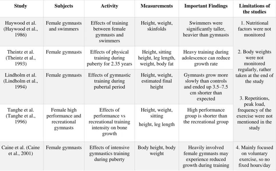
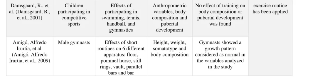
Table 2.1 Clinical studies investigating the effect of loading on growing bones (short term period) ... 26 Table 2.2 Clinical studies investigating the effect of loading on growing bones (long term period) ... 28 Table 2.3 Experimental animal studies investigating the effect of loading on growing bones (short
term period) ... 31 Table 2.4 Summary of previous experimental studies on the effect of loading on growing bones
(long term period) ... 32 Table 4.1 Image acquisition and reconstruction parameters of the rat proximal tibiae for the three
doses groups ... 59 Table 4.2 Longitudinal assessment of trabecular microarchitecture of the right proximal tibial
metaphysis in three doses groups of rats. ... 72 Table 4.3 Longitudinal assessment of cortical microarchitecture of the right proximal tibial
metaphysis in three doses groups of rats. ... 73 Table 4.4 ANOVA test with Tukey’s multiple comparisons for the trabecular and cortical bone
structural properties of the irradiated rat tibiae for three radiation groups on the 14th week. 74 Table 4.5 Percentage of unaffected bone marrow cells for 0.83, 1.65 and 2.47 Gy radiation groups
extracted from trypan blue test (mean value ± SD). ... 75 Table 5.1 Results from Bland-Altman test between experimental and FE modeling strain results
for rat tibiae of three age groups. ... 103 Table 5.2 Cortical bone structural parameters for rat tibiae (mean ± SD). ... 108 Table 6.1 ANOVA test with Tukey’s multiple comparisons for the trabecular microarchitecture of
the right proximal tibial metaphysis in control, sham, LI, MI and HI groups of rats after 4 weeks and 8 weeks of loading regime. ... 130
Table 6.2 ANOVA test with Tukey’s multiple comparisons for the cortical microarchitecture of the right tibial mid-diaphysis in control, sham, LI, MI and HI groups of rats after 4 weeks and 8 weeks of loading regime. ... 131 Table 6.3 ANOVA test with Tukey’s multiple comparisons for structural and intrinsic mechanical
properties of the right tibiae from control, sham, LI, MI and HI groups of rats derived from three-point bending tests of the mid-diaphysis. ... 132 Table 7.1 Muscle weights (g) for control, sham, LI, MI and HI groups evaluated at the end of
experiment. ... 175 Table 7.2 Structural and intrinsic mechanical properties of the right tibiae for control, sham, LI, MI
LIST OF FIGURES
Figure 2.1 Functions of the skeletal system (Adapted from Anatomy and Physiology, Oregon State University, 2019) ... 4 Figure 2.2 Anatomy of a typical long bone (Adapted from Anatomy and Physiology, Rice
University, 2013). ... 5 Figure 2.3 (a) Structure of cortical (compact) bone; (b) Structure of trabecular bone. (Iannotti &
Parker, 2013) ... 7 Figure 2.4 Intramembranous ossification comprises four steps: (a) mesenchymal cells group into
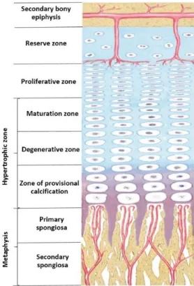
clusters and ossification centers form; (b) secreted osteoid traps osteoblasts, which then become osteocytes; (c) trabecular matrix and periosteum form; (d) compact bone develops superficial to the trabecular bone, and crowded blood vessels condense into red marrow (Adapted from Anatomy and Physiology, Rice University, 2013) ... 9 Figure 2.5 Typical section of a growth plate showing its reserve, proliferative and hypertrophic
zones (Iannotti & Parker, 2013) ... 11 Figure 2.6 Schematic of physiological bone remodeling (Siddiqui & Partridge, 2016) ... 15 Figure 2.7 The load-deformation curve illustrates the performance strength characteristic of a
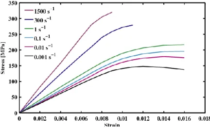
material when subjected to the load. During the load application, an (A) initial elastic response prevails and leads to a (B) yield point. With further loading, it goes into the (C) plastic response when the material is deformed permanently or is broken. The strength of the material is determined by the (D) energy or area under the curve. The elasticity module (or rigidity) is determined by the (E) slope of the curve during the elastic response phase (Bankoff, 2012). ... 18 Figure 2.8 Viscoelasticity of bone tissue (cortical bone) (Johnson, Socrate, & Boyce, 2010) ... 19 Figure 2.9 Compression test setup for trabecular bone samples (Adapted from Mechanics of Bone
by Bethany Jacobs, 2017) ... 20 Figure 2.10 Tensile strength test setup (Adapted from the European Space Agency) ... 21
Figure 2.11 (a) Three-point bending test configuration, (b) 4-point bending test, F = applied forces; d = resulting displacement; a and L = lengths (Oksztulska-Kolanek, Znorko, Michałowska, & Pawlak, 2016). ... 23 Figure 2.12 Schematic diagram of shear and torsion loading conditions that can be imparted to a
bone or bone region (Adapted from Shear Resistance-Priority Hypothesis) ... 24 Figure 2.13 QCT of the forearm using a dedicated peripheral scanner (Adams, 2009) ... 34 Figure 2.14 SCANCO Medical's XtremeCT (HR-pQCT scanner) with a Scout View of a region to
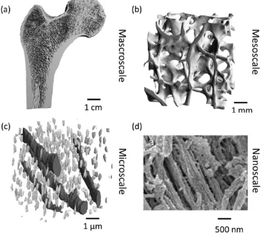
be scanned (Adapted from SCANCO Medical) ... 35 Figure 2.15 SKYSCAN 1176 (in vivo micro-CT scanner) (Adapted from www.bruker.com) ... 38 Figure 2.16 Schematic overview of the hierarchical levels of bone (Ruffoni & Van Lenthe, 2017).
(a) At the macroscale, a micro-CT image femur section (image courtesy of Thomas Mueller, ETH Zurich, Switzerland). (b) At the mesoscale, micro-CT image of a femoral head trabecular bone section. (c) At the microscale, canal network and osteocyte lacunae are visible with synchrotron radiation nano-computed tomography (Adapted from Schneider, P.; Stauber, M.; Voide, R.; et al. Journal of Bone and Mineral Research 2007) (d) At the nanoscale, scanning electron micrographs of mineralized collagen fibrils in a human bone specimen (image courtesy of Paul Hansma, UCSB, Santa Barbara, USA). ... 41 Figure 2.17 Continuum-level FE model of a human proximal femur obtained from CT images
(Marangalou, J. H., 2013) ... 43 Figure 2.18 Micro FE model of a distal radius section (9 mm long) scanned with HR-pQCT. The
nodes at the proximal side were fixed, while axial compression was applied on the distal end (Zysset, Dall'ara, Varga, & Pahr, 2013) ... 45 Figure 3.1 An overview of the project timeline for Objective 1 ... 48 Figure 3.2 An overview of the project timeline for Objective 3 ... 49 Figure 3.3 An overview of the project timeline for Objective 4 ... 51 Figure 4.1 Rat positioning on the Skyscan 1176 scanner for in vivo scanning. The rat was placed
sideways on the scanning bed while kept anesthetized (anesthesia mask not shown). This configuration was adapted to facilitate the positioning of the irradiated leg (right) into the
iso-center of the scanning chamber. The right tibia was secured into a Styrofoam holder (1 cm thick) of cylindrical shape and firmly held with a medical adhesive tape. The non-radiated leg (left) was folded towards the animal’s head and placed alongside the animal with its tail. .. 57 Figure 4.2 Bone growth rates (μm/day) measurements. (A) 5x magnified microscopic images of
the tibial metaphysis labeled twice with calcein for representative irradiated and control tibiae from three doses groups (Ⅰ-ⅤⅠ). Bone growth (ΔX, μm) measured as the mean distance between the two calcein lines, which were modeled as splines and divided by the time interval (3 days) between the two applied injections. (B) Growth rates (μm/day) of rat proximal tibiae for 0.83, 1.65 and 2.47 Gy radiation groups (mean value ± SD). *: a significant difference (p < 0.05) between the control (left) and irradiated (right) tibiae for each radiation dose. ... 62 Figure 4.3 Histomorphometry measurement. (A) Growth plate section embedded in MMA and
stained with toluidine blue (10x). Evaluation of the hypertrophic and proliferative zonal thicknesses for three doses groups. (B) Growth plate section embedded in MMA and stained with toluidine blue (20x). Evaluation of the hypertrophic cell height and number of proliferative cells per column for three doses groups. ... 63 Figure 4.4 In vivo scanning of proximal tibia and bone segmentation process. (a) A representative
3D reconstructed tibia showing the total tibial length (L). (b) Scanned proximal tibial cross-section (10 mm in height) of the rat tibia. This representative image was acquired from a 17.48-μm pixel size scanning at 0.83 Gy radiation dose. VOI consisting trabecular and cortical bone, for morphometric parameters evaluation, beginning at ~1mm distal to the growth plate and extending for 10% of the total tibial length (L). Proximal (f) and distal (c) tibial sections are illustrated. The cortical (d, g) and trabecular (e, h) bone regions were segmented using a semi-automatic bone segmentation algorithm. ... 64 Figure 4.5 Histomorphometry measurements comparison for control and irradiated tibiae. (a-d)
Growth plate histomorphometry measurements of rat proximal tibiae for 0.83, 1.65 and 2.47 Gy radiation groups (mean value ± SD). *: a significant difference (p < 0.05) between the control (left) and irradiated (right) tibiae for each radiation dose. ... 67 Figure 4.6 Mean values and standard deviations of the trabecular bone parameters for the left
Figure 4.7 Mean values and standard deviations of the cortical bone parameters for the left (hatched columns), and right tibiae (black columns) at 14th week of age (n = 11/group). ... 69 Figure 4.8 Body weight of male Sprague Dawley rats for three doses groups over the adolescent
period. ANOVA test (general linear model) was performed to determine time effects, radiation dose, and their interaction on body weight. N = 11 rats per group (mean value ± SD). ... 71 Figure 4.9 Trabecular and cortical bone representation after the 9-weekly in vivo micro-CT scans.
(a - f) Representative 3D micro-CT images of metaphyseal bone structure of the irradiated (right) and non-irradiated control (left) tibiae at 14th week of age after 0.83, 1.65 and 2.47 Gy radiation doses during the rat adolescent period. 3D micro-CT images within each radiation dose portray tibiae from the same rat, randomly selected to be representative of its respective dose group. ... 77 Figure 5.1 (A) Experimental setup for the in vivo rat tibial compression with strain gauge attached
to the tibia. (B) Overlying skin and muscles of the tibia were retracted to expose the strain gauge with lead wires. (C) Geometric and boundary conditions of a representative rat tibia illustrating the strain gauge site at 35% of the tibial length (highlighted in red). A compressive force (F) was applied at the top. At the distal end, pin conditions were applied to all nodes. A = anterior, P = posterior. ... 96 Figure 5.2 (A) Stiffness of the finite element model of the tibia under compression for different
voxel sizes. (B) Computational strain at the strain gauge site of the tibia under compression for different voxel sizes. (C) Distribution of longitudinal strains (ƐZZ) in the representative
tibial section for different voxel sizes. ... 98 Figure 5.3 Linear regression between experimental strains and longitudinal strains evaluated from
finite element models of the rat tibiae for the three different age groups (A-C). Correlation coefficient between the two approaches is calculated based on the 95% confidence interval for the regression line. ... 101 Figure 5.4 Bland-Altman plots comparing experimental and finite element model derived strains
of the rat tibiae for the three different age groups. (A, B, C)* The difference between experimental and FE modeling strains plotted against the mean of experimental and FE modeling strains. ... 102
Figure 5.5 (Ⅰ) Longitudinal strain distribution throughout the whole rat tibia loading model (12 week old). (ⅠⅠ) 3D section of the rat tibial geometry showing locations of the experimental (AM) and FE modeling (AL, PL and AM) strains in the transverse cross-section containing the strain gauge. ... 104 Figure 5.6 Experimental strain at the strain gauge location vs FE modeling strain at the AL, PL and
AM regions of the rat tibia for three different age groups (A-C). ... 107 Figure 5.7 (A) FE modeling strains (mean value ± SD) measured at +0.5 mm toward the proximal
or distal side of the tibiae with respect to the experimentally measured strains (mean value ± SD) under 35N load. (B) FE modeling strains (mean value ± SD) measured at +0.5 mm toward the anterior or posterior side of the tibiae with respect to the experimentally measured strains (mean value ± SD) under 35N load. Asterisk represents a significant difference with *p < 0.05. ... 107 Figure 6.1 Rat body weight (g) and food consumption (g/day) during experimental period. (A)
ANOVA test (general linear model) was performed to determine time effects, group effects, and their interaction on body weight. (B) ANOVA test (general linear model) was performed to determine time effects, group effects, and their interaction on food consumption. ... 126 Figure 6.2 Bone growth rates (µm/day) and longitudinal tibial lengths (mm). (A) 2.5x magnified
microscopic images of the tibial metaphysis labeled twice with calcein and representative images of tibiae for control, sham, LI, MI and HI groups (Ⅰ-Ⅴ). Bone growth (ΔX, μm) measured as the mean distance between the two calcein lines, which were modeled as splines and divided by the time interval (3 days) between the two applied injections. (B) Bone growth rates (μm/day) of rat proximal tibiae for control, sham, LI, MI and HI groups. (C) Relative (control minus individual group) gross tibial length (mm) of the tibiae. MI and HI groups exhibited approximately three and four times reduction in tibial length difference. ... 127 Figure 6.3 Growth plate histomorphometric parameters for control, sham, LI, MI and HI tibiae. (A)
Growth plate section embedded in MMA and stained with toluidine blue (10x). Evaluation of the hypertrophic and proliferative zonal thicknesses (µm) for control, sham, LI, MI and HI groups (Ⅰ-Ⅴ). (B) Growth plate section embedded in MMA and stained with toluidine blue (20x). Evaluation of the hypertrophic cell height (µm) and number of proliferative cells per column (cells) for control, sham, LI, MI and HI groups (Ⅰ-Ⅴ). (C) Growth plate
histomorphometry measurements of rat proximal tibiae for control, sham, LI, MI and HI groups (Ⅰ-Ⅴ). ... 128 Figure 6.4 Impact loading setup and strain gauge calibration. (A) With the rats under anesthesia,
the right tibiae from LI, MI and HI groups were loaded using a waveform generating 450, 850, and 1250 µε at the medio-proximal tibial surface from the 4 to 11 week period. (B) The loading profile consisted of haversine waveform displacements at 2Hz and characterized by symmetric loading/unloading with a 0.10 sec of rest insertion between loading cycles. Loadings were repeated for 1200 cycles, yielding a daily (5 days/week) loading period of 10 minutes. (C) Strain gauge positioned at the medio-proximal surface of the tibia for allowing strain assessment for 0.5 mm to 3.5 mm of displacement. (D) Linear relationship between applied displacement and resulting strain at the medio-proximal surface of 4, 8 and 12 week old rat tibiae (mean value ± SD) (N = 6 rats/group). ... 141 Figure 6.5 Trabecular and cortical volume of interests and experimental setup for three-point
bending tests. (A) Representative 3D reconstructed tibia showing the total tibial length (L). The trabecular VOI started at ~0.35 mm distal to the growth plate and extended for 12% of the overall bone length (L). The VOI for cortical bone was centered at the tibial mid-diaphysis and extended proximally and distally for 5% of the tibial length (L). Volumes of interest including only trabecular and cortical bone were semi-automatically segmented using an in-house algorithm. (B) Ⅰ-Experimental setup for the three-point bending tests. A distance of 50% of the total tibial length was fixed between the supports, while the remaining 50% was distributed equally between the external sides of the supports. Ⅱ- Representative image of the fractured tibia after the bending test. ... 144 Figure 7.1 (A) In vivo loading of the right tibia of a 8 w.o. rat. (B) Strain gauge calibration curves
at the medioproximal tibial surface for 4, 8 and 12 week old rats. Error bars represent standard deviations (n = 6 rats/age group). (C) Representative in vivo loading profile including 1200 repetitions over approximately 10 min/day. Peak-to-peak displacements were chosen based on the strain gauge calibration curves previously obtained for the three age groups. ... 163 Figure 7.2 (A) Five rat groups (n=42 total) were used: control (C; n=6), sham (S; n=6), low impact
(LI; n=10), medium impact (MI; n=10), and high impact (HI; n=10). The right tibia of each rat from LI, MI and HI groups were loaded using the waveform respectively triggering 450,
850, and 1250 με tensile strain at the medio-proximal tibial surface from 4 to 11 weeks of age, corresponding to rat adolescence. (B) Impact loadings were applied 5 days/week from 4 to 11 weeks of age. Rats were detrained from the 11th to 52nd week. At the end of the experiment (52 w.o.), rats were sacrificed, both structural and estimated tissue-level mechanical properties were obtained. Right tibiae were scanned during the entire experimental period, at different time intervals, for acquiring in vivo bone microstructural parameters. ... 164 Figure 7.3 (A) (Ⅰ) Rat positioning for the in vivo micro CT scanning. While anesthetized, the rat
was placed sideways securing the right tibia into a Styrofoam holder and firmly held with medical adhesive tape. The left tibia was folded towards the animal’s head and placed alongside with the tail. (Ⅱ) Representative longitudinal section of a rat tibial CT scan showing the total tibial length (L). The trabecular VOI started at ~0.35 mm distal to the growth plate and extended for 12% L. The cortical VOI was fixed at the tibial mid-diaphysis and equally spanned proximally and distally for a total of 5% L. Using a semi-automatic segmentation algorithm, trabecular and cortical sections were extracted to further evaluate bone morphometric parameters. (B) (Ⅰ) Three-point bending test experimental setup, before and after bone fracture. Half of the total tibial length (L) was set between supports, with the remaining length equally distributed between the external sides of the supports. (Ⅱ) Representative force vs. displacement curves for a HI tibia and sham tibia after detraining (52 week old). ... 165 Figure 7.4 (A) Rat body weight (g). ANOVA test (general linear model) was performed to
determine time effects, group effects, and their interactions on body weight. (B) Absolute daily food intake (g/day). ANOVA test (general linear model) was performed to determine time effects, group effects, and their interactions on food consumption. (C) Relative quantity of food intake per unit body weight (g/kg. day-1). ANOVA test (general linear model) was performed to determine time effects, group effects, and their interactions on food intake per unit body weight. ... 170 Figure 7.5 Trabecular bone morphometric parameters (means and standard deviations) for the five
experimental groups at the end of training (11 week of age) and at selected detraining time points (14, 22, 34, and 52 week of age). ... 172
Figure 7.6 Cortical bone morphometric parameters (means and standard deviations) for the five experimental groups at the end of training (11 week of age) and at selected detraining time points (14, 22, 34, and 52 week of age). ... 173 Figure 7.7 (A) Principal tensile strain distribution within a representative 52 w.o. rat tibia and
within corresponding transverse sections of proximal trabecular and mid-diaphysis cortical bone VOIs. (B) Principal compressive strain distribution within a representative 52 w.o. rat tibia and within corresponding transverse sections of proximal trabecular and mid-diaphysis cortical bone VOIs. (C) Principal compressive and tensile strains within in the 52 w.o. rat tibial proximal trabecular VOIs and mid-diaphysis cortical VOIs for the five experimental groups. ... 176 Figure B.1 Mean values and standard deviations of the periosteal and endocortical perimeter for
the five experimental groups at the end of training (11 week of age) and at selected detraining time points (14, 22, 34, and 52 week of age) ... 227
LIST OF SYMBOLS AND ABBREVIATIONS
AL Antero-lateralAM Antero-medial
BMD Bone mineral density BV/TV Bone volume fraction CaHA Calcium hydroxyapatite Conn.D Connectivity density
CT Computed tomography
Ct.Ar Cortical bone area
CTDI Computed tomography dose index Ct.Th Cortical thickness
Ec.Pm Endocortical perimeter
HU Hounsfield Unit
Ma.Ar Medullary area PL Postero-lateral Ps.Pm Periosteum perimeter Tb.N Trabecular number Tb.Sp Trabecular Spacing Tb.Th Trabecular thickness TMD Tissue mineral density Tt.Ar Total area
LIST OF APPENDICES
Appendix A Calculation of radiation doses ... 225 Appendix B Cortical bone morphometry ... 227
INTRODUCTION
A fundamental precept for bone biomechanics is the adaptation of its microstructure in response to mechanical stimuli regularly imposed on it (Ahn & Grodzinsky, 2009; Duncan & Turner, 1995; M. K. Karlsson, 2004; S. J. Warden, Fuchs, Castillo, & Turner, 2005; Wolff, 1892). Mechanical loadings in the form of physical activity are considered beneficial for bone tissues and for the skeletal system, with enhanced bone quality as well as increased bone mass and mineral content (Chamay & Tschantz, 1972; S. J. Warden, Fuchs, Castillo, Nelson, & Turner, 2007; S. J. Warden et al., 2014; Wolff, 1892). Adolescence or pubertal period is a prime period for bone development, and bones respond more sensitively to the mechanical loadings at this particular growth period (Sievänen, 2012; Spengler, Morey, Carter, Turner, & Baylink, 1983; Weaver, 2002). Considering the many skeletal changes that could occur during adolescence, maintaining these loading effects at adulthood would greatly benefit the skeletal system, improving the overall bone health condition (Health & Services, 2004; Rizzoli, Bianchi, Garabédian, McKay, & Moreno, 2010).
Previous clinical and experimental studies on the effects of loading on bone microstructure hold contradictory results. Few clinical studies reported that normal physiologic activities during childhood could negatively affect bone growth (Bernink, Erich, Peltenburg, Zonderland, & Huisveld, 1983; Caine, Lewis, O'Connor, Howe, & Bass, 2001; Haywood, Clark, & Mayhew, 1986; Lindholm, And, & Ringertz, 1994; Tanghe et al., 1996; Theintz, Howald, Weiss, & Sizonenko, 1993). However, the studies were not well controlled and failed to separate the effects of nutritional factors from mechanical factors. Several experimental studies reported contradictory findings (positive and negative) on the effects of mechanical loading during adolescence (Bourrin, Palle, Pupier, Vico, & Alexandre, 1995; Forwood & Parker, 1987; Niehoff, Kersting, Zaucke, Morlock, & Brüggemann, 2004; Snyder, Zierath, Hawley, Sleeper, & Craig, 1992). Hence, changes in bone microstructure and longitudinal bone growth due to the application of impact loading during adolescence remain to be determined. Moreover, both clinical and experimental studies have been conducted for assessing the possible remaining effects of pubertal exercise on bone structure at adulthood (Bass et al., 1998; Duckham et al., 2014; Gunter et al., 2008; Honda, Sogo, Nagasawa, Kato, & Umemura, 2008; Iwamoto, Yeh, & Aloia, 2000; M. Karlsson et al., 2000; Kontulainen et al., 2001; Kontulainen, Sievänen, Kannus, Pasanen, & Vuori, 2003; Nordström, Olsson, & Nordström, 2005; Pajamäki et al., 2003; S. J. Warden et al., 2007; S. J. Warden et al., 2014).
However, results are again inconsistent on the effects prevailing at adulthood period. Few studies reported the positive effects of pubertal exercise in the long term period (Bass et al., 1998; Duckham et al., 2014; Honda et al., 2008; M. Karlsson et al., 2000; Kontulainen et al., 2001; Kontulainen et al., 2003; S. J. Warden et al., 2007; S. J. Warden et al., 2014), whereas others reported the absence of any skeletal benefits at adulthood (Gunter et al., 2008; Iwamoto et al., 2000; Nordström et al., 2005; Pajamäki et al., 2003). Hence, it is not clearly determined whether impact loading applied during adolescence would affect bone development, quality, and mechanical strength at maturity or how long these effects would remain.
The main purpose of this thesis was to investigate the effects of in vivo dynamic impact loadings applied during the adolescence on bone growth, quality and mechanical strength at the end of the growing period as well as effects of loadings on bone quality and mechanical strength after a detraining period at adulthood. To do so, several complementary studies, including investigating a safe radiation doses limit for growing bone, establishing a displacement-strain relationship in the growing bone, developing a non-invasive micro-CT based finite element tool, and completing a symmetry analysis of contralateral limb, etc. were performed.
This thesis includes ten chapters and is submitted as an article-based thesis. Following the Introduction, Chapter 2 presents a literature review on the context of the research, the state of knowledge on mechanobiology of the longitudinal bone growth, bone biomechanics, and bone adaptability to mechanical loading in the form of physical exercise. Chapter 3 introduces the rationale, research questions and objectives of the project. The body of this thesis is composed of four principal articles presented in Chapters 4 to 7. Chapter 4 presents the first article entitled: “Can repeated in vivo micro-CT irradiation during adolescence alter bone microstructure, histomorphometry and longitudinal growth in a rodent model?”, which was published in PloS one Journal. This article investigates the radiation effects on bone morphometry, bone marrow cells, bone growth rate and growth plate histomorphometry in growing tibiae for three radiation doses from repeated in vivo micro-CT scanning in adolescent rats to determine a safe dose level for repeated use in the adolescent period. Chapter 5 introduces the second article entitled: “Experimental and finite element analyses of bone strains in the growing rat tibia induced by in vivo axial compression”. This article was published in Journal of the Mechanical Behavior of Biomedical Materials and develops the displacement-strain relationship in the growing rats and introduces a finite element modeling tool for validating the experimental bone strains. Chapter 6
presents the third article entitled: “High Impact Exercise Improves Bone Microstructure and Strength in Growing Rats”, submitted to the Scientific Reports. In this chapter, the effects of in vivo impact loadings applied during puberty on bone growth, quality, and mechanics of the bone microstructure at the end of the growing period are discussed. Chapter 7 presents the last article of this thesis entitled: “Impact Exercise during Adolescence Improves Bone Microstructure and Strength at Adulthood”, submitted to the Journal of Bone and Mineral Research. In this chapter the effects of in vivo impact loadings applied during puberty on longitudinal bone development, morphometry and biomechanics are evaluated at the end of puberty as well as at the adulthood. Chapter 8 discusses the overall results of the project and establishes connections between the four articles and the reviewed literature. Chapter 9 summarizes the key contributions of this thesis to the advancement of knowledge in mechanical engineering. Finally, Chapter 10 presents the overall contributions of this thesis and recommendations that future studies.
LITERATURE REVIEW
2.1 Skeletal system
The skeletal system, which is composed of bones, cartilages, ligaments, and other tissues, undertake fundamental functions for the human body. Bones are considered rigid tissues, which contain embedded cells with abundant hard intercellular material, blood, nerves, and other connective tissues (Currey, 2014; Hinwood, 1997). At birth, the human body consists of 300 soft bones. During the growing period and throughout the adolescence, the fusion of some soft bones takes place, and eventually, a total of 206 bones compose the human skeleton at adulthood (Markings, 1995). Bone continues to grow in length and width throughout the adolescent period or childhood. Our skeleton continues to repair itself at the microstructural level as a part of the bone remodeling process (Bourne, 2014). The major functions of bone include: (1) structural support for our body, (2) protection of vital organs and tissues, (3) storage for minerals, and (4) protective environment for marrow (the location for producing white blood cells) (D. B. Burr & Allen, 2019).
Figure 2.1 Functions of the skeletal system (Adapted from Anatomy and Physiology, Oregon State University, 2019)
2.2
Bone
Structure and Composition
2.2.1 Bone Structure
A typical long bone is shown in Figure 2.2. A long bone generally comprises two main parts: the diaphysis and the epiphyses. The epiphysis consists in the main or midsection (tubular shaft) of the bone. It is located between the proximal and distal side of the bone and is mainly composed of dense compact bone. This compact bone surrounds a central marrow cavity structure, which contains red or yellow marrow (Figure 2.2). The epiphyses are the rounded ends of the long bone. Proximal and distal epiphyses are filled with red bone marrow. The narrow area between the epiphysis and diaphysis is called the metaphysis, which contains the epiphyseal plate (or growth plate) (Figure 2.2). The epiphysis is covered with articular cartilage and the zone situated below this region known as the subchondral bone.
Figure 2.2 Anatomy of a typical long bone (Adapted from Anatomy and Physiology, Rice University, 2013)
2.2.2 Bone Composition
Bone structural pattern is nonhomogeneous. At the macroscopic level, bone is composed of two types of osseous tissue that are identified as trabecular (or cancellous or spongy) and cortical (or compact) bones. Bone is mainly composed of cells embedded in an organic extracellular matrix of fibers. A ground substance also consists of a large portion in the tissue structure. Constitutive cells for bone formation are three types: Osteoblasts, Osteocytes and Osteoclasts. Osteoblasts are bone-forming cells. They are connective tissue cells and found at the bone surface. Osteoblasts generate osteoid, a protein mixture, which eventually mineralizes to become bone. Osteoblasts can be differentiated as osteocytes under stimulation. Osteocytes are mature bone cells. They are trapped and surrounded by the bone matrix. Bone formation, maintenance of matrix and homeostasis of Calcium are the primary functions of osteocytes. Osteoclasts are responsible for bone resorption and remodeling. They are large, multinucleate cells located on bone surfaces in resorption pits. Cortical bone has a dense structure and forms the outer shell or cortex of the bone. At the cortical bone microscopic level, the structural unit responsible for bone composition is called the osteon, or haversian system (Figure 2.3a). A small canal, the Haversian canal, is situated at the center of each osteon. This canal accommodates blood vessels and nervous fibers. The structural components of the osteon comprise of a concentric series of layers (lamellae) of the mineralized matrix, which surrounds the central canal. Small cavities known as lacunae can be found near the boundaries of each layer, or lamella. The lacunae contain an osteocyte, which is a bone cell and has entombed itself within the bony matrix (Figure 2.3). Also, there exists a number of small channels, called canaliculi. These canals radiate from each lacuna, then connect the lacunae of adjacent lamellae and finally reach the haversian canal (Figure 2.3a). Bone’s resistance behavior to mechanical stress is largely contributed from the effective cross-liking of the collagen fibers within the osteon. This feature also accounts for the vulnerability of the cement line, which is considered the weakest portion of the bone microstructure. The cement line is also responsible for the fatigue behavior of the cortical bone, which takes place by dissipating energy through crack propagation. Cracks and micro damages are thus confined to more densely mineralized interstitial bone located between osteons (Hernandez & Keaveny, 2006).
Trabecular bone is composed of trabeculae, which are thin rods or plate shape structure. Red marrow is located between the trabeculae (Figure 2.3b). Trabecular bone does not contain
Haversian canals. Its tissue structure is arranged in a concentric lacunae pattern containing lamellae. Osteocytes receive nutrients through blood vessels, which are located in the red marrow zones. Trabecular bone is surrounded by cortical bone, but its relative thickness varies significantly among bones in accordance with varying functional tasks. Bone marrow is made of blood vessels, nerves, and various cells. This marrow is of importance because of its contribution to blood cells generation during bone load application. It is highly osteogenic in nature and is capable of stimulating bone formation in any parts of the body.
The periosteum is a fibrous layer covering the bones, everywhere except at the joint surfaces, where bones are covered with articular cartilage. Periosteum is permeated by blood vessels and nerve fibers, which lead to the bone via Volkman's canals.
Figure 2.3 (a) Structure of cortical (compact) bone; (b) Structure of trabecular bone. (Iannotti & Parker, 2013)
2.3 Bone Growth and Remodeling
2.3.1 Bone Growth
Bones are important part of the skeletal system, and they begin to form before we are born. After birth, bones grow very fast, then the growth slows down rapidly and again increases the growth later in infancy. Bones continue growing throughout childhood and adolescence.