The Basic Property of Lys385 Is Important for Potentiation of
the Human
␣1 Glycine Receptor by Ethanol
Patricio A. Castro, Maximiliano Figueroa,
1Gonzalo E. Yevenes,
2Loreto S. San Martin,
and Luis G. Aguayo
Laboratory of Neurophysiology, Department of Physiology, University of Concepcion, Concepcion, Chile Received July 11, 2011; accepted October 28, 2011
ABSTRACT
Ethanol alters the function of several members of the Cys-loop ligand-gated ion channel superfamily. Recent studies have shown that the sensitivity of the␣1 glycine receptor (GlyR) to ethanol can be affected by the state of G protein activation mediated by the interaction of G␥ with intracellular amino acids in the GlyR. Here, we evaluated the physicochemical property of Lys385 that con-tributes to ethanol modulation by using mutagenesis, patch-clamp, and biochemical techniques. A conserved substitution (K385R) did not affect either the apparent glycine EC50 (40⫾ 1 versus 41⫾ 0.5M) or the ethanol-induced potentiation (53 ⫾ 5 versus 46 ⫾ 5%) of the human ␣1 GlyR. On the other hand, replacement of this residue with glutamic acid (K385E), an acidic
amino acid, reduced the potentiation of the GlyR to 10⫾ 1%. Furthermore, mutations with a hydrophobic leucine (K385L), a hydrogen bond donor glutamine (K385Q), or a neutral residue (K385A) also reduced ethanol modulation. Finally, substitution by a large and hydrophobic residue (K385F) and deletion of 385 (Lys385_) reduced ethanol modulation to 10⫾ 4 and 17 ⫾ 0.4%, respectively. Experiments using dynamic cysteine substitution with a methanethiosulfonate reagent and homology modeling in-dicate that the basic property and the position of Lys385, probably because of its interaction with G␥, is critical for ethanol potenti-ation of the receptor.
Introduction
Glycine is the principal inhibitory neurotransmitter pres-ent in the spinal cord and brain stem, and it has significant action in motor and respiratory control (Legendre, 2001; Lynch, 2004). It activates homomeric and heteromeric gly-cine receptors (GlyRs), which are composed of five subunits (␣1– 4 and). GlyRs, such as GABAA receptors,
5-hydroxy-tryptamine type 3 (5-HT3) receptors, and nicotinic
acetylcho-line receptors (nAChRs), belong to the Cys-loop ligand-gated ion channel superfamily (Cys-loop LGICs) (Lester et al., 2004). It is noteworthy that these receptors share several structural similarities, such as the presence of a large
N-ter-minal extracellular domain that contains the ligand binding site and four transmembrane (TM) segments (TM1– 4) with
the pore formed by TM2 and one large intracellular loop (IL) connecting TM3 and TM4 that participate in intracellular
modulations (Lester et al., 2004; Unwin, 2005).
Similar to other members of the family, the function of GlyRs can be altered by several modulators and intracellular signals, such as neurosteroids (␣-xalone), Zn2⫹, protein kinase C, cAMP-dependent protein kinase (Legendre, 2001; Lynch, 2004), and G␥ (Ahmadi et al., 2002; Yevenes et al., 2006). Further-more, general anesthetics and alcohols are potent modulators of this inhibitory receptor (Aguayo and Pancetti, 1994; Yamakura et al., 1999; Harris et al., 2008). It is believed that the effect of ethanol on the GlyR is relevant because of its implications in human health, including alterations in motor control, respira-tion, and cardiovascular depression.
Several studies have intended to elucidate the mechanisms by which ethanol can affect LGICs such as GlyRs (Harris et al., 2008; Yevenes et al., 2008), 5-HT3 receptors (Lovinger
and White, 1991), nAChRs (Godden et al., 2001), GABAA
receptors (Mihic et al., 1997), and N-methyl-D-aspartate re-ceptors (Ren et al., 2008). In the case of GlyRs, for example, This work was supported by the National Institutes of Health National
Institute on Alcohol Abuse and Alcoholism [Grant AA15150]; and the Comisio´n Nacional de Investigacio´n Científica y Tecnolo´gica [Grant AT-24080125].
1Current affiliation: Unit of Molecular Biology and Genetic Engineering, Groupe Interdisciplinaire de Génoprotéomique Appliquée-Research, Univer-sity of Lie`ge, Lie`ge, Belgium.
2Current affiliation: Institute of Pharmacology and Toxicology, University of Zurich, Winterthurerstrasse, Switzerland.
Article, publication date, and citation information can be found at http://jpet.aspetjournals.org.
http://dx.doi.org/10.1124/jpet.111.185140.
ABBREVIATIONS: GlyR, glycine receptor; IL, intracellular loop; LGIC, ligand-gated ion channel; nAChR, nicotinic acetylcholine receptor; 5-HT3, 5 hydroxytryptamine type 3; TM, transmembrane; HEK, human embryonic kidney; PDB, Protein Data Bank; MTS, 3-(4,5-dimethylthiazol-2-yl)-5-(3-carboxymethoxyphenyl)-2-(4-sulfophenyl)-2H-tetrazolium, inner salt; SCAM, substituted-cysteine accessibility method; WT, wild type; MTSEA, methanethiosulfonate ethylammonium; ESP, electrostatic surface potential.
THEJOURNAL OFPHARMACOLOGY ANDEXPERIMENTALTHERAPEUTICS Vol. 340, No. 2
Copyright © 2012 by The American Society for Pharmacology and Experimental Therapeutics 185140/3742105 JPET 340:339–349, 2012
339
at Divine Sce Express Cafs(Uinv/Medecine/Lieg/22827242) on February 7, 2012
jpet.aspetjournals.org
the presence of binding sites in TM regions (Mihic et al., 1997; Ye et al., 1998; Yamakura et al., 1999) and the activa-tion of G␥ proteins (Yevenes et al., 2008) have been postu-lated to be important for ethanol sensitivity. In addition, a critical residue in loop 2 of the extracellular domain, Ala52, was reported to modulate ethanol sensitivity on the receptor (Perkins et al., 2008).
It was previously shown that several residues in the IL play roles on the physiological and pharmacological proper-ties of various LGICs. For example, it was reported that the IL regulates nAChR and GlyR channel gating via G␥ bind-ing (Fischer et al., 2005; Yevenes et al., 2006). In addition, a single residue (Arg436) in the IL of the human 5-HT3A
re-ceptor was found to regulate channel conductance (Deeb et al., 2007; Carland et al., 2009). Furthermore, another study showed that the most important residue controlling the ac-tions of G␥ and ethanol on GlyRs was Lys385 (Yevenes et al., 2008). Therefore, in the present study we characterized which physicochemical property of K385 is correlated to eth-anol-induced modulation of this receptor by using site-di-rected mutagenesis, patch clamp, cysteine-recognizing MTS reagents, and structural modeling.
Materials and Methods
cDNA Constructs. The cDNA construct encoding the human
glycine receptor␣1 subunit with a C-terminal hexahistidyl tag
sub-cloned in a pCI vector (Promega, Madison, WI) for expression in HEK 293 cells has been described previously (Yevenes et al., 2006). Mu-tations were inserted by using the QuikChange site-directed mu-tagenesis kit (Agilent Technologies, Santa Clara, CA). All construc-tions were confirmed by full sequencing. The glycine receptor amino acids were numbered according to their positions in the mature protein sequence.
Cell Culture and Transfection. HEK 293 cells were cultured by
using standard methodologies. HEK 293 cells were transfected by
using Lipofectamine 2000 (Invitrogen, Carlsbad, CA) with 2g of
DNA for each plasmid studied per well (35 mm). Expression of green fluorescent protein was used as a marker of positively transfected cells, and recordings were made after 18 to 24 h.
Electrophysiology. Whole-cell recordings were performed as
de-scribed previously (Yevenes et al., 2006). A holding potential of⫺60
mV was used. Patch electrodes were filled with 140 mM CsCl, 10 mM
1,2-bis(O-aminophenoxy)ethane-N,N,N⬘,N⬘-tetraacetic acid, 10 mM
HEPES, pH 7.4, 4 mM MgCl2, 2 mM ATP, and 0.5 mM GTP. The
external solution contained 150 mM NaCl, 5.4 mM KCl, 2.0 mM
CaCl2, 1.0 mM MgCl2, 10 mM HEPES, pH 7.4, and 10 mM glucose.
For G protein activation experiments, GTP␥S (0.5 mM;, Sigma, St.
Louis, MO) was added directly to the internal solution, replacing GTP. The amplitude of the glycine current was assayed by using a brief (1– 6 s) pulse of glycine every 60 s. The modulation of the glycine current by 100 mM ethanol (Sigma) was assayed by using a short
application of glycine (EC20) coapplied with ethanol to each receptor
studied without any preapplication. Although the effect of ethanol was evident at 10 mM (Aguayo et al., 1996), in this study we used 100
Fig. 1. Intracellular residues in the
intra-cellular loop of␣1 GlyR important for eth-anol-induced potentiation. A, primary se-quence of the large IL between TM3 and TM4. The red and blue colors indicate the positive or negative charged residues, re-spectively. B, the graph summarizes concentration-response relationships for WT and 316 –320Ala, 326 –328Ala, 371– 375Ala, 376 –380Ala, and 385–386Ala mutations. C, effects of mutations of basic and acidic residues on GlyR potentiation induced by 100 mM ethanol. ⴱⴱⴱ, p ⱕ 0.001. Each bar represents the mean⫾ S.E.M. obtained from at least five cells. D, homology ribbon model for the IL where the residues colored in A are indicated.
at Divine Sce Express Cafs(Uinv/Medecine/Lieg/22827242) on February 7, 2012
jpet.aspetjournals.org
mM to facilitate statistical analysis. In all of the experiments, a brief pulse of 1 mM glycine was performed at the end of the recording period to test that the glycine concentration corresponded to the
actual EC20in each single experiment. Cells having responses⬍EC5
or ⬎EC20 were discarded. Strychnine (1 M) blocked all of the
current elicited by wild-type and mutant glycine receptors (data not shown). For substituted-cysteine accessibility method (SCAM) stud-ies, stock solutions of MTS reagents (200 mM) obtained from Toronto Research Chemicals Inc. (North York, ON, Canada) were stored at ⫺20°C. Before each experiment, MTS reagents were diluted in the electrode solution for whole-cell experiments to yield a final
concen-tration of 200M.
Immunofluorescence, Image Visualization, and Analysis.
HEK 293 cells were first fixed with 4% paraformaldehyde (0.1 M phosphate buffer, pH 7.4) and then permeabilized (0.3% Triton X-100) and blocked with 10% horse serum. Subsequently, all night incubation with a polyclonal hexa-histidine antibody (His Tag; US Biological, Swampscott, MA) was carried out. Epitope visualization was performed by incubating the sample with secondary antibodies conjugated to fluorescein isothiocyanate and Cy5 (1:600; Jackson ImmunoResearch Laboratories Inc., West Grove, PA). Finally, the cells were coverslipped by using Fluorescence Mounting Medium (Dako North America, Inc., Carpinteria, CA). For quantitative anal-ysis, cells were chosen randomly for imaging using Nikon confocal
Fig. 2. Expression and localization of
Lys385 mutants overexpressed in HEK cells. A, the Western blots were obtained from HEK cell lysates expressing the WT and mutant versions of␣1 GlyR. Each letter corresponds to the substitution (R, K385R; Q, K385Q; L, K385L, E, K385E, C, K385C; F, K385F), and _ represents deletion. B, confocal microscopy obtained in HEK cells transfected with the␣1 GlyR WT and single point mutations in Lys385. A, Ala; E, Glu; L, Leu; R, Arg; Q, Gln. The red signal corresponds to GlyR detected with an anti-His antibody, and the green color corresponds to green fluorescent protein used for the identification of transfected cells for patch clamp record-ings. The bar corresponds to 20m. C, the graph summarizes the fluorescence ratio between membrane (periphery) and cyto-plasm (center) associated to GlyR WT and mutant expression using an anti-His antibody.
TABLE 1
Electrophysiological parameters of WT and Lys385 mutations of ␣1 GlyR
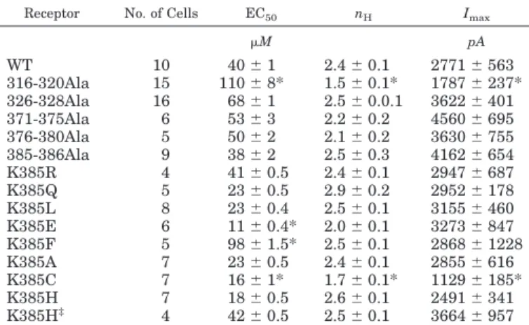
Shown is a summary of the values for glycine sensitivity (EC50), Hill coefficient (nH), and maximal currents (Imax) for WT and the different mutations of␣1 GlyR. The EC50, nH, and Imaxvalues were measured from the glycine concentration-response relationships by using 1 to 1000M. K385H‡corresponds to values obtained at pH 6.0. Each value represents the mean⫾ S.E.M.
Receptor No. of Cells EC50 nH Imax
M pA WT 10 40⫾ 1 2.4⫾ 0.1 2771⫾ 563 316-320Ala 15 110⫾ 8* 1.5⫾ 0.1* 1787⫾ 237* 326-328Ala 16 68⫾ 1 2.5⫾ 0.0.1 3622⫾ 401 371-375Ala 6 53⫾ 3 2.2⫾ 0.2 4560⫾ 695 376-380Ala 5 50⫾ 2 2.1⫾ 0.2 3630⫾ 755 385-386Ala 9 38⫾ 2 2.5⫾ 0.3 4162⫾ 654 K385R 4 41⫾ 0.5 2.4⫾ 0.1 2947⫾ 687 K385Q 5 23⫾ 0.5 2.9⫾ 0.2 2952⫾ 178 K385L 8 23⫾ 0.4 2.5⫾ 0.1 3155⫾ 460 K385E 6 11⫾ 0.4* 2.0⫾ 0.1 3273⫾ 847 K385F 5 98⫾ 1.5* 2.5⫾ 0.1 2868⫾ 1228 K385A 7 23⫾ 0.5 2.4⫾ 0.1 2855⫾ 616 K385C 7 16⫾ 1* 1.7⫾ 0.1* 1129⫾ 185* K385H 7 18⫾ 0.5 2.6⫾ 0.1 2491⫾ 341 K385H‡ 4 42⫾ 0.5 2.5⫾ 0.1 3664⫾ 957 * Pⱕ 0.05 with respect to WT.
at Divine Sce Express Cafs(Uinv/Medecine/Lieg/22827242) on February 7, 2012
jpet.aspetjournals.org
microscopy (TE2000; Nikon, Melville, NY). Single stacks of optical sections in the z-axis were acquired, and dual-color immunofluores-cent images were captured in simultaneous two-channel mode. The ratio of receptor fluorescence intensities obtained in the periphery and cytoplasm in transfected HEK cells was analyzed as a way of indirectly evaluating receptor membrane expression of the mutants by using an ImageJ software plugin (National Institutes of Health, Bethesda, MD).
Western Blot. HEK cells cultured in 35-mm wells were lysed with
standard solution. The amount of protein was quantified by using microBSA (Thermo Fisher Scientific, Waltham, MA). Two micro-grams of total protein was charged per lane in a polyacrylamide denaturating 12% gel and run for 2 h. The proteins were transferred to a nitrocellulose membrane for immunodetection by using a pri-mary antibody against His Tag (US Biological; 1:500, 4°C overnight) and a secondary antibody against IgG coupled to horseradish perox-idase (1:1000; room temperature; 2 h). Signal detection was mea-sured by using a Luminescence kit (Thermo Fisher Scientific).
Molecular Modeling. The GlyR model was constructed by
ho-mology using coordinates from the Torpedo nAChR at 4-A° resolution
(Unwin, 2005) (PDB ID code2BG9) and acetylcholine-binding protein
structure (PDB ID code1UV6) (Celie et al., 2004) using the software
Modeler (Eswar et al., 2006). The models were relaxed by energy minimi-zation using a Conjugate Gradient protocol with GROMACS (Hess et al., 2008). To optimize the H-Bond net, the models were processed by the server REMO (http://zhanglab.ccmb.med.umich.edu/REMO/; Li and Zhang, 2009).
Intracellular Loop Modeling. Because of the lack of sequence
identity with a protein having a defined structure that allows build-ing a protein model by “homology modelbuild-ing,” we decided to model this region by a “folding recognition” technique. For this, we ana-lyzed the intracellular loop sequence in the web server for folding recognition, 3D-Jury (Ginalski et al., 2003). We obtained a template in the SecG subunit of the preprotein translocase (PDB ID code 3DIN). Using Modeler and the E chain of3DINas a template, we developed the 3D model of the GlyR intracellular loop for WT and mutants.
Electrostatic Potential Surface. Using APBS (Baker et al.,
2001) together with the input files generated by PDB2PQR (Dolinsky et al., 2007), the electrostatic potential surface was generated. To evaluate the difference between wild type and mutants on electro-static potential surfaces, we used the server VisualDEP (http:// 152.74.15.29). All the images were generated with the software Py-mol (for reference see Yevenes et al., 2008) and retouched with GIMP 2.6.10 (http://www.gimp.org/). To determine the partial charge dis-tribution in the MTS-modified cysteine residue, Ghemical 2.99 (http://www.brothersoft.com/ghemical-349099.html) and MPQC/6 – 31** (http://www.mpqc.org/) as quantum mechanic engines were used (Hassinen and Pera¨kyla¨, 2001).
Data Analysis. Statistical analyses were performed by using
analysis of variance and are expressed as arithmetic mean⫾ S.E.M.
Values of Pⱕ 0.05 were considered statistically significant. For all
the statistical analysis and plots, Origin 6.0 software (MicroCal LLC, Northampton, MA) was used. Normalized values were obtained by
dividing the current amplitude obtained with the time of GTP␥S
dialysis by the current at minute one.
Results
Several studies have shown that positively charged resi-dues are important for the modulation exerted by G␥ in several effectors, such as phospholipase C and G protein-coupled inwardly rectifying potassium channel (Touhara et al., 1995; Ford et al., 1998; Cantí et al., 1999; Barr et al., 2000). Clusters of charged residues present in the IL of the GlyR were analyzed to determine whether their mutations modified the extent of G␥-dependent ethanol-induced
po-tentiation in human␣1 GlyRs. Five clusters of charged res-idues exist in the IL (316 –320, 326 –328, 374 –375, 377–378, and 385–386) (Fig. 1A), and all the mutations of these resi-dues to alanine were able to form functional membrane re-ceptors whose properties are shown in Fig. 1B and Table 1. Whereas ethanol potentiated the WT GlyR by 53 ⫾ 5%, alanine substitutions in the 316 to 320 and 385 to 386 clus-ters significantly decreased the effect of ethanol to 7⫾ 3 and 9⫾ 3%, respectively. On the other hand, alanine substitu-tions in the acidic 326 to 328 cluster, or in posisubstitu-tions 374 to 378, did not alter the potentiation by ethanol (Fig. 1C). After these results, we focused on the Lys385 residue because it was reported that it displayed the largest contribution to ethanol potentiation (Yevenes et al., 2008). The localization of Lys385 in the intracellular loop is relevant because it has been suggested that the sensitivity to ethanol depends on protein-protein interactions between G␥ and GlyRs (Yevenes et al., 2008). This potential interaction would im-plicate complementary charges with G␥ and secondary structure requirements (Yevenes et al., 2008). In addition, it is known that the IL in other LGICs participates in ion channel function and receptor intracellular modulation
(Fi-Fig. 3. Macroscopic electrophysiological properties of WT and mutant
GlyRs recorded in HEK 293 cells. A, the current traces correspond to WT and the K385E mutation using several glycine concentrations in micro-molar. B, the graph summarizes concentration-response relationships for WT and K385R, K385E, K385Q, K385A, K385H‡, K385L, K385F, and K385C mutations.
at Divine Sce Express Cafs(Uinv/Medecine/Lieg/22827242) on February 7, 2012
jpet.aspetjournals.org
scher et al., 2005; Deeb et al., 2007). Using homology models for LGICs (Unwin, 2005; Yevenes et al., 2006, 2008) and a “threading” approach to predict structure in the less charac-terized IL, we constructed a model for this region. The results show that Lys385 is facing outward (Fig. 1D) and forms a common region with this basic cluster facing the other basic region of the IL (positions 316 –320).
Expression and Localization of Lys385 Mutants. We decided to use a mutagenesis approach that would not sig-nificantly alter the␣-helix structure proposed to exist in this region (Anderson et al., 2005; Unwin, 2005; Livesey et al., 2008). In addition, previous studies have shown that modifi-cations in basic residues present in the IL can affect the correct topology and alter the accurate folding of the receptor
Fig. 4. The presence of a basic residue in 385 determines ethanol
poten-tiation in␣1 GlyR. A, the current traces show the effects of ethanol (100 mM) in WT, K385R, and K385L mutations recorded at an equipotent concentration of glycine (EC20). B, the graph illustrates the ethanol-induced potentiation (100 mM) on WT and the 385 mutants. R, K385R; L, K385L; Q, K385RQ; E, K385E; F, K385F; C, K385C; A, K385A. H and H‡ are the unprotonated and protonated forms of histidine residue, respec-tively. C, the graph shows the percentage of potentiation obtained in the presence of 500M intracellular GTP␥S at 15 min in WT, K385R (R), K385A (A), and K385E (E).ⴱ, p ⱕ 0.05; ⴱⴱ, p ⱕ 0.01; ⴱⴱⴱ, p ⱕ 0.001.
Fig. 5. Substitutions of residues with distinct physicochemical properties
in 385 did not affect propofol and␣-xalone modulations. A, current traces in the absence and presence of␣-xalone (50 M) and propofol (30 M) in WT and the K385Q mutation at EC20glycine. B, the graph summarizes the percentage potentiation of WT and mutants in the presence of ␣-xalone. C, the graph summarizes the percentage of potentiation of WT and mutants in presence of propofol. R, K385R; L, K385L; Q, K385RQ; E, K385E; F, K385F; C, K385C; A, K385A.
at Divine Sce Express Cafs(Uinv/Medecine/Lieg/22827242) on February 7, 2012
jpet.aspetjournals.org
in the membrane (Sadtler et al., 2003). This mutagenesis approach ranks substitutions to preserve the appropriate secondary structure in the domain of interest while altering a single physicochemical property of Lys385 (Anderson et al., 2005). Western blot analysis done after 24 h post-transfection, using tubulin as a housekeeping control, showed that WT and mutants (Arg, Leu, Gln, Glu, Phe, Cys, and deletion) were expressed to very similar levels (Fig. 2A). Additional confocal microscopy analysis using 0.2-m sec-tions revealed that the expressed receptors (Fig. 2B, shown in red) were located mainly in the cell periphery, possibly associated with the cellular membrane (Fig. 2, B and C).
Sensitivity of Lys385 Mutants to Glycine. Previous studies showed that the effects of ethanol on␣1 GlyRs highly depend on glycine concentration, starting at 10 mM (Aguayo et al., 1996; Ye et al., 1998). Therefore, before testing the effect of ethanol on the distinct Lys385 mutations, we eval-uated basic electrophysiological properties by using the whole-cell patch-clamp technique with various concentra-tions of glycine. Macroscopic parameters for the Cl⫺current, such as EC50, Hill coefficient (nH), and maximum current
(Imax), were compared in HEK 293 cells expressing the WT
and mutant receptors. Figure 3A illustrates representative traces of currents recorded for WT and K385E receptors in the presence of increasing glycine concentrations. The data show that the shape of the current was very similar in both WT and the K385E mutant. Full concentration-response curves for substituted receptors (K385R, K385Q, K385L, K385E, K385F, K385H‡, K385A, and K385C) are shown in
Fig. 3B. Data for the values of EC50, nH, and Imaxfor WT and
mutants are given in Table 1, and these results show that the conserved substitution (K385R) displayed a concentration-response curve with similar characteristics to WT (Fig. 3B,䡺 and f, respectively). The K385H mutant was studied at pH 6.0 and 7.4 to evaluate whether its level of protonation was able to alter the receptor physiological properties. It is
note-worthy that the EC50was 42 ⫾ 1 at 6.0 and 18 ⫾ 1 at 7.4
(Table 1). On the other hand, K385E (Fig. 3B, F) and K385C (Fig. 3B,多) substitutions shifted the curve to the left, reduc-ing the EC50values with respect to WT. The K385F mutant
(Fig. 3B, ‚), on the other hand, showed the largest increase in the EC50of the series, and this is in correlation with its
position in the amino acid priority substitution rank (Ander-son et al., 2005). In summary, all Lys385 mutants were expressed in the cell membrane and were functional.
Effects of Substitutions in Lys385 to Ethanol Sensi-tivity. Representative traces of the Cl⫺current activated by using an equipotent concentration of glycine (EC20) for WT,
K385R, and K385L mutations in the presence or absence of 100 mM ethanol are shown in Fig. 4A. The data show that only the K385R substitution maintained the WT ethanol sensitivity, whereas all of the other substitutions abolished this potentiation (Fig. 4B). Additional studies with the pro-tonated K385H mutation (H‡; pH 6.0) showed that ethanol
was able to potentiate the current, although to a lower degree (31⫾ 2%) than the WT. K385H was insensitive to ethanol at pH 7.4 (H), suggesting that the positive charge is important for the ethanol modulation (Fig. 4B). Finally, in agreement with previous studies showing that ethanol requires G␥ protein activation to potentiate the WT GlyR (Yevenes et al., 2008, 2010), we found that the current elicited by activation of K385R was positively modulated by intracellular GTP␥S, whereas that in K385E was insensitive to modulation by G␥ (Fig. 4C).
Specificity of the Mutations for Ethanol Sensitivity. Previous studies have shown that mutations in a single res-idue can affect the receptor sensitivity to different modula-tors (Mihic et al., 1997). Therefore, we tested whether mod-ifications in the physicochemical property at position 385 could render the mutants insensitive to neurosteroids and general anesthetics. Figure 5 shows that␣-xalone and propo-fol produced the same degree of potentiation in WT and mutant receptors (Fig. 5).
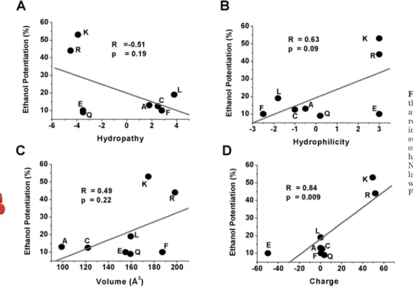
Fig. 6. The effects of ethanol depend on
the basic property of the residue present at position 385. The graphs illustrate the relationships between the different phys-icochemical properties of the residues substituted in position 385 and the effect of 100 mM ethanol. A, hydropathy. B, hydrophilicity. C, volume. D, charge. Note that only polarity was highly corre-lated to ethanol modulation (p⬍ 0.009). R was 0.84. R, Arg; L, Leu; Q, Gln; E, Glu; F, Phe; C, Cys; A, Ala.
at Divine Sce Express Cafs(Uinv/Medecine/Lieg/22827242) on February 7, 2012
jpet.aspetjournals.org
Correlation between Ethanol Sensitivity and Physicochemical Properties. Next, we examined whether the presence of potentiation was correlated to one or more physicochemical properties of the substitutions to establish correlations that could help to understand the chemical nature of ethanol sensitivity in the GlyR. To construct these correlations, the values for volume, hydro-philicity, hydropathy, and charge for each of the seven substitutions were plotted against the potentiation of the receptor by ethanol (Ye et al., 1998; Ren et al., 2008). The analyses showed that the residue charge was the only property statistically related to the potentiation of␣1 sub-units by ethanol (p⫽ 0.009; r ⫽ 0.84). The other properties did not show any significant correlation with the degree of current potentiation (Fig. 6).
Evaluation of the Influence of Lys385 on the Surface Electrostatic Potential. To analyze the contribution of Lys385 on the surface electrostatic potential, we analyzed the predicted three-dimensional model of the IL by using APBS analysis (Baker et al., 2001) and VisualDep (http:// 152.74.15.29). The region containing Lys385 showed a highly positive electrostatic potential (Fig. 7, A and B, shown in blue inside the circle). On the other hand, the mutant K385E displayed a shift in the surface electrostatic potential, indi-cating that the nature of the amino acid in this position is important for the region polarity (Fig. 7, C and D, shown in red). The K385F mutation showed a similar pattern to that of K385E, which is interesting because the nonlocalized elec-trons in the phenylalanine ring add a negative charge to the region (Fig. 7, E and F). These analyses, together with the
Fig. 7. Effects of substitutions at 385 on
the region electrostatic surface potential. A and B, electrostatic surface potential (ESP) representation showing a positive region (blue) surrounding the residue Lys385 (green circle) in two different views. C and D, the mutation K385E in-duced a large decrease in the positive ESP in the region surrounding position 385. E and F, mutation K385F shows a decrease in the positive ESP and suggests a struc-tural modification caused by the presence of a phenylalanine residue in the region surrounding position 385. Red and blue represents negative and positive regions, respectively.
at Divine Sce Express Cafs(Uinv/Medecine/Lieg/22827242) on February 7, 2012
jpet.aspetjournals.org
experimental results, suggest that the charge of this amino acid is critical for determining the sensitivity to ethanol.
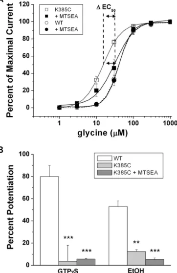
Dynamic Cysteine Modification. To evaluate whether a protein residue is exposed to a hydrophilic environment, cys-teine substitution and chemical reagents that are able to covalently transfer radical groups to the cysteine are used (Deeb et al., 2007). In this study, a reagent with a basic group (MTSEA) was used in the cysteine-substituted mutation. The K385C mutation was characterized by an EC50shifted
to-ward the left (15⫾ 0.4 M; Fig. 8A, 䡺) with respect to the WT (Fig. 8A, E). This shift is advantageous because we can monitor whether the MTS reagent, intracellularly applied, can modify this parameter as expected by the addition of a positive charge. From a structural view no large changes in volume are predicted with respect to lysine (147 versus 145 Å3
). When the reagent was applied to WT GlyRs (Fig. 8A, F), no significant changes on the current were detected, which is interesting because the WT receptor has a cysteine in posi-tion 344 that could potentially interfere with the interpreta-tion of the results. Figure 8A shows that 15 min of applica-tion of the positively charged reagent MTSEA to the K385C
mutant shifted the EC50toward the right (32⫾ 4 M), which
is consistent with a partial rescue of function by the positive charge (f). We then evaluated the response of the K385C mutant to ethanol (100 mM) and GTP␥S (0.5 mM) after intracellular application of MTSEA for 15 min (Fig. 8B). Unlike the WT GlyR (Fig. 8B, white bars), ethanol and GTP␥S were unable to potentiate the current (6 ⫾ 0.7 and 5⫾ 1.4%, respectively) after the treatment, indicating that although MTSEA is transferring a positive charge to K385C, the receptor remained insensitive to ethanol and GTP␥S.
Discussion
The present study examined which physicochemical prop-erty of Lys385, located in the IL of GlyR, was correlated with ethanol modulation of this inhibitory receptor. Our results are consistent with the idea that positive charges in the IL need complementary negative charges in G for interaction and functional modulation.
Importance of the IL for LGIC Functions. LGICs are macromolecules found in neuronal membranes, and they play critical roles in cell signaling (i.e., excitability, neurotransmitter release). They selectively recognize the neurotransmitter that binds to extracellular domains and subsequently undergoes restricted transmembrane rearrangement that allows channel opening. Ion channels show different properties regarding channel conductance and gating kinetics, and only recently it became apparent that intracellular domains are able to regu-late the functions of LGICs. For example, it was shown that Arg436 in the “MA” stretch of the intracellular loop of the 5-HT3Areceptor regulates channel conductance (Livesey et al.,
2008). For example, replacing three arginines with QDA en-hanced single-channel conductance. In␣42 nACh receptors, similarly, changing residues in homologous positions to 5-HT3A
receptor (Glu584, Phe588, and Glu592) with positive amino acids caused a reduction in channel conductance (Peters et al., 2010). Regarding GlyRs, it was shown that replacement of all basic amino acids present in the IL to negatively charged resi-dues produced a strong impact on the GlyR, rendering it non-functional. In addition, simultaneous mutations of arginine and lysine at positions 377, 378, 385, and 386 to glutamate reduced the conductance of the channel, showing their importance in channel function (Carland et al., 2009). Furthermore, the cor-rect assembly of GlyR seems to depend on the presence of the RFRRKRR cluster in the intracellular loop (Sadtler et al., 2003).
Other studies have shown that the IL in␣1 GlyRs interacts with G␥ via two intracellular motifs (Yevenes et al., 2006). Furthermore, it was found that the Lys385 mutation caused the biggest impact on G protein modulation. Similar attenu-ations in ethanol actions were found with these mutattenu-ations, supporting the existence of a high correlation between these two modulations (Yevenes et al., 2008). In agreement, the present study showed that this basic residue was the most important for ethanol action on GlyRs (Fig. 1B). The presence of basic residues in the IL of GlyR and their interaction with G␥ are in agreement with previous studies that showed that lysine and arginine were important for modulation of volt-age-gated calcium channels, phospholipase C, and adrenergic receptor kinase (Touhara et al., 1995; Cantí et al., 1999; Barr et al., 2000).
Fig. 8. SCAM modifications of 385. A, concentration-response curves of WT
␣1 GlyR (circles) and K385C mutant (squares) in the absence (open symbols) or presence (filled symbols) of MTSEA. B, the graph summarizes the per-centage of potentiation of WT (white bars), K385C (light gray bars), and K385C in the presence of MTSEA (dark gray bars) using GTP␥S (500 M) and ethanol (EtOH) (100 mM).ⴱⴱ, p ⱕ 0.01; ⴱⴱⴱ, p ⱕ 0.001.
at Divine Sce Express Cafs(Uinv/Medecine/Lieg/22827242) on February 7, 2012
jpet.aspetjournals.org
A Positive Charge in the 385 Residue Is Important for Ethanol Action on GlyRs. Building from a previous study (Yevenes et al., 2008), the present analyses indicate that the basic property of Lys385, reproduced in Arg385 and His385, is relevant for ethanol-induced modulation. All of the other mutations (K385E, K385Q, K385L, K385F, K385C, and K385A) were less sensitive to the allosteric actions of etha-nol. The data also showed that mutants sensitive to ethanol were potentiated by G protein activation (WT and K385R) and mutants insensitive to ethanol (K385A and K385E) were insensitive to G␥ modulation. These data are in good agree-ment with previous studies that examined a large number (n⫽ 20) of chimeric and mutant GlyRs and showed a high correla-tion between GTP␥S and ethanol modulations (Yevenes et al., 2008, 2010).
The correlation analysis between physicochemical proper-ties (i.e., polarity, charge, hydropathy, hydrophilicity, and volume) and ethanol modulation showed that only charge was highly correlated to ethanol modulation of the GlyR. Furthermore, analysis of electrostatic potential and pre-dicted structure of the region neighboring Lys385 showed
that the substitution altered only charge distribution in the region (Fig. 7), without changes on the ␣-helix secondary structure. Taken together, these results show a critical role for the basic charge of Lys385, providing charge distribution for interaction with G␥, leading to ethanol modulation of GlyRs.
The importance of charge in Lys385 is distinct to the mo-lecular volume property reported for residues 267 and 288 in GlyRs (Ye et al., 1998; Yamakura et al., 1999) associated to the binding pocket for both ethanol and anesthetics. Other studies in nAChR showed a significant correlation between molecular volume and n-alcohol potentiation actions, sup-porting the idea of a site for alcohol in the receptor (Godden et al., 2001). Studies in N-methyl-D-aspartate receptors have shown that two residues, Phe637 and Met823, were involved in ethanol sensitivity, and the molecular volume and hydro-phobicity of these two residues participate on this interaction (Ren et al., 2008). A similar type of analyses in ATP-gated P2X purinoceptor 4 receptors identified two residues (Asp331 and Met 336) whose hydropathy and polarity influenced eth-anol inhibitory actions (Popova et al., 2010). Our hypothesis
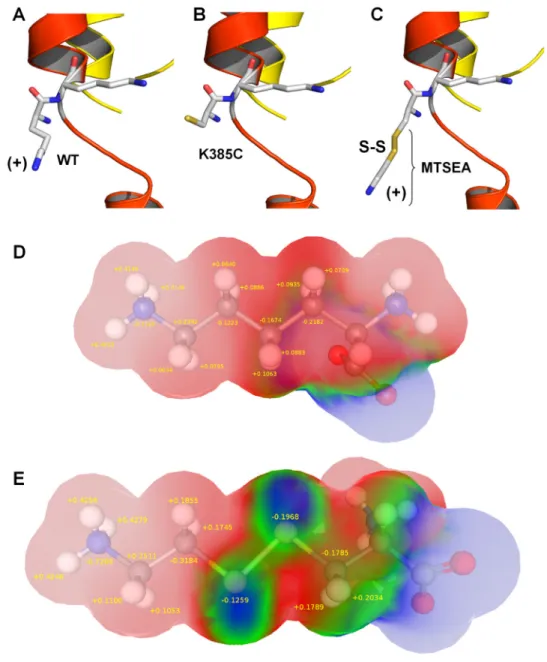
Fig. 9. Structural differences between
WT and K385C-MTSEA-modified recep-tors. A, a stick model of Lys385 and Lys386 in WT ␣1 GlyR. The protein is shown as a ribbon. B, a representation of the mutant K385C. The cysteine residue allows for chemical modification. C, chem-ical modification of K385C with MTSEA gives the residue an electropositive (MTSEA) character, but with a different charge distribution and structure to lysine. D, the charge distribution present in the lateral chain of lysine. The Van der Waals surface is represented and colored according to atomic charge (blue, nega-tive; red, positive). It can be noticed that the lateral chain is positive because of the sum of all partial charges. E, the charge distribution of the lateral chain in the MTS-modified cysteine. The disulfide bond presents a negative (blue) charge unlike the natural residue.
at Divine Sce Express Cafs(Uinv/Medecine/Lieg/22827242) on February 7, 2012
jpet.aspetjournals.org
proposing that G␥ interacts directly with the IL of the GlyR should depend on classic protein-protein interactions, regu-lated by hydrophobic, hydrogen bonding, and electrostatic forces (for review see Leckband, 2000), and this agrees with the complex interaction of G␥ with several effectors (Ford et al., 1998). Studies in residues in loop 2 of the GlyR ectodo-main, which together with the interface to TM2 is believed to serve as a binding site for ethanol (Crawford et al., 2007), showed that polarity at position 52 plays a key role in deter-mining sensitivity to ethanol (Perkins et al., 2008).
We recently postulated that the molecular requirements needed to modulate the GlyR by ethanol are positioned along the structure of GlyR (Yevenes et al., 2010), reconciling sults from different laboratories in terms of identifying re-gions important for ethanol actions (Mihic et al., 1997; Craw-ford et al., 2007). For example, Ala52 in loop 2 of␣1 might transfer binding energy into conformational changes to open the ion channel affecting residues in TM2 (Gly254) and TM3 (Ser296). Finally, basic intracellular residues are required to bind G␥, thereby facilitating channel opening (i.e., increase in open probability).
A Positive MTS Reagent Affected GlyR Activation but Not Its Modulation by Ethanol. The K385C mutation, in addition to being resistant to ethanol, showed differences in activation with respect to WT (Fig. 3; Table 1).Therefore, we wanted to rescue WT properties by using the SCAM in this mutation. This reaction involves the sulfur atom from the reagent and the cysteine amino acid, which generate a disulfide bridge, thereby modifying the substituted residue (Deeb et al., 2007). For instance, the MTSEA reagent pos-sesses a basic radical group (-S-CH2-CH2-NH3⫹) similar to
the lysine residue (Fig. 9, C and E). Concentration-response curves obtained after application of the reagent showed no differences when the WT GlyR was treated with MTSEA, indicating that Cys344 was not accessible to the reagent. It is noteworthy that, in agreement with aqueous intracellular accessibility, MTSEA significantly modified the properties of the K385C mutant receptor, producing a shift in the curve in the direction of the WT receptor, with EC50values of 32⫾ 4
and 40⫾ 1 M, respectively. Thus, the addition of a basic group to the K385C mutant was sufficient to partly recover the phenotype of the GlyR. On the other hand, the MTSEA-treated K385C mutant receptor remained insensitive to GTP␥S and ethanol (Fig. 8B). This lack of modulation can be explained because, despite the gain of a positive charge in the conserved␣-helix region, the resulting disulfide bond gener-ated an alteration in charge distribution on the modified residue (Fig. 9). This is probably because of the nature of the unnatural amino acid formed with the reagent because the added positive charge would be somewhat dissipated by the electronegative environment produced by the disulfide bond. This in turn should modify the pattern of the positive charge in the amine added in the lateral peptide chain. The data suggest that ethanol sensitivity in GlyRs depends on precise electrostatic interactions between G␥ and the GlyR IL. Future experiments with crystallized proteins will con-tribute to elucidating a more precise molecular interaction between G and Lys385.
Acknowledgments
We thank Laurie Aguayo and Ariel Avila for technical assistance in molecular biology and text editing.
Authorship Contributions
Participated in research design: Castro and Aguayo. Conducted experiments: Castro, Figueroa, and San Martin. Performed data analysis: Castro.
Wrote or contributed to the writing of the manuscript: Castro,
Yevenes, and Aguayo.
References
Aguayo LG and Pancetti FC (1994) Ethanol modulation of the␥-aminobutyric acid A-and glycine-activated Cl⫺current in cultured mouse neurons. J Pharmacol Exp
Ther 270:61– 69.
Aguayo LG, Tapia JC, and Pancetti FC (1996) Potentiation of the glycine-activated Cl⫺current by ethanol in cultured mouse spinal neurons. J Pharmacol Exp Ther 279:1116 –1122.
Ahmadi S, Lippross S, Neuhuber WL, and Zeilhofer HU (2002) PGE2selectively blocks inhibitory glycinergic neurotransmission onto rat superficial dorsal horn neurons. Nat Neurosci 5:34 – 40.
Anderson RJ, Weng Z, Campbell RK, and Jiang X (2005) Main-chain conformational tendencies of amino acids. Proteins 60:679 – 689.
Baker NA, Sept D, Joseph S, Holst MJ, and McCammon JA (2001) Electrostatics of nanosystems: application to microtubules and the ribosome. Proc Natl Acad Sci
U S A 98:10037–10041.
Barr AJ, Ali H, Haribabu B, Snyderman R, and Smrcka AV (2000) Identification of a region at the N terminus of phospholipase C-3 that interacts with G protein ␥ subunits. Biochemistry 39:1800 –1806.
Cantí C, Page KM, Stephens GJ, and Dolphin AC (1999) Identification of residues in the N terminus of␣1B critical for inhibition of the voltage-dependent calcium channel by G␥. J Neurosci 19:6855–6864.
Carland JE, Cooper MA, Sugiharto S, Jeong HJ, Lewis TM, Barry PH, Peters JA, Lambert JJ, and Moorhouse AJ (2009) Characterization of the effects of charged residues in the intracellular loop on ion permeation in␣1 glycine receptor chan-nels. J Biol Chem 284:2023–2030.
Celie PH, van Rossum-Fikkert SE, van Dijk WJ, Brejc K, Smit AB, and Sixma TK (2004) Nicotine and carbamylcholine binding to nicotinic acetylcholine receptors as studied in AChBP crystal structures. Neuron 41:907–914.
Crawford DK, Trudell JR, Bertaccini EJ, Li K, Davies DL, and Alkana RL (2007) Evidence that ethanol acts on a target in loop 2 of the extracellular domain of␣1 glycine receptors. J Neurochem 102:2097–2109.
Deeb TZ, Carland JE, Cooper MA, Livesey MR, Lambert JJ, Peters JA, and Hales TG (2007) Dynamic modification of a mutant cytoplasmic cysteine residue modulates the conductance of the human 5-HT3A receptor. J Biol Chem 282:6172– 6182. Dolinsky TJ, Czodrowski P, Li H, Nielsen JE, Jensen JH, Klebe G, and Baker NA
(2007) PDB2PQR: expanding and upgrading automated preparation of biomolec-ular structures for molecbiomolec-ular simulations. Nucleic Acids Res 35:W522–W525. Eswar N, Webb B, Marti-Renom MA, Madhusudhan MS, Eramian D, Shen MY,
Pieper U, and Sali A (2006) Comparative protein structure modeling using Mod-eller. Curr Protoc Bioinformatics, Chapter 5, Unit 5.6.
Fischer H, Liu DM, Lee A, Harries JC, and Adams DJ (2005) Selective modulation of neuronal nicotinic acetylcholine receptor channel subunits by Go-protein sub-units. J Neurosci 25:3571–3577.
Ford CE, Skiba NP, Bae H, Daaka Y, Reuveny E, Shekter LR, Rosal R, Weng G, Yang CS, Iyengar R, et al. (1998) Molecular basis for interactions of G protein␥ subunits with effectors. Science 280:1271–1274.
Ginalski K, Elofsson A, Fischer D, and Rychlewski L (2003) 3D-Jury: a simple approach to improve protein structure predictions. Bioinformatics 19:1015– 1018.
Godden EL, Harris RA, and Dunwiddie TV (2001) Correlation between molecular volume and effects of n-alcohols on human neuronal nicotinic acetylcholine recep-tors expressed in Xenopus oocytes. J Pharmacol Exp Ther 296:716 –722. Harris RA, Trudell JR, and Mihic SJ (2008) Ethanol’s molecular targets. Sci Signal
1:re7.
Hassinen T and Pera¨kyla¨ M (2001) New energy terms for reduced protein models implemented in an off-lattice force field. J Comput Chem 22:1229 –1242. Hess B, Kutzner C, van der Spoel D, and Lindahl E (2008) GROMACS 4: algorithms
for highly efficient, load-balanced, and scalable molecular simulation. J Chem
Theory Comput 4:435– 447.
Leckband D (2000) Measuring the forces that control protein interactions. Annu Rev
Biophys Biomol Struct 29:1–26.
Legendre P (2001) The glycinergic inhibitory synapse. Cell Mol Life Sci 58:760 –793. Lester HA, Dibas MI, Dahan DS, Leite JF, and Dougherty DA (2004) Cys-loop
receptors: new twists and turns. Trends Neurosci 27:329 –336.
Li Y and Zhang Y (2009) REMO: a new protocol to refine full atomic protein models from C-␣ traces by optimizing hydrogen-bonding networks. Proteins 76:665– 676.
Livesey MR, Cooper MA, Deeb TZ, Carland JE, Kozuska J, Hales TG, Lambert JJ, and Peters JA (2008) Structural determinants of Ca2⫹permeability and conduc-tion in the human 5-hydroxytryptamine type 3A receptor. J Biol Chem 283:19301– 19313.
Lovinger DM and White G (1991) Ethanol potentiation of 5-hydroxytryptamine3 receptor-mediated ion current in neuroblastoma and isolated adult mammalian neurons. Mol Pharmacol 40:263–270.
Lynch JW (2004) Molecular structure and function of the glycine receptor chloride channel. Physiol Rev 84:1051–1095.
Mihic SJ, Ye Q, Wick MJ, Koltchine VV, Krasowski MD, Finn SE, Mascia MP, Valenzuela CF, Hanson KK, Greenblatt EP, et al. (1997) Sites of alcohol and volatile anaesthetic action on GABA(A) and glycine receptors. Nature 389:385– 389.
Perkins DI, Trudell JR, Crawford DK, Alkana RL, and Davies DL (2008) Targets for
at Divine Sce Express Cafs(Uinv/Medecine/Lieg/22827242) on February 7, 2012
jpet.aspetjournals.org
ethanol action and antagonism in loop 2 of the extracellular domain of glycine receptors. J Neurochem 106:1337–1349.
Peters JA, Cooper MA, Carland JE, Livesey MR, Hales TG, and Lambert JJ (2010) Novel structural determinants of single channel conductance and ion selectivity in 5-hydroxytryptamine type 3 and nicotinic acetylcholine receptors.
J Physiol 588:587–596.
Popova M, Asatryan L, Ostrovskaya O, Wyatt LR, Li K, Alkana RL, and Davies DL (2010) A point mutation in the ectodomain-transmembrane 2 interface eliminates the inhibitory effects of ethanol in P2X4 receptors. J Neurochem 112:307–317. Ren H, Salous AK, Paul JM, Lamb KA, Dwyer DS, and Peoples RW (2008)
Func-tional interactions of alcohol-sensitive sites in the N-methyl-D-aspartate receptor M3 and M4 domains. J Biol Chem 283:8250 – 8257.
Sadtler S, Laube B, Lashub A, Nicke A, Betz H, and Schmalzing G (2003) A basic cluster determines topology of the cytoplasmic M3–M4 loop of the glycine receptor ␣1 subunit. J Biol Chem 278:16782–16790.
Touhara K, Koch WJ, Hawes BE, and Lefkowitz RJ (1995) Mutational analysis of the pleckstrin homology domain of the-adrenergic receptor kinase. Differential ef-fects on G␥ and phosphatidylinositol 4,5-bisphosphate binding. J Biol Chem 270:17000 –17005.
Unwin N (2005) Refined structure of the nicotinic acetylcholine receptor at 4Å resolution. J Mol Biol 346:967–989.
Yamakura T, Mihic SJ, and Harris RA (1999) Amino acid volume and hydropathy of a transmembrane site determine glycine and anesthetic sensitivity of glycine receptors. J Biol Chem 274:23006 –23012.
Ye Q, Koltchine VV, Mihic SJ, Mascia MP, Wick MJ, Finn SE, Harrison NL, and Harris RA (1998) Enhancement of glycine receptor function by ethanol is inversely correlated with molecular volume at position␣267. J Biol Chem 273:3314–3319. Yevenes GE, Moraga-Cid G, Avila A, Guzma´n L, Figueroa M, Peoples RW, and Aguayo LG (2010) Molecular requirements for ethanol differential allosteric modulation of glycine receptors based on selective G␥ modulation. J Biol Chem 285:30203–30213. Yevenes GE, Moraga-Cid G, Guzma´n L, Haeger S, Oliveira L, Olate J, Schmalzing G,
and Aguayo LG (2006) Molecular determinants for G protein␥ modulation of ionotropic glycine receptors. J Biol Chem 281:39300 –39307.
Yevenes GE, Moraga-Cid G, Peoples RW, Schmalzing G, and Aguayo LG (2008) A selective G␥-linked intracellular mechanism for modulation of a ligand-gated ion channel by ethanol. Proc Natl Acad Sci U S A 105:20523–20528.
Address correspondence to: Luis G. Aguayo, Department of Physiology, University of Concepcion, P. O. Box 160-C, Concepcion, Chile. E-mail: [email protected]
at Divine Sce Express Cafs(Uinv/Medecine/Lieg/22827242) on February 7, 2012
jpet.aspetjournals.org