Research Institute of Manitoba, Winnipeg, Manitoba, Canada, 4 Department of Medicine, de Groote School of Medicine, McMaster University, Hamilton, Ontario, Canada, 5 Department of Pediatrics, Child & Family Research Institute and BC Children’s Hospital, University of British Columbia, Vancouver, British Columbia, Canada, 6 Department of Pediatrics, University of Alberta, Edmonton, Alberta, Canada, 7 Department of Pediatrics, Hospital for Sick Children, University of Toronto, Toronto, Ontario, Canada, 8 CHILD (Canadian Healthy Infant Longitudinal Development Study) Investigators, McMaster University, Hamilton, Canada
¶ Membership of the CHILD Principal Investigators is provided in the Acknowledgments.
*kent.hayglass@umanitoba.ca
Abstract
Changes in maternal innate immunity during healthy human pregnancy are not well
under-stood. Whether basal immune status in vivo is largely unaffected by pregnancy, is
constitu-tively biased towards an inflammatory phenotype (transiently enhancing host defense) or
exhibits anti-inflammatory bias (reducing potential responsiveness to the fetus) is unclear.
Here, in a longitudinal study of healthy women who gave birth to healthy infants following
uncomplicated pregnancies within the Canadian Healthy Infant Longitudinal Development
(CHILD) cohort, we test the hypothesis that a progressively altered bias in resting innate
immune status develops. Women were examined during pregnancy and again, one and/or
three years postpartum. Most pro-inflammatory cytokine expression, including CCL2,
CXCL10, IL-18 and TNF
α
, was reduced in vivo during pregnancy (20–57%, p
<
0.0001).
Anti-inflammatory biomarkers (sTNF-RI, sTNF-RII, and IL-1Ra) were elevated by ~50–100%
(p
<
0.0001). Systemic IL-10 levels were unaltered during vs. post-pregnancy. Kinetic
studies demonstrate that while decreased pro-inflammatory biomarker expression (CCL2,
CXCL10, IL-18, and TNF
α
) was constant, anti-inflammatory expression increased
progres-sively with increasing gestational age (p
<
0.0001). We conclude that healthy resting
mater-nal immune status is characterized by an increasingly pronounced bias towards a systemic
anti-inflammatory innate phenotype during the last two trimesters of pregnancy. This is
resolved by one year postpartum in the absence of repeat pregnancy. The findings provide
enhanced understanding of immunological changes that occur in vivo during healthy human
pregnancy.
a1111111111
a1111111111
a1111111111
a1111111111
a1111111111
OPEN ACCESSCitation: Graham C, Chooniedass R, Stefura WP,
Becker AB, Sears MR, Turvey SE, et al. (2017) In vivo immune signatures of healthy human pregnancy: Inherently inflammatory or anti-inflammatory? PLoS ONE 12(6): e0177813.https:// doi.org/10.1371/journal.pone.0177813
Editor: Virginia J Vitzthum, Indiana University,
UNITED STATES
Received: September 27, 2016 Accepted: May 3, 2017 Published: June 21, 2017
Copyright:© 2017 Graham et al. This is an open access article distributed under the terms of the
Creative Commons Attribution License, which permits unrestricted use, distribution, and reproduction in any medium, provided the original author and source are credited.
Data Availability Statement: All relevant data are
within the paper and its supporting information files.
Funding: The Canadian Institutes of Health
Research (CIHR) and the Allergy, Genes and Environment (AllerGen) Network of Centres of Excellence provided core funding for the CHILD Study. Support for this work has also been provided by Canada Research Chairs, Children’s Hospital Research Foundation of Manitoba. The funders had no role in study design, data collection
Introduction
For healthy pregnancy to proceed to term, changes need to occur to prevent immune mediated
rejection of the semi-allogenic fetus. At the same time, the immune system must maintain, or
enhance, protection of mother and fetus from external pathogens. There is extensive literature
concerning immunity at the maternal-fetal interface and its role in progression of fetal
devel-opment [
1
–
5
]. Similarly, many studies have examined pathologic conditions that can arise
during pregnancy (i.e. preeclampsia, infection, hypoxia), often with small, cross-sectional,
healthy control groups for comparison.
Surprisingly, healthy pregnancy that leads to healthy infants has not been a major research
focus and is not well understood. Publications [
6
] and recent NIH workshops [
7
,
8
] identify
the need for better insight into the biology of normal pregnancy. Attention needs to be given
to (i) understanding maternal adaptations and (ii) creating a biological definition of an
opti-mal pregnancy phenotype from fetal, maternal and paternal standpoints. Understanding
puta-tive changes in women’s
in vivo innate immune status during normal pregnancy–the healthy
phenotype–will strengthen efforts to understand linkages between
in vivo maternal status and
the subsequent development of healthy vs. chronic inflammatory phenotypes such as asthma
or autoimmunity in children, or their mothers, later in life [
9
–
13
].
The changes, if any, that occur in innate immune status during a healthy pregnancy are
controversial. Existing evidence supports several mutually exclusive conclusions. Some data
are consistent with the concept that basal maternal systemic immunity exhibits a mild bias
towards inflammatory phenotypes (hence, transiently enhancing host defense). Others support
the notion that immunosuppressed phenotypes (reducing potential responses to the fetus)
are normally dominant during pregnancy [
14
]. A third school of thought argues that innate
immune function is largely unchanged in pregnant and non-pregnant women [
3
,
15
].
Interest in healthy pregnancy is driven by at least three other rationales. Exclusion of
preg-nant women from clinical research, while well intentioned, can be counterproductive [
7
].
Their inclusion requires better understanding of maternal health norms during healthy
preg-nancy. Secondly, identifying and understanding differences in basal innate immune status
in
vivo during healthy pregnancy will provide better understanding of mechanisms that underlie
difficult pregnancies [
15
]. Finally, with extensive efforts to link systemic innate immunity
in
vivo and clinical outcomes later in life for both mother and fetus [
16
,
17
], we need to better
define immunity in healthy human pregnancy as the entry point to childhood.
The Canadian Healthy Infant Longitudinal Development (CHILD) birth cohort was
initi-ated to study the development of allergy and asthma, with a strong focus on clinical,
immuno-logic and environmental assessments of infants and parents. The 3,624 participating infants
and families are predominantly from urban centres; over 80% of the Canadian population is
urban. The cohort is multicultural and ethnically varied. Its value is enhanced by extensive
phenotyping of both children and parents, characterization of their environments and an
extensive repository of biological samples [
18
].
Here we test the hypothesis that resting systemic pro-/anti-inflammatory bias
in vivo is
transiently shifted during pregnancy. In this longitudinal study of 251 randomly selected
healthy women who gave birth to healthy infants, pairwise comparisons were used to assess
innate immune biomarker levels
in vivo during the second/third trimester then again at one
and three years postpartum. An extensive panel of pro-inflammatory cytokines that are
consti-tutively present in most healthy individuals (CCL2, CXCL10, CXCL8, IL-18, IL-6, and TNFα)
was examined. While studies of inflammatory processes often include few or no
anti-inflam-matory regulators, endogenous levels of a broad panel of anti-inflamanti-inflam-matory cytokines (IL-10,
IL-1Ra, sTNF-RI, and sTNF-RII) were incorporated to provide a better immune signature of
and analysis, decision to publish, or preparation of the manuscript.
Competing interests: The authors have declared
The Canadian Healthy Infant Longitudinal Development (CHILD) Study is a prospective
national population-based longitudinal birth cohort of 3,624 neonates (
http://www.
canadianchildstudy.ca/knowledge.html
) [
18
]. Following written, informed consent,
non-fasting venous blood was obtained at the University of Manitoba recruitment site to yield
plasma samples from 499 women. Median age at recruitment was 31.1 years. Women were
recruited between conception and delivery, as possible, with most recruitment taking place
during the second and third trimester. Inclusion criteria included uneventful pregnancy
without documented concerns about hypertension, proteinuria or gestational diabetes that
resulted in a healthy singleton baby. Exclusion criteria included in vitro fertilization (IVF),
twins, miscarriage, intrauterine growth restriction (IUGR), clinically discernible upper
respi-ratory tract (URT) or gastrointestinal (GI) infection within a week of study visit or repeat
pregnancy evident at or within two months following one year and three year postpartum
visits. This study was approved by the University of Manitoba Human Research Ethics
Board.
To increase power, rather than incorporating unrelated non-pregnant female controls, a
longitudinal study design was used where a woman’s status during pregnancy was compared
with her own at subsequent times. Samples were obtained once at initial recruitment (a visit
between gestational weeks 10–38) and again one year postpartum. Major clinical
characteris-tics of the women studied are provided in
Table 1
.
251 of the mothers for whom paired plasma samples were available from prenatal (PN) and
one year post-partum (1YPP) time points (n = 331) were randomly selected for analysis of
in
vivo pro- and anti-inflammatory cytokine expression (CCL2 and sTNF-RI). These biomarkers
were chosen because published, and our own preliminary data, demonstrated that readily
quantified levels are evident in >95% of healthy individuals (cf. IL-6 or IL-10 where a
substan-tial proportion of healthy individuals exhibit sub pg/ml plasma levels). For more extensive
analyses, 8 additional
in vivo biomarkers were determined using approximately every second
individual (n = 120) from the original study group. Investigators were blind to any
immuno-logical data at the time of randomization and sample selection.
Table 1. Demographic and clinical characteristics of study population.
No. of Women in Study 251
Maternal Age at Delivery (years) 31.1 (18.4–43.2)
Gestational Age at PN Visit (weeks) 27.0 (9.9–38.4) Gestational Age at Delivery (weeks) 38.0 (35.0–42.1)
Maternal Tobacco Use 5%
Preeclampsia 0% (excluded)
Gestational Diabetes Mellitus (GDM) 0% (excluded)
Values are presented as median (range) or as a percentage of the total population.
Among women who provided plasma samples at both one and three years post-partum,
(and who did not experience pregnancy in the interim) 32 were randomly selected to further
compare potential changes in selected plasma biomarker levels post-pregnancy (see
Results
).
Immunological assays
Samples were processed as described [
19
]. Briefly, whole blood samples were kept at room
temperature during transport and prior to processing later that day. Whole blood plasma was
collected from 10 mL sodium heparin Vacutainer tubes (BD, Mississauga, Canada) by
centri-fugation at 500g for 10 minutes, aliquoted and stored at -20˚C. Immediately prior to use in
immunological assays, thoroughly mixed plasma was subjected to a quick spin (500g, 1
min-ute) to remove protein/lipid precipitate. Samples for pregnant and one year postpartum, or
one and three years postpartum, were analyzed as pairs on the same assay plates.
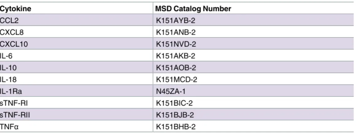
Meso Scale Discovery (MSD, Rockville, Maryland) singleplex assays were used to quantify
analyte concentrations following the manufacturer‘s instructions. Catalog numbers for the
reagent kits used are provided at
Table 2
.
For all assays, concentrations were determined based on standard curves created using
serial dilutions of fresh aliquots of constant recombinant lab standards prepared in culture
medium and stored at -80˚C in individual 400 uL aliquots (Cedarlane, Burlington, Canada;
PeproTech, Quebec, Canada; R&D Systems, Minneapolis, Minnesota). Longitudinal sample
sets from each individual were always paired on the same assay plates. Median intra-assay
vari-ation was typically below 5%.
Statistics
Group results are presented as medians, with each point representing the average (mean)
value obtained from duplicate or triplicate analyses of an individual woman’s plasma at that
timepoint. Supporting information
S1 File
contains the raw duplicates or triplicates used to
calculate the mean values for each cytokine or chemokine, for each woman, at each visit,
under each condition examined, that were then used to create the figures presented. Data for
each population were analyzed using GraphPad Prism (La Jolla, California). Pairwise
compari-sons (Wilcoxon Matched Pairs/Signed Rank tests) were used for most data sets. The Spearman
rank order correlation coefficient (non-parametric) was used to assess potential associations
between plasma biomarker levels and increasing gestational age. Differences were considered
significant at the 95% confidence level (two-tailed p<0.05).
Table 2. Catalog numbers for Meso Scale Discovery kits used in this publication.
Cytokine MSD Catalog Number
CCL2 K151AYB-2 CXCL8 K151ANB-2 CXCL10 K151NVD-2 IL-6 K151AKB-2 IL-10 K151AOB-2 IL-18 K151MCD-2 IL-1Ra N45ZA-1 sTNF-RI K151BIC-2 sTNF-RII K151BJB-2 TNFα K151BHB-2 https://doi.org/10.1371/journal.pone.0177813.t002
Results
Alterations in basal innate immune balance are characteristic of healthy
human pregnancy
This longitudinal study aimed to determine whether the constitutive immune status of healthy
pregnant women exhibits a pro- or anti-inflammatory bias and whether it changes during
pregnancy. As described in detail at Material and Methods, plasma samples were obtained at a
single time point during pregnancy, again one year postpartum, and for a randomly obtained
subset of individuals who did not experience further pregnancies in the interim, at three years
postpartum. Two representative biomarkers of pro- and anti-inflammatory responses were
assessed initially (
Fig 1
). Pro-inflammatory chemokine CCL2 was readily detectible in all
indi-viduals. Among the 251 women examined, systemic CCL2 was found to be reduced by 40%
during pregnancy compared to levels one year postpartum (p<0.0001, Wilcoxon matched
pairs, medians 110 vs. 154 pg/mL). Conversely, sTNF-RI was 38% elevated during pregnancy
(medians 1,266 vs. 913, p<0.0001). Thus, the median ratio of these
anti-inflammatory:pro-inflammatory innate biomarkers (sTNF-RI:CCL2) changed from 11.4 during pregnancy to 5.9
a year following delivery. This suggests that constitutive maternal status may incline towards
an anti-inflammatory balance over the course of pregnancy.
Given the large number of pro- and anti-inflammatory cytokines and inhibitors involved in
human immune regulation, this analysis was extended in approximately every second woman
(n = 120) to a panel of five additional pro-inflammatory biomarkers (CXCL10, TNFα, IL-18,
IL-6 and CXCL8) as well as three more anti-inflammatory biomarkers (IL-10, sTNF-RII and
IL-1Ra). The cytokines selected were chosen on the basis of being (i) routinely present in the
plasma of healthy humans and (ii) having been extensively implicated in immune regulation
of inflammation in murine and human studies. These large panels of biomarkers were used in
preference to relying upon a single biomarker (CRP or IL-6 or IL-10, as is commonly done) in
order to strengthen the capacity to draw conclusions about pro- vs. anti- inflammatory bias
in
vivo during pregnancy. A brief overview of the major cellular sources and biological functions
of the cytokines, chemokines and receptors studied is provided in
Table 3
.
Fig 2
demonstrates that during healthy pregnancy, most pro-inflammatory cytokines were
indeed reduced relative to levels in the same women one year postpartum. Thus, constitutive
CXCL10, TNFα and IL-18 were reduced during pregnancy. Median IL-6 trended lower by 42%,
but the difference did not reach statistical significance. There was no evidence of changes in
con-stitutive CXCL8 levels during healthy pregnancy. Note that due to the inherent immunological
Fig 1. Among healthy women, a systemic bias towards a resting anti-inflammatory phenotype is evident in vivo during pregnancy. Paired plasma samples from 251 women are assessed during and one year following healthy pregnancy. Data are presented as pg/mL. Bars represent median values of each data set. Wilcoxon paired analyses are shown.
Table 3. Cytokine, chemokine and soluble receptor biomarkers examined in this publication.
Cytokine Major Sources Major Functions Major role as a Pro- or
Anti-Inflammatory immune response modifier CCL2
(MCP-1)
Macrophages, monocytes, endothelial cells, fibroblasts, epithelial cells, smooth muscle cells, mesangial cells, astrocytic cells, monocytic and microglial cells [20].
CCL2 recruits monocytes, memory T cells, and dendritic cells to sites of inflammation [21–23]. It has been Implicated in pathogenesis or exacerbation of many inflammatory diseases including asthma, atherosclerosis, gestational diabetes mellitus, hypertension and rheumatoid arthritis [24,25].
Pro-Inflammatory
CXCL8 (IL-8)
Macrophages, monocytes, epithelial cells, airway smooth muscle cells and endothelial cells [26,27].
CXCL8 is an inflammatory chemokine involved in neutrophil (and other granulocyte) recruitment/ chemotaxis towards sites of infection and subsequent phagocytosis/ degranulation [27]. It is also a potent promoter of angiogenesis [28–30].
Pro-Inflammatory
CXCL10 (IP-10)
Macrophages, monocytes, endothelial cells and fibroblasts [31].
CXCL10 is involved in chemoattraction for monocytes/ macrophages, T cells, NK cells, and dendritic cells [32, 33]. It is also involved in the promotion of T cell adhesion to endothelial cells, enhances Th1 activity and has antitumor activity [32–35].
Pro-Inflammatory
IL-6 Macrophages, monocytes, T cells, endothelial cells, placental cells and adipocytes [20,35,36].
In response to infection (e.g. viruses, bacteria) or trauma (e.g. surgery, burns, tissue damage), IL-6 is secreted and stimulates inflammation [37]. It is an important mediator of fever, the acute phase response, neutrophil production in bone marrow, supports B cell growth and is antagonistic to regulatory T cells [35]. Clinically, it stimulates inflammatory processes in host defense and many diseases including diabetes [38], atherosclerosis [39], depression [40], Alzheimer’s [41], systemic lupus erythematosus [42], multiple myeloma [43], prostate cancer [44], and rheumatoid arthritis [45].
Pro-Inflammatory
IL-10 Macrophages, monocytes, T cells, B cells, NK cells, mastocytes and adipocytes [20,46,47].
IL-10 inhibits T cell derived cytokine production, MHC class II and co-stimulatory molecule expression, and enhances B cell survival, proliferation, and antibody production [48–52]. It inhibits TLR mediated induction of multiple pro-inflammatory cytokines including TNFα, IL-1β, IL-12, and IFNγ[46,47].
Anti-Inflammatory
IL-18 Macrophages, monocytes, dendritic cells, endothelial cells, keratinocytes and intestinal epithelial cells [53,54].
IL-18 induces cell-mediated immunity following infection with microbial products (e.g. LPS) [53]. IL-18 stimulation of NK and T cells stimulates IFNγrelease, activating macrophages and other cells [55–57].
Pro-Inflammatory
IL-1Ra Macrophages, monocytes, neutrophils, mast cells, epithelial cells and adipocytes [46,54,58–60].
IL-1Ra inhibits the activities of pro-inflammatory cytokines IL-1αand IL-1β, modulating IL-1 related immune and inflammatory responses [58,61–63].
Anti-Inflammatory
sTNF-RI Macrophages, monocytes, T cells, B cells, NK cells, dendritic cells, endothelial cells and epithelial cells [46,54,64].
sTNF-RI is generated by the shedding of its
membrane-expressed counterpart (TNF-RI) [65]. Thus, sTNF-RI has inherent anti-inflammatory activity, as it competes with membrane-associated receptors for the binding of free cytokines, confining the
pro-inflammatory cytokine activity to the local site of inflammation [65,66].
Anti-Inflammatory
sTNF-RII Macrophages, monocytes, T cells, B cells, NK cells, dendritic cells, endothelial cells and epithelial cells [46,54,64].
sTNF-RII is generated via shedding of a membrane-expressed counterpart (TNF-RII) [65]. As for sTNF-RI, it competes with membrane-associated signaling receptors for the TNF binding, confining the pro-inflammatory cytokine activity to the local site of inflammation [65,66], it has inherent anti-inflammatory activity.
Anti-Inflammatory
TNFα Macrophages, monocytes, T cells, NK cells, neutrophils, mast cells, eosinophils and neurons [20, 67].
TNFαis a major player in chronic and acute
inflammation [67,68]. It also inhibits tumorigenesis and viral replication [67]. TNFαdysregulation has been implicated in a broad variety of human inflammatory diseases [68,69].
Pro-Inflammatory
diversity characteristic of human populations, and to avoid compression of clearly positive but
low levels of cytokine expression in some individuals, y-axis breaks are included.
Anti-inflammatory biomarkers (sTNF-RI, sTNF-RII, and IL-1Ra) demonstrated that unlike
the reductions seen in constitutive pro-inflammatory cytokine expression, anti-inflammatory
biomarker levels were elevated during pregnancy (Figs
1
and
3
, p<0.0001). Interestingly,
Fig 2. Basal levels of multiple pro-inflammatory innate biomarkers are reduced in vivo during healthy pregnancy. n = 120 longitudinal sample pairs. Bars represent median values of each data set. Wilcoxon paired analyses are shown.
https://doi.org/10.1371/journal.pone.0177813.g002
Fig 3. Constitutive expression of multiple anti-inflammatory biomarkers is increased during pregnancy. n = 120 longitudinal sample pairs. Bars represent median values of each data set. Wilcoxon paired analyses are shown.
plasma IL-10, readily quantified in ~85% of the population, did not differ. Taken together,
the data suggest that healthy pregnancy is characterized by transiently decreased pro-, and
increased anti-inflammatory expression. This self-resolves by one year postpartum.
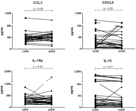
Validity of one year postpartum as a surrogate for the non-pregnant state
It is not logistically feasible to recruit women one year prior to an anticipated conception in
order to assess their then basal systemic immune status. We therefore addressed the possibility
that one year postpartum does not allow sufficient time for the innate immune system to return
to the non-pregnant status, hence is not an appropriate comparison group. 32 randomly
selected women who remained non-pregnant over the two years following the one year
post-partum visit were examined. At this year three visit, plasma samples were obtained and
com-pared (at the same time, in parallel assays) with plasma obtained from the same women at one
year postpartum. One pro- and one anti-inflammatory biomarker that had exhibited
differen-tial expression during pregnancy (CCL2 and IL-1Ra) and one pro- and one anti-inflammatory
biomarker that had been unchanged during pregnancy (CXCL8 and IL-10) were examined.
Fig
4
demonstrates that the level of each cytokine was stable within an individual at one and three
years post-pregnancy. This supports the concept that one (or three) year postpartum samples
offer an appropriately stable comparator for non-pregnant status in this longitudinal study.
Anti-inflammatory bias is evident at least as early as the second
trimester
The comparisons above test the hypothesis that resting constitutive innate immune status is
distinct during healthy pregnancy. To better understand the kinetics of this shift in resting
immune status, the population was next stratified by trimester of recruitment. With only a
small number of women recruited during the first trimester, analysis centred on the second
Fig 4. Stability of in vivo pro- and anti-inflammatory plasma biomarkers. n = 32 longitudinal sample pairs. Wilcoxon paired analyses are shown.
and third trimesters.
Fig 5
demonstrates basal (i.e. healthy, resting) innate immune status is
skewed towards an anti-inflammatory phenotype at least as early as the second trimester.
The intensity of anti-inflammatory bias increases with increasing
gestational age
Finally, to determine if the
intensity of anti-inflammatory phenotypic changes are altered with
the progression of pregnancy, we assessed relationships between plasma biomarker
concentra-tions and gestational age. sTNF-RI, IL-1Ra (
Fig 6
) and sTNF-RII (not shown) all exhibited
increasingly intense
in vivo expression with increased gestational age. Conversely, the decrease
in pro-inflammatory cytokine expression (
Fig 6
: CCL2 and IL-18; data not shown: CXCL8,
CXCL10, IL-6 and TNFα) was constant and exhibited no relationship with increasing
gesta-tional age. Thus, the shift towards an increasingly anti-inflammatory bias intensified with
increasing gestational age during healthy pregnancy.
Fig 5. Longitudinal analysis of differences in pro- and anti-inflammatory cytokines in second and third trimesters. Independent panels of volunteers were examined during the second or third trimester and compared with levels one year postpartum. Bars represent median values of each data set. Wilcoxon paired analyses are shown.
https://doi.org/10.1371/journal.pone.0177813.g005
Fig 6. Relationships between gestational age and intensity of anti-inflammatory or pro-inflammatory plasma biomarker expression. Spearman regression analyses are shown.
Discussion
Although most pregnancies lead to healthy infants, prior research has focused heavily on
dis-eases of pregnancy. Here, maternal health was examined in 251 healthy pregnant women over
up to three years. The data reveal that at least as early as the second trimester, women exhibit
increasingly strong constitutive expression of anti-inflammatory mediators and reduced
expression of many, but not all, cytokines linked to host defense and inflammatory immune
capacity. This transient anti-inflammatory phenotype intensifies with increasing gestational
age and is self-resolving by one year postpartum.
Our primary focus here is maternal health and the systemic, constitutive innate immune
sta-tus exhibited by women during pregnancy. This is not an investigation of factors which enable
pregnancy to become established, to proceed to term, or to respond to acute viral or bacterial
infection. There is a longstanding lack of agreement regarding constitutive innate immune
sta-tus during a healthy pregnancy. Small cohorts of healthy pregnant women are incorporated in
most studies that examine diseases of pregnancy. In those studies, cross-sectional comparisons
are drawn with a broad range of difficult pregnancies, rather than explicitly using well-powered
longitudinal study designs to examine the status of healthy women during vs. post-pregnancy.
Not surprisingly, such studies do not provide a clear consensus. One school argues that systemic
immune status
in vivo, unlike immune status at the maternal/fetal interface, reflects a
general-ized mild inflammatory response [
70
–
73
]. Others argue for the dominance of
anti-inflamma-tory responses [
74
,
75
]. These apparent contradictions may be due to cross-sectional study
designs, cohorts of relatively small size, the resulting impact of subject to subject variability, and
reliance on a small number of biomarkers (often one) to assess innate immune bias
in vivo. For
example, two of the largest longitudinal studies available [
76
,
77
] are composed of 48 and 21
healthy women respectively who are followed through pregnancy and postpartum. Many
stud-ies involve different ethnicitstud-ies: Kraus included a 94% Hispanic/Black population [
76
], while
Szarka utilized a Caucasian population [
78
]. As pointed out more than two decades ago [
79
],
due to high inter-subject variability inherent to any outbred population, a longitudinal,
matched study design involving a large n is logistically challenging, but highly important.
This research has important caveats. Firstly, because a longitudinal, multi-year study design
was selected to achieve increased sensitivity, it was not socially or logistically feasible to recruit
individuals prior to an anticipated pregnancy. For this reason, pregnant women were
com-pared with their immune status at one year and, for a subset, three years postpartum.
Pre-preg-nancy immune status could not be directly determined. Secondly, a potential confounder that
cannot be ruled out is the impact of specific environmental antigens and pathogens on these
findings. Women with complicated pregnancies with known linkages to altered innate
immu-nity (i.e. preeclampsia) are excluded from this study. Among the women studied, clinical
assessments at each blood draw excluded women with active transient URT or GI infections.
The data reported above, characterizing the constitutive
in vivo phenotype of healthy normal
pregnancy positions us to better assess the impact of environmental and genetic influences in
pregnancy in subsequent studies. A third caveat is that the impact of menstrual stage
postpar-tum at sampling could influence some values obtained. This underlines the need for sufficient
power because the impact of such variables is increasingly reduced as the number of
individu-als examined increases.
A potential confounder to interpretation of the data obtained was day-to-day stability of
biomarker expression within a given individual. We found this to have minimal impact
because, as shown in
Fig 4
, and as was previously reported in short-term studies conducted in
men and non-pregnant women [
76
,
80
], systemic levels of most biomarkers are remarkably
stable across time (days-weeks) in healthy individuals.
cessful pregnancy [
75
].
These data may stand in contrast to
in vivo expression of acute phase proteins during
healthy pregnancy. Multiple studies have found increased plasma/serum CRP levels vs.
non-pregnant controls [
83
–
86
]. The extent to which changes in this largely IL-6 and TNFα driven
biomarker of inflammation are due to activation of classical innate immune responses or
other cells (i.e. adipocytes or necrotic processes associated with placenta ageing) is under active
investigation [
83
,
87
,
88
]. Interestingly, several groups have found undetectable changes or
decreases in CRP over the course of healthy pregnancy [
83
,
89
–
92
]. This inconsistency, and
the many pregnancy independent factors (i.e. obesity) that can influence CRP levels,
under-lines a need for caution and continued research prior to drawing mechanistic conclusions
about the role any of these mediators play in successful conclusion of pregnancy.
In this study, we do not address putative differences in maternal immune responses upon
infection
in vivo or acute in vitro activation. Studies examining in vitro responses to various
pattern recognition receptor (PRR) ligands are currently underway as a complementary
approach to understanding changes in maternal health that can occur during pregnancy.
With increasing attention given to defining what constitutes a healthy pregnancy [
93
], this
study provides valuable insight into systemic innate immune changes that result in expression
of an increasingly intense anti-inflammatory phenotype
in vivo during pregnancy. It
under-lines the need for further characterization of what constitutes a successful environment for
healthy human pregnancy.
Supporting information
S1 File. Raw duplicates and triplicates from immunological assays performed.
(XLSX)
Acknowledgments
We are grateful to all the families who took part in this study and the entire CHILD research
team. We also thank Saiful Huq for database assistance and Paul Lopez for editorial assistance.
CHILD (Canadian Healthy Infant Longitudinal Development Study, headquartered at
McMaster University, Hamilton, Canada) investigators include: MR Sears (Director),
McMas-ter University; P Subbarao (co-Director), The Hospital for Sick Children; R Allen, Simon
Fra-ser University; SS Anand, McMaster University; AB Becker, University of Manitoba; AD
Befus, University of Alberta; M Brauer, University of British Columbia; JR Brook, University
of Toronto; E Chen, Northwestern University, Chicago; M Cyr, McMaster University; D
Daley, University of British Columbia; S Dell, Sick Children’s Hospital; JA Denburg, McMaster
University; S Elliott, University of Waterloo; H Grasemann, Sick Children’s Hospital; K
Hay-Glass, University of Manitoba; R Hegele, Sick Children’s Hospital; DL Holness, University of
Toronto; WYW Lou, University of Toronto; MS Kobor, University of British Columbia; TR
Kollman, University of British Columbia; AL Kozyrskyj, University of Alberta; C Laprise,
Uni-versite du Quebec a Chicoutimi; M Larche, McMaster University; J Macri, McMaster
Univer-sity; PM Mandhane, University of Alberta; G Miller, Northwestern University, Chicago; R
Moqbel (deceased), University of Manitoba; T Moraes, Sick Children’s Hospital; PD Pare,
University of British Columbia; C Ramsey, University of Manitoba; F Ratjen, Sick Children’s
Hospital; A Sandford, University of British Columbia; JA Scott, University of Toronto; J Scott,
University of Toronto; F Silverman, University of Toronto; T Takaro, Simon Fraser
Univer-sity; P Tang, University of British Columbia; S Tebbutt, University of British Columbia; T To,
Sick Children’s Hospital; SE Turvey, University of British Columbia, all in Canada.
Author Contributions
Conceptualization: KTH ABB CG RC.
Data curation: CG RC KTH MRS.
Formal analysis: KTH CG ABB RC.
Funding acquisition: MRS ABB KTH SET PS PJM.
Investigation: KTH ABB CG RC WPS.
Methodology: CG RC WPS ABB MRS SET PJM PS KTH.
Project administration: KTH ABB RC MRS.
Resources: CG RC WPS ABB MRS SET PJM PS KTH.
Supervision: KTH ABB MRS.
Validation: KTH ABB CG RC WPS.
Visualization: CG WPS.
Writing – original draft: KTH CG.
Writing – review & editing: CG RC WPS ABB MRS SET PJM PS KTH CHILD.
References
1. Chaouat G, Tranchot Diallo J, Volumenie JL, Menu E, Gras G, Delage G, et al. Immune suppression and Th1/Th2 balance in pregnancy revisited: a (very) personal tribute to Tom Wegmann. Am J Reprod Immunol. 1997 Jun; 37(6):427–34. PMID:9228297
2. St Louis D, Romero R, Plazyo O, Arenas-Hernandez M, Panaitescu B, Xu Y, et al. Invariant NKT Cell Activation Induces Late Preterm Birth That Is Attenuated by Rosiglitazone. J Immunol. 2016 Feb 01; 196(3):1044–59.https://doi.org/10.4049/jimmunol.1501962PMID:26740111
3. PrabhuDas M, Bonney E, Caron K, Dey S, Erlebacher A, Fazleabas A, et al. Immune mechanisms at the maternal-fetal interface: perspectives and challenges. Nat Immunol. 2015 Apr; 16(4):328–34. https://doi.org/10.1038/ni.3131PMID:25789673
4. Svensson-Arvelund J, Mehta RB, Lindau R, Mirrasekhian E, Rodriguez-Martinez H, Berg G, et al. The human fetal placenta promotes tolerance against the semiallogeneic fetus by inducing regulatory T cells and homeostatic M2 macrophages. J Immunol. 2015 Feb 15; 194(4):1534–44.https://doi.org/10. 4049/jimmunol.1401536PMID:25560409
5. Baylis F. Pregnant women deserve better. Nature. 2010 Jun 10; 465(7299):689–90.https://doi.org/10. 1038/465689aPMID:20535185
6. Catalano PW, MA. Wise, PH. Bianchi, DW. Saade, GR. Scientific Vision Workshop on Pregnancy and Pregnancy Outcomes. Bethesa, Maryland2011 [updated 2011; cited 2016 02/18/2016];https://www. nichd.nih.gov/vision/vision_themes/pregnancy/Documents/Vision_Pregnancy_WP_042811.pdf. 7. Foulkes MA, Grady C, Spong CY, Bates A, Clayton JA. Clinical research enrolling pregnant women: a
12. von Mutius E, Martinez FD. Inconclusive Results of Randomized Trials of Prenatal Vitamin D for Asthma Prevention in Offspring: Curbing the Enthusiasm. JAMA. 2016 Jan 26; 315(4):347–8.https://doi.org/10. 1001/jama.2015.18963PMID:26813205
13. Tyrrell J, Richmond RC, Palmer TM, Feenstra B, Rangarajan J, Metrustry S, et al. Genetic Evidence for Causal Relationships Between Maternal Obesity-Related Traits and Birth Weight. JAMA. 2016 Mar 15; 315(11):1129–40.https://doi.org/10.1001/jama.2016.1975PMID:26978208
14. Aagaard-Tillery KM, Silver R, Dalton J. Immunology of normal pregnancy. Semin Fetal Neonatal Med. 2006 Oct; 11(5):279–95.https://doi.org/10.1016/j.siny.2006.04.003PMID:16784908
15. Visser N, van Rijn BB, Rijkers GT, Franx A, Bruinse HW. Inflammatory changes in preeclampsia: cur-rent understanding of the maternal innate and adaptive immune response. Obstet Gynecol Surv. 2007 Mar; 62(3):191–201.https://doi.org/10.1097/01.ogx.0000256779.06275.c4PMID:17306041 16. Aye IL, Lager S, Ramirez VI, Gaccioli F, Dudley DJ, Jansson T, et al. Increasing maternal body mass
index is associated with systemic inflammation in the mother and the activation of distinct placental inflammatory pathways. Biol Reprod. 2014 Jun; 90(6):129.https://doi.org/10.1095/biolreprod.113. 116186PMID:24759787
17. Chawes BL, Stokholm J, Bonnelykke K, Brix S, Bisgaard H. Neonates with reduced neonatal lung func-tion have systemic low-grade inflammafunc-tion. J Allergy Clin Immunol. 2015 Jun; 135(6):1450–6 e1. https://doi.org/10.1016/j.jaci.2014.11.020PMID:25579483
18. Subbarao P, Anand SS, Becker AB, Befus AD, Brauer M, Brook JR, et al. The Canadian Healthy Infant Longitudinal Development (CHILD) Study: examining developmental origins of allergy and asthma. Thorax. 2015 Oct; 70(10):998–1000.https://doi.org/10.1136/thoraxjnl-2015-207246PMID:26069286 19. Moraes TJ, Lefebvre DL, Chooniedass R, Becker AB, Brook JR, Denburg J, et al. The Canadian healthy
infant longitudinal development birth cohort study: biological samples and biobanking. Paediatr Perinat Epidemiol. 2015 Jan; 29(1):84–92.https://doi.org/10.1111/ppe.12161PMID:25405552
20. Pendeloski KP, Ono E, Torloni MR, Mattar R, Daher S. Maternal obesity and inflammatory mediators: A controversial association. Am J Reprod Immunol. 2017 Mar 22;
21. Carr MW, Roth SJ, Luther E, Rose SS, Springer TA. Monocyte chemoattractant protein 1 acts as a T-lymphocyte chemoattractant. Proc Natl Acad Sci U S A. 1994 Apr 26; 91(9):3652–6. PMID:8170963 22. Xu LL, Warren MK, Rose WL, Gong W, Wang JM. Human recombinant monocyte chemotactic protein
and other C-C chemokines bind and induce directional migration of dendritic cells in vitro. J Leukoc Biol. 1996 Sep; 60(3):365–71. PMID:8830793
23. Lam VC, Lanier LL. NK cells in host responses to viral infections. Curr Opin Immunol. 2016 Dec 13;4443–51.
24. Xia M, Sui Z. Recent developments in CCR2 antagonists. Expert Opin Ther Pat. 2009 Mar; 19(3):295– 303.https://doi.org/10.1517/13543770902755129PMID:19441905
25. O’Connor T, Borsig L, Heikenwalder M. CCL2-CCR2 Signaling in Disease Pathogenesis. Endocr Metab Immune Disord Drug Targets. 2015 15(2):105–18. PMID:25772168
26. Hedges JC, Singer CA, Gerthoffer WT. Mitogen-activated protein kinases regulate cytokine gene expression in human airway myocytes. Am J Respir Cell Mol Biol. 2000 Jul; 23(1):86–94.https://doi. org/10.1165/ajrcmb.23.1.4014PMID:10873157
27. Harada A, Sekido N, Akahoshi T, Wada T, Mukaida N, Matsushima K. Essential involvement of interleu-kin-8 (IL-8) in acute inflammation. J Leukoc Biol. 1994 Nov; 56(5):559–64. PMID:7964163
28. Baggiolini M, Walz A, Kunkel SL. Neutrophil-activating peptide-1/interleukin 8, a novel cytokine that acti-vates neutrophils. J Clin Invest. 1989 Oct; 84(4):1045–9.https://doi.org/10.1172/JCI114265PMID: 2677047
29. Hosoki K, Itazawa T, Boldogh I, Sur S. Neutrophil recruitment by allergens contribute to allergic sensiti-zation and allergic inflammation. Curr Opin Allergy Clin Immunol. 2016 Feb; 16(1):45–50.https://doi. org/10.1097/ACI.0000000000000231PMID:26694038
30. Tan SY, Weninger W. Neutrophil migration in inflammation: intercellular signal relay and crosstalk. Curr Opin Immunol. 2016 Dec 09;4434–42.
31. Luster AD, Unkeless JC, Ravetch JV. Gamma-interferon transcriptionally regulates an early-response gene containing homology to platelet proteins. Nature. 1985 Jun 20–26; 315(6021):672–6. PMID: 3925348
32. Dufour JH, Dziejman M, Liu MT, Leung JH, Lane TE, Luster AD. IFN-gamma-inducible protein 10 (IP-10; CXCL10)-deficient mice reveal a role for IP-10 in effector T cell generation and trafficking. J Immu-nol. 2002 Apr 01; 168(7):3195–204. PMID:11907072
33. Angiolillo AL, Sgadari C, Taub DD, Liao F, Farber JM, Maheshwari S, et al. Human interferon-inducible protein 10 is a potent inhibitor of angiogenesis in vivo. J Exp Med. 1995 Jul 01; 182(1):155–62. PMID: 7540647
34. Campbell JD, Gangur V, Simons FE, HayGlass KT. Allergic humans are hyporesponsive to a CXCR3 ligand-mediated Th1 immunity-promoting loop. FASEB J. 2004 Feb; 18(2):329–31.https://doi.org/10. 1096/fj.02-0908fjePMID:14657006
35. Hamidzadeh K, Christensen SM, Dalby E, Chandrasekaran P, Mosser DM. Macrophages and the Recovery from Acute and Chronic Inflammation. Annu Rev Physiol. 2017 Feb 10;79567–92. 36. Hunter CA, Jones SA. IL-6 as a keystone cytokine in health and disease. Nat Immunol. 2015 May; 16
(5):448–57.https://doi.org/10.1038/ni.3153PMID:25898198
37. van der Poll T, Keogh CV, Guirao X, Buurman WA, Kopf M, Lowry SF. Interleukin-6 gene-deficient mice show impaired defense against pneumococcal pneumonia. J Infect Dis. 1997 Aug; 176(2):439–44. PMID:9237710
38. Kristiansen OP, Mandrup-Poulsen T. Interleukin-6 and diabetes: the good, the bad, or the indifferent? Diabetes. 2005 Dec; 54 Suppl 2S114–24.
39. Dubinski A, Zdrojewicz Z. [The role of interleukin-6 in development and progression of atherosclerosis]. Pol Merkur Lekarski. 2007 Apr; 22(130):291–4. PMID:17684929
40. Dowlati Y, Herrmann N, Swardfager W, Liu H, Sham L, Reim EK, et al. A meta-analysis of cytokines in major depression. Biol Psychiatry. 2010 Mar 01; 67(5):446–57.https://doi.org/10.1016/j.biopsych.2009. 09.033PMID:20015486
41. Swardfager W, Lanctot K, Rothenburg L, Wong A, Cappell J, Herrmann N. A meta-analysis of cytokines in Alzheimer’s disease. Biol Psychiatry. 2010 Nov 15; 68(10):930–41.https://doi.org/10.1016/j. biopsych.2010.06.012PMID:20692646
42. Tackey E, Lipsky PE, Illei GG. Rationale for interleukin-6 blockade in systemic lupus erythematosus. Lupus. 2004 13(5):339–43.https://doi.org/10.1191/0961203304lu1023oaPMID:15230289
43. Gado K, Domjan G, Hegyesi H, Falus A. Role of INTERLEUKIN-6 in the pathogenesis of multiple mye-loma. Cell Biol Int. 2000 24(4):195–209.https://doi.org/10.1006/cbir.2000.0497PMID:10816321 44. Smith PC, Hobisch A, Lin DL, Culig Z, Keller ET. Interleukin-6 and prostate cancer progression.
Cyto-kine Growth Factor Rev. 2001 Mar; 12(1):33–40. PMID:11312117
45. Nishimoto N. Interleukin-6 in rheumatoid arthritis. Curr Opin Rheumatol. 2006 May; 18(3):277–81. https://doi.org/10.1097/01.bor.0000218949.19860.d1PMID:16582692
46. Banchereau J, Pascual V, O’Garra A. From IL-2 to IL-37: the expanding spectrum of anti-inflammatory cytokines. Nat Immunol. 2012 Oct; 13(10):925–31.https://doi.org/10.1038/ni.2406PMID:22990890 47. Gabrysova L, Howes A, Saraiva M, O’Garra A. The regulation of IL-10 expression. Curr Top Microbiol
Immunol. 2014 380157–90.
48. Mosser DM, Zhang X. Interleukin-10: new perspectives on an old cytokine. Immunol Rev. 2008 Dec;226205–18.
49. Pestka S, Krause CD, Sarkar D, Walter MR, Shi Y, Fisher PB. Interleukin-10 and related cytokines and receptors. Annu Rev Immunol. 2004 22929–79.
50. Saraiva M, O’Garra A. The regulation of IL-10 production by immune cells. Nat Rev Immunol. 2010 Mar; 10(3):170–81.https://doi.org/10.1038/nri2711PMID:20154735
51. de Waal Malefyt R, Abrams J, Bennett B, Figdor CG, de Vries JE. Interleukin 10(IL-10) inhibits cytokine synthesis by human monocytes: an autoregulatory role of IL-10 produced by monocytes. J Exp Med. 1991 Nov 01; 174(5):1209–20. PMID:1940799
52. Moore KW, de Waal Malefyt R, Coffman RL, O’Garra A. Interleukin-10 and the interleukin-10 receptor. Annu Rev Immunol. 2001 19683–765.
53. Dinarello CA, Novick D, Kim S, Kaplanski G. Interleukin-18 and IL-18 binding protein. Front Immunol. 2013 Oct 08;4289.
54. Banchereau R, Cepika AM, Banchereau J, Pascual V. Understanding Human Autoimmunity and Auto-inflammation Through Transcriptomics. Annu Rev Immunol. 2017 Jan 30;
17097645
59. Di Paolo NC, Shayakhmetov DM. Interleukin 1alpha and the inflammatory process. Nat Immunol. 2016 Jul 19; 17(8):906–13.https://doi.org/10.1038/ni.3503PMID:27434011
60. Ito Y, Kaneko N, Iwasaki T, Morikawa S, Kaneko K, Masumoto J. IL-1 as a target in inflammation. Endocr Metab Immune Disord Drug Targets. 2015 15(3):206–11. PMID:26333726
61. Dinarello CA. The interleukin-1 family: 10 years of discovery. FASEB J. 1994 Dec; 8(15):1314–25. PMID:8001745
62. El-Omar EM, Carrington M, Chow WH, McColl KE, Bream JH, Young HA, et al. Interleukin-1 polymor-phisms associated with increased risk of gastric cancer. Nature. 2000 Mar 23; 404(6776):398–402. https://doi.org/10.1038/35006081PMID:10746728
63. Aksentijevich I, Masters SL, Ferguson PJ, Dancey P, Frenkel J, van Royen-Kerkhoff A, et al. An autoin-flammatory disease with deficiency of the interleukin-1-receptor antagonist. N Engl J Med. 2009 Jun 04; 360(23):2426–37.https://doi.org/10.1056/NEJMoa0807865PMID:19494218
64. Willrich MA, Murray DL, Snyder MR. Tumor necrosis factor inhibitors: clinical utility in autoimmune dis-eases. Transl Res. 2015 Feb; 165(2):270–82.https://doi.org/10.1016/j.trsl.2014.09.006PMID: 25305470
65. Ware CF, Santee S, Glass A. Tumor necrosis factor-related ligands and receptors. In: Thomson A, edi-tor. The Cytokine Handbook. Third ed. San Diego, CA: Academic Press; 1998. p. 549–92.
66. Wei M, Kuukasjarvi P, Laurikka J, Pehkonen E, Kaukinen S, Laine S, et al. Inflammatory cytokines and soluble receptors after coronary artery bypass grafting. Cytokine. 2001 Aug 21; 15(4):223–8.https:// doi.org/10.1006/cyto.2001.0920PMID:11563882
67. Locksley RM, Killeen N, Lenardo MJ. The TNF and TNF receptor superfamilies: integrating mammalian biology. Cell. 2001 Feb 23; 104(4):487–501. PMID:11239407
68. Baxter AE, Kaufmann DE. Tumor-necrosis factor is a master of T cell exhaustion. Nat Immunol. 2016 May; 17(5):476–8.https://doi.org/10.1038/ni.3436PMID:27092797
69. Green WD, Beck MA. Obesity altered T cell metabolism and the response to infection. Curr Opin Immu-nol. 2017 Mar 27;461–7.
70. Challis JR, Lockwood CJ, Myatt L, Norman JE, Strauss JF 3rd, Petraglia F. Inflammation and preg-nancy. Reprod Sci. 2009 Feb; 16(2):206–15.https://doi.org/10.1177/1933719108329095PMID: 19208789
71. Sacks G, Sargent I, Redman C. An innate view of human pregnancy. Immunol Today. 1999 Mar; 20 (3):114–8. PMID:10203701
72. Sacks GP, Studena K, Sargent K, Redman CW. Normal pregnancy and preeclampsia both produce inflammatory changes in peripheral blood leukocytes akin to those of sepsis. Am J Obstet Gynecol. 1998 Jul; 179(1):80–6. PMID:9704769
73. Blank V, Hirsch E, Challis JR, Romero R, Lye SJ. Cytokine signaling, inflammation, innate immunity and preterm labour—a workshop report. Placenta. 2008 Mar; 29 Suppl AS102–4.
74. Munn DH, Zhou M, Attwood JT, Bondarev I, Conway SJ, Marshall B, et al. Prevention of allogeneic fetal rejection by tryptophan catabolism. Science. 1998 Aug 21; 281(5380):1191–3. PMID:9712583 75. Chatterjee P, Chiasson VL, Bounds KR, Mitchell BM. Regulation of the Anti-Inflammatory Cytokines
Interleukin-4 and Interleukin-10 during Pregnancy. Front Immunol. 2014 5253.
76. Kraus TA, Sperling RS, Engel SM, Lo Y, Kellerman L, Singh T, et al. Peripheral blood cytokine profiling during pregnancy and post-partum periods. Am J Reprod Immunol. 2010 Dec; 64(6):411–26.https:// doi.org/10.1111/j.1600-0897.2010.00889.xPMID:20712812
77. Bjorkander S, Bremme K, Persson JO, van Vollenhoven RF, Sverremark-Ekstrom E, Holmlund U. Preg-nancy-associated inflammatory markers are elevated in pregnant women with systemic lupus
erythematosus. Cytokine. 2012 Aug; 59(2):392–9.https://doi.org/10.1016/j.cyto.2012.04.046PMID: 22633082
78. Szarka A, Rigo J Jr., Lazar L, Beko G, Molvarec A. Circulating cytokines, chemokines and adhesion molecules in normal pregnancy and preeclampsia determined by multiplex suspension array. BMC Immunol. 2010 1159.
79. Johnstone FD, Thong KJ, Bird AG, Whitelaw J. Lymphocyte subpopulations in early human pregnancy. Obstet Gynecol. 1994 Jun; 83(6):941–6. PMID:8190436
80. Campbell JD, Stinson MJ, Simons FE, Rector ES, HayGlass KT. In vivo stability of human chemokine and chemokine receptor expression. Hum Immunol. 2001 Jul; 62(7):668–78. PMID:11423172 81. Chaouat G, Assal Meliani A, Martal J, Raghupathy R, Elliott JF, Mosmann T, et al. IL-10 prevents
natu-rally occurring fetal loss in the CBA x DBA/2 mating combination, and local defect in IL-10 production in this abortion-prone combination is corrected by in vivo injection of IFN-tau. J Immunol. 1995 May 01; 154(9):4261–8. PMID:7722286
82. Thaxton JE, Sharma S. Interleukin-10: a multi-faceted agent of pregnancy. Am J Reprod Immunol. 2010 Jun; 63(6):482–91.https://doi.org/10.1111/j.1600-0897.2010.00810.xPMID:20163400
83. Belo L, Santos-Silva A, Rocha S, Caslake M, Cooney J, Pereira-Leite L, et al. Fluctuations in C-reactive protein concentration and neutrophil activation during normal human pregnancy. Eur J Obstet Gynecol Reprod Biol. 2005 Nov 01; 123(1):46–51.https://doi.org/10.1016/j.ejogrb.2005.02.022PMID: 16260340
84. Miller EM. Changes in serum immunity during pregnancy. Am J Hum Biol. 2009 May-Jun; 21(3):401–3. https://doi.org/10.1002/ajhb.20882PMID:19189417
85. Perucci LO, Carneiro FS, Ferreira CN, Sugimoto MA, Soriani FM, Martins GG, et al. Annexin A1 Is Increased in the Plasma of Preeclamptic Women. PLoS One. 2015 10(9):e0138475.https://doi.org/10. 1371/journal.pone.0138475PMID:26398190
86. McDade TW, Borja JB, Largado F, Adair LS, Kuzawa CW. Adiposity and Chronic Inflammation in Young Women Predict Inflammation during Normal Pregnancy in the Philippines. J Nutr. 2016 Feb; 146 (2):353–7.https://doi.org/10.3945/jn.115.224279PMID:26764318
87. Romem Y, Artal R. C-reactive protein in pregnancy and in the postpartum period. Am J Obstet Gynecol. 1985 Feb 01; 151(3):380–3. PMID:3970105
88. Rebelo F, Schlussel MM, Vaz JS, Franco-Sena AB, Pinto TJ, Bastos FI, et al. C-reactive protein and later preeclampsia: systematic review and meta-analysis taking into account the weight status. J Hyper-tens. 2013 Jan; 31(1):16–26.https://doi.org/10.1097/HJH.0b013e32835b0556PMID:23188419 89. Christian LM, Porter K. Longitudinal changes in serum proinflammatory markers across pregnancy and
postpartum: effects of maternal body mass index. Cytokine. 2014 Dec; 70(2):134–40.https://doi.org/10. 1016/j.cyto.2014.06.018PMID:25082648
90. Friis CM, Paasche Roland MC, Godang K, Ueland T, Tanbo T, Bollerslev J, et al. Adiposity-related inflammation: effects of pregnancy. Obesity (Silver Spring). 2013 Jan; 21(1):E124–30.
91. Stewart FM, Freeman DJ, Ramsay JE, Greer IA, Caslake M, Ferrell WR. Longitudinal assessment of maternal endothelial function and markers of inflammation and placental function throughout pregnancy in lean and obese mothers. J Clin Endocrinol Metab. 2007 Mar; 92(3):969–75.https://doi.org/10.1210/ jc.2006-2083PMID:17192290
92. Simavli S, Derbent AU, Uysal S, Turhan NO. Hepcidin, iron status, and inflammation variables among healthy pregnant women in the Turkish population. J Matern Fetal Neonatal Med. 2014 Jan; 27(1):75– 9.https://doi.org/10.3109/14767058.2013.804054PMID:23662610
93. NICHD. Pregnancy: For Researchers and Health Care Practitioners 2013;https://www.nichd.nih.gov/ health/topics/pregnancy/resources/Pages/providers.aspx.