Université de Montréal
Behavioral And Muscular Deficits Induced By Muscimol Injection Into The Primate Primary Motor Cortex During A Reach-To-Grasp Task
Par Eléonore Serrano
Département de Neurosciences, Faculté de Médecine
Mémoire présenté à la Faculté de Médecine en vue de l’obtention du grade de Maîtrise
en Neurosciences
Décembre 2019
Université de Montréal
Département de Neurosciences, Faculté de Médecine
Ce mémoire intitulé
Behavioral And Muscular Deficits Induced By Muscimol Injection Into The Primate Primary Motor Cortex During A Reach-To-Grasp Task
Présenté par Eléonore Serrano
A été évalué(e) par un jury composé des personnes suivantes
Dr Trevor Drew Président-rapporteur Dr Numa Dancause Directeur de recherche Dr Daniel Bourbonnais Membre du jury
i
Résumé
Le contrôle moteur fin et précis des doigts est une habileté importante dans la vie quotidienne pour écrire ou manger par exemple. Ce contrôle moteur est pris en charge par le cortex moteur primaire (M1) qui transmet le signal neuronal à la moelle épinière via la voie corticospinale. Le macaque rhésus est un excellent modèle pour étudier ce système moteur car, comme chez l’humain, il possède cette voie cortico-motoneuronale directe. Bien que les déficits du contrôle moteur de la main suite à des inactivations de M1 aient été étudiés sur des modèles de singes, peu d’études ont décrit les changements musculaires sous-tendant ces déficits. Le but de cette étude était d’évaluer les effets d’une inactivation partielle de M1 sur le comportement et l’activation du patron musculaire du membre supérieur chez le macaque rhésus. Pour ce faire, nous avons effectué des injections intra-corticales de Muscimol, un agoniste du GABA, pour inactiver temporairement l’aire de représentation de la main de M1. Des singes ont été entrainés à réaliser une tâche d’atteinte et de préhension qui requière l’utilisation du pouce et de l’index pour attraper une pastille de nourriture. En parallèle, les activités électromyographiques (EMG) des muscles proximaux et distaux du membre supérieur contralatéral aux sites d’injections ont été enregistrées. L’inactivation partielle de M1 entraine différents déficits moteurs comme une diminution du taux de succès, une perte des mouvements indépendants des doigts, une première flexion de l’index plus lente, et l’apparition de nouvelles stratégies de préhension pour attraper la pastille. Dans le cas de trouble sévère, les singes ont présentés tous ces déficits comportementaux. Ces troubles moteurs étaient sous-tendus par des activités musculaires anormales. En effet, les analyses EMG ont mis en évidence des changements dans les latences et les patrons d’activations musculaires des muscles proximaux et distaux au cours de la phase d’atteinte, d’ajustement et de préhension. Dans le cas de trouble modéré, les patrons d’activations musculaires étaient préservés malgré certain déficits visibles. Cependant, les patrons d’activations musculaires étaient altérés si la tâche demandait une rotation de l’avant-bras et de la main. Ces résultats montrent que les déficits comportementaux et les changements musculaires dépendent de la sévérité des troubles moteurs et/ou de la difficulté de la tâche (i.e. une rotation de l’avant-bras).
Mots-clés : cortex moteur primaire, Muscimol, contrôle fin des doigts, modèle de macaque, électromyographie
ii
Abstract
Fine digit movements contribute to many different aspects of our daily life and require appropriate muscle coordination. The main pathway through which M1 sends motor commands to spinal motor neurons is via the corticospinal tract. The rhesus macaque, like humans, have this direct corticomotoneuronal pathway of M1, making it a useful model to study this system. Although the effect of M1 inactivation on the control of the hand in term of behavioral changes has been studied in monkeys, little is known of how muscle activation patterns of the upper limb during reaching and grasping in monkeys becomes altered. The goal of this study was to evaluate the effect of a partial inactivation of the primary motor cortex (M1) in rhesus macaques on both behavioral performance and muscle activations. To do so we performed intra-cortical injections of Muscimol, a GABA agonist, to inactivate the hand area of M1. Monkeys performed a reach-to-grasp task that required a precision grip to retrieve a food pellet from a well. Electromyographic (EMG) activity of the proximal and distal muscles of the contralateral upper limb were recorded and quantified relative to the behavioral performance. We found that depending on the severity of the impairment, the Muscimol injection could induce several different movement abnormalities, such as decrease in the success rate, loss of independent finger movements, longer duration of the first flexion of the index finger, and use of alternate types of grasp to retrieve the food pellet. In cases of severe impairment, monkeys displayed all these movement abnormalities concurrently. In addition, we observed that behavioral deficits were associated with muscle discoordination. Indeed, EMG analysis revealed that the latencies and the muscle activation patterns were altered during the reach, hand preshaping and the grasp phases of the movement. These inappropriate EMG activities were visible on both proximal and distal muscles of the upper limb. In cases of mild impairment, monkeys had fewer behavioral deficits, but still showed some changes in the temporal muscle activation patterns. In contrast to the severe cases, the muscle activation patterns were more preserved. Interestingly, in the mild cases, the muscle activation patterns were altered if a rotation of the forearm was required by the task. Thus, we found that behavioral and muscular activation changes were dependent on the severity of the impairment and/or the difficulty of the task (i.e. required a rotation of the forearm).
Keywords: primary motor cortex, Muscimol, precision grip, macaque model, electromyography
iii
Table of contents
Résumé ... i
Abstract ... ii
Table of contents ... iii
List of table ... v
List of figures ... vi
List of abreviations ... viii
Remerciements ... x
Chapter 1 – General Introduction ... 1
1. Stroke, a burden on the health system ... 1
2. The motor system ... 2
2.1 Organization of primary motor cortex ... 2
2.2 Premotor areas ... 3
2.3 The corticospinal pathway ... 3
3. Motor cortical control and synergy ... 5
4. Motor impairments following stroke ... 7
5. Muscimol: a tool to evaluate cortical motor control ... 10
6. Objectives of the study... 15
Chapter 2 – Materials and methods ... 17
Training ... 17
Behavioral task ... 18
Surgical procedures ... 18
Mapping of the motor cortex and sites of injection ... 21
Protocol ... 21
Behavioral data analysis ... 22
EMG signals ... 24
Muscles onsets and offsets criteria and selection ... 24
Muscles latencies ... 25
iv
Chapter 3 – Results ... 28
The execution of the movement before inactivation ... 28
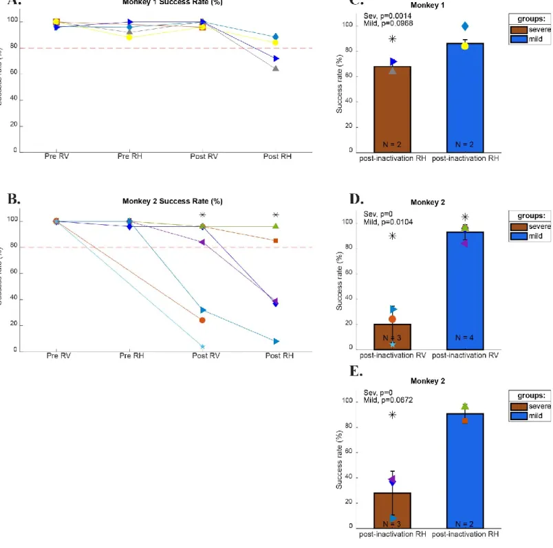
1. Evaluation of the success rate of the reach-to-grasp task ... 29
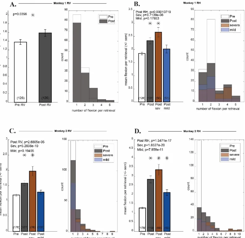
2. Number of flexions executed to grasp the pellet ... 32
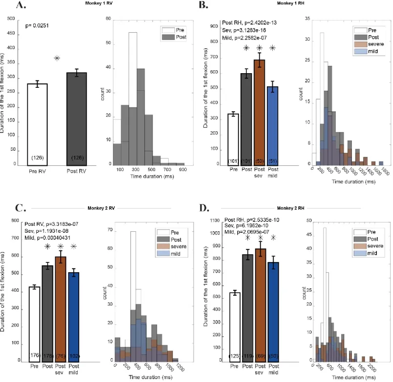
3. Evaluation of the duration of the first flexion to grasp the pellet ... 35
4. The effect of inactivation on the grasping configuration of the contralateral arm ... 38
5. The effect of inactivation on the reach and grasp phase duration ... 43
6. Example of EMG signals with Monkey 2 in the Vertical condition ... 46
7. The effects of partial M1 inactivation on the temporal activation of the contralateral arm’s muscles ... 48
8. The effect of partial M1 inactivation on muscles coordination of the contralateral arm ... 58
Chapter 4 – General Discussion ... 72
1. General summary ... 72
2. Dexterous motor control of the contralateral hand by the primary motor cortex (M1) ... 73
3. Reach versus Grasp ... 76
4. Impact of the inactivation on muscle activation patterns ... 78
5. Future direction ... 80
Conclusion ... 82
v
List of table
vi
List of figures
Figure 1: Overview of the experimental setup………20
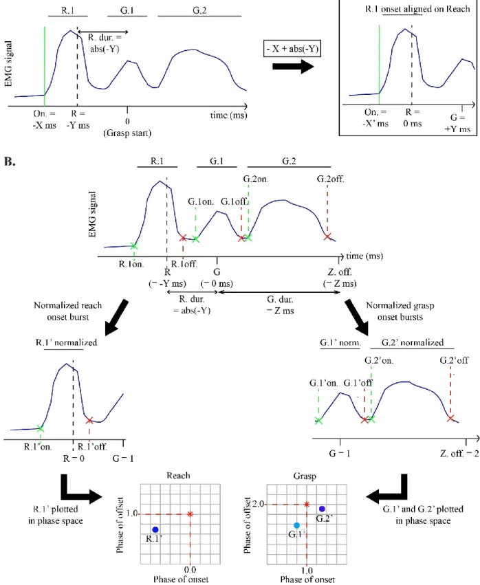
Figure 2: A schematic illustrating how burst onset and offset times for latencies and phase-plot were calculated………27
Figure 3: Monkeys Success Rate before and after inactivation………..31
Figure 4: Monkeys’ number of flexions before and after inactivation………34
Figure 5: Monkeys duration of the first flexion before and after inactivation………37
Figure 6: The hand start position and the grasping configurations used during the reach-to-grasp task before and after inactivation……….41
Figure 7: Changes in the reach and grasp phase duration after inactivation………45
Figure 8: Comparison of the EMG activity in Pre and Post of Monkey 2 during the reach-to-grasp task in Vertical condition……….47
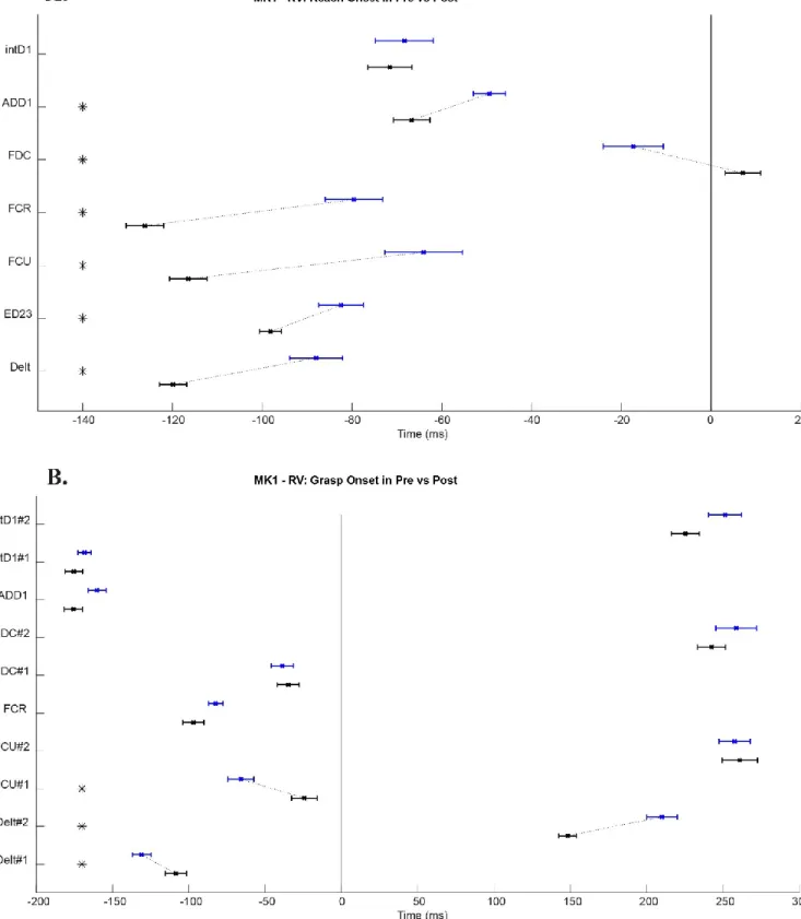
Figure 9: The muscles latencies before (Pre) and after (Post) inactivation for the Monkey 1 in the vertical condition………54
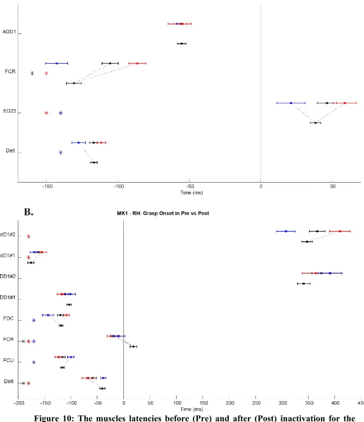
Figure 10: The muscles latencies before (Pre) and after (Post) inactivation for the Monkey 1 in the horizontal condition………55
Figure 11: The muscles latencies before (Pre) and after (Post) inactivation for the Monkey 2 in the vertical condition………56
Figure 12: The muscles latencies before (Pre) and after (Post) inactivation for the Monkey 2 in the horizontal condition………57
Figure 13: Monkey 1 phase-plot of EMGs activity for the Vertical condition……….65
Figure 14: Monkey 1 phase-plot of EMGs activity for the Horizontal condition……….66
Figure 15: Monkey 1 phase-plot of EMGs activity for the Horizontal condition for each post-inactivation group………...67
Figure 16: Monkey 2 phase-plot of EMGs activity for the Vertical condition……….68
Figure 17: Monkey 2 phase-plot of EMGs activity for the Vertical condition for each post-inactivation group………...69
vii
Figure 19: Monkey 2 phase-plot of EMGs activity for the Horizontal condition for each post-inactivation group………...71
viii
List of abreviations
CM: corticomotoneuron CMA: cingulate motor area CNS: central nervous system CS: corticospinal
CST: corticospinal tract EMG: electromyography
GABA: gamma-aminobutyric acid HRP: horseradish peroxidase
ICMS: intra-cortical micro-stimulation LED: light-emitting diode
M1: primary motor cortex MCP: metacarpophalangeal joint
NNMF: non-negative matrix factorization PMD: dorsal premotor area
PMV: ventral premotor area PTN: pyramidal tract neuron S1: primary sensory cortex SMA: supplementary motor area STA: spiked trigger-averaging TIA: transient ischemic attack
ix
« Ce qu’on s’éclate au Canada» John McClane, Die hard 3 : Une journée en enfer
x
Remerciements
Au terme de ce travail, je souhaite adresser une pensée à tous ceux qui, de quelque manière que ce soit, par un conseil, une idée, un coup de main, ou tout simplement leur amitié, m’ont aidée à le réaliser.
Je voudrais tout d’abord exprimer ma reconnaissance au Docteur Numa Dancause, mon directeur de maîtrise, pour m’avoir accueillie dans son laboratoire et permis de réaliser ce projet ambitieux chez un modèle de primate. Merci à Stephan Quessy pour l’aide octroyée au cours de cette maîtrise. Je vous remercie pour vos conseils, votre disponibilité et votre patience qui ont contribués à rendre cette expérience enrichissante sur les plans professionnel et personnel.
J’adresse aussi mes plus vifs remerciements aux membres de mon comité de parrainage, le Docteur Trevor Drew et le Docteur Jean-Pierre Gossard, pour votre présence et l’attention que vous avez portée à mon projet. J’associe à ces remerciements le Docteur Daniel Bourbonnais pour avoir accepté de composer mon jury. Je les prie de retrouver ici le témoignage de ma respectueuse reconnaissance.
Les prochains remerciements sont destinés à mes camarades du laboratoire. A Ian, mon binôme, merci pour ton amitié qui m’est chère, pour tes conseils, pour avoir contribué à améliorer mon anglais et pour ton soutien infaillible. Merci à Boris, Charles, et Maxime pour votre assistance et votre bonne humeur, que ce soit au travail ou autour d’une bière. Ce fut un plaisir de vous côtoyer tous les jours, de parler de sciences, ou par moment de philosophie.
J’adresse également mes remerciements à mes collègues de bureau et amies, Loyda et Lucie. Merci pour votre amitié, votre humour, de m’avoir soutenue et d’avoir contribué à rendre cette expérience agréable. Je souhaite aussi remercier la gentillesse et le soutien de Blanche Perraud et Elsa Tremblay, mes guerrières.
Je tiens à remercier les membres de mon équipe de boxe française, club de savate l’Escouade. Merci de m’avoir aidé à décompresser, déstresser, et maintenir une bonne garde.
xi
Un grand merci à mes parents et mon frère pour leur soutien et leur amour tout au long de ces années. C’est grâce à votre appui et présence que je me suis rendue jusqu’ici. Enfin, je tiens à remercier Marien pour sa patience, son soutien et sa bienveillance quotidienne à mon égard.
1
Chapter 1 – General Introduction
1. Stroke, a burden on the health system
Stroke injuries pose a heavy burden on the Canadian health care system. It is one of the leading causes of persistent physical disability and at least 405 000 Canadians are currently living with the consequences of stroke. In addition, the incidence of stroke is expected to increase by 80% by 2038 (Krueger et al. 2015).
A stroke happens when blood stops flowing to any part of the brain, damaging brain cells. There are two types of strokes, hemorrhagic and ischemic. Hemorrhagic stroke is caused by the rupture of a blood vessel. This type of stroke accounts for 20% of all stroke cases. In contrast, ischemic stroke is caused by a blockage or clot in a blood vessel. This either significantly slows down the blood flow or stops it completely, interrupting vital oxygen and nutrient supply to the brain. This type of stroke is much more common and accounts for approximately 80% of all stroke cases (Heart & Stroke Foundation). Finally, there is also a related condition called a transient ischemic attack (TIA), where the blood supply to the brain is temporarily interrupted. This causes what’s known as a mini-stroke. It can last a few minutes or persist up to 24 hours.
In the stroke cases due to blockage of the middle cerebral artery occlusion, there is often damage to M1 which leads to a loss of fine motor control mainly in the contralateral limb (Bamford et al. 1991). In addition, unilateral stroke affects the upper limb more than the lower limb, and recovery of the upper limb is worse (Twitchell 1951). Stroke survivors live with motor impairments in the upper limb that cause decrease of productivity, autonomy and quality of life (Olsen 1990).
Thus, the study of the functional role of M1 in terms of hand control is important not just for a better understanding of the role of M1 in the control of movements, but also for a larger clinical importance.
2
2. The motor system
2.1 Organization of primary motor cortex
The ability to organize complex motor acts and execute fine movements with precision depends on control signals from the motor areas in the cerebral cortex. The motor areas of the cerebral cortex are subdivided into a M1 and several premotor areas. Brain mapping studies have shown that M1 is organized somatotopically in humans (Penfield and Boldrey 1937) and monkey (Woolsey et al. 1952). These studies used surface stimulation to evoke movements of different segments of the body such as leg, trunk, arm, neck, face and mouth. The amount of cortical space devoted to any particular body part represents the amount of control that M1 has over that body part. Therefore, a large part of the motor cortex is dedicated to move the muscles of the fingers and the muscles related to speech (Becker 1953).
In 1975, Asanuma proposed an organization based on cortical columns in the motor cortex. These studies used intracortical microstimulation (ICMS) in monkeys, an invasive technique that delivered a train of cathodal current pulses that evoked muscle contractions. They observed that stimulation at threshold current induced contractions to a single muscle. In this point of view, each cortical column in M1 would project to a single muscle (Asanuma 1975).
However, this columnar organization described by Asanuma was challenged. In a study of Fetz and Cheney (1980), M1 neuron’s discharge timing were correlated to muscles contractions while monkeys executed a ramp-and-hold wrist movements. They used spike-triggered averaging (SpTA) to assess the influence of a single M1 neuron on a population of spinal cord motoneurons. The results showed that a given M1 neuron could directly facilitate a number of different muscles, usually two-three muscles per M1 neuron. In addition, an anatomical study with horseradish peroxidase (HRP) showed that identified corticospinal (CS) axons spread collaterals at several levels of the spinal cord and individual CS axons made connections with motoneurons of different muscles (Shinoda, Yokota, and Futami 1981). These studies demonstrated the existence of divergent projection and connectivity of motor cortical neurons.
The divergent projection of motor cortical neurons was also tested during fine control movement. In a study of Schiber and Hibbard (1993), isolated M1 neurons were recorded while
3
monkeys executed independent finger movements. They observed that neuronal populations were active with movement of different fingers and with extensive overlap. Therefore, control of finger movements involved population of neurons distributed throughout the M1 hand area rather than a somatotopically segregated population (Schieber and Hibbard 1993).
2.2 Premotor areas
In addition to M1, six premotor areas are involved in the production of motor outputs. These premotor areas are defined as areas from the frontal lobe with a direct access to M1 and the spinal cord (Dum and Strick 2002). These 6 premotor areas are: the dorsal (PMd) and ventral (PMv) premotor area, the supplementary motor area (SMA), and the three cingulate (CMA) motor areas (Morecraft and Van Hoesen 1992; Picard and Strick 1996; Barbas and Pandya 1987; Matelli, Luppino, and Rizzolatti 1985). These premotor areas are the main source of inputs to M1 (Dancause et al. 2006; Dum and Strick 2002; Stepniewska, Preuss, and Kaas 1993). Premotor areas are also a major source of corticospinal fibers (⁓44%) (Dum and Strick 1991).
Further subdivisions were identified in the PMd and the SMA premotor areas. These premotor areas contain a rostral portion called pre-PMd and pre-SMA, and a caudal portion called PMd proper and SMA proper (Geyer et al. 2000; Picard and Strick 2001). These caudal subdivisions have more connections with M1 and more corticospinal projections than their rostral counterparts (Dum and Strick 1991; He, Dum, and Strick 1993). In contrast, the pre-PMd and the pre-SMA have more connections with frontal and prefrontal areas (Bates and Goldman-Rakic 1993; Lu, Preston, and Strick 1994; Luppino et al. 1993).
2.3 The corticospinal pathway
The corticospinal fibers originate from the layer V of the motor cortex, the axons descend through the internal capsule, the cerebral peduncle, and then form the medullary pyramids where these projection is called the pyramidal tract. In humans, most of those axons (75-90%) (Davidoff 1990; Nathan, Smith, and Deacon 1990; Jang 2014) cross the midline in the pyramidal decussation at the junction of the medulla and spinal cord. The crossed fibers descend in the dorsolateral columns of the spinal cord, forming the lateral corticospinal tract. The lateral CST project to motoneurons in the lateral part of the ventral horn of the spinal cord and to interneurons of the
4
intermediate zone. In contrast, the uncrossed axons (⁓10%) descend in the ventral columns of the spinal cord, forming the ventral corticospinal tract. The ventral CST project bilaterally to motoneurons that innervate axial muscles in the intermediate zone of the spinal cord (Kuypers 2011; Kuypers 1962).
In seminal studies using retrograde transneuronal transport of rabies virus, Rathelot and Strick examined the distribution of corticomotoneuronal (CM) cells in M1 that make monosynaptic connections with the motoneurons of the hand muscles. In these experiments, virus was injected in a single digit muscle of macaque monkeys and then was transported retrogradely across one synapse to label M1 cells. The distribution of CM cells projecting to motoneurons of the hand muscles was mainly located in the caudal part of M1 and the sulcus. In addition, these CM cells of one digit muscle were widely distributed and overlapped extensively with the representation of other muscles in M1 (Rathelot and Strick 2006, 2009).
It was demonstrated there was a causal relationship between CM cells and muscle activity, with CM cells that directly facilitate different distal muscles (Fetz and Cheney 1980). Moreover, approximately half of these cells could facilitate proximal and distal muscles (McKiernan et al. 1998). In the study of McKiernan et al. (1998), they tested whether the CM cells produce postspike effects in both proximal and distal muscles. They used SpTA while macaque monkeys performed a reach-and-grasp task that required multijoint coordination of the forelimb. The results showed that 45.5% of the CM cells produced postspike effects in both proximal and distal forelimb muscles. In addition, 44.7% of those produced postspike effects in distal muscle only, while 9.8% produced postspike effects in proximal muscle only. Therefore, CM cells make more frequent and potent connection with motoneuron pools of distal compared with proximal muscles. Interestingly, the results also showed that while 70.7% of the CM cells induced facilitation effects, 29.3% of those induced suppression effects, on mostly distal muscles. Accordingly, those CM cells involved in coordinated forelimb movements facilitate and/or suppress muscles activities at multiple joints. Indeed, the extensive representation of a single hand muscle in M1 was close to different muscles of elbow and shoulder movement as shown by stimulus-triggered averaging (StTA) (Park et al. 2001). This proximity could provide neural substrate to favor a broader range of functional synergies involving multijoint movement (McKiernan et al. 1998).
5
This direct CM system of M1 is crucial for the control of complex finger movements such as precision grip. It is well developed in humans, great apes, and some non-human primates including the macaque (Porter R 1985).
Indeed, lesion of the descending pathway lead to a permanent loss of precision movement (Lawrence and Kuypers 1968a, 1968b). Lawrence and Kuypers (1968a) made the first systematic investigation of bilateral pyramidal lesions and tested the motor skills of eight macaque monkeys. They observed that monkeys recovered part of the general motor behavior (run, jump, climb), but lost the ability to execute independent finger movements. These lesions studies support the importance of M1 and the corticospinal projections in the control of hand movements.
3. Motor cortical control and synergy
Successful execution of voluntary movements depend on the integrity of M1 but also many different part of the central nervous system (CNS). These cortical areas produce neural signals descending to the spinal cord to innervate the spinal interneurons and motoneurons, and finally activate different muscles into motor behaviors (Dum and Strick 1996; Rathelot and Strick 2009).
The role of M1 in the control of arm and hand movements has been extensively studied and highlights a diversity of functions. The discharge of M1 neurons in a variety of experimental paradigms correlates with the direction, speed, and movement, as well as to joint posture and muscle force (Georgopoulos et al. 1982; Georgopoulos and Stefanis 2007; Evarts 1968; Kalaska et al. 1989; Scott and Kalaska 1997). In effect, almost every movement parameter that has been tested has been found to be encoded by motor cortex neurons. This plethora of correlations make it difficult to understand how the spinal circuitries interpret descending cortical signals for a variety of motor tasks. In addition, motor cortical areas need to assure the proper coordination between a large number of muscles and multiple joint motions even for simple movements. Presumable, the CNS simplify the control of goal directed movement by combining discrete elements. Recent work suggest that these discrete elements are muscle synergies (Bizzi, Mussa-Ivaldi, and Giszter 1991; d'Avella, Saltiel, and Bizzi 2003).
6
The idea that complex patterns of muscle activity may be constructed from a few synergies produced by a limited number of spinal modules was first detailed by Bizzi and his collaborators (Bizzi, Mussa-Ivaldi, and Giszter 1991; Tresch, Cheung, and d'Avella 2006; Tresch, Saltiel, and Bizzi 1999; Tresch et al. 2002; d'Avella and Bizzi 2005). In this approach, a mathematical decomposition methods, like non-negative matrix factorization (NNMF), used on EMG data identified a small number of muscle synergies that was sufficient to explain a wide range of movements. The NNMF was used as a weighting matrix that differentially activates all the muscles recorded during a movement, and linearly combined with a time-varying activation coefficient. Therefore a variety of movement can be produced by modifying few muscle synergies (4-6 synergies in most studies). Such synergies have been described in normal reaching movements in primates and humans (d'Avella et al. 2008; d'Avella et al. 2006; Overduin et al. 2008). Other studies used it to evaluate the deficits in movement and changes in synergies after stroke (Cheung et al. 2009; Cheung et al. 2012; Clark et al. 2010).
Another approach to identify synergies was adopted by Drew and his collaborators (Krouchev and Drew 2013; Krouchev, Kalaska, and Drew 2006; Drew, Kalaska, and Krouchev 2008; Yakovenko and Drew 2015). This approach used a more restricted definition of a synergy: a group of muscles that are temporally co-activated and in which muscle activity begins and ends synchronously. Indeed, based on their works on the contribution of the motor cortex to the control of locomotion, they showed that pyramidal tract neurons (PTNs) in the cat were activated sequentially during locomotion. Most of PTNs discharge during only a restricted part of the step cycle, some discharged at the onset of swing and others discharged later in the swing phase (Drew 1993; Lavoie and Drew 2002). Other studies have demonstrated that PTNs discharge sequentially during reaching movements in cats (Yakovenko, Krouchev, and Drew 2011), and M1 neurons also discharge sequentially to coordinate reaching movement in primates (Murphy, Wong, and Kwan 1985). On the basis of these studies, Yakovenko and collaborators (2011) suggested that that the sequential activation of PTNs would modify the muscle synergies by specifying the changes in the spatiotemporal pattern of EMG activity needed to coordinate the movement of the limb. In this approach, a novel cluster analysis identified a greater number of synergies (up to 11) (Krouhev 2006). Each synergy contained only a small number of muscle bursts recorded, and was active during only a small part of the phase movement (i.e. swing or stance phase during locomotion, or reach phase). These synergies could provide a flexible substrate by which descending commands
7
can regulate and modify limb movements. In a more recent study (Yakovenko 2015), they demonstrated that PTNs cells make a similar motor control to muscle activity during reaching and during stepping over an obstacle.
Muscles synergies have been proposed as building blocks that could simplify the construction of motor behaviors. The muscle synergies represent a repertoire of motor subtasks that can be differently recruited by the nervous system to produce diverse complex movements. Analysis in hemiparetic stroke patients showed differences in the number of muscles synergies, which reflect disruptions in descending neural commands and correlated to motor deficits (Clark et al. 2010). Therefore muscle synergies patterns could be used as physiological markers by clinicians, and elucidate which interventions work best for certain patients and not for others.
4. Motor impairments following stroke
Hemiparetic stroke is accompanied by abnormalities of muscle tone, muscle weakness, and disturbances of muscular coordination. However, in many patients, when weakness and spasticity are treated effectively or resolved spontaneously, motor dysfunction remains severe. Therefore, the primary source of movement dysfunction or global disability in many hemiparetic stroke patients is likely neither spasticity nor muscular weakness, but abnormal movement coordination (Dewald et al. 2001; Hesse et al. 1996). Accordingly, understanding the impaired motor movement and muscle coordination following stroke is essential for the design of effective rehabilitation.
Different research groups have shown alterations of muscle activation patterns following stroke. One such change is inappropriate muscles co-activation (Kamper and Rymer 2001). Indeed, numerous clinical studies have shown inappropriate co-activation of agonist muscles (Hammond et al. 1988), frequently accompanied by co-contraction of antagonist muscles (Bourbonnais et al. 1989; Kamper and Rymer 2001). These alterations were observed in flexors and extensors of the shoulder and elbow (Dewald et al. 1995), wrist (Hammond et al. 1988) and digits (Kamper and Rymer 2001) in hemiparetic patients. Kamper and Rymer assessed the extent of co-activation during voluntary extension of the metacarpophalangeal (MCP) joint through three
8
sets of experiments: isometric, isokinetic and free contractions. The torque, position and EMG analysis were compared between stroke and control subjects. The results confirmed the presence of chronic motor dysfunction in the fingers that involved improper co-activation of finger flexors and extensors across the three sets of experiments. Indeed, an extension torque to the MCP joint resulted in the production of an inappropriate flexion torque instead. This inappropriate co-activation was also observed with the flexors and extensors of the wrist (Hammond et al. 1988). Other studies among stroke survivors confirmed the presence of muscle co-activation patterns in the paretic limb, and that abnormal coordination correlated with the severity of upper limb impairment (Dewald et al. 1995; Lee et al. 2013).
Another change in muscle activation patterns following stroke is a loss of selective activation of sets of muscles needed to perform skilled movement tasks (Lang and Schieber 2004). Agonist-antagonist co-activation may explain the reduced ability to selectively recruit upper arm and forearm muscles. This is consistent with the results of experiments performed by Lang and Schieber using humans with pure motor hemiparesis. Kinematics analysis of individuated finger movements (i.e., the ability to move each finger while keeping the other fingers still) were compared between patients and controls subjects. They first showed independent finger movements were differentially impaired. In the affected hands, the independence of the thumb was normal, the independence of the index finger was slightly impaired, while the independence of the middle, ring and little fingers was substantially impaired. The hemiparetic subjects received an extensive rehabilitation training that could explain this differential impairments. The rehabilitation process required independent movements of the index finger and the thumb that could lead to plastic changes in M1, compensation by other cortical motor areas, and/or compensation by other descending pathways (Lang and Schieber 2003). Second, they showed that the loss of independent finger movement was due to a reduced selectivity of finger muscle activation during an individuate finger movement task (Lang and Schieber 2004). Furthermore, this study showed that the loss was beyond the agonist or antagonist muscle activation. Indeed, they observed that a muscle of the little finger (the ADQ, abductor digit quinti) was also activated during movement of the thumb and index fingers in hemiparetic subjects (Lang and Schieber 2004).
9
Electromyographic (EMG) studies have shown delayed initiation and termination of muscle activity as another alteration of muscle control observed in hemiparetic subjects (Chae et al. 2002; Dewald et al. 1995; Hammond et al. 1988). These alterations also correlated significantly with clinical measures of motor impairment, suggesting there is relationship between muscle timing and motor impairment. In Chae and collaborators (2002), stroke survivors were instructed to contract the wrist flexor or extensor as forcefully and quickly as possible against the confinement of an apparatus in response to an audible sound, and to relax the muscle as quickly as possible as soon as the end of this sound. The data showed significantly greater delay in initiation or termination of muscle contraction in the paretic and non-paretic limbs.
Interestingly, clinical studies also revealed more subtle ipsilateral motor deficits that emerged acutely (Sunderland et al. 1999) and persist chronically (Yarosh, Hoffman, and Strick 2004; Wetter, Poole, and Haaland 2005). In the study of Yarosh and collaborators (2004), abnormalities in the temporal activation of the muscles were observed in wrist muscles ipsilateral to a stroke in hemiparetic subjects during a reaching task. Step-tracking movements of the wrist and associated EMG activity (extensor carpi radialis longus i.e. ECRL, extensor carpi radialis brevis i.e. ECRB, extensor carpi ulnaris i.e. ECU, and flexor carpi radialis i.e. FCR) of stroke subjects were examined and compared to control subjects. Stroke subjects showed great difficulty or were unable to perform the task with the contralateral wrist, as expected. The results showed that patients had also marked impairments in the ipsilesional step-tracking movement of the wrist with abnormal EMG patterns. First, these abnormalities in the EMG patterns consisted in movement initiated by only the onset times of agonist muscle (i.e. ECU) without the co-activation of the onset times of synergist muscles (i.e. ECRB and ECRL) during wrist extension and ulnar deviation movements. Second, the activity was inappropriately timed if the activity with synergist muscles was observed. The onset times of synergist muscles, ECRB and ECRL, occurred well before or after the onset times of the agonist muscle ECU. Third, the alternation of agonist and antagonist activity was disrupted as observed during a flexion and ulnar deviation of the wrist. Either patients initiated the movement using the onset times of antagonist ECU instead of the agonist FCR, or the onset times of antagonist ECU occurred well later or did not occurred. These abnormal activations of antagonist muscle bursts produces misdirected and irregular trajectories due to inappropriate temporal coordination of muscle activity. This study demonstrated that unilateral lesion can produce bilateral effect on the generation and control of distal movement.
10
Changes in the temporal activation of muscles are visible from days to months after a stroke, and can change with time. Wagner and associates (2007) explored how the muscle activations in the arm contralateral to a stroke in hemiparetic subjects evolved from the “acute phase” (mean= 9 days post-stroke) to the “subacute phase” (mean=109 days post-stroke) compared to healthy subjects by looking at the changes in the muscles’ activation onset times during a reaching task. The muscle onset time was used to reflect the temporal activation of the muscles during recovery. In the acute phase, the muscle onset times occurred later and were more variable relative to healthy subjects. In contrast to healthy subjects, the most distal muscles (wrist extensor carpi radialis and wrist flexor carpi radialis) and the triceps onset times occurred after the start of the movement. Subsequently, the onset times decreased in the subacute phase relative to the acute phase, such that they became more similar to those observed in the control subjects. Furthermore, muscle onsets occurred prior to the start of movement for all muscles except the distal muscle of wrist flexor carpi radialis. Nonetheless in both time points, the hemi-paretic group had considerably more variability in muscle onset times compared to controls. The changes observed in the muscle activation were differentially related to changes in reaching performance. Their results suggest that improvement in muscle timing and a better level of volitional activation might underlie improvement in reaching performance following a stroke (Wagner et al. 2007).
5. Muscimol: a tool to evaluate cortical motor control
Stroke often disrupts descending motor commands due to a loss of cortical/subcortical input to muscles, contributing to impaired movements and coordination (Twitchell 1951). In addition, stroke could also induced damages in brain regions that have synaptic connections with the primary lesion site. For example, following cerebral infarction in the middle cerebral artery, neuronal death, gliosis, and axonal degeneration have been observed in the thalamus, substantia nigra, and distal pyramidal tract all of which lie outside of the middle cerebral artery territory (Ogawa et al. 1997; Forno 1983; Buss et al. 2004; Buss et al. 2005; Schmitt et al. 1998; Schmitt et al. 2000).
The use of animal models helped to evaluate the solely role of M1 in the generation of voluntary movements without the induced damage in other structures observed after a stroke. In the literature, monkey models have been used only occasionally to measure the behavioral deficits and the impact
11
on muscles activities induced by lesions to the motor cortex (Hoffman and Strick 1995) or corticospinal tract (Hepp-Reymond and Wiesedanger 1972, Lawrence and Kuyper 1968). Large lesions of the primary motor cortex (M1) make voluntary movements of the contralateral side weaker and slower. For example, it has been shown that after a lesion of the M1 arm area there was a marked change in the movement kinematics and the patterns of activity in agonist, antagonist and synergist wrist muscles during step-tracking movements (Hoffman and Strick 1995). Fine motor control deficits were also visible after small lesions of the M1 hand area (Friel and Nudo 1998). Following 1 month of rehabilitative training, skilled use fingers appeared to recover. Despite the apparent recovery of skilled fingers dexterity, the study provide evidence of compensatory strategies. This study demonstrated that even after small infarct that produce mild and transient motor deficits, the monkeys used compensatory movement patterns as a recovery of motor function. These studies highlight the contribution of M1 in the generation of appropriate muscle activation patterns and fine motor control of the finger.
However, to evaluate the contribution of M1 in the motor control of the hand, without the secondary effects of lesion that could involve mechanical damage and tissue inflammation, animal studies used Muscimol injection. Indeed, reversible inactivation of cortical areas by using drug injections has been a powerful tool to elucidate the differential functional contributions of cortical forelimb representations to limb movement control. Muscimol, a pharmacological GABA-A agonist agent, was used to temporarily inactivate different areas of the cortex. Intra-cortical injections of Muscimol have been realized in different animal models such as the rat, cat and monkey (Martin 1991; Martin and Ghez 1993; Matsumura et al. 1991; Brochier et al. 1999; Schieber and Poliakov 1998).
Monkey studies showed that the injection of Muscimol in M1 induced impairments such as decreased speed, weakness of muscles, and loss of independent finger movement (Kubota 1996; Matsumura et al. 1991; Schieber and Poliakov 1998; Brochier et al. 1999; Fogassi et al. 2001). During a reach-to-grasp task, a reduction of movement speed, of movement time, and reaction time was observed after injection of Muscimol (Kubota 1996; Fogassi et al. 2001). Based on the video analysis, the movement time almost double with respect to the control condition (Fogassi et al. 2001) and the reaction time was significantly longer (Kubota 1996). Another study with a visual reaction-time task, where the animal need to press a lever and release it after a go signal to obtain
12
a reward, confirmed the significant increase in reaction time after Muscimol injection (Matsumura 1991).
Moreover, muscle weakness was also often observed together with the slowness of movement (Matsumura et al. 1991; Kubota 1996; Schieber and Poliakov 1998; Brochier et al. 1999; Fogassi et al. 2001). The weakness in the finger muscles was measured by Brochier et al. 1999. In this study they evaluated the effects of a small inactivation (10 µg) of the primary somatosensory (S1) and motor (M1) cortex on the control of fingers forces in a precision grip task. The animal needed to grasp a metal tab between the thumb and the index finger, lift it into a vertical position window and hold it stationary for two seconds to obtain a reward. In addition, the monkey’s performance was assessed with a modified Klüver board. The precision grip was differentially impaired by the injection either into S1 or M1. The M1 injections produced impairment in the ability to perform independent digit movements and a general weakness in the digit muscles. Indeed, all the fingers moved together in a whole-hand grasping configurations, and this loss of precision grip resulted in inappropriate digits positions on the metal tab surfaces. In addition, the monkey was unable to oppose the thumb and index digit to enter the food well of the Klüver board. The motor deficit was restricted to the most distal segments of the hand because the monkey could still execute the reach phase of the movement. In contrast to M1, the S1 injections induced a loss of manual coordination in order to position the thumb and index finger for grasping, and an increase in the grip force. In addition, during the Klüver board task, the monkey was able to execute some crude independent finger movements to retrieve and eat food morsels under visual control. These results showed that the main effect of small inactivation into the thumb and index representation area of M1 was the inability to perform independent finger movements in association with muscular weakness of the hand.
Similarly, Fogassi et al (2001) realized small and large injection (15 and 90 µg of total Muscimol injected respectively) of Muscimol in M1 and the ventral premotor cortex (PMv). The size of injection was an important factor that caused different impact with deficits restricted to the most distal segment and/or also more proximal segment. The small inactivations of M1 hand field area produced paresis in the fingers of the contralateral hand, in association with a reduction of the grip force, and hypotonia. The capacity to grasp was also strongly impaired; monkeys reached with a “flat” hand posture similar to a whole-hand grasping observed previously by Brochier et al (1999). Therefore, the precision grip couldn’t be executed. The larger inactivation produced
13
deficits that were the same as those after a small and single injection, but more severe. The grasping was completely disrupted and one monkey showed impairment of more proximal movements (Fogassi et al. 2001).
The injection of Muscimol induced also specific deficits in the digits and was studied in detail with partial inactivation of M1 hand area during a visually cued individuated finger movement task (Schieber and Poliakov 1998). This study tested the existence of a somatotopical finger representations in M1. If the M1 hand area contains this somatotopic organization, then partial inactivation might produce weakness, slowness and loss of independence of one or two adjacent digits while the other fingers remained unaffected. Monkeys were trained to perform visually cued individuated finger movements. The right hand was placed in a pistol-grip manipulandum that separated each finger into a different slot. Monkeys were instructed with different light-emitting diodes (LEDs) which digit to flex or extend. By flexing or extending a digit, a ventral or dorsal switch was closed respectively. The monkey received a water reward if the instructed switch was closed and hold in a proper response time without closing any other switches. In addition, monkeys were trained to retrieve small food morsels from a large well that permitted entry of the whole hand and a small well that permit entry of only one finger. The results showed several deficits: (i) increased failure of instructed individuated finger movement, (ii) prolongation of response time (that included both the pre-movement reaction time and the movement time itself), and (iii) decrease of individuated finger movement. Interestingly, this study observed a wide variety of possible combinations of these deficits. Indeed in some inactivation sessions all these three deficits were observed. In other sessions, however, increase in movement failure (i) could be observed without significant prolongation of response time (ii) or decrease in individuation (iii). In still other cases, response time and/or decrease individuation could be observed without a significant increase in failure. Moreover, in some cases there was prolonged response time without decrease individuation and vice-versa. Based on these different combinations, the authors suggested that prolongation of response time and decrease in individuation could be to some extent dissociable. In addition, the injection impaired performance of retrieving food morsels from the two wells and the contralateral hand failed to preshape. They found little evidence that which finger movements were affected after each injection was related to the injection location along the central sulcus. These findings showed that the M1 hand area was not so somatotopically organized as implied by the homunculus, but suggest a distributed organization for control of the different fingers
14
One study, in awake cat, evaluated the changes in kinematics of reaching and grasping after single injection of Muscimol (1µg) (Martin and Ghez 1993). This study showed that local inactivation within the forelimb representation in the M1 motor cortex of the cat produced measurable deficits in reaching behavior with slower movements, reaching errors, and defect in kinematic planning or in trajectory adaptation to avoid an obstacle, and in grasping movements during food retrieval.
While the studies described above presented detailed descriptions of behavioral deficits following M1 inactivation with kinematics, forces, and response time, few studies have attempted to provide a description of electromyographic (EMG) muscle activity. Matsumura et al. (1991) performed an experiment looking not just at the behavioral deficits but also the change in EMG activity induced by injection of agonist (Muscimol) and antagonist (Bicuculline) of GABA in the primate motor and premotor cortex during a raisin pick-up test and a visual reaction-time task. They showed the local injection of Muscimol in either area caused deficits in hand and arm movements. Injection of Muscimol in M1 deteriorated the digit manipulation in the raisin pick up test, with an increase in retrieval attempt and in raisin drop. The movements were weaker and slower, with monkeys that spent more time for single trials. The EMG analysis were not possible with this task due to vigorous arm movement that created too much noise in the EMG recordings. As previously described, the results of the visual reaction-time task showed a significant increase of reaction time after Muscimol injection, and this deficit was in association with changes in muscle activities. The Muscimol caused a transient decrease in the total muscle activity of hand muscles like the flexor carpi ulnaris (FCU) and extensor carpi radialis (ECR) showed in the article. The EMG activity decreased with a similar time course of the manipulation deficits. This study demonstrated the behavioral deficits were associated with changes in muscle activity as probably a reflection of plastic changes in cortical motor area (Matsumura et al. 1991).
Taken together, these studies suggest the motor cortex is crucial for the regulation of spatiotemporally organized ensembles of muscles activity. In addition, inactivation of the M1 hand area could produce main effect as slowness and weakness of the movement, and/or a loss of independent finger movements. However, none of these studies have explored how the activation across a set of muscles was changed.
15
6. Objectives of the study
The general goal of the current work is to further our understanding of how the hand area of M1 contributes to the execution and coordination of movement of the upper limb during a reach-to-grasp task. As outlined in the previous sections, M1 plays a critical role in the motor of the hand and fine digit movements, and the main pathway through which M1 sends motor commands to spinal motor neurons is via the corticospinal tract. Clinical studies have shown that cortical damage, such as stroke, interferes with the descending signals to the spinal cord, resulting in abnormal movements. To explore the relationship between cortical damage and motor control, fundamental research has often used Muscimol to temporarily inactivate different areas of the cortex and measure the induced motor impairments in different animal models. In spite of extensive studies of behavioral deficits following Muscimol injections, the correlation of these motor deficits with changes in muscle activities of the upper limb have not been described. The current project set out to address this gap in our knowledge. The present experiments were conducted in macaques, a species in which, like humans, M1 has direct corticomotoneuronal projections to hand motoneurons. We used Muscimol to test the contributions of the M1 hand area to the production of a reach-to-grasp movement and the evoked motor patterns that constitute this movement.
We hypothesized that behavioral deficits in reach-to-grasp movements after partial inactivation of M1 would be associated with abnormal muscle co-activation patterns, and that these abnormalities would depend on the severity of the inactivation (Fogassi et al. 2001; Cheung et al. 2012). Our first objective was to investigate the behavioral performance of the paretic hand both prior to and after partial inactivation of the hand area of M1. The behavior was evaluated based on the monkeys’ ability to retrieve a food pellet from a well using video recordings and task sensors. Two monkeys were trained to perform a reach-to-grasp task that culminated in a precision grip. The target could be oriented in a vertical or horizontal orientation and, as such, required monkeys to perform a rotation of the forearm or not. The rotation of the forearm added more difficulty to the motor control of the movement. Our second objective was to describe the muscle activation patterns relative to the behavioral performance. Muscles of different segments of the paretic limb, from proximal muscles such as the deltoid to distal muscles such as the first interosseous, had their electrical activity recorded from using chronic subcutaneously implanted wires (i.e. EMG). The behavior and the EMG activities were evaluated and compared before and after the Muscimol
16
injections. First, the behavior was assessed using the task success rate, the number of digit flexions required to retrieve the pellet, the duration of the first flexion of the index finger, the duration of the reach and the grasp phases of the movement, and the type of grasp used. These measures allowed us to test whether the partial inactivation of M1 could lead to deficits in one or both phases of the movement and whether there was an induced loss of manual dexterity. Second, the EMG activities were compared relative to the behavior to determine whether the execution of reach-to-grasp movements underwent changes in the coordination of voluntary muscle activities. The latencies and the muscle activation patterns was analyzed based on the onsets and offsets of proximal muscles (deltoid, biceps) and distal muscles (wrist flexors and extensors, and intrinsic hand muscles). The results will provide a better understanding of the contribution of M1 to the control of fine digit movements and how muscle patterns changed after partial inactivation of the M1 hand area.
17
Chapter 2 – Materials and methods
The experiments were conducted on 2 adult female monkeys (Macaca mulatta), Monkey 1 (5.5 kg) and Monkey 2 (5.7 kg). All surgical and experimental protocols were performed in accordance with the guidelines set forth by the Canadian council on Animal Care, and were approved by the Comité de Déontologie de l’Expérimentation sur les Animaux de l’Université de Montréal.
Training
The monkeys were trained for up to 6 months on the reach-to-grasp task (Fig. 1A). The monkeys were trained to be brought out of the cage, sit quietly in our custom-made primate chair, and brought back to the cage every day for 2 months. The custom-made chair had two removable panels for the arms and an opening near the mouth allowing the monkeys to eat the pellet rewards. After monkeys’ habituation to sit in the chair, they were brought into our training room and installed in front of the task. Then, they were trained to realize a reach-to-grasp task.
The apparatus consisted of a box with a home plate and a precision target for each hand. The two home plates were placed on a plane surface at 10 cm in front of the monkey and contained two sensors each. The two targets were placed vertically to face monkeys at approximately 10 cm height above the home-plates. Each target contained a small well in the middle to hold a banana flavor food pellet as a reward (190mg Dustless Precision Pellets; BioServ, Flemington, NJ, USA), and a slot that permitted the monkeys to grasp pellets only by performing a precision grip. Sensors were installed at each extremity of slots. In addition, the slots could be oriented horizontally or vertically to retrieve the pellet from the well. All sensors were used to monitor the proper sequential execution of reach and grasp phases of the movement.
First, monkeys were trained every day for 2-3 months to place and keep their hands between the home plates sensors before subsequently performing a reach and grasp movement to grasp the pellet. The reward was remotely controlled and one pellet was given when monkeys placed the hand between the home plates sensors.
The second parts of the training started after monkeys learned to place their hands on the home plates and executed a sequential movement of reaching, grasping, and retrieve/eating the pellet. Then, monkeys were trained to perform the task with our automatic program that included
18
5 different delays before the pellet drop (from 800 ms to 2 s). Monkeys were trained daily to keep the hand on the home plate and execute the reach-to-grasp task with a high (<80%) and constant (<2 weeks) success rate.
Behavioral task
In the day-to-day experiment, the monkeys sat in a the chair installed in front of the behavioral task consisting of one sensor home plate and one precision well per arm (Fig. 1A left panel). The monkeys had to perform a reach-to-grasp movement culminating in a precision pinch with either hand. The precision grip was done using the thumb in opposition with the index digit. The trial sequence was as follows: (i) monkeys first placed their hands in the start position (Enter start). (ii) Then, after a variable delay (baseline) a food pellet dropped into the well. (iii) The sound of the pellet being dropped served as a signal to the monkey to move (Pellet drop; a “GO” signal). (iv) The monkey’s hand left the start position (Exit start; reach start) to reach towards the food well, until its fingers crossed the sensors around the slot (Enter slot; grasp start). (v) After performing a successful precision pinch to grasp the food, the fingers left the slot (Exit slot; grasp end) and the animal brought its hand to its mouth to consume the food pellet (Return) (Fig. 1A right panel).
Surgical procedures
Once the training was completed, the monkeys were prepared for surgeries under aseptic conditions, and anesthesia was induced with an intramuscular injection of ketamine hydrochloride (Ketaset; Pfizer, Inc, New York, NY, USA). The animals were transitioned to 2-3% isoflurane (Furane; Baxter, Deerfield, IL, USA) with oxygen. To help prevent inflammation and swelling of the brain, the animal received an intramuscular injection of Dexamethasone 2 (Vetoquinol; 0.5 mg/kg), a sub-cutaneous injection of Atropine (0.04 mg/kg) and intravenous injection of Mannitol 20% (1500 mg/kg) at the beginning of the surgery. To keep the animal’s body temperature near 36.5°C throughout the surgery a self-regulating heating blanket was installed under the animal (Harvard Apparatus, Holliston, MA). Blood oxygen saturation and heart rate were continuously monitored.
19
In a first surgery, the monkeys were implanted with a head post on the skull to allow fixation of the head during the experiment. The head posts were fixed to the skull with screws covered with dental acrylic (Palacos R, Heraeus Medical, Germany) that contained a mix of antibiotics (Vancomycin hydrochloride, Alfa Aesar A Johnson Matthey Compagny and Tobramycin, TCI America Thermo Fisher Scientific).
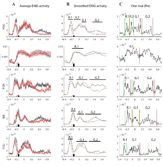
A second surgery was done 4 to 8 months later, allowing time for animals’ habituation of the head post and for the skin around the implant to heal and thicken. Muscles were intramuscularly implanted with electromyographic (EMG) insulated, multistranded microwires (Cooner Wire, Chatsworth, CA, USA). Using various sizes of stainless steel tunneling needles, all the wires were passed subcutaneously from the head posts’ acrylic/skin junction at the back of the head with a small incision (Fig. 1B). Pairs of wires were then tunneled subcutaneously to the appropriate target muscles and each wire was then inserted intramuscularly (Park, Belhaj-Saif et al. 2000). Accurate placement of the EMG wires was tested by electrical stimulation of the muscles through implanted wires and observation of the evoked movements both during the surgery and in later sessions in the awake monkey. A total of 16 muscles per arm was implanted: Deltoid (musculus deltoideus), BB (biceps brachii), BR (brachioradialis), PT (pronator teres), PL (palmaris longus), FCU (flexor carpi ulnaris), FCR (flexor carpi radialis), FDC (flexor digit communis), FDS (flexor digit superficialis), ECU (extensor carpi ulnaris), ECR (extensor carpi radialis), EDC (extensor digit communis), ED23 (extensor digitorum 2 and 3), ADD1 (adductor of the thumb), ABD1 (abductor of the thumb), intD1 (first dorsal interosseus), and FPB (flexor pollicis brevis superficialis).
A third surgery was conducted to implant intra-cortical multi-electrode arrays in several cortical areas in both hemispheres and one chronic inactivation chamber (Fig. 1B) one month following the EMG implantation. Multi-electrodes arrays (Utah Array, Blackrock microsystems; and Floating Microelectrode Array, MicroProbes for Life Science) were implanted in the ventral and dorsal premotor cortex (PMv, PMd) in both hemispheres and the primary motor cortex (M1) in the right hemisphere. These neural data will not be presented in this study. In the left hemisphere, the dura was left intact over the central sulcus and a chronic chamber (2x2 cm opening; plexiglass) was positioned to provide access to the dura over the M1 hand representation. The chamber allowed us to perform Muscimol reversible inactivation experiments in the left hemisphere, which we refer to as the “ipsilesional” hemisphere. By extension the right hemisphere is referred to as the “contralesional” hemisphere in both monkeys.
20
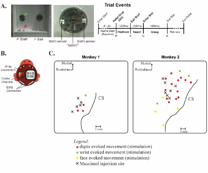
Figure 1: Overview of the experimental setup. A, photos of the experimental task (left panel) and a zoom on the target with the well and slot (middle panel). The different events during one trial as measured by the sensors’ target (right panel): Enter start; Pellet drop; Exit start; Enter slot; Exit slot; Eat pellet. B, Illustration of the EMG connectors and cortex chamber on the monkey’s head. C, cartoon of the chamber of Monkey 1 (left) and Monkey 2 (right) with injections sites in the M1 hand area (black cross) and stimuli sites (red dot: digit, green dot: wrist, yellow dot: face evoked movement). A total of 4 and 20 sites were mapped in Monkey 1 and 2, respectively. For each session, 1 x 0.75 µL of Muscimol was delivered using a Hamilton syringe. CS=Central Sulcus.
21
Mapping of the motor cortex and sites of injection
A brief topographic motor map of M1 was constructed inside the inactivation chamber by using standard intra-cortical micro-stimulation trains (i.e. ICMS), while the awake animal sat quietly in the primate chair in order to confirm the location of the hand representation area (Fig. 1C). ICMS stimulation consisted of a 40 ms train of 13 monophasic cathodal pulses of 200 µs delivered at 350 Hz from an electrically isolated, constant current stimulator (Mansoori, Jean-Charles et al. 2014). A glass coated tungsten microelectrode was placed on a micromanipulator mounted on a stereotaxic frame fixed to the primate chair and descend in the cortex through the chamber of inactivation for electrical stimulation. In the chamber, stimulations were done medio-laterally to locate the face and proximal representation and antero-posteriorly to locate the central sulcus (CS) and primary somatosensory cortex (S1). All cortical sites retained for the Muscimol inactivation protocols evoked digit or wrist movements in the contralateral arm with ICMS trains. For every site, the type of evoked movement, the depth and the lowest stimulus intensity was noted. There were 4 sites mapped in Monkey 1 and 20 sites mapped in Monkey 2 (Fig. 1C, dots). This allowed us to identify digit and wrist representation area for both monkeys, and face representation area for Monkey 2 (Fig. 1C). A total of 5 Muscimol injections were done in Monkey 1, and 7 Muscimol injections were done in Monkey 2 (Fig. 1C, black crosses).
Protocol
Data were collected in a sequential order with the two hands and slot orientations presented in separate blocks of 25 trials (in order: Left arm – Vertical orientation = LV; Right arm – Vertical orientation = RV; Left arm- Horizontal orientation = LH; Right arm – Horizontal orientation = RH). These blocks (LV-RV-LH-RH) of data were recorded at 5 different time points: before the inactivation (Pre-injection block, i.e. Pre), 15 to 40 min after the inactivation (i.e. Post), 3 hours after the inactivation (i.e. T03), 10 hours (i.e. T10), and 24 hours after inactivation (i.e. T24). Only data in Pre and POST were analyzed and presented in this study.
Prior to the Muscimol inactivation protocol, we first recorded baseline behavioral, and EMG (Pre). Then, based on our motor map of M1, Muscimol was intra-cortically injected in M1 hand representation. The drug was loaded in a 5 µL Hamilton syringe with a bevelled tip 26 gauge needle (Hamilton Robotics, Reno, NV, USA). The syringe was placed on a micromanipulator
22
mounted on a stereotaxic frame fixed to the primate chair. That allowed us to descend the syringe in the cortex accessible within the chamber of inactivation. One injection of 0.75µL of Muscimol solution (concentration 5µg/µL) was injected in all individual experiments presented in this study (Fig. 1C). This concentration of Muscimol (5 µg/µL) has been already successfully used to temporally inactivate cortical motor areas on Macaque models (Schieber and Poliakov 1998, Brochier 1999, Fogasse 2001). Each individual injection of 0.75µL Muscimol was delivered at a constant rate of 4 nl/s with a micro-injector (Harvard Apparatus, Holliston, MA, USA). The spread of one injection of Muscimol had an estimated maximum radius of 1.5 mm (Martin and Ghez 1999) and therefore was restricted to the M1 hand area.
Behavioral data analysis
BEHAVIORAL ASSESSMENT: A frame-by-frame video analysis was made to extract behavioral data. We calculated the percentage of successful trials to obtain a success rate per block of trials. A trial was considered successful if the animal was able to retrieve the pellet and eat it. The numbers of trials in which the monkey successfully retrieved pellets were counted. If during the retrieval the pellet fell or was not retrieved successfully, the trial was considered a failure.
The number of flexions executed with the index finger to retrieve the pellet was also assessed. A total mean number of flexions were calculated based on trials from each block of data. For each trial, the number of flexions executed by the monkey while their finger was inside the well until the retrieval or falling of the pellet was counted.
The duration of the first flexion of the index finger during the grasp phase of the movement was also calculated. This parameter was defined as the time beginning when the distal tip of the index finger was in contact with the pellet and started to flex until the time the hand moved to bring the pellet towards the mouth (i.e. in the case of successful trials), or until the time the distal tip of the index finger stopped to be in contact with the pellet (i.e. in the case of several flexions or unsuccessful trials). For each trial, the duration of the first flexion of the index finger was calculated based on the number of frames between the start and the end of the first flexion (1 second = 30 frames).
23
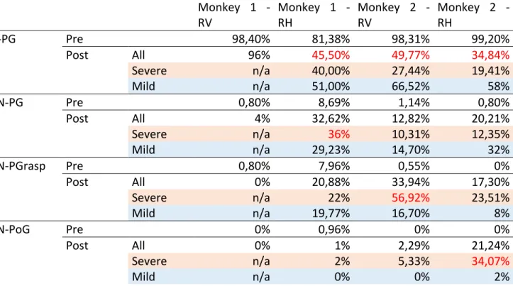
DESCRIPTION OF GRASPING CONFIGURATION: Different grasping configuration could be identified after inactivation in the vertical and horizontal slot orientation (Macfarlane and Graziano 2009, Castiello and Dadda 2019). The movement pattern to retrieve the pellet was reported based on the video analysis. The categorization was defined by the shape of the grasp used to successfully dislodge pellets from the slot based on the frame-by-frame video analysis for each trial. A total of 4 categories were identified in percentage: (i) Normal precision grip (“N-PG”): the pellet is pinched between the distal pad of the first digit (D1) and the index digit (D2). (ii) Abnormal precision grip (“AN-PG”): the pellet is grasped between the thumb and the index digit but no longer between the distal pad of D1 and D2. The monkey does the precision grip but the pellet is between the distal pad of D1 and along the radial side of D2, or between the distal pad of D2 and another part of D1. (iii) Abnormal precision grasp (“AN-PGrasp”): the pellet is grasped with the collaboration of the second and third digits in opposition to the thumb. The pellet is held between these three digits (tri-digital) or between D1-D2 and the palm of the hand. Occasionally the pellet was lodged between lateral surfaces of two adjacent fingers. (iv) Abnormal power grip (“AN-PoG”): the pellet is grasped by several digits or into the palm (see Fig. 6B).
DURATION OF REACHING AND GRASPING: To assess the impact of the inactivation on the reaching and grasping components of the movement we compared the duration of the reaching and the grasping phases before and after Muscimol injection. The duration of the phases was calculated from all trials based on the signals from the sensors of the task. The reach duration was defined as the time between the hand leaving the platform (Home plate sensors off) and the digits entering the slot (enter of the slot sensors on). The grasp duration, i.e. Contact time, was defined as the time between the digits entering the slot (enter of the slot sensors on) and the digits leaving the slot (enter of the slot sensors off). The Contact time did not take into account the number of flexions inside the slot, only the time spent by the monkeys’ digits inside the slot to retrieve the pellet. Some Contact time trial onsets and offsets were misplaced by the task because of multiple activations of the sensors. All trials were inspected, and either corrected or excluded based on visual inspection. This included trials for which the reach duration was abnormal, for example if the animal was distracted by some noises and interrupted the movement.