Université de Montréal
Increased spinal pain sensitization:
A new explanation for highly prevalent painful somatic symptoms
in major depressive disorder?
par Andràs Tikàsz
Département de Psychiatrie Faculté de Médecine
Mémoire présenté à la Faculté des études supérieures en vue de l’obtention du grade de M.Sc.
en Sciences Biomédicales option Sciences psychiatriques
Août 2015
Université de Montréal Faculté des études supérieures
Ce mémoire intitulé: Increased spinal pain sensitization:
A new explanation for highly prevalent painful somatic symptoms in major depressive disorder?
Présenté par Andràs Tikàsz
A été évalué par un jury composé des personnes suivantes : Kieron O’Connor, Président rapporteur
Stéphane Potvin, Directeur de recherche Valérie Tourjman, Codirecteur Marc Corbière, Membre de jury
Résumé
Objectifs: Malgré que les patients souffrant de dépression majeure (DM) rapportent souvent
des symptômes douloureux, la relation entre la douleur et la dépression n’est pas encore claire. Ce n’est que récemment que des études employant des paradigmes de sommation temporelle ont pu offrir une explication préliminaire de la cooccurrence de la douleur et de la dépression. Notre étude vise à évaluer la contribution des procédés spinaux et surpraspinaux dans la sensibilisation de la douleur dans la DM en utilisant un paradigme de sommation temporelle.
Participants : Treize sujets sains et quatorze patients souffrant de DM ont été inclues dans
l’analyse finale. Méthodes : Pour induire une sommation temporelle, nous avons utilisé des stimulations intermittentes du nerf sural de basses et hautes fréquences. La sensibilisation spinale de la douleur a été quantifiée en mesurant la variation de l’amplitude du réflex de retrait nociceptif (NFR) entre les deux conditions de stimulations, ainsi que la sensibilisation supraspinale de la douleur a été obtenue en mesurant le changement dans l’appréciation verbale de la douleur entre ces deux conditions. Résultats : Nous avons observé une sensibilisation plus élevée de la réponse NFR chez les patients dépressifs durant la condition de stimulation à haute fréquence, un effet qui n’a pas été reflété par une sensibilisation amplifiée des appréciations subjectives de la douleur durant l’expérience. Néanmoins, nous avons observé une association entre la sensibilisation spinale et les symptômes somatiques douloureux chez les patients DM. Conclusion : Ces résultats suggèrent une sensibilisation spinale amplifiée dans la DM, ce qui pourrait expliquer la prévalence élevée des symptômes somatiques douloureux chez ces patients.
Abstract
Objectives: Although patients suffering from major depressive disorder (MDD) often
complain from painful symptoms, the relationship between pain and depression has yet to be clearly characterized. Only recently have studies employing temporal summation paradigms offered some preliminary insight into the co-occurrence of pain and depression. This study sets out to evaluate the contribution of spinal and supraspinal processes in pain sensitization in MDD using a temporal summation paradigm. Subjects: Thirteen healthy controls and fourteen MDD patients were included in the final analysis. Methods: To induce temporal summation, we used low- and high-frequency intermittent stimulations of the sural nerve. Spinal pain sensitization was quantified by measuring the change in the amplitude of the nociceptive-specific flexion reflex (NFR) response, and supraspinal pain sensitization was obtained by measuring change in subjective pain rating, from the low- to high-frequency stimulation condition. Results: We found an increased sensitization in the NFR response in MDD patients in the high-frequency condition, which did not translate into an increased amplification of their subjective responses during testing. However, we found a positive association between spinal sensitization and painful somatic symptoms in MDD patients. Conclusion: Together, these results suggest increased spinal pain sensitization in MDD, which might explain the high prevalence of painful somatic symptoms in these patients.
Table of contents
Résumé ... i
Abstract ... ii
Table of contents ... iii
List of tables ... v
List of figures ... vi
List of abbreviations ... vii
Acknowledgements ... viii 1 Introduction ... 1 1.1 Depression... 1 1.1.1 Epidemiology ... 1 1.1.2 Consequences ... 2 1.1.3 Clinical features ... 3 1.1.4 Etiology ... 5 1.1.5 Neurobiology ... 6 1.1.6 Treatment ... 9 1.2 Pain ... 12 1.2.1 Epidemiology ... 12 1.2.2 Clinical features ... 13 1.2.3 Physiology... 14
1.2.4 Endogenous inhibitory pain modulation ... 16
1.2.5 Endogenous excitatory pain modulation ... 18
1.2.6 Treatment ... 19
1.3 Comorbidity of pain and depression ... 21
1.3.1 Epidemiology ... 21
1.3.2 Clinical features ... 23
1.3.4 Pain modulation in depression ... 26
1.3.5 Treatment ... 28
1.4 Objectives ... 29
2 Article accepted for publication in Pain Medicine ... 31
3 Discussion ... 58
3.1 Pain threshold and NFR threshold ... 58
3.2 Spinal pain sensitization ... 59
3.3 Supraspinal pain sensitization ... 61
3.3.1 Pain sensitization and anxiety ... 62
3.4 Limitations ... 63
3.4.1 Sample size ... 63
3.4.2 NFR paradigm ... 64
3.4.3 Medication ... 65
3.5 Recommendation for future research ... 66
Conclusion ... 70
List of tables
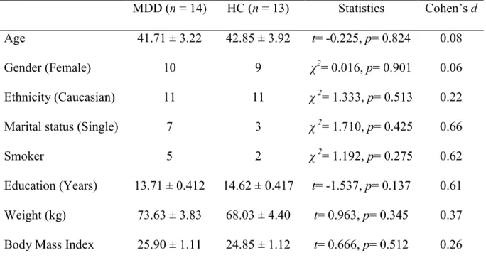
Table 1 Demographic characteristics of participants, given as mean ± standard error. ... 54 Table 2 Mean (± standard error) response to self-report questionnaires. ... 55 Table 3 Thresholds, NFR response and subjective pain rating, given as mean ± standard error.
... 56
List of figures
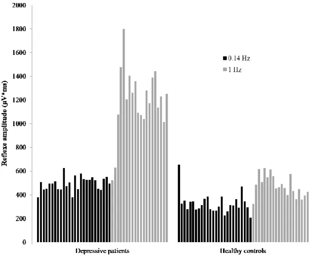
Figure 1 Mean nociceptive-specific flexion reflex amplitude obtained at low- (0.14 Hz) and
high- (1 Hz) frequency stimulation for MDD patients and healthy controls. All 26 stimulations in the 0.14 Hz stimulation condition and all 20 stimulation in the 1 Hz stimulation condition are shown. ... 53
List of abbreviations
CBT : Cognitive-behavioral therapy dlPFC : Dorsolateral prefrontal cortex DNIC : Diffuse noxious inhibitory controlsDSM-IV-TR : Fourth edition of the Diagnostic and Statistical Manual of Mental Disorders DSM-5 : Fifth edition of the Diagnostic and Statistical Manual of Mental Disorders
ECT : Electroconvulsive therapy EMG : Electromyography
fMRI : Functional magnetic resonance imaging IASP : International Association for the Study of Pain ICPM : Inhibitory conditioned pain modulation MAOIs : Monoamine oxidase inhibitors
MDD : Major Depressive Disorder MRI : Magnetic resonance imaging NFR : Nociceptive-specific flexion reflex NMDA : N-methyl-D-aspartate
NRM : Nucleus raphe magnus PET : Positron emission tomography RVM : Rostro-ventral medulla SES : Socioeconomic status
SNP : Single nucleotide polymorphism
SNRIs : Serotonin-norepinephrine reuptake inhibitors SSRIs : Serotonin reuptake inhibitors
TCAs : Tricyclic antidepressants
TDCS : Transcranial direct current stimulation TENS : Transcutaneous electrical nerve stimulation TMS : Transcranial magnetic stimulation
5-HTR1A : Serotonin 1A receptor gene 5-HTR2A : Serotonin 2A receptor gene
Acknowledgements
I would like to thank Dr. Stéphane Potvin for his support and mentoring throughout my masters. I would also like to thank Dr. Valérie Tourjman for her support and supervision in the short time we worked together. Finally, I would like to thank my family.
1
Introduction
1.1 Depression
1.1.1 Epidemiology
With over 350 million people affected worldwide, depression is one of the most prevalent mental health disorder in the general population (Public Health Agency of Canada, 2014) and is currently considered the leading cause of disability by the World Health Organization (WHO, October 2012). The latest Canadian Community Health Survey on Mental Health estimates that 11.3 % of Canadians have experienced symptoms consistent with depression in their lifetime (Pearson et al., 2013), a rate similar to previous estimates from the 2002 Canadian National Survey (Patten et al., 2006). With depression being widespread, many personally face or are impacted by the disorder, hence the necessity to address it.
Across most nations and ethnicities, depression was shown to be twice as prevalent in women than in men (Angst et al., 2002; Kuehner, 2003), a gender difference that emerges in adolescence (Essau et al., 2010; Hyde et al., 2008). The mean age of onset is estimated to be between 25 and 32 years old (Kessler et al., 2005; National Institue of Mental Health, 2015). Having experienced family instability during childhood (Gilman et al., 2003), or being currently separated/divorced increases the risk of developing depression (Andrade et al., 2003). Depression is almost twice as prevalent in highincome countries as in low/middle -income countries (Bromet et al., 2011). Conversely, in high -income countries, depression is more frequently reported among people of lower socioeconomic status (SES) (Lorant et al., 2003). In fact, lower SES during childhood (Gilman et al., 2002), and worsening SES (Lorant et al., 2007) were associated with increasing rates of depression. Unfortunately, a poorer SES
has not only been linked with poor mental health, but with poor physical health as well (Everson et al., 2002).
1.1.2 Consequences
In a comprehensive review of epidemiological studies, Evans et al. (2005) detail the extensive repercussions of depression on medical conditions, which include among others an increased risk of cardiac disease (Rudisch & Nemeroff, 2003), diabetes (Musselman et al., 2003), a perturbation of recovery from cerebrovascular diseases (Krishnan, 2000), a higher cancer mortality rate (Satin et al., 2009), poor human immunodeficiency virus and acquired immune deficiency syndrome treatment adherence (Gonzalez et al., 2011), and an increased severity of chronic pain (Arnow et al., 2006). The consequences of this disorder are manifested as well in socio-professional functioning impairments (Kessler et al., 2003) and an overall diminished quality of life (IsHak et al., 2015).
Depression is also a public health issue, with absenteeism (Druss et al., 2000), loss of productivity (Stewart et al., 2003), and a substantial increase in costs related to medical resource consumption (Luppa et al., 2007) as some of the disorder’s economic implications. With 33 % of total costs related to brain disorders in Europe attributable to depression (Sobocki et al., 2006), and ranking in sixth place in terms of economic costs in an international review (Berto et al., 2000), depression indubitably constitutes both a great individual and societal burden.
An important concern regarding this disorder is the 52 percent greater risk of mortality in depressed individuals compared to the general population (Cuijpers et al., 2014). Other than the negative influence of depression on other medical illnesses which can offer a partial explanation, more than half of suicide victims are thought to suffer from depression
(Cavanagh et al., 2003). Furthermore, depressed individuals are at 2.6 fold higher risk of death by homicide (Crump et al., 2013b), and at 2 fold higher risk of accidental death (Crump et al., 2013a).
1.1.3 Clinical features
To be diagnosed with Major Depressive Disorder (MDD) according to the recent fifth edition of the Diagnostic and Statistical Manual of Mental Disorders (DSM-5) (American Psychiatric Association, 2013a), an individual has to meet/display five or more of the following nine signs or symptoms continuously for a period of at least two weeks: a) depressed mood, most of the day as indicated by subjective reports or observations made by others; b) diminished interest or pleasure in most activities, most of the day; c) significant weight change, including weight loss when not dieting or weight gain, or change in appetite, increase or decrease; d) change in sleep, insomnia or hypersomnia; e) change in activity, as manifested by psychomotor agitation or retardation observable by others; f) fatigue or loss of energy; g) feelings of worthlessness or excessive or inappropriate guilt; h) diminished ability to think or concentrate, or more indecisiveness as indicated by subjective reports or observations made by others; and i) recurrent thoughts of death, or suicidal ideation without a plan, or suicidal ideation with a specific plan, or a suicide attempt. The symptoms have to cause clinically significant distress or impairments in functioning, as well as the episode should not be attributable to the effects of a substance or another medical condition.
The DSM-5 criteria for MDD remain, for the most part, unchanged from the definition of the previous fourth edition of the Diagnostic and Statistical Manual of Mental Disorders (DSM-IV-TR) (American Psychiatric Association, 2000). However, the bereavement exclusion in the DSM-IV-TR, which specified that individuals presenting depressive
symptoms lasting less than 2 months after the death of a loved one did not meet the diagnostic criteria for MDD, was removed from the DSM-5. Evidence showed that depressive symptoms emerging as a consequence of bereavement were not different from those caused by other stressors (American Psychiatric Association, 2013b).
Although only 2 symptomatic weeks are necessary for the diagnosis of MDD, the median duration of a depressive episode was observed to vary between 3 to 6 months in large cohort studies (Eaton et al., 2008; Richards, 2011). Following the first episode of MDD, close to half of MDD patients were found to recover with no relapse (Eaton et al., 2008). It is estimated that 15 % to 42 % of MDD patients will experience the recurrence of the disorder within 20 years following a depressive episode (Eaton et al., 2008; Hardeveld et al., 2013). Additionally, the risk of a subsequent depressive episode increases with each recurrence of a depressive episode (Solomon et al., 2000).
An important issue with diagnosing MDD is the difficulty of differentiating core MDD symptoms from symptoms specific to other psychiatric disorders because of the often significant overlap. It is estimated that half of the individuals suffering from bipolar disorder have a depressive episode before the manifestation of manic symptoms (Etain et al., 2012), as well as patients suffering from bipolar disorder were shown to be more likely to consult for depressive symptoms than for manic symptoms (Hirschfeld, 2004). Furthermore, generalized anxiety disorder and MDD share a number of somatic symptoms (Zbozinek et al., 2012), and depressive mood is very common in prodromal schizophrenia (Hafner et al., 2005). Taking into account the overlap of symptoms is definitely crucial when establishing a MDD diagnosis.
1.1.4 Etiology
Similar to other psychiatric disorders, the consensus is that a gene x environment interaction is involved in the etiology of MDD. An estimated third of the risk for developing depression is attributed to genetic factors (inherited) and two-thirds of the risk to environmental factors (Sullivan et al., 2000). In line with the gene x environment interaction, much of the theories describing MDD follow a diathesis-stress model (Willner et al., 2013), where the vulnerability (diathesis) of an individual for MDD can be precipitated by adverse life events (stress). Complementary to this model, stress-inflammation pathways have been proposed as potential underlying processes in depression (Saveanu & Nemeroff, 2012), where stress induced immune response may play a role in the pathogenesis of MDD (Slavich & Irwin, 2014). Psychosocial stress originating from interpersonal, legal, or work related events (Brigitta, 2002), stress from the aftermath of a major trauma (O'Donnell et al., 2004), or the loss of loved ones (Zisook & Shuchter, 1991) were reported as some of the potential sources that might trigger the onset of MDD. Stressful events and physical or sexual abuse in childhood were shown to increase the predisposition of an individual towards developing the disorder (Saveanu & Nemeroff, 2012).
Twin studies have been consistent in demonstrating the moderate heritability of MDD (Kendler et al., 2006; Kendler & Prescott, 1999), yet the genetic basis of the disorder has proven to be difficult to define. The Genome Wide Association Study conducted by the Cross-Disorder Group of Psychiatric Genomics Consortium (Lee et al., 2013) found a shared genetic etiology for attention deficit hyperactivity disorder, bipolar disorder, schizophrenia, depression and autism. This might be in part the consequence of overlapping or broad symptoms, and
heterogeneous disorders. Nonetheless, studies have succeeded to a certain extent in supporting specific neurobiological mechanisms underlying MDD with genetics.
1.1.5 Neurobiology
The monoamine deficiency hypothesis, a popular neurobiological model of depression, was developed to explain the efficacy of monoamine oxidase inhibitors (MAOIs) and tricyclic antidepressants (TCAs) in treating MDD patients (Hindmarch, 2001). Considering that both MAOIs and TCAs facilitate monoamine neurotransmission in the brain (Hindmarch, 2001), the model postulates that monoamine neurotransmitters are depleted in MDD (Delgado, 2000), especially: serotonin and norepinephrine. The serotonergic system, involved in impulsivity and vigilance, originates in the raphe nuclei of the brainstem and projects to a wide area in the brain, including the frontal cortex, limbic system (amygdala and hippocampus), and hypothalamus (Savitz & Drevets, 2009). Similarly, the noradrenergic system originates in the brainstem (locus coeruleus) and projects diffusely across the brain to the prefrontal cortex, thalamus, hypothalamus, hippocampus, and amygdala; it is, however, associated to arousal and stress response (Goddard et al., 2010). Overall, the anatomy of the monoaminergic systems suggest they are involved in the regulation of a broad spectrum of behaviors and brain functions, such as mood, attention, motivation, psychomotor agitation, sleep, appetite and cognition (Brigitta, 2002; Hasler, 2010).
Human studies in genetics and positron emission tomography (PET) imaging have provided partial support to the monoamine hypothesis, particularly with regards to the implication of serotonergic neural transmission abnormalities in the pathophysiology of depression. Notably, the less functional short (s) allele variation in the 5-hydroxytryptamine transporter gene (5-HTTLPR) polymorphic promoter region, associated with lower serotonin
transporter protein expression than the long (l) allele, was repeatedly linked with increased risk for MDD in response to adverse life events (Daniele et al., 2011; Karg et al., 2011). Consistent with these findings, studies in PET observed lower serotonin transporter density in the brain of depressed suicide attempters (Miller et al., 2013), and medication-free MDD patients (Selvaraj et al., 2011), also mirroring post-mortem results suggesting decreased serotonin transporter density in depressed suicide victims (Stockmeier, 2003). However, in both PET imaging and post-mortem studies, these results appear to be related to suicidal behavior more so than depression (Miller et al., 2013; Stockmeier, 2003). Furthermore, certain PET studies observed the opposite relation between serotonin transporter binding and MDD (Cannon et al., 2007), or failed to find an association (Meyer et al., 2004). In turn, serotonin receptors have been associated to MDD with similarly mixed results. A single nucleotide polymorphism (SNP) located in the serotonin 1A receptor gene (5-HTR1A) promoter region, associated with increased expression of the 5-HTR1A receptor, was significantly associated with mood disorders (Kishi et al., 2013). However, PET studies investigating the 5-HTR1A receptor density in MDD patients have either found that MDD was associated with decreased binding potential (Savitz & Drevets, 2009), or were inconclusive (Shrestha et al., 2012). SNPs in the promoter region of the serotonin 2A receptor gene (5-HTR2A) have been inconsistently linked with MDD, with meta-analyses reporting an association between certain SNPs and depression (Zhao et al., 2014), and no relationships with other SNPs (Jin et al., 2013).
The involvement of the noradrenergic system in the pathophysiology of depression has received some attention as well, albeit to a lesser extent (Moret & Briley, 2011). Although reduced levels of norepinephrine transporters were found in the locus coeruleus of MDD patients post-mortem (Klimek et al., 1997), no associations between MDD and SNPs in the
promoter region of the norepinephrine transporter gene were observed in a short meta-analysis (Zhao et al., 2013). Increased α2- and β1-adrenoceptor densities were reported in post-mortem brain of MDD suicide victims (Rivero et al., 2014), which is in line with the monoamine hypothesis (Goddard et al., 2010), although the evidence is limited. Overall, studies in genetics and PET imaging investigating the monoamine hypothesis of MDD remain for the most part inconclusive.
Recently, the involvement of the glutamatergic system in the pathophysiology of depression has received considerable attention (Mitchell & Baker, 2010), motivating a number of studies to investigate the prophylactic effect of low-dose ketamine, targeting N-methyl-D-aspartate (NMDA) receptors, in treating symptoms of depression (Aan Het Rot et al., 2012). Although most compelling evidence for the implication of the glutamatergic system in MDD comes from clinical trials with glutamatergic agents such as ketamine, there are preliminary results in post-mortem and magnetic resonance spectroscopy studies in humans suggesting a decrease/dysregulation of glutamate metabolites in the prefrontal cortex and the limbic system of individuals suffering from MDD (Mitchell & Baker, 2010). Because of both the limited and delayed effect of current monoamine treatments in MDD, the complementary role of glutamate, which is not a monoamine neurotransmitter, appears to be worthwhile to pursue in future research.
Studies in magnetic resonance imaging (MRI) and functional MRI (fMRI) have identified brain regions that may be implicated in the neurobiology of MDD. One of the most consistently documented result in MDD is the increased reactivity of the amygdala in response to negative stimuli compared to healthy individuals (Hamilton et al., 2012), an effect that has been associated to the s allele of the 5-HTTLPR gene (Savitz & Drevets, 2009). The insula
and the dorsal anterior cingulate cortex were also shown to be overactive in response to negative stimuli in MDD patients (Hamilton et al., 2012), thereby indicating the potential involvement of hyperactive (para)limbic structures in the pathophysiology of MDD. Conversely, studies found negative stimuli to elicit decreased response in the dorsolateral prefrontal cortex (dlPFC) and the dorsal striatum in MDD (Hamilton et al., 2012), results that are corroborated by studies suggesting a disrupted fronto-limbic connectivity in depression (Savitz & Drevets, 2009). Given the implication of the dlPFC in cognitive control/ inhibition, these results support the cognitive model that posits a deficit in the regulation of emotional processing in MDD (Gotlib & Joormann, 2010; Wolkenstein & Plewnia, 2013). The role of the hippocampus in MDD has also received some attention, as biased memory processes towards negative stimuli have been reported in MDD (Gotlib & Joormann, 2010), and fMRI studies have observed less recruitment of this region during memory tasks in depressed patients compared to healthy individuals (Milne et al., 2012). Reduced hippocampal gray matter volumes were also observed in MDD patients, which might potentially be explained by decreased hippocampal neurogenesis (Eisch & Petrik, 2012). Authors hypothesize that these functional and structural changes in the hippocampus of MDD patients might be involved in the pathophysiology of MDD (Campbell & Macqueen, 2004). Further abnormalities have been identified in the brain morphology of MDD patients, including gray matter reductions in the limbic regions (anterior cingulate cortex), frontal regions (middle and frontal gyrus), and the thalamus (Du et al., 2012).
1.1.6 Treatment
According to the National Health and Nutrition Examination Surveys in the United States, up to 11 % of Americans are prescribed antidepressants, making it the third most common
prescription drug in the U.S. (Pratt et al., 2011) and amounting to an estimated 11 billion dollars in spending in 2011 (Institute for Healthcare Informatics, April 2012). Even though antidepressants are also used to treat other disorders, which might explain the elevated costs, pharmacotherapy seems to be the preferred treatment for MDD. Although the efficacy might vary depending on the severity of the depression (Kirsch et al., 2008), antidepressants have only shown modest efficacy compared to placebo treatments (Lima & Moncrieff, 2000; von Wolff et al., 2013), with less than 50 % of MDD patients achieving adequate response to treatment with antidepressants (Fava, 2003).
Currently, there are over five classes of antidepressants prescribed to MDD patients. MAOIs, the first class of antidepressants that was developed, inhibit the activity of the monoamine oxidase enzyme involved in the catabolism of monoamines such as serotonin, noradrenaline and dopamine (Shulman et al., 2013). By inhibiting their breakdown, MOAIs increase the availability of monoamine neurotransmitters in the brain. TCAs and tetracyclic antidepressants, the second class of antidepressants, exert their effect by inhibiting the reuptake of serotonin and norepinephrine from the synaptic cleft, thereby increasing the availability of those neurotransmitters (Hindmarch, 2001). Together, cyclic antidepressants and MAOIs constitute the first-generation of antidepressant medication, which led to the advent of the monoamine-deficiency hypothesis in MDD. Of the second-generation antidepressants, selective serotonin reuptake inhibitors (SSRIs) and serotonin-norepinephrine reuptake inhibitors (SNRIs) have similar mechanisms of action, where both selectively block the reuptake of serotonin and/or norepinephrine and have little affinity to histamine and muscarinic receptors (Horst & Preskorn, 1998), the latter being potentially the reason why these second generation antidepressants have less side-effects than MAOIs and TCAs.
Because they increase synaptic availability of monoamines, the efficacy of SSRIs and SNRIs in MDD corroborates the monoamine-deficiency hypothesis. Moreover, the efficacy of SSRIs in MDD has been associated with serotonin transporter gene promoter polymorphisms (Porcelli et al., 2012; Serretti et al., 2007). Further second-generation antidepressants include atypical antidepressants, which are more tolerable than first-generation antidepressants despite having multiple sites of actions (Horst & Preskorn, 1998).
Likewise, there are many psychotherapies that are employed to treat MDD patients. Out of the common therapies, which include cognitive-behavioral therapy (CBT), nondirective supportive treatment, behavioral activation treatment, psychodynamic treatment, problem-solving therapy, interpersonal therapy, and social skills training, meta-analyses indicate that there is little difference between the efficacy of these psychotherapies (Barth et al., 2013; Cuijpers, van Straten, Andersson, et al., 2008), yet most studies agree that interpersonal psychotherapy (Schramm et al., 2007) and cognitive-behavioral therapy (Luty et al., 2007) are the most efficacious. Physical exercise is also often recommended to MDD patients, and has shown similar efficacy to the above mentioned therapies (Cooney et al., 2013). Nondirective supportive treatment is, however, less efficacious than the other available therapies (Cuijpers, van Straten, Andersson, et al., 2008). Overall, pharmacotherapy and psychotherapy individually appear to be equally effective (Cuijpers, van Straten, van Oppen, et al., 2008), and Cuijpers et al. (2012) recommend a combination of pharmacotherapy and psychotherapy, as it results in better outcome than any individual therapy.
There are few alternatives to pharmacotherapies and psychotherapies, namely somatic therapies (Cusin & Dougherty, 2012). Although mostly used for acute refractory depression, a recent meta-analysis reported a 50.9 % remission rate in unipolar depression using
electroconvulsive therapy (ECT) (Dierckx et al., 2012). In ECT, bifrontal electrode placement was found to elicit less of the cognitive impairments previously reported with ECT, such as memory loss, while remaining as efficacious as other electrode placements (Bailine et al., 2000). Interestingly, electrical stimulation of the frontal brain regions using transcranial direct current stimulation (TDCS) resulted in enhanced cognitive control in MDD patients (Wolkenstein & Plewnia, 2013), which the authors attributed to the activation of the hypoactivated dlPFC that is characteristic of MDD (Hamilton et al., 2012). Studies using transcranial magnetic stimulation (TMS) have also focused on activating and/or inhibiting the dlPFC in MDD patients with the same purpose as TDCS (Cusin & Dougherty, 2012), although remission rates from MDD using TMS are lower than treatments employing ECT (Carpenter et al., 2012).
1.2 Pain
1.2.1 Epidemiology
Primarily an adaptive multidimensional sensory experience providing relevant information for the protection of the organism from injury, and promoting healing when injury has occurred, pain can become a disease when it is maladaptive and/or chronic (Woolf, 2004). An estimated 20 % of adults report experiencing pain (Goldberg & McGee, 2011), and between 10 % and 19 % of adults, representing more than 1.5 million Canadians and 39.4 million Americans, suffer from chronic/persistent pain according to Canadian and U.S. national health surveys (Kennedy et al., 2014; Ramage-Morin & Gilmour, 2010; Reitsma et al., 2011). Pain is a major health care concern, as it accounts for 20 % of medical consultations (Alford et al., 2008) and between 4 and 6 billion dollars in direct costs in Canada (Lynch et al., 2009; Philips &
Schopflocher, 2008). Chronic pain has been linked with increased absenteeism, lost productivity at work, as well as interference with daily activities (Reitsma et al., 2011). Higher chronic pain prevalence was associated with sex (women), increasing age, marital status (separated/divorced), and lower SES (Johannes et al., 2010). Although predominantly perceived as a symptom rather than a disease (Goldberg & McGee, 2011), pain and chronic pain have received increasing attention due to their extensive impact on both patients and health care system.
1.2.2 Clinical features
The International Association for the Study of Pain (IASP) defines pain as “an unpleasant sensory and emotional experience associated with actual or potential tissue damage, or described in terms of such damage” (IASP, 2012a). Chronic pain is characterized as pain that may arise from injury, or manifest itself without clear cause, and last longer than 3 to 6 months (American Psychiatric Association, 2013a; IASP, 1986; National Institute of Health, 2011). Both acute and chronic pain are multi-facetted experiences of heterogeneous aetiologies (Woolf, 2004).
Pain experience can be divided into three psychological dimensions: sensory-discriminative, affective-motivational and evaluative-cognitive (Melzack & Casey, 1968). The sensory-discriminative aspect of pain refers to the intensity, duration, location and quality of the pain experienced. The affective-motivational aspect of pain refers to the unpleasantness of pain and the urge towards escaping the unpleasantness or attacking its source. The intensity of the pain experienced along the sensory-discriminative and affective-motivational dimensions are influenced by the central evaluative-cognitive dimension, which refers to the appraisal of the input from the first two dimensions based on past experiences.
Pain is commonly divided into four distinct types of pain: nociceptive, inflammatory, neuropathic, and functional (Marchand, 2008; Woolf, 2004). Nociceptive pain is the transient response to noxious stimuli indicating the presence of damaging or potentially damaging stimuli. Following an injury, inflammatory pain can be experienced as an increased sensitivity to the affected area which prevents contact or movement of the injured part, in order to promote healing. Neuropathic pain is the consequence of lesions or diseases to the peripheral nervous system. Lastly, functional pain occurs in the absence of detectable lesions or abnormalities. This latter type of pain may occur as a result of abnormal responsiveness of the nervous system.
1.2.3 Physiology
Nociception is the encoding of noxious stimuli in the nervous system (IASP, 2012a; Loeser & Treede, 2008). It is initiated by nociceptive stimuli, which are damaging or potentially damaging mechanical, thermal or chemical events (IASP, 2012b; Mitra et al., 2013), activating nociceptors, which are specialized sensory free nerve endings, located in peripheral tissues responding almost exclusively to pain stimuli (Woolf, 2004). Nociceptor terminals transduce noxious stimuli into action potentials, that are then conducted along primary afferent fibers from peripheral terminals through the dorsal root ganglion, entering the spinal cord via the dorsal root, to finally reach the central nervous system (Mitra et al., 2013; Woolf, 2004).
There are two distinct types of peripheral afferent fibers that carry pain sensation: myelinated Aδ-fibers and thinly/un-myelinated C-fibers (Woolf, 2004). Afferent fibers with myelinated axons (Aδ-fibers) have rapid conduction velocity, and are therefore responsible for the immediate transient sharp pain sensation, or first pain sensory input (Marchand, 2008). Afferent fibers with unmyelinated axons (C-fibers) constitute approximately 75 % of
peripheral fibers, and have slow conduction velocity (Marchand, 2008). C-fibers are responsible for the delayed and prolonged diffuse dull pain sensation, or second pain sensory input (Mitra et al., 2013). Both Aδ-fibers and C-fibers (first order neurons) transport the information from the affected area to the spinal cord. On the other hand, non-noxious sensations, including vibration, movement and light touch, are carried from the periphery to the spinal cord by large myelinated Aα-fibers and Aβ-fibers, which have faster conduction velocity than Aδ-fibers (Marchand, 2008).
Peripheral afferent fibers synapse in the dorsal horn of the spinal cord, where the signal may be modulated via synaptic contact with inhibitory and excitatory interneurons before being transmitted to second order neurons (Woolf, 2004). Second order neurons project from the dorsal horn of the spinal cord to supraspinal systems through two separate ascending pathways: the sensory spinothalamic tract and the affective spinoreticulothalamic/ spinoreticular tract (Basbaum et al., 2009; Marchand, 2008).
Only a fraction of the input from nociceptors will be transmitted to the thalamus, and then relayed to the cortex by third order neurons (Woolf, 2004). The spinothalamic tract projects directly from the dorsal horn of the spinal cord to the contralateral nuclei of the ventrobasal thalamus (Marchand, 2008). From the thalamus, the information is then transmitted to the somatosensory cortex as well as (para-)limbic regions (Marchand, 2008). The projection neurons in the spinothalamic tract have fast conducting axons, small receptive fields, as well as somatotopic organization (Hong et al., 2011) making this pathway essential for the sensory-discriminative aspects (location, duration and intensity) of pain (Basbaum et al., 2009). The spinoreticular tract projects from the dorsal horn of the spinal cord to the reticular formation which in turn projects to the medial nuclei of the thalamus (and then to the
primary somatosensory cortex), the hypothalamus and the limbic system (Patestas & Gartner, 2009). The projection neurons in this latter tract have large receptive fields and are essential in the affective-evaluative aspects (emotional and memory) of pain (Mitra et al., 2013). From the dorsal horn, the spinoreticular tract also projects to the periaqueductal gray (PAG) area and the nucleus raphe magnus (NRM) located in the brainstem (Marchand, 2008).
Studies in PET and fMRI have shown a distributed network of cortical structures participating in pain perception (Marchand, 2008). The primary somatosensory cortex and secondary somatosensory cortex are believed to be involved in the sensory-discriminative aspects of pain (Ossipov, 2012; Peyron et al., 2000), whereas the anterior cingulate cortex, the insula (Apkarian et al., 2005), as well as the amygdala (Simons et al., 2014) are thought to be involved in the affective-emotional aspects of pain. Finally, the prefrontal cortex may be involved in the cognitive-evaluative aspects of pain perception (Apkarian et al., 2005), such as the modulation of the intensity of the pain subjectively perceived through the reappraisal of the painful stimuli (Wiech et al., 2008). Specifically, fMRI studies examining the influence of cognitive processes on pain perception have shown that regions in the prefrontal cortex such as the orbitofrontal cortex (Bantick et al., 2002), dlPFC (Lorenz et al., 2003), and the ventrolateral prefrontal cortex (Wiech et al., 2008) potentially exert control on the activity of regions responsible for the affective-emotional qualities of pain (e.g. amygdale, insula).
1.2.4 Endogenous inhibitory pain modulation
The ascending nociceptive pathways describe the way in which the nociceptive signal is transmitted from the periphery to the central nervous system. However, nociception and pain perception are separate processes, as the afferent nociceptive signal can be modulated by inhibitory and excitatory mechanisms before being perceived by the organism (Marchand,
2008). These endogenous mechanisms modulating the nociceptive signal are categorized into spinal, descending and supraspinal processes.
The inhibitory modulation of nociceptive input at the spinal cord was first described by Melzack and Wall (1965) in the gate control theory of pain, and has been well document since then (Marchand, 2008). The theory asserts that non-nociceptive peripheral input can suppress nociceptive signal from travelling to the central nervous system (Melzack & Wall, 1965). The input from non-nociceptive Aα-fibers and Aβ-fibers in the dorsal horn of the spinal cord will recruit inhibitory interneurons in the substantia gelatinosa, producing localized analgesia, thereby limiting or preventing the transmission of afferent nociceptive signal from Aδ and C fibers to second order neurons (Calvino & Grilo, 2006). Transcutaneous electrical nerve stimulation (TENS) is a clinical application of the gate control theory of pain, as it employs electrical stimulations applied to the skin to relieve pain (Sluka & Walsh, 2003).
A growing number of supraspinal processes influencing descending inhibitory mechanisms have been reported in the literature. Descending inhibitory mechanisms from the brainstem were described by Lebars et al. (1979) as diffuse noxious inhibitory controls (DNIC). Lebars et al. (1979) postulated that a nociceptive stimulation will inhibit another spatially distant nociceptive stimulation by producing diffuse analgesia throughout the rest of the body (i.e. counter-irritation) (Marchand, 2008). Studies showed that DNIC recruit opioids in the PAG, therefore triggering the release of serotonin from rostro-ventral medulla (RVM) neurons. The release of serotonin decreases the input from nociceptive afferent fibers at the dorsal horn of the spinal cord (Stavro & Potvin, 2014). Noradrenergic projections from the locus coeruleus were shown to produce similar descending inhibitory effects (Ossipov et al., 2010). Notably, a deficit in inhibitory conditioned pain modulation (ICPM) (Yarnitsky, 2010),
a term referring to DNIC in human experimental setting, was shown to be a key element in chronic pain disorders, such as fibromyalgia (Marchand, 2008).
Other supraspinal inhibitory mechanisms have also been reported, involving prefrontal and limbic brain activity in particular. Multiple supraspinal processes, including cognitive modulators such as attention (i.e. distraction), reappraisal, hypnosis (i.e. suggestion), and placebo analgesia (Marchand, 2008; Wiech et al., 2008), were shown to inhibit pain perception. Studies in brain imaging have suggested that these cognitive factors exert their analgesic effect by recruiting prefrontal regions, which in turn inhibit brain regions that are involved in the emotional component of pain (Ochsner & Gross, 2005). In addition to these cognitive factors, positive and agreeable emotions induced by music, odors or films (Roy et al., 2008) were found to have analgesic properties as well. Preliminary studies in fMRI suggest that the analgesic effect might be produced by recruiting the reward system (e.g. ventral striatum) (Schweinhardt et al., 2009).
1.2.5 Endogenous excitatory pain modulation
The excitatory modulation of spinal cord neurons can be distinguished in two separate, albeit related, mechanisms: windup and central sensitization (Woolf, 2011). Windup consists of a progressive increase in action potential discharge in second order neurons during high-frequency (≥0.3 to 5 Hz) stimulation of C-fibers (first order neurons) at constant intensity (Latremoliere & Woolf, 2009; Li et al., 1999). It is a process that recruits NMDA glutamate receptors (Herrero et al., 2000). Windup can be elicited by temporal summation paradigms, where increasing the frequency of repeated identical noxious stimulation will produce a heightened sensation of pain, although the intensity of the stimulation remains unchanged (Marchand, 2008). While windup can induce central sensitization, it is a transient excitability
of the spinal cord neurons that disappears quickly following the end of the stimulation as the membrane returns to its resting state, whereas central sensitization is a state that remains after the end of stimulation (Woolf, 2011). The clinical manifestations of central sensitization can be observed in hyperalgesia (amplified response to nociceptive input) and allodynia (perception of pain in the absence of nociceptive stimulation) (Marchand, 2008). Temporal summation paradigms, including paradigms employing the nociceptive-specific flexion reflex (NFR), are advantageous in an experimental setting as they allow investigating the contribution of spinal cord neurons to pain sensitization in humans (Arendt-Nielsen et al., 1994; Lévesque et al., 2012).
Finally, certain supraspinal processes were shown to increase the pain perceived. Notably, anxiety was shown to exacerbate the intensity of the pain sensation (Goffaux et al., 2011), an effect potentially mediated by the hippocampal formation (Ploghaus et al., 2001). Sad mood was also shown to increase pain unpleasantness, which was associated to a greater inferior frontal gyrus and amygdala response (Berna et al., 2010). Moreover, catastrophizing was shown to increase pain sensitivity (Kristiansen et al., 2014), which was associated with greater activity in the prefrontal and anterior cingulate cortex (Quartana et al., 2009). Therefore, these supraspinal processes appear to modulate the response of brain regions associated with affective and cognitive processes involved in pain perception.
1.2.6 Treatment
The American Chronic Pain Association (2015) identifies pharmacotherapy as the most common treatment for chronic pain. In fact, over 7 million Canadians are estimated to take pain medication (Lynch & Watson, 2006). Given the significant variability in the cause and the severity of pain (Food and Drug Administration, 2009), there are many classes of
medication with distinct mechanisms of actions available to treat the various types of pain (i.e. nociceptive, inflammatory, neuropathic, and functional). First line pharmacological treatments for nociceptive and inflammatory pain involve non-narcotics (e.g. acetaminophen) and nonsteroidal anti-inflammatory drugs. These medications target the inflammatory process at the origin of the peripheral nociceptor sensitivity (Lynch & Watson, 2006; Vane & Botting, 1998). For more severe/chronic nociceptive and inflammatory pain, opioids are recommended (World Health Organization, 1996). Opioids are, however, only employed as second line treatment for neuropathic pain because of their addictive qualities, and antidepressants and anticonvulsants are recommended instead (Attal et al., 2010; Attal et al., 2006). It is hypothesized that antidepressants exert their analgesic effect by increasing the availability of neurotransmitters that are essential to the ICPM (Portenoy & Ahmed, 2013), whereas anticonvulsants dampen spinal sensitization by reducing neuronal hyperactivity via glutamatergic and GABAergic mechanisms (Marchand, 2008; Tremont-Lukats et al., 2000). Antidepressants and anticonvulsants are prescribed to treat functional pain as well (Marchand, 2008).
Similarly to MDD, alternatives to pharmacotherapy for pain treatment include psychotherapies, somatic therapies and physical therapies (American Chronic Pain Association, 2015; Turk & Gatchel, 2002). Among psychotherapies, CBT is most commonly recommended, although mindfulness therapy as well as acceptance and commitment therapy appear as equally good alternatives for pain management (Veehof et al., 2011). Although TENS is often used to induce ICPM in research settings, the literature supporting the use of TENS in a clinical setting is still inconclusive (DeSantana et al., 2008; Nnoaham & Kumbang, 2008). Other somatic therapies in pain treatment, such as acupuncture, are employed in pain
management (American Chronic Pain Association, 2015). However, only limited research has investigated the efficacy of such treatments in pain treatment (Sun et al., 2008). Finally, physical exercise and other forms of physical therapies (e.g. yoga, tai chi, qigong) are also recommended for patients suffering from chronic pain (American Chronic Pain Association, 2015).
1.3 Comorbidity of pain and depression
1.3.1 Epidemiology
Studies show that between 50 to 65 % of patients suffering from MDD report pain symptoms (Bair et al., 2003; Katona et al., 2005), and as many as 92 % of MDD patients report at least one pain-related symptom (Corruble & Guelfi, 2000). Evidently, pain and depression co-occur frequently, and seem to be mutually exacerbating. In fact, pain in joints, limbs, back, and abdomen (Corruble & Guelfi, 2000; Garcia-Cebrian et al., 2006), as well as headaches (Mathew et al., 1981) constitute some of the common somatic symptoms that negatively impact treatment response (Bair et al., 2004), time to remission (Karp et al., 2005), daily functioning (Ohayon & Schatzberg, 2010), sleep (Ohayon, 2004), and quality of life of MDD patients (Lin et al., 2014), as well as predict disorder chronicity (Gerrits et al., 2012). Conversely, depressive symptoms were associated with poor prognosis (Bair et al., 2003), longer time to recovery (Henschke et al., 2008), higher probability of pain chronicity (Pincus et al., 2002), and poor response to treatment (Bair et al., 2003) in individuals suffering from pain.
Individuals presenting both pain and depression were found by a national survey in the U.S. to be older, with lower income, and reporting an increased use of medical services
compared to depressed individuals without comorbid pain (Bao et al., 2003), which in turn is associated with a substantial increase in medical costs (Emptage et al., 2005; Greenberg et al., 2003). Interestingly, Bao et al. (2003) also observed that depressive individuals suffering from pain were 21 % less likely to see a mental health specialist, alluding to the problem of overlapping symptomatology between MDD and certain pain conditions (Wilson et al., 2001). When presented simultaneously, patients and healthcare professionals might attribute depressive symptoms to a painful condition and vice-versa, consequently focusing on the primary condition while the secondary condition remains unattended (Katona et al., 2005; Wilson et al., 2001). Nevertheless, attending to both pain and depression when these conditions are comorbid is crucial, as they influence negatively one another.
In order to explain the frequent co-occurrence of depression and pain, certain authors have postulated that pain might be a risk factor for developing symptoms of depression, and others have suggested the opposite, that it is depression that might lead to painful symptoms. Regarding the former hypothesis, authors have posited that chronic pain might operate as a stressor activating the hypothalamic-pituitary-adrenal axis (Munro & Blackburn-Munro, 2001), or as a precipitating factor for a pre-existing vulnerability to develop a psychiatric disorder (Dersh et al., 2002), and therefore symptoms of depression would be the consequence of pain. General population studies have observed that pain and chronic pain might indeed precede depression (Gerrits et al., 2014; McBeth et al., 2002), and there seems to be more support for this direction of the relation between pain and depression (Fishbain et al., 1997; Goesling et al., 2013). The alternative hypothesis, where depression could bring about biological and cognitive changes that would potentially facilitate the subsequent emergence of pain (Goesling et al., 2013), has also received some attention. Painful symptoms might be the
somatization of depression, which would be a way to communicate distress (Dersh et al., 2002). In fact, a national survey has found that depression increased the risk of future chronic pain (Currie & Wang, 2005; Gupta et al., 2007).
Although the relationship between pain and depression appears to be reciprocal, only a few studies have demonstrated within the same experiment the two conditions having an equally adverse impact on one another (Goesling et al., 2013). In a population based study, pain at baseline predicted depression, and similarly depression at baseline predicted pain over 2 years (Chou, 2007). Furthermore, changes in pain severity were shown to predict subsequent depression severity over 12 month in primary care patients, as well as the converse (Kroenke et al., 2011). Overall, the relationship between pain and depression seems to be bidirectional.
1.3.2 Clinical features
Given the quantity of studies suggesting a reciprocal and bidirectional relation between depression and pain, a growing number of authors consider the frequent comorbidity between these two conditions as a result of common cognitive and affective factors. In fact, common cognitive factors were found to be influenced by pain and depression, and to mediate the relationship between these conditions. Specifically, deficits in working memory and attention were independently noted in pain (Moriarty & Finn, 2014; Moriarty et al., 2011) as well as in depression (Gotlib & Joormann, 2010), suggesting that individuals suffering from either conditions would have a certain difficulty to attend to stimuli that is not congruent with their condition, whereas they would display a bias towards disorder-salient stimuli (Erickson et al., 2005; Khatibi et al., 2009). Studies suggest that reduced gray matter density and altered activation of the prefrontal cortex in both conditions might be the neural substrate of this cognitive deficit (Robinson et al., 2009). The well documented implication of catastrophizing,
which is a maladaptive cognitive distortion involving processes such as rumination and magnification, in pain and depression reinforces the importance of shared cognitive processes between the two conditions (Goesling et al., 2013; Linton & Bergbom, 2011; Quartana et al., 2009). In fact, research in cognitive-behavioral interventions suggests that reduction in catastrophizing would also reflect improvements in pain severity and depression in comorbid patients (Quartana et al., 2009). Helplessness, self-efficacy, and pessimism were also shown to mediate the relationship between pain and depression (Campbell et al., 2003; Goesling et al., 2013). Negative affect, such as sadness, distress and fear have also been associated with both pain and MDD (Goesling et al., 2013). The affective component of pain is increasingly supported by results in imaging as being partially independent from the sensory component of pain. Specifically, the anterior cingulate cortex and the amygdala seem to integrate negative affect and pain (Neugebauer et al., 2004; Robinson et al., 2009; Shackman et al., 2011).
Pain processing and depression appear to share multiple neurobiological mechanisms, which could explain the frequent co-occurrence of these conditions. Serotonergic and noradrenergic signaling pathways have been implicated in the pathogenesis of MDD, and are thought to be involved in the emergence of pain when dysfunctional. The existence of common serotonergic and noradrenergic alterations in pain and depression is also suggested by the mechanism of action of pharmacological treatments common to both pain and depression (Goesling et al., 2013). Although first considered as a treatment for the affective symptoms of chronic pain, antidepressants, including SSRIs and SNRIs, are receiving increasing support as analgesics that are hypothesized to act on endogenous pain control by enhancing descending inhibitory pain mechanisms which rely on serotonin and norepinephrine (Jann & Slade, 2007; Mico et al., 2006). Still, pain management with antidepressants is most
recommended in patients presenting comorbid chronic pain and depression (Campbell et al., 2003; Goesling et al., 2013).
Alternatively, an increasing number of studies have proposed inflammatory mechanisms that could explain the link between pain and depression (Walker et al., 2014). Inflammation is pertinent in the context of pain. In fact, it is an inflammatory process that is at the origin of the sensitization of the peripheral nociceptors. In theory, a neuropathic inflammatory process might be implicated in central pain sensitization (Clark et al., 2013). Inflammatory processes might also be implicated in the pathophysiology of depression. A meta-analysis has shown interleukin-6 and C-reactive protein elevations in MDD (Haapakoski et al., 2015). Therefore, common inflammatory alterations could potentially be at the origin of the frequent co-occurrence of the two phenomena.
1.3.3 Pain perception in depression
In order to better understand the reasons underlying the elevated prevalence of painful somatic symptoms in major depression, several psychophysical studies have been performed in patients with MDD. However, current experimental studies have failed to offer a clear insight into the relationship between the two conditions (Potvin, 2011). Most studies agree that MDD patients perceive pain to be subjectively less intense than healthy individuals when administered the same intensity of pain (Boettger et al., 2010; Dworkin et al., 1995; Lopez-Sola et al., 2010). However, a number of studies have found the reverse relationship or no differences between MDD and healthy individuals (Frew & Drummond, 2009; Normand et al., 2011; Strigo et al., 2008a). Furthermore, a majority of studies investigating pain threshold found a higher pain threshold across most nociceptive stimulation modalities (e.g. thermic, electric, mechanic, and ischemic) in MDD compared to controls (Bär et al., 2007; Bar et al.,
2005; Boettger et al., 2013; Dickens et al., 2003; Schwier et al., 2010), suggesting that MDD patients might tolerate pain better than healthy individuals. Even so, substantial literature report decreased pain threshold in MDD, or no significant differences between MDD and healthy individuals (Bar et al., 2005; Klauenberg et al., 2008; Normand et al., 2011; Spernal et al., 2003; Strigo et al., 2008a; Terhaar et al., 2010). Finally, a similar pattern can be observed in studies investigating pain tolerance in MDD, with studies suggesting a higher (Bar et al., 2005; Bar et al., 2003), lower (Gormsen et al., 2004), or identical (Frew & Drummond, 2009) pain tolerance in depression. In sum, psychophysiological studies investigating pain perception found MDD patients to be hypoalgesic, hyperalgesic or normal when compared to healthy individuals (Dickens et al., 2003; Lautenbacher & Krieg, 1994; Potvin, 2011), which are overall inconsistent results that fail to determine the impact of MDD on pain perception (Dickens et al., 2003) and to explain the elevated frequency of pain complaints observed in a depressive population.
1.3.4 Pain modulation in depression
Considering that the literature investigating pain perception, pain threshold, and pain tolerance in depression is for the most part inconclusive, authors have suggested that the issue in MDD might lie in the endogenous inhibitory or excitatory pain modulation (Potvin, 2011). Taking into account that serotonin and norepinephrine were shown to be implicated in pain inhibition (Millan, 2002; Ossipov et al., 2010), as well as substantial research has identified both serotonergic and noradrenergic neural transmission abnormalities in the pathophysiology of depression (Moret & Briley, 2011; Selvaraj et al., 2011; Stockmeier, 2003), a deficit of endogenous pain inhibition in MDD seems probable. Such deficit in descending inhibitory pathways would allow for painful sensations that are normally supressed to be interpreted as
pain by the brain (Stahl & Briley, 2004), which would offer an explanation for the prevalence of painful somatic symptoms in depression. To date, few studies have investigated descending pain inhibition, with inconsistent results. Measuring the jaw-opening reflex, Wang et al. (2000) observed no deficit in inhibitory mechanisms, whereas Bar et al. (2003) found better pain inhibition in MDD than in controls. However, the association between the jaw-opening reflex and pain processing has been questioned by certain authors (Forkmann et al., 2009). In a recent study from our group, Normand et al. (2011) set out to measure deficits in pain inhibition in individuals suffering from MDD using an experimental thermal pain paradigm. Participants were administered two temporal summation tests using heat pulses, and a cold pressor test was conducted in between to induce inhibitory conditioned pain modulation. Remarkably, our group found no deficits in pain inhibition in MDD patients (Normand et al., 2011).
Although very little literature has addressed the alternative hypothesis, painful somatic symptoms in MDD might emerge from overactive excitatory pain mechanisms. Endogenous excitatory pain mechanisms were found to be mediated by glutamatergic mechanisms (Gebhart, 2004; Herrero et al., 2000; Porreca et al., 2002; Woolf, 2004), and they are most frequently studied using temporal summation paradigms (Potvin et al., 2012; Potvin et al., 2008). Notably, recent studies suggest an implication of glutamatergic disturbances in the pathophysiology of depression (Hashimoto, 2009; Mitchell & Baker, 2010; Sanacora et al., 2008), which might result in a potential disruption of endogenous excitatory pain mechanisms in MDD. To date, Klauenberg et al. (2008) are the only group to have assessed endogenous excitatory mechanisms in depression. Using a mechanical temporal summation paradigm, the authors observed an increase in pain sensitization in depressive disorders and found no
differences in pain threshold between depressive participants and controls (Klauenberg et al., 2008). These results might indicate overactive excitatory pain mechanisms in depression. However, the sample recruited by Klauenberg et al. (2008) also included patients suffering from mood disorders other than MDD, such as bipolar disorder, adjustment disorder and dysthymia, thereby limiting the generalizability of the results. Moreover, the paradigm employed to induce temporal summation did not allow the separation of the spinal and supraspinal aspects of pain sensitization in depression.
1.3.5 Treatment
There are currently no set guidelines available for the treatment of unexplained painful somatic symptoms in patients with MDD, and there are only few suggestions for the treatment of comorbid pain and MDD. The National Institute of Mental Health (2015) proposes SSRIs and SNRs as pharmacotherapies, and CBT as psychotherapy for the treatment of chronic pain and depression. Similarly, Nicolson (2010) suggests antidepressants (SNRIs and TCAs), as it appears to relieve neuropathic pain. However, these are circumstantial evidences that do not indicate whether the aforementioned treatments are optimal in MDD patients presenting unexplained somatic symptoms.
1.4 Objectives
The neurobiological mechanisms underlying the high prevalence of painful somatic symptoms in major depression are yet to be fully understood. Considering that studies investigating pain perception in MDD have yielded inconsistent results, recent studies have focused on characterizing endogenous inhibitory and excitatory pain mechanisms in MDD. Normand et al. (2011) found no deficits in endogenous inhibitory pain modulation in MDD. Conversely, Klauenberg et al. (2008) showed that pain sensitization might be disrupted in patients with mood-disorder, which is an indication of potentially altered excitatory pain mechanisms. However, no studies to date have investigated simultaneously the contribution of spinal and supraspinal processes in pain sensitization in MDD.
The primary purpose of this study was to investigate excitatory pain mechanisms in major depression. We set out to replicate the increased pain sensitization observed by Klauenberg et al. (2008) in MDD patients exclusively. In order to study endogenous excitatory pain mechanisms, we employed a temporal summation paradigm that enabled us to assess the contribution of spinal and supraspinal processes in pain sensitization in MDD. To this effect, we measured the NFR triggered by a transcutaneous electrical stimulation to assess the contribution of spinal processes to pain sensitization. Furthermore, by measuring the subjective pain during the temporal summation paradigm, we assessed the contribution of supraspinal processes to pain sensitization. Therefore, our second objective was to evaluate whether the alteration of pain sensitization in MDD has spinal and/or supraspinal origins. Given the substantial literature addressing the subject of supraspinal disturbances in depression, we hypothesize that MDD patients will demonstrate an altered supraspinal pain
sensitization. Finally, we set out to assess the clinical correlates of the experimentally-induced pain response.
2
Article accepted for publication in Pain Medicine
Andràs Tikàsz’s contribution to the article consists of data acquisition, analyzing and interpreting the data, as well as writing this manuscript.
Valérie Tourjman was involved in patient recruitment and assessment, as well as revision of the manuscript.
Philippe Chalaye was involved in participant testing and provided critical comments about the manuscript.
Serge Marchand was involved in study design and provided critical comments about the manuscript.
Stéphane Potvin was involved in study design, supervised Andràs Tikàsz, and was involved in the revision of the manuscript
Increased spinal pain sensitization: A new explanation
for highly prevalent painful somatic symptoms in major depressive disorder?
Andràs Tikàsz, MSc c 1, Valérie Tourjman, MD PhD c 1, Philippe Chalaye, PhD 2, Serge Marchand, PhD 2, Stéphane Potvin, PhD 1
1 Centre de recherche de l’Institut Universitaire en Santé Mentale de Montréal; Department of psychiatry, Faculty of medicine, University of Montreal; Montreal, Canada
2 Centre Hospitalier de l’Université de Sherbrooke; Department of surgery, Faculty of medicine, University of Sherbrooke; Sherbrooke, Canada
Corresponding author
Stéphane Potvin, PhD; Centre de recherche de l’Institut Universitaire en Santé Mentale de Montréal; 7331 Hochelaga; Montreal, Quebec, Canada; H1N 3V2; Tel: (514) 251-4015
Disclosures
This study was supported by a grant from Les Instituts Servier to Dr. Marchand and Dr. Potvin. AT was supported by a fellowship from Eli Lilly. SP is holder of the Eli Lilly Chair of Schizophrenia from University of Montreal. The authors have no conflicts of interest to declare.
Abstract
Objectives. Although patients suffering from major depressive disorder (MDD) often
complain from painful symptoms, the relationship between pain and depression has yet to be clearly characterized. Only recently have studies employing temporal summation paradigms offered some preliminary insight into the co-occurrence of pain and depression. This study sets out to evaluate the contribution of spinal and supraspinal processes in pain sensitization in MDD using a temporal summation paradigm.
Subjects. Thirteen healthy controls and fourteen MDD patients were included in the final
analysis. Diagnosis was made according to the Diagnostic and Statistical Manual of Mental Disorders (DSM)-IV-TR.
Methods. To induce temporal summation, we used low- and high-frequency intermittent
stimulations of the sural nerve, as previously employed by our laboratory. Spinal pain sensitization was quantified by measuring the change in the amplitude of the nociceptive-specific flexion reflex (NFR) response, and supraspinal pain sensitization was obtained by measuring change in subjective pain rating, from the low- to high-frequency stimulation condition.
Results. We found an increased sensitization in the NFR response in MDD patients in the
high-frequency condition, which did not translate into an increased amplification of their subjective responses during testing. However, we found a positive association between spinal sensitization and painful somatic symptoms in MDD patients.
Conclusion. Together, these results suggest increased spinal pain sensitization in MDD, which
might explain the high prevalence of painful somatic symptoms in these patients.
Introduction
Pain and depression often co-occur, with an estimated 65 % of patients suffering from major depressive disorder (MDD) reporting pain symptoms [1], and as many as 92 % of MDD patients reporting at least one pain-related symptom [2]. Pain in joints, limbs, back, and abdomen [2, 3], as well as headaches [4] constitute some of the common somatic symptoms that negatively impact treatment response [5], time to remission [6], daily functioning and quality of life of MDD patients [7].
Despite the high prevalence and negative consequences of these symptoms, current experimental studies have failed to offer a clear insight into the relationship between pain and depression [8]. Psychophysiological studies investigating pain thresholds found MDD patients to be hypoalgesic, hyperalgesic or normal when compared to healthy individuals [8-10]. These inconsistent results hardly account for the reported frequency of pain complaints in MDD. As an alternative explanation, authors have suggested that the issue in MDD might lie in the endogenous inhibitory or excitatory modulation of pain and not with pain perception [8]. Considering that serotonin and norepinephrine have been associated with pain inhibition [11, 12], as well as substantial research has identified both monoamines as crucial in depression [13, 14], a deficit in endogenous pain inhibition in MDD seems plausible. However, our group found no deficits in pain inhibition in MDD patients [15]. Although a largely unexplored hypothesis, painful somatic symptoms in MDD might arise from overactive excitatory pain mechanisms. Pain facilitation is usually studied using temporal summation paradigms [16], and is known to be dependent on glutamatergic mechanisms [17, 18]. Interestingly, recent evidence suggests that glutamatergic disturbances play a key role in the pathophysiology of MDD [19, 20], hinting at a possible disruption of endogenous excitatory pain mechanisms in