HAL Id: hal-03160847
http://hal.univ-nantes.fr/hal-03160847
Preprint submitted on 5 Mar 2021HAL is a multi-disciplinary open access archive for the deposit and dissemination of sci-entific research documents, whether they are pub-lished or not. The documents may come from teaching and research institutions in France or abroad, or from public or private research centers.
L’archive ouverte pluridisciplinaire HAL, est destinée au dépôt et à la diffusion de documents scientifiques de niveau recherche, publiés ou non, émanant des établissements d’enseignement et de recherche français ou étrangers, des laboratoires publics ou privés.
Modeling sudden cardiac death risks factors in
COVID-19 patients -the hydroxychloroquine and
azithromycin case
Jérôme Montnach, Isabelle Baró, Flavien Charpentier, Michel de Waard,
Gildas Loussouarn
To cite this version:
Jérôme Montnach, Isabelle Baró, Flavien Charpentier, Michel de Waard, Gildas Loussouarn. Modeling sudden cardiac death risks factors in COVID-19 patients -the hydroxychloroquine and azithromycin case. 2021. �hal-03160847�
Modeling sudden cardiac death risks factors in COVID-19 patients – the hydroxychloroquine and azithromycin case
Jérôme Montnach PhD1, Isabelle Baró PhD1, Flavien Charpentier PhD1, Michel De Waard PhD1,2, Gildas Loussouarn PhD1*.
1. Université de Nantes, CNRS, INSERM, l’institut du thorax, F-44000 Nantes,
France.
2.
Laboratory of Excellence « Ion Channels, Science & Therapeutics », F‐06560
Valbonne, France.
* Address correspondence to: Dr. Gildas Loussouarn
L'institut du thorax
INSERM UMR 1087 / CNRS UMR 6291 IRS-UN, 8 Quai Moncousu BP 70721 44007 Nantes cedex 1, France Tel: +33 (0)2 2808 0150 Fax: +33 (0)2 2808 0130
Abstract
Aims. Coronavirus disease of 2019 (COVID-19) has rapidly become a worldwide pandemic. Many clinical trials have been initiated to fight the disease. Among those, hydroxychloroquine and azithromycin had initially been suggested to improve clinical outcomes. Despite any demonstrated beneficial effects, they are still in use in some countries but have been reported to prolong the QT interval and induce life-threatening arrhythmia. Since a significant proportion of the world population may be treated with such COVID-19 therapies, evaluation of the arrhythmogenic risk of any candidate drug is needed. Methods Using the O'Hara-Rudy computer model of human ventricular wedge, we evaluate the arrhythmogenic potential of clinical factors that can further alter repolarization in COVID-19 patients in addition to HCQ and AZM such as tachycardia, hypokalemia, and subclinical to mild long QT syndrome.
Results. HCQ and AZM drugs have little impact on QT duration and do not induce any substrate prone to arrhythmia in COVID-19 patients with normal cardiac repolarization reserve. Nevertheless, in every tested condition in which this reserve is reduced, the model predicts larger ECG impairments, as with dofetilide. In subclinical conditions, the model suggests that mexiletine limits the deleterious effects of AZM and HCQ.
Conclusion. By studying the HCQ and AZM co-administration case, we show that the easy-to-use ORd model can be applied to assess the QT-prolongation potential of off-label drugs, beyond HCQ and AZM, in different conditions representative of COVID-19 patients and to evaluate the potential impact of additional drug used to limit the arrhythmogenic risk.
Keywords: COVID-19; QT duration; arrhythmia; predictive model; asymptomatic.
What’s new?
O'Hara-Rudy (ORd) computer model can be used to assess, at the ECG level, COVID-19 off-label drug pro-arrhythmic potential in conditions when the repolarization is impaired.
Patients with impaired repolarization reserve are at high risk of arrhythmias with such treatments.
ORd model may help select anti-arrhythmic therapy in addition to COVID-19 treatments.
Introduction
1
2
The coronavirus disease of 2019 (COVID-19) caused by the Severe Acute Respiratory
3
Syndrome Coronavirus 2 (SARS-CoV-2), and first identified in Wuhan, China, in December
4
2019, has rapidly become a global pandemic, with more than 69.5 million confirmed cases
5
and over 1,580,000 deaths on December 12, 2020 (WHO COVID-19 Dashboard). The high
6
transmission rate of the virus and the lack of collective immunization and therapy have made
7
it a threat to public health, despite its low morbidity in a large part of the population (1).
Pre-8
existing cardiovascular disease, including cardiac arrhythmias, is associated with a
9
prognosis worsening (2-5). Arrhythmias were reported in 17% of patients affected by
10
COVID-19 and this percentage reaches 44% for patients in intensive care unit (ICU) (2-4). In
11
absence of approved drugs to prevent or treat COVID-19, many clinical trials have been
12
initiated to test the efficiency of drugs already approved for other diseases on this new
13
pathology. Among those, more than 260 focused on hydroxychloroquine (HCQ)
14
(clinicaltrials.gov), a chloroquine (CQ) derivative historically used to treat malaria and
15
autoimmune diseases (6). HCQ has shown potent in vitro activity against both SARS-CoV-1
16
and SARS-CoV-2 (7-9). Two small, non-randomized, open-label clinical trials in France,
17
suggested that the combination of HCQ and azithromycin (AZM) drugs may reduce the viral
18
load of infected patients and improve clinical outcomes (10, 11). Despite accumulation of
19
studies questioning the clinical efficacy of HCQ, the topic remains highly debated (12-17).
20
HCQ has been occasionally reported to prolong the QT interval on surface ECG and
21
provoke torsades de pointes (TdP), a life-threatening arrhythmia (18-22). AZM has been
22
developed for the treatment of respiratory tract infections (23-25) because the related
23
macrolide, erythromycin, induced prolonged QT intervals and TdP. Nevertheless, AZM has
24
been occasionally reported as a triggering factor of QT prolongation (26, 27), arrhythmias
25
(25, 28, 29) and increased risk for sudden death (25, 30, 31). Both HCQ and AZM are
26
categorized as being at ‘torsades de pointes’ risk (crediblemeds.org) and their administration
27
is not recommended to patients presenting with congenital long QT syndrome (LQTS) (32).
28
On the other hand, large population studies indicate that AZM use was not associated with
an increased risk of death from cardiovascular causes in a general population of young and
30
middle-aged adults (33), and 85 out-patients treated with HCQ for connective tissue
31
diseases for a minimum of 1 year did not show QTc interval and heart rate different from
32
those in a population of healthy young adults (34). Last, in two recent studies investigating
33
HCQ and AZM treatment of COVID-19 patients, subsets of 9.2% (11/119 patients) and 16%
34
(40/251) of the treated patients presented severely prolonged QTc to values >500 ms, a
35
known marker of high risk of malignant arrhythmia and sudden cardiac death (35, 36). A
36
more recent meta-analysis reported major QTc prolongation above 60 ms in about 13% of
37
the COVID-19 patients treated with both drugs, with an overall considerable heterogeneity,
38
though (37).
39
In face of this variability, we exploited a computer model of human ventricular wedge to test
40
the arrhythmogenic potential of a combination of several factors: (i) HCQ and/or AZM
41
treatments, (ii) events occurring in COVID-19 patients that can contribute to alter
42
repolarization: hypokalemia, tachycardia, and (iii) subclinical LQTS phenotypes. We chose
43
the O’Hara and Rudy pseudo-ECG computer model, based on non-diseased human
44
ventricular data (38). This model has been previously used and thoroughly validated by
45
many laboratories, including ours, to study cardiac pathophysiological mechanisms in
46
multiple diseases such as inherited and acquired long QT, short QT and Brugada syndrome
47
(39-46). The model was adapted to incorporate off-target effects of HCQ and AZM on
48
cardiac ion currents (27, 47).
49
50
Methods
51
Transmural wedge simulations
52
We computed the pseudo-ECG using a 1-dimensional model of a transmural wedge
53
consisting in 165 human ventricular myocytes (ORd model) (38). Cells 1–60 were
sub-54
endocardium type, 61–105 were mid-myocardium type, and 106–165 were sub-epicardium
55
type (Supplemental figure 1). The spatially weighted sum of the voltage gradient was
56
determined at a point 2 cm from the sub-epicardium end of a heterogeneous multicellular
fiber, along the fiber axis. The number of computed beats needed to reach convergence in
58
ECG and action potential (AP) mathematical parameters, was determined by following at
59
each beat, computed single cardiomyocyte AP and Ca2+ transient evolution at 1000-ms
60
cycle length, starting from the model initial default conditions. Of note, the number of
61
iterations needed to reach steady-state cannot be used to predict the number of action
62
potentials necessary to reach biological steady-state. At first, AP duration decreased and
63
Ca2+ transient amplitude increased to reach a constant value at the 250th beat
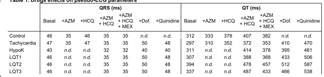
64
(Supplemental figure 2;https://models.cellml.org/e/71). A value of 300 beats was chosen for
65
all the tested conditions as it reflected stability of the modelling conditions. The healthy
66
condition was modeled at 1000-ms cycle length, and tachycardia was modeled at 700-ms
67
cycle length, that is commonly observed in COVID-19 patients (3) and at 500-ms cycle
68
length.
69
To model cardiac response of COVID-19 patients with moderate hypokalemia, external K+
70
concentration has been decreased from 5.4 to 3.4 mM.
71
We reasoned that LQTS patients with major alterations in repolarization would not be
72
prescribed QT lengthening compounds. Thus, we operated moderate modifications of the
73
implicated currents to model long QT syndromes. For type 1 LQTS, we reduced the
74
conductance of the slow component of the delayed rectifier K+ current (IKs) to 50% of the
75
wild-type condition to mimic moderate loss-of-function of mutated KCNQ1-encoded
76
channels, without any dominant negative effect usually associated with severe LQT (48, 49).
77
Similarly, for type 2 LQTS (LQT2), we reduced the conductance of the rapid component of
78
the delayed rectifier K+ current (I
Kr) to 50% of the wild-type condition to mimic moderate
loss-79
of-function (50). For type 3 LQTS (LQT3), we reproduced the consequences of the
80
QKP1507-1509 mutant on SCN5A-encoded channel, Nav1.5, with 4-fold increase in the
81
conductance of the late component of the Na+ current (51, 52).
82
Effects of 3 µM HCQ have been chosen based on the serum concentration measured in
83
COVID-19 patients treated with 600 mg/day (10). HCQ effects on ion channels have been
84
modeled as follows: 35% decrease of IKr conductance and 12% decrease of the
conductance of the L-type Ca2+ current, I
Ca,L (47). For AZM, data on serum concentrations
86
from SARS-Cov2 patients are not available so far. Peak plasma AZM concentrations during
87
oral dosing range from ≈0.4 to 1.1 μmol/L. However, plasma concentrations are misleading,
88
as the drug accumulates within cells, achieving concentrations approaching 900 μmol/L in
89
leukocytes and pulmonary tissue (27). A previous study by the pharmaceutical sponsor,
90
Pfizer Inc., reported similar accumulation of the drug in cardiac cells for mice receiving oral
91
AZM (200 mg∙kg−1∙d−1 for 10 days), with ≈200-fold increase in concentration compared with
92
plasma at day 10 (53). Based on that, Yang et al. used an in vitro concentration of 50 µM,
93
which seems reasonable to estimate the effect on cardiac currents (27). Of Note, AZM has
94
different effects with regard to acute (instantaneous) or “chronic” 24-hour exposure,
95
regarding the Na+ current. Acute exposure to AZM was shown to decrease both the peak
96
sodium current and peak L-type calcium current. It also decreases the inward rectifier
97
potassium current IK1, and the delayed potassium currents IKr and IKs. In contrast, 24-hour
98
exposure increases the peak and late sodium currents. Unfortunately, the effects of 24-hour
99
exposure AZM on IK1, IKr, IKs and the L-type calcium current were not tested in this earlier
100
report (27). Interestingly, even in the case of acute exposure, Yang et al. showed an
101
enlargement of QTc duration in mice (see figure 2 of their publication), suggesting that the
102
decrease in L-type calcium current is counterbalanced or even exceeded by the decrease in
103
IK1 (IKr and IKs being absent in adult mice). Because AZM is administered for several days in
104
the COVID-19 context, we considered only reported 24-hour effects of this compound on ion
105
channels, as follows: 1.8-fold increase in sodium peak current conductance and 2.5-fold
106
increase in late sodium current conductance. Of note, adding the other current alterations,
107
based on their studied acute effects (65% reduction in L-type calcium current, 30% reduction
108
in IKr and IKs, 66% reduction in IK1) further increased QT duration (supplemental figure 3).
109
Since the equivalence between acute and 24-hour effects is hypothetical, we chose to keep
110
the condition with the minimal effect (only clearly established 24-hour effect of AZM on
111
sodium channel). This point stresses out the importance of the characterization of longer
112
application (at least 24-hour) of a given molecule on the currents.
State-dependent effects of mexiletine (MEX) were modeled at therapeutic concentration
114
(0.8-2 µg/mL) (54) by a 40% decrease in only the late sodium current conductance (55).
115
Two antiarrhythmic drugs, that prolong QT interval and have been reported to induce
116
torsades de pointes, were also tested as positive controls. Dofetilide is mostly active on IKr
117
and Ito at about 2 nM corresponding to the free plasma Cmax concentration (factors applied:
118
0.45, 0.98, 0.98, 0.85, 0.98, and 0.95 to IKr, ICa,L, INa fast, Ito, IKs and IK1 conductances,
119
respectively) (55). Quinidine has a larger spectrum and was tested at its free plasma Cmax
120
concentration of about 850 nM (factors applied: 0.3, 0.9, 0.98, 0.85, 0.9, and 0.95 to IKr, ICa,L,
121
INa fast, Ito, IKs and IK1 conductances, respectively) (55).
122
Combined effects (such as LQT mutation+AZM+HCQ+MEX) were obtained by applying
123
each factor respective of each drug or condition to the appropriate conductance(s). Models
124
were processed with C++ code.
125
Electrophysiological determinations
126
Pseudo-ECG time parameters were determined as previously described (56). As expected,
127
this model adapts to frequency by decreasing QT duration when frequency increases (38).
128
As presented above, pseudo-ECG models are obtained from 1-dimensional strand of 165
129
cells reporting left ventricle transmural activity. For example, apex-to-base and right
130
ventricle-to-left ventricle gradients are absent in this model. Therefore, generated
pseudo-131
ECG time parameters are lower than human ECG values. QRS and QT durations are 46
132
and 312 ms, respectively, in this model compared to 90-100 and 370-440 ms in patients (i.e.
133
-60 ms), suggesting that difference in QT between the model and patients is mainly due to
134
the difference in QRS, not ST duration. For the sake of comparison, we arbitrarily added the
135
empirical value of 60 ms to the model QT duration to obtain ‘clinical-like’ QT values (in figure
136
7). QRS widening induced by HCQ may occur in COVID-19 patients. It is a slight median
137
increase of 4 ms of borderline significance (57). However, we kept this 60-ms value constant
138
in every tested condition.
139
Arrhythmogenic risks were assessed by the repolarization time from APD30 to APD90 (APD
90-140
30) measured from the beginning of AP upstroke until 30% and 90% of repolarization as
previously described (58) and by QT duration (59). The breaks in the repolarization slope in
142
early phase 3 of the computed APs were considered as early afterdepolarizations (EADs) by
143
analogy with the EAD originally defined as a depolarizing afterpotential that begins prior to
144
the completion of repolarization and causes (or constitutes) an interruption or retardation of
145
normal repolarization, in the princeps publication by Cranefield (60). Data were analyzed
146
using R3.6.2 and GraphPad8.
147
148
Results
149
We started with the most general case of COVID-19 patients, presenting no arrhythmia risk
150
factor that reduces the repolarization reserve. Thus, we first investigated the effects of HCQ
151
or AZM alone and their combined effects on the ventricular repolarization on simulated
152
‘normal’ ECG. At a cycle length of 1,000 ms, we observed that AZM alone induced a
153
shortening of the QRS complex (-24%) and an increase in QT duration (+7%) due to the
154
increased contribution of the peak and late sodium currents, respectively (Figure 1A-B and
155
Table 1). HCQ alone induced a larger increase in QT duration (+21% vs. baseline) without
156
affecting the QRS duration (Figure 1A-B). The combined AZM and HCQ synergistically
157
prolonged the QT interval (30%; figure 1A-B). These drugs target two different types of ion
158
channels. HCQ reduces a repolarizing current (IKr), while AZM increases a depolarizing
159
current (late INa). Their effects are thus more than additive on the action potential duration as
160
described by previous studies (61). Looking at specific cell levels, modeled action potentials
161
from sub-endocardium (cell #19), mid-myocardium (cell #84) and sub-epicardium (cell #144)
162
underwent major modifications when the effects of HCQ alone or combined with AZM were
163
simulated (Figure 1C).
164
165
Because COVID-19 patients admitted in ICU are frequently tachycardic, we investigated the
166
effects of the treatment at a faster rate (cycle length, CL = 700 ms). The resulting effects of
167
the three treatments on pseudo-ECG and APs parameters were in the same range as at
1,000-ms CL (Figure 2A-B and Table 1). When higher heart rate was tested (CL = 500 ms),
169
similar results were observed (Supplemental figure 4).
170
171
Because hypokalemia can precipitate acquired LQTS (62), we investigated AZM and HCQ
172
effects when a moderate hypokalemia (3.4 mM of extracellular K+) commonly observed in
173
COVID-19 patients (63) was implemented in the model in addition to tachycardia. As shown
174
in Figure 2C, hypokalemia induced a QT prolongation (+5% compared to baseline at 700-ms
175
CL) and exacerbated the effects of the AZM+HCQ combination (+33% of increase in QT
176
compared to +25% of increase in normokalemia at 700-ms CL; figure 2C). Hypokalemia also
177
hyperpolarized the diastolic membrane potential of each cardiomyocyte layer (-99.2 mV in
178
hypokalemia vs. -86.9 mV in normokalemia) leading to increased sodium channel
179
availability. This increased availability caused QRS shortening. The combination of both
180
drugs induced a triangulation of the AP shape as assessed by the prolongation of the
181
repolarization time from APD30 to APD90 (APD90-30; 195 ms vs. 110 ms with no treatment, in
182
the sub-endocardium; figure 2D), which is known to favor early afterdepolarizations (64, 65).
183
In summary, the model suggests that COVID-19 patients with tachycardia and hypokalemia,
184
even ‘sub-clinical’, have to be closely monitored due to the potentiation of HCQ and AZM
185
arrhythmogenic effects.
186
187
QT and AP duration lengthening were also observed when the reference drugs dofetilide
188
and quinidine were applied (Supplemental figures 5 and 6). The deleterious effects of
well-189
known arrhythmogenic drugs can be clearly identified. It appears that AZM+HCQ have
190
similar effects as dofetilide, a high risk torsadogenic drug. In summary, our results confirm
191
the AZM+HCQ-induced QT prolongation observed in patients and validate the use of the
192
model to investigate the arrhythmogenic consequences of drugs to treat COVID-19.
193
In order to validate the use of this model to predict arrhythmogenic susceptibility of patients
194
with moderate long QT syndrome, we first tested AZM and HCQ effects in a LQT2 model
replicating hERG haplo-insufficiency in normokalemia. As expected, a 33% prolongation of
196
the QT was obtained. AZM+HCQ combined effects further prolonged it by 21% vs.
197
‘untreated’ LQT2 condition (Figure 3A). In LQT2 conditions, AP repolarization relies mostly
198
on IKs. As expected, the AZM-HCQ combined effects were major in the mid-myocardium
199
where IKs is of small amplitude. Mid-myocardium APD90-30, already prolonged by IKr
200
decrease, was severely prolonged from 145 to 203 ms by AZM+HCQ treatment and
201
associated with the occurrence of a subthreshold early afterdepolarization (Figure 3B).
202
Again, AZM+HCQ treatment had effects in the same range as those observed with dofetilide
203
(Supplemental figures 5 and 6). In the same conditions, quinidine application led to more
204
pronounced QT prolongation and EADs particularly at the mid-myocardium level. These sets
205
of data show that the ORd model replicated the impact of proarrhythmic drugs on LQT2 AP
206
and ECG. These results confirm the absolute proscription of the use of such proarrhythmic
207
drugs in COVID-19 patients with baseline long QT (66-68).
208
Then, we used the model to predict the effects of AZM and HCQ in the context of a
sub-209
clinical QT prolongation as seen in parents of patients with autosomal recessive Jervell and
210
Lange-Nielsen LQTS, for instance (69). Despite a 50% reduction in IKs amplitude, a minimal
211
3% prolongation of the QT duration was observed (Figure 4A). However, the combination of
212
AZM and HCQ induced a 26% increase in QT duration (vs. ‘untreated’ LQT1 condition) as
213
well as APD90 prolongation (Figure 4). This approach suggests that COVID-19 patients with
214
primary moderate hypokalemia or asymptomatic LQT1 have a slightly higher risk to develop
215
drug-induced arrhythmias when treated with AZM and HCQ than patients without these
co-216
morbidities (+11% and +4% QT prolongation in hypokalemia and LQT1, respectively,
217
compared to QT values of ‘treated’ ‘normal’ ECG at the same heart rhythm). These patients
218
have to be followed closely and additional preventive anti-arrhythmic therapy might be
219
proposed in this case.
As AZM increases the late component of the sodium current, we also investigated the
221
effects of the combined therapy in a model in which the late component of the Na+ current
222
was already increased i.e. in the model replicating LQT3. A 13% QT prolongation was
223
obtained, to a lesser extent than in the LQT2 condition, though. However, a dramatic QT
224
prolongation of 44% was induced by AZM+HCQ treatment (Figure 5A). At the ‘cellular level’,
225
combining both drugs effects favored AP triangulation (APD90-30 duration increased from 102
226
to 195 ms) and occurrence of early afterdepolarizations in mid-myocardium, close to what
227
was obtained with the LQT2 model (Figure 5B).
228
Since it appears that the ORd model confirmed the observed and expected results regarding
229
HCQ and AZM effects on ECG, we used the model to predict the effect of mexiletine
230
treatment. Mexiletine, a well-known anti-arrhythmic drug used in LQTS patients was
231
proposed to be associated with HCQ and AZM treatment of COVID-19 patients to limit
232
excessive QT prolongation (70, 71). As shown in Figure 6, mexiletine reversed
AZM+HCQ-233
induced QT prolongation in all tested conditions (+19% vs. +25% in tachycardia, +22%
234
vs.+33% in hypokalemia, +16% vs.+21% in LQT2, +20% vs.+26% in LQT1, and +28%
235
vs.+45% in LQT3 model). In hypokalemia, LQT2 and LQT3 models, mexiletine reduced the
236
AZM+HCQ-induced early afterdepolarization susceptibility in mid-myocardium (APD90-30 of
237
131 ms vs.148 ms, 186 ms vs. 203 ms and 159 vs.195 ms for in hypokalemia, LQT2, and
238
LQT3 models, respectively). Of note, the model predicts that mexiletine supplementation to
239
shorten the prolonged QT has a mild but not negligible effect. Moreover, the model may be
240
robust enough to evaluate the combined effects of new additional drugs (with known effects
241
on ion channels) to limit AZM+HCQ arrhythmogenic consequences.
242
Figure 7 summarizes the QT duration values obtained at 700-ms cycle length. The ORd
243
transmural wedge model values are arbitrarily transposed to clinical-like values by adding 60
244
milliseconds (right Y-axis). A QTc cut-off of 500 ms is clinically considered as pathological
(66-68). At 700 ms of cycle length, the corresponding absolute QT duration according to
246
Bazett’s formula is 418 ms.247
Discussion248
Our study confirms that treating COVID-19 patients with HCQ and AZM drugs has, in most
249
patients, little impact on QT duration (72) and does not induce any substrate prone to
250
arrhythmia. However, in clinical conditions in which the repolarization reserve is reduced, the
251
model predicts larger ECG impairments including QT > 418 ms at 700 ms of cycle length,
252
corresponding to QTc > 500 ms (figure 7). Such dramatic QT prolongations are potentially
253
enabling the occurrence of life-threatening events, such as ventricular fibrillation. In addition,
254
the model allows the dissection of the relative contribution of each drug to the establishment
255
of pro-arrhythmic conditions, as well as their synergic effects to the mechanisms involved.
256
We also show that, mexiletine can limit only partly the dramatic increase in QT duration for
257
patients with tachycardia, hypokalemia or reduced conduction reserve, but can bring it back
258
to manageable duration for the mildest phenotypes. These results are in agreement with
259
observations reported by Badri et al. after mexiletine treatment on acquired-LQT syndrome
260
patients (73). The use of lidocaine, another class I antiarrhythmic drug has shown some
261
benefits in a COVID-19 patient treated with AZM and HCQ (74). Therefore, the ORd model
262
may be used to evaluate the potential impact of additional drug, with known effects on ion
263
channels, to limit the arrhythmogenic risks.
264
265
There is currently an explosion of proposed therapies for treating the virus but none of them
266
have clearly demonstrated their efficacy (75). Among these therapies, hydroxychloroquine
267
combined with azithromycin is still being used based on in vitro studies indicating their ability
268
to inhibit virus-cell fusion (7-9) and despite accumulation of studies questioning their clinical
269
efficacy, the topic is still debated (12-16). A major concern of this therapy has been the risk
270
of QT prolongation and TdP. The proarrhythmic mechanism of HCQ is thought to be due to
its ability to inhibit hERG potassium channel and L-type calcium channel, which can result in
272
early afterdepolarization triggered activity (47). Association of well-timed early
273
afterdepolarization and QT prolongation results in TdP. The proarrhythmic mechanism of
274
AZM is thought to be due to its ability to increase cardiac sodium current and promote
275
intracellular sodium loading (27). Obviously, clinical decision cannot rely on the results
276
obtained with this ECG model, but, by comparing the effects obtained with AZM+HCQ, and
277
two proarrhythmic drugs, it can be suspected that the treatment has deleterious effects in
278
vivo. Indeed, based on the proposed mechanisms we confirmed, using this in silico model,
279
recent reports indicating QT prolongation (72, 76) and high risk of TdP (77) in COVID-19
280
patients treated with HCQ and/or AZM. The discrepancy between occasional reports of QT
281
prolongation and life-threatening arrhythmias triggered by HCQ and AZM and the absence
282
of QT prolongation effects in large population studies (especially with AZM (33)), is probably
283
due to the necessity, for triggering arrhythmia, of the combination of factors such as
284
tachycardia, hypokalemia, and subclinical LQTS as substrate.
285
286
More than 280 drugs have been reported to induce QTc prolongation (78). Among them
287
several are antiarrhythmic drugs, but also non-cardiovascular drugs, that are widely used in
288
ICU (79). Clear recommendations have been established to avoid their administration to
289
patients with symptomatic and well-established congenital long QT syndrome. In addition,
290
IKr, IKs, ICa,L, INa late and more generally Ca2+ homeostasis, are differentially impaired in various
291
cardiopathies and cardiomyopathies frequently associated with aging, and also in hypoxia,
292
much more frequent conditions in hospitalized COVID-19 patients. This is of concern,
293
especially since INa late increase, most frequently associated with these acquired diseases,
294
appears to lead to severe ECG changes (LQT3).
295
296
Regardless of genetic aspects or pre-existing chronic pathologies, clinical case series have
297
also identified risk factors for drug-induced LQTS including hypokalemia as commonly
298
observed in COVID-19 patients (80). Hypokalemia prolongs QT and is a risk factor for
induced LQTS. In addition to direct consequences on IKr current (81, 82), hypokalemia may
300
activate CaMKII leading to an increase in late sodium current and further prolongation of
301
ventricular repolarization (83). Moderate to severe hypokalemia has been reported in
302
COVID-19 patients (63). SARS-CoV-2 virus invades cells through binding to angiotensin I
303
converting enzyme 2 (ACE2) that enhances ACE2 degradation. The final effect of this
304
degradation is a continuous renal K+ loss that makes it difficult to correct hypokalemia (63).
305
Noteworthy, low levels of potassium have been correlated with QTc > 500 ms occurrence in
306
COVID-19 patients under HCQ and AZM medication (36). Consistent with these
307
observations, this model emphasizes the fact that kalemia of COVID-19 patients has to be
308
followed very carefully, particularly in case of medication with drugs such as HCQ or AZM.
309
More generally, this model could be used to evaluate in a pre-clinical approach, the risk of
310
drug-induced QT prolongation in this context. In addition to electrolyte imbalance, there is
311
also a greater prevalence of risks factors among COVID-19 patients in ICU, including older
312
age, presence of underlying heart disease, and co-treatment with other QT prolonging
313
medications.
314
315
With the possibility that a significant proportion of the world population may receive
SARS-316
CoV-2 drugs with torsadogenic potential, the risk to treat patients with asymptomatic and
317
undiagnosed long QT syndrome is increasing. These patients have a QTc duration in the
318
limit of the general population variability and are not identified as such. Indeed, in a recent
319
study, patients with extreme QTc prolongation when treated with HCQ and AZM, presented
320
a baseline QTc around 431 ms only, within the normal QTc range (36). As modeled in the
321
present study, cardiomyocytes harboring mutations leading to haplo-insufficiency in KCNQ1
322
may present very minimal action potential prolongation because of a normal IKr (84), but IKr
323
blockers such as HCQ, can lead to marked action potential prolongation in limited
324
repolarization reserve. All guidelines for QT management in COVID-19 context (66-68)
325
recommend to avoid QT prolonging drugs in individuals with a QTc > 500 ms due to a
two-326
fold to three-fold increase in risk for TdP (85). Nevertheless, those asymptomatic patients
might receive these drugs based on this criterion. As modeled in this study, despite the
328
absence of QT prolongation in baseline conditions because of a normal IKr (84) and
329
regardless of the origin of low repolarization reserve, these patients are at high risk of TdP
330
when IKr blockers such as HCQ are used. However, the model shows that, in a borderline
331
condition such as moderate LQT1, mexiletine can limit to some extent the deleterious effects
332
of AZM and HCQ. Therefore, we propose that the ORd model can be used to evaluate the
333
potential impact of other additional drugs, with known effects on ion channels, that may be
334
used in the future to limit arrhythmogenic risk of COVID-19 therapies.
335
336
In summary, the ORd model appears to be an easy-to-use tool to assess off-label drug
337
arrhythmia potential in different conditions representative of COVID-19 patients at risk for
338
arrhythmia and life-threatening torsades de pointes.
339
340
Limitations
341
We used the original ORd model based on its more realistic conductance values compared
342
to others. This model may underestimate IKs amplitude even if obtained from human
343
cardiomyocytes (86). In some rare cases (heterozygous non-dominant-negative LQT1
344
mutations) a minimal reduction (less than 50%) of the channel activity leads to severe QTc
345
prolongation (very minor cases, cf. for instance (87)). The model we use is simple, robust,
346
and incorporates pseudo ECGs but not inter-individual variability, to remain affordable in
347
time and resources. Since the model does not include the population variability (e.g., due to
348
genetic background), it cannot reproduce the LQTS phenotype heterogeneity. Other studies
349
focusing on single cell AP, adjusted IKs amplitude to compensate for this insufficiency leading
350
to more severe LQT1 phenotype (88). It would be interesting to try optimizing pseudo-ECG
351
models using the same strategy.
352
One-dimension strip of 165 cardiomyocytes simulates only the transmural gradient.
Apex-to-353
base and right-to-left gradients are absent in this model. There are 3D models but (i) they
are highly computationally demanding thus requiring simpler alternative approaches to
355
model single cell action potential (89) and (ii) they are less realistically adaptive because
356
each current is not individually modelled. Therefore, we preferred to use a 1D-model in
357
which precise biophysical equations representing the biological currents can be finely tuned
358
to model the drug effects at the AP then ECG levels. Thus, the resulting caveat is the lower
359
QT duration values. In order to allow translational approach, we suggest adding an
360
empirically estimated value of 60 ms. The calculated QT values can then be roughly
361
compared to clinical ECG values. In addition, T wave shape results more from regional
362
heterogeneity than from transmural gradients (90). As another limit of the 1D-model, it
363
cannot simulate changes in T wave amplitude.
364
In this study, we investigated potential effects of drugs prescribed to patients with COVID-19
365
on AP with reduced repolarization reserve, in order to detect any arrhythmogenic substrate.
366
To do so, we used well-defined conditions with “pure” repolarization reserve decrease such
367
as LQT syndrome with various genetic origins. Cardiopathies and cardiomyopathies
368
frequently associated with aging, and in hypoxia, are much more frequent conditions in
369
hospitalized COVID-19 patients. However, instead of adding another condition, generic for
370
these pathologies, which is difficult to establish since conductance decreases are not the
371
same for all the pathologies (91), conditions with “pure” repolarization-reserve decrease as
372
LQT syndromes were preferred. Similarly, the complex modifications induced by systemic
373
inflammation and oxidative stress observed in COVID-19 patients have not been introduced
374
at the level of the ion currents in the modeling. Such complex alterations of expression
375
and/or activity of ion channels are hardly quantifiable and cannot be mimicked. In any case,
376
it can be suspected that the addition of pre-existing pathologic conditions and
COVID-19-377
related modifications would exacerbate the arrhythmia susceptibility.
378
The effects of adrenergic stimulation were not evaluated for the following reason. An
379
observational study of 138 patients affected by COVID-19 reported moderate tachycardia
380
with a median heart rate of 88 bpm (3) indicating that the adrenergic tone is not high in those
381
patients. Thus, in this study, we evaluated, during moderate tachycardia, the theoretical
effect of the drugs on AP with reduced repolarization reserve, in order to detect any
383
arrhythmogenic substrate. Interestingly, a very recent work, complementary to ours, used a
384
modified version of the ORd model to study the β-adrenergic receptor stimulation on the
385
cellular proarrhythmic effects of chloroquine and azithromycin, at the single AP level (92). In
386
this paper, Sutanto and Heijman suggest that sympathetic stimulation limits drug-induced
387
APD prolongation. Therefore, at least for CQ and AZM, the unstimulated situation that we
388
studied may represent the most critical situation.
389
It has to be mentioned that the ECG ORd model is conservative. Arrhythmogenic
390
mechanisms such as triggered activities are hardly induced. Indeed, significant impairment
391
of the Ca2+ current window was needed to induce repolarization failure in the recent study of
392
Sutanto and Heijman (92). However, the deleterious effects of arrhythmogenic drugs can be
393
clearly identified with the ECG model, namely EADs and QT lengthening.
394
Gender differences, resulting from multiple intersecting processes implying complex
395
regulations of ion channels, cannot be easily modeled and was not investigated in this study.
396
This would be indeed another improvement of the model.
397
398
Acknowledgments399
None400
401
Sources of Funding402
M. De Waard and G. Loussouarn thank the Agence Nationale de la Recherche for its
403
financial support to the Région Pays de la Loire (ANR FLASH Covid-19 - CoV2-E-TARGET)
404
and the laboratory of excellence “Ion Channels, Science and Therapeutics” (grant N°
ANR-405
11-LABX-0015). The fellowship of J. Montnach is provided by a National Research Agency
406
Grant to M. De Waard entitled OptChemCom (grant N° ANR-18- CE19-0024-01). Genavie
407
foundation supported J. Montnach and G. Loussouarn.
408
Disclosures
None