O
pen
A
rchive
T
OULOUSE
A
rchive
O
uverte (
OATAO
)
OATAO is an open access repository that collects the work of Toulouse researchers and
makes it freely available over the web where possible.
This is an author-deposited version published in :
http://oatao.univ-toulouse.fr/
Eprints ID : 18543
To link to this article : DOI:10.1016/j.otsr.2015.11.005
URL :
https://doi.org/10.1016/j.otsr.2015.11.005
To cite this version : Pham, Tao T. and Bayle-Iniguez, Xavier and
Mansat, Pierre and Maubisson, Laurent and Bonnevialle, Nicolas
Postoperative pain after arthroscopic versus open rotator cuff repair.
A prospective study. (2016) Orthopaedics & Traumatology: Surgery &
Research, vol. 102 (n° 1). pp. 13-17. ISSN 1877-0568
Any correspondence concerning this service should be sent to the repository
administrator:
[email protected]
Original article
Postoperative pain after arthroscopic versus open rotator cuff repair.
A prospective study
T.T. Pham
a,∗, X. Bayle Iniguez
a, P. Mansat
a, L. Maubisson
b, N. Bonnevialle
aaInstitut de l’appareil locomoteur, hôpital Pierre-Paul-Riquet, place du Docteur-Baylac, 31059 Toulouse, France bEA CNRS 6296, IAE de Tours, université Franc¸ois-Rabelais, 37200 Tours, France
Keywords: Postoperative pain Rotator cuff repair Open
Arthroscopic
a b s t r a c t
Introduction: Although the arthroscopic technique is becoming the gold standard for rotator cuff tendon repair, there is no proof that this technique results in less postoperative pain compared to open repair. The aim of this study was to prospectively compare the postoperative pain level after arthroscopic or open rotator cuff repair and to define factors that could influence its course.
Materials and methods: Between January 2012 and January 2013, 95 patients were operated for a rota-tor cuff tear: 45 using an arthroscopic technique and 50 an open technique. Daily analgesic use and self-evaluation of pain level using a visual analogic scale were recorded preoperatively and twice a day postoperatively during the first 6 weeks. These data were compared between the two groups and analyzed according to patients’ demographic data and preoperative evaluation of the tear.
Results: The preoperative pain level was equivalent in the two groups (P = 0.22). Postoperatively, level-2 analgesic medication use was greater in the arthroscopic group after the 4th week (P = 0.01). A pain-free shoulder was obtained before the 6th week in 75% and 66% of the patients after arthroscopic or open repair, respectively (P = 0.34). There was a positive correlation between the preoperative and postoperative pain level (r = 0.25; P = 0.02). Work compensation patients experienced more pain postop-eratively (P = 0.08). Level-III analgesic medication use was greater for patients with massive rotator cuff tear (P = 0.001).
Conclusion: No evidence was found on the superiority of arthroscopy versus open repair of rotator cuff tear concerning the postoperative pain level. The choice of the surgical technique should not be based on this argument.
Level of evidence: II.
1. Introduction
Since Codman’s first description in 1911[1]of a rotator cuff tear repair technique, different studies have reported satisfactory clinical results in 70–95% of cases after open tendon reinsertion, with a 45–90% healing rate[2–15]. Since the advent of arthroscopy, endoscopic repairs of rotator cuff tendon lesions have been widely studied[16–21]. Postoperative comfort in terms of pain symptoms, notably the absence of muscle disinsertion, seems to be one of the arguments orienting the surgeons’ choice toward arthroscopic repairs. However, few comparative studies have precisely assessed the functional results between these two techniques, notably in terms of postoperative pain.
∗ Corresponding author.
E-mail address:[email protected](T.T. Pham).
The objective of the present study was to prospectively compare immediate postoperative pain after arthroscopic and open rotator cuff repair and to determine the factors influencing this parameter. We hypothesized that arthroscopic repair of the rotator cuff would lead to less postoperative pain than open repair.
2. Material and methods
A prospective, consecutive, single-center study was conducted in our university orthopaedics-traumatology department from Jan-uary 2012 to JanJan-uary 2013. The study included all patients who underwent surgical repair of the rotator cuff associated with acromioplasty and tenotomy or tenodesis of the long head of biceps tendon. The repair was either arthroscopic (group A) or performed with an open technique (group O), non randomized, performed by two experienced operators in these techniques: NB for group A and PM for group O. Patients with a history of rotator cuff repair
Table 1
Characteristics of the population of the two groups studied.
Arthroscopy Open P-value Mean age (years) 55 (37–72) 60 (43–78) 0.001 Females 37.5% 44.7% 0.323*
Trauma context 37.5% 38.3% 0.558*
Work accident or occupational disease
20% 17% 0.466*
Moderate or heavy manual labor
62.5% 48.9% 0.146*
Dominant side 82.5% 63.8% 0.043 Active smoking 20% 21.3% 0.549*
* P > 0.05: no significant difference.
in the same shoulder were excluded, as were those who had not undergone acromioplasty or bicipital surgery or those presenting associated pathologies.
2.1. Comparability of the groups
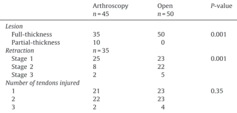
Ninety-five patients underwent rotator cuff repair, 45 arthro-scopic repair and 50 with the open technique. The mean age was 55 years (range: 37–72 years) in group A and 60 years (range: 43–78 years) in group O. The mean hospital stay was 2.2 days in group A and 3.5 days in group O (P = 0.001). Groups A and O were comparable in terms of gender, the trauma context, the occupational accident or occupational disease context, moderate or heavy manual labor, the dominant side injured, and active smoking (Table 1). The distribution of tendon involvement (type, retraction, extension) is summarized inTable 2. In group A, there were 10 par-tial tears greater than 50% of the tendon thickness, eight of which were on the deep side and two on the superficial side. No partial-thickness lesions were found in group O. There were more stage 1 non-retracted lesions in the arthroscopic group. The two groups were comparable in terms of the number of tendons injured. 2.2. Surgical techniques
All the patients included were given preoperative interscalene block in a single injection of ropivacaine 7.5 mg/mL (20 mL) or in a single injection of ropivacaine 7.5 mg/mL (20 mL) and dexa-methasone 4 mg or associated with placement of a perineural catheter with an initial injection of ropivacaine 7.5 mg/mL (20 mL), then continuous postoperative perfusion of ropivacaine 2 mg/mL to 5 mL/h, associated with on-demand boluses. The catheter was removed 48 h after surgery[22,23]. The choice of perioperative analgesics was made by the anesthesiologist.
The patient was installed in the semi-sitting position. The lesion was assessed intraoperatively either with arthroscopy in group A or in the open procedure in group O. The frontal extension of the lesion was evaluated in three stages according to Patte[24].
Table 2
Distribution of rotator cuff tendon lesions between the two groups studied.
Arthroscopy n = 45 Open n = 50 P-value Lesion Full-thickness 35 50 0.001 Partial-thickness 10 0 Retraction n = 35 Stage 1 25 23 0.001 Stage 2 8 22 Stage 3 2 5 Number of tendons injured
1 21 23 0.35
2 22 23
3 2 4
Fig. 1. Tension-band suture technique.
2.3. Arthroscopic technique
No traction system was used and manual traction was used as needed. The intervention began with glenohumeral joint explo-ration. An intra-articular tenotomy of the long head of biceps tendon (LHBT) was performed at the supraglenoid tubercle. In cases undergoing tenodesis, the transverse humeral ligament was resected after anterior bursectomy, and tenodesis of the LHBT was carried out in the bicipital groove using a bioabsorbable screw anchorage system or a bioabsorbable interference screw. An acromioplasty was performed via the posterior approach using a motorized reamer after complete bursectomy and resection of the coracoacromial ligament. The rotator cuff was repaired using the tension band technique. Mattress sutures mounted on a resorbable screw anchorage system were performed (Fig. 1). Depending on the extent of the injury, a technique using a double row of anchors was sometimes used[25]. In cases with partial-thickness tear, release and then reinsertion following the same tension band technique was performed.
2.4. Open technique
The superior approach with an incision measuring 4–5 cm was used (Fig. 2). The deltoid muscle was incised following the muscle fibers between the anterior and middle bundles. The coracoacro-mial ligament was resected and then, an anterior and inferior acromioplasty was performed with a chisel. The rotator cuff was explored after a bursectomy. The rotator interval was opened to explore the LHBT and the tendon of the subscapularis muscle
Fig. 3. Transosseous suture using Mason-Allen stitch.
whenever the LHBT was not visible through the tendon lesion. A tenotomy-tenodesis of the LHBT was performed with stitches join-ing the fibrous ceiljoin-ing of the bicipital groove to the tendon. The intra-articular part of the tendon was then resected and the rotator interval was closed. The rotator cuff was repaired by transosseous reinsertion and suture using modified Mason-Allen stitches (Fig. 3).
2.5. Recovery
The rehabilitation program was identical for both techniques. Immobilization consisted of a shoulder scarf. Rehabilitation began on the 1st postoperative day with the patient learning the pendular rehabilitation exercises. Beginning on the 15th postoperative day, passive mobilization was used to progressively recuperate joint range of motion. Active work was only authorized beginning in the 6th week after removal of the scarf.
2.6. Pain assessment
Pain was assessed preoperatively and then on the 2nd day to the 45th day after surgery. It began on the 2nd day to palliate the confusion factor caused by the interscalene block. The distri-bution of the perioperative analgesia mode was comparable in the two groups (P = 0.37). Pain was assessed using a self-evaluation in which the patient completed a booklet provided and explained by the investigator the day before the intervention. Pain was evaluated using a visual analog scale (VAS) twice a day. The daily VAS was the mean of the morning and evening VAS. Patients were consid-ered totally pain-free when the VAS was 0 for 3 consecutive days. Morphine was prescribed depending on pain. Other pain medica-tions (NSAIDs, corticoids) were taken as prescribed by the family physician or in self-medication. The patient noted the daily con-sumption of pain medications with the name of the medication and the number of times taken. Based on data collected from the patient’s booklet, the consumption of analgesics was classified by level (I–III, others: NSAIDs, corticoids, etc.) and quantified by the number of days, these different levels of analgesics were consumed per week.
2.7. Statistical analysis
The statistical analysis was done using the Chi2test for the
qual-itative variables and the Student t-test and the Fisher test for the quantitative variables. The Pearson correlation coefficient was used to evaluate the correlation between the continuous variables. The significance threshold of P < 0.05 was considered significant.
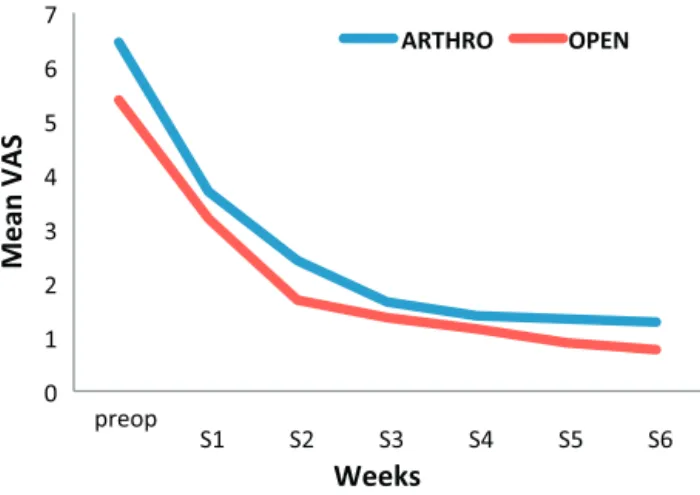
Fig. 4. Mean VAS changes.
3. Results
3.1. Postoperative pain
The mean preoperative pain was greater in group A (6.5 points) than in group O (5.4 points) (P = 0.005). During the 6 weeks of follow-up, mean postoperative pain was equivalent in the two groups (P = 0.22) (Fig. 4). At the 6th week of evaluation, the mean pain level was 1.2 (range: 0–8) for the arthroscopic group and 0.7 (range: 0–4) for the open surgery group (P = 0.1). The mean reduc-tion in pain after rotator cuff repair was 5.3 points in group A and 4.7 points in group O (P = 0.2). The reduction in VAS was maximum during the 1st week in both groups, with a decrease of 2.8 and 2.2 points, respectively, in group A and group O (P = 0.26). A pain-free shoulder was achieved before the 6th week in 75% and 66% of the patients in group A and group O, respectively (P = 0.34).
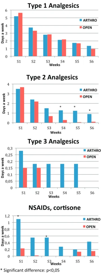
Levels I and III analgesic intake was equivalent in the two groups over the 6 weeks of assessment (P > 0.05). Nevertheless, in group A, there was greater consumption of level II analgesics beyond the 4th week and of anti-inflammatories during the 1st and 3rd weeks after surgery (P = 0.01) (Fig. 5).
3.2. Predictive factors of pain
There was a positive correlation between the preoperative and postoperative pain level (r = 0.25; P = 0.02). Work compensation patients tended to need more pain relief postoperatively (P = 0.08) (Table 3). A trauma context, the dominant side, active smoking, age, gender, tendon retraction, partial- or full-thickness tendon lesions, and a bicipital intervention did not influence mean postoperative pain (Table 3). Within group A, we found no significant difference between partial- and full-thickness lesions, with the mean VAS for pain at 2.67 and 1.73 points (P = 0.195), respectively. There were six cases of retractile capsulitis in group A and three in group O (P = 0.58). This complication had no impact on postoperative pain (Table 3). No infection occurred in either group.
Consumption of level III analgesics was greater in patients pre-senting massive rotator cuff tear, i.e., involving two or more tendons (P = 0.001) (Table 4).
4. Discussion
The initial hypothesis, suggesting that the arthroscopic tech-nique resulted in less postoperative pain than the open techtech-nique, was not confirmed in this study because postoperative pain as evaluated using a VAS over 6 weeks was equivalent in the two groups. However, there was greater consumption of level II
Fig. 5. Analgesic use.
analgesics beginning in the 4th postoperative week in the arthro-scopic group.
These results are comparable to those found in the literature. In a retrospective series of 100 patients operated on for full-thickness rotator cuff tear (50 with an open procedure and 50 with
Table 3
Comparison of postoperative pain as related to epidemiological factors.
Factor studied n VAS (mean) P-value Smoking Yes 18 2.06 0.338 No 77 1.64 Trauma context Yes 33 1.87 0.522 No 62 1.64
Work accident or occupational disease
Yes 16 2.37 0.08 No 79 1.58 Dominant side Yes 63 1.80 0.486 No 32 1.53 Manual labor Yes 48 1.94 0.181 No 47 1.47 Lesion Partial 10 2.67 0.212 Full-thickness 85 1.61 Bicipital operation Tenodesis 74 1.69 0.692 Tenotomy 21 1.87 Retractile capsulitis Yes 6 2.98 0.329 No 89 1.64 Table 4
Consumption of level III analgesics in relation to number of tendons injured. No. of tendons
injured
n Consumption of level III analgesics (no. of days/week)
P 1 44 0.85
2 45 0.02 0.0001 3 6 1.75
arthroscopy), Ide et al.[26]showed equivalent clinical results for all lesion sizes. Bishop et al.[27]compared 40 arthroscopic repairs and 32 open repairs and found no significant difference in the pain score between the two groups. The preoperative VAS was 8.2 in the open technique group and 5.2 in the arthroscopic technique group; the VAS at last follow-up was 1.1 and 1.5 (P = 0.41), respectively. In a retrospective study comparing open repair (n = 30) and arthro-scopic repair (n = 66), Buess et al.[28]found a significantly greater difference between preoperative VAS and at the last follow-up (24 months) in the arthroscopy group with a reduction of 6.4 points, whereas in the open surgery group, it was 5.7 points. However, the mean duration of postoperative pain (3 months) was the same in both groups. In the present study, the decrease in mean pain after rotator cuff repair was 5.3 points in the arthroscopy group and 4.7 points in the open surgery group (P = 0.2). The meta-analysis reported by Ji et al.[29]on five prospective randomized studies, comparing the clinical results of arthroscopic and mini-open rota-tor cuff repair did not demonstrate a significant difference in terms of postoperative VAS between the two groups.
In a comparative study evaluating postoperative pain, Williams et al.[30]found equivalent results between the arthroscopic group (n = 50) and the open group (n = 52). The number of postopera-tive days to obtain a pain-free shoulder was similar between the open group (mean: 28.8 days) and the arthroscopic group (mean: 27.6 days; P = 0.69). The authors showed that high preoperative pain, female gender, and small lesions were predictive factors of greater postoperative pain. Our study showed a positive correlation between preoperative and postoperative pain, but gender and small lesions were not found to be predictive factors. On the contrary, the patients presenting a massive rotator cuff lesion consumed more level III painkillers. Partial and stage 1 tears, more frequent in group A, therefore did not influence the results. The work accident or occupational disease context was a factor tending toward higher
postoperative pain levels, which confirmed the results reported by Stiglitz et al.[31].
The limitations of this study were marked by the absence of ran-domization and the lack of statistical power. In addition, the groups were not strictly comparable, notably in terms of the mean age and the size of the lesions treated. Nevertheless, as demonstrated by Thomazeau et al.[32], since the lesions were evaluated intraopera-tively, there was probably an underestimation of the lesions when using arthroscopy, explaining the higher number of stage 1 lesions in group A. The absence of standardization of early postoperative analgesics made it impossible to use the data from day 0 and day 1. The strength of this study lies in its prospective design. The sur-gical techniques were validated and performed by two surgeons who are specialists in shoulder surgery. Finally, pain and analgesic consumption were assessed by the patient him- or herself and not by the examiner, who was independent of the two surgeons.
5. Conclusion
Superiority of arthroscopy over conventional open surgery was not demonstrated in terms of postoperative pain from day 2 to day 45 after repair of a rotator cuff tendon lesion. The choice of the rotator cuff repair technique cannot be based on this argument.
Disclosure of interest
The authors declare that they have no competing interest.
References
[1]Codman EA. Complete rupture of the supraspinatus tendon. Operative treat-ment with report of two successful cases. Bost Med Surj J 1911;164:708.
[2]Cofield RH. Rotator cuff disease of the shoulder. J Bone Joint Surg Am 1985;65:974.
[3]Calvert PT, Packer NP, Stoker DJ, Bayley JI, Kessel L. Arthrography of the shoulder after operative repair of the torn rotator cuff. J Bone Joint Surg Br 1986;68:147.
[4]Galatz LM, Griggs S, Cameron BD, Iannotti JP. Prospective longitudinal analysis of postoperative shoulder function: a 10-year follow-up study of full-thickness rotator cuff tears. J Bone Joint Surg Am 2001;83–A:1052–6.
[5]Gazielly DF, Gleyze P, Montagnon C. Functional and anatomical results after rotator cuff repair. Clin Orthop Relat Res 1994;304:43–53.
[6]Gerber C, Fuchs B, Hodler J. The results of repair of massive tears of the rotator cuff. J Bone Joint Surg Br Am 2000;82:505.
[7]Klepps S, Bishop J, Lin J, Cahlon O, Strauss A, Hayes P, et al. Prospective evalu-ation of the effect of rotator cuff integrity on the outcome of open rotator cuff repairs. Am J Sports Med 2004;32:1716.
[8]Zumstein MA, Jost B, Hempel J, Hodler J, Gerber C. The clinical and structural long-term results of open repair of massive tears of the rotator cuff. J Bone Joint Surg Am 2008;90:2423–31.
[9]Adamson GF, Tibone JE. Ten-year assessment of primary rotator cuff repairs. J Shoulder Elbow Surg 1993;2:57–63.
[10]Harryman DT, Mack LA, Wang KY, Jackins SE, Richardson ML, Matsen FA. Repairs of the rotator cuff. Correlation of functional results with integrity of the cuff. J Bone Joint Surg Am 1991;73(7):982–9.
[11]Bigliani LU, Cordasco FA, McIlveen SJ, Muso ES. Operative repairs of massive rotator cuff tears: long-term results. J Shoulder Elbow Surg 1992;1:120–30.
[12]Cofield RH, Parvizi J, Hoffmeyer PJ, Lanzer WL, Ilstrup DM, Rowland CM. Surgical repair of chronic rotator cuff tears. A prospective long-term study. J Bone Joint Surg Am 2001;83–A(1):71–7.
[13]Ellman H, Hanker G, Bayer M. Repair of the rotator cuff. End-result study of factors influencing reconstruction. J Bone Joint Surg Am 1986;68(8):1136–44.
[14]Hawkins RJ, Misamore GW, Hobeika PE. Surgery for full-thickness rotator-cuff tears. J Bone Joint Surg Am 1985;67(9):1349–55.
[15]Bassett RW, Cofield RH. Acute tears of the rotator cuff. The timing of surgical repair. Clin Orthop Relat Res 1983;175:18–24.
[16]Burkhart SS. Arthroscopic treatment of massive rotator cuff tears. Clin Orthop Relat Res 2001;390:107–18.
[17]Gartsman GM. Arthroscopic rotator cuff repair. Clin Orthop Relat Res 2001;390:95–106.
[18]Wolf EM, Pennington WT, Agrawal V. Arthroscopic rotator cuff repair: 4- to 10-year results. Arthroscopy 2004;20:5.
[19]Wilson F, Hinov V, Adams G. Arthroscopic repair of full-thickness tears of the rotator cuff: 2- to 14-year follow-up. Arthroscopy 2002;18:136.
[20]Marrero LG, Nelman KR, Nottage WM. Long-term follow-up of arthroscopic rotator cuff repair. Arthroscopy 2011;27:885–8.
[21]Gleyze P, Thomazeau H, Flurin PH, Lafosse L, Gazielly D, Allard M. Arthro-scopic rotator cuff repair: a multicentric retrospective study of 87 cases with anatomical assessment. Rev Chir Orthop Repar Appar Mot 2000;86:566.
[22] Recommandations pour la pratique clinique« Les blocs périphériques des membres chez l’adulte». SFAR 2001.
[23]Delaunay L, Bonnet F. Choix d’une technique de bloc pour la chirurgie du mem-bre supérieur. Conférences d’actualisation. © 2002 Éditions scientifiques et médicales Elsevier SAS et SFAR; 2002. p. 125–46.
[24]Patte D. Classification of rotator cuff lesions. Clin Orthop Relat Res 1990;254:81–6.
[25]Boileau P, Brassard N, Watkinson DJ, Carles M, Hatzidakis AM, Krishnan SG. Arthroscopic repair of full-thickness tears of the supraspinatus: does the ten-don really heal? J Bone Joint Surg Am 2005;87(6):1229–40.
[26]Ide J, Maeda S, Takagi K. A comparison of arthroscopic and open rotator cuff repair. Arthroscopy 2005;21:1090–8.
[27]Bishop J, Klepps S, Lo IK, Bird J, Gladstone JN, Flatow EL. Cuff integrity after arthroscopic versus open rotator cuff repair: a prospective study. J Shoulder Elbow Surg 2006;15:290–9.
[28]Buess E, Steuber KU, Waibl B. Open versus arthroscopic rotator cuff repair: a comparative view of 96 cases. Arthroscopy 2005;21:597.
[29]Ji X, Bi C, Wang F, Wang Q. Arthoscopic versus mini-open rotator cuff repair: an up-to-date meta-analysis of randomized controlled trials. Arthroscopy 2015;31(1):118–24.
[30]Williams Jr G, Kraeutler MJ, Zmistowski B, Fenlin Jr JM. No difference in post-operative pain after arthroscopic versus open rotator cuff repair. Clin Orthop Relat Res 2014;472(9):2759–65.
[31]Stiglitz Y, Gosselin O, Sedaghatian J, Sirveaux F, Molé D. Pain after shoulder arthroscopy: a prospective study on 231 cases. Orthop Traumatol Surg Res 2011;97(3):260–6.
[32]Thomazeau H, Gleyze P, Lafosse L, Walch G, Kelbérine F, Coudane H. Arthroscopic assessment of full-thickness rotator cuff tears. Arthroscopy 2000;16(4):367–72.