UNIVERSITÉ DES SCIENCES ET TECHNOLOGIES DE LILLE (FRANCE) ÉCOLE DOCTORALE DE BIOLOGIE-SANTÉ
UNIVERSITÉ DE SHERBROOKE (CANADA) DÉPARTEMENT DE BIOCHIMIE
THÈSE DE DOCTORAT
En vue de l’obtention du grade de Docteur en Science
de l’Université des Sciences et Technologies de Lille et de l’Université de Sherbrooke
Présentée par
JUSAL QUANICO
Development of On-Tissue Mass Spectrometric Strategies for Protein
Identification, Quantification and Mapping
Thèse sous la cotutelle internationale du Pr Isabelle FOURNIER et du Pr Robert DAY
Soutenue à Lille le 11 Juillet 2014
Devant le jury composé de :
Dr Philippe BULET Rapporteur
Pr Xavier ROUCOU Rapporteur
Pr. Klaus Klarskov Rapporteur
Isabelle FOURNIER Co-Directrice de Thèse
This manuscript is dedicated to all graduate students, who indefatigably burned their candles all through the night just to”keep the science going”.
I shall be telling this with a sigh Somewhere ages and ages hence: Two roads diverged in a wood, and I— I took the one less traveled by, And that has made all the difference.
1A
CKNOWLEDGEMENTThis work would not have been possible without the contributions of the following:
Prof. Isabelle Fournier, my thesis adviser in University Lille 1, whose tutelage and guidance permitted me to explore the world of Mass Spectrometry and its infinite possibilities;
Prof. Robert Day, my thesis adviser in University of Sherbrooke, for his continued support in this co-supervised thesis despite the complications it entailed. For giving me the opportunity to study in Canada and learn from his expertise;
Prof. Michel Salzet, head of our laboratory in Lille, who made this co-supervised thesis possible in the first place. For dealing with all of the administrative issues and logistics, and especially for handling the difficulties brought about by my sojourn as an international student in France;
My thesis reporters, Prof. Xavier Roucou, Prof. Klaus Klarskov and Dr. Philippe Bulet, for accepting to evaluate my work and taking time out of their busy schedules to peruse through the manuscript;
University Lille 1 for granting me the Bourse President and Fonds de recherche en santé du Québec (FRSQ), my sources of funding in this research, and in extension, the École Doctorale de Biologie Santé de Lille and the Biochemistry Department of University of Sherbrooke;
Dr. Julien Franck, my immediate supervisor in Lille, for dealing with my day to day struggles and being there during experiments that last through the night. For coming up with out-of-the-box ideas when I had none, thus allowing me to start anew in otherwise dead-end directions. For training me not only with physical manipulations necessary for work with MS, but also for helping me to develop critical thinking and question the status quo;
Dr. Maxence Wisztorski, my first immediate supervisor when I started working in Lille, who is always there when his assistance is needed and never fails to accommodate you in spite of his tons of work;
Members of the laboratory in Lille, namely, Benoit, Francesco, Jean Pascal, Annie, Stephanie, Pierre Eric, Jacopo, Dounia, Marie, Françoise, Christophe, Krystelle, Franck and Lucie for keeping a convivial atmosphere that stimulates productive work;
Members of Prof. Day’s lab, namely, Frédéric, Kevin, Ania, Christine, Hugo, Frédérik, Roxane, Sandra, Xue Wen, Evelyne and Prof. Witold Neugebauer, for the warm welcome in
Sherbrooke. For their constructive critcisms and aid which were crucial in fulfilling the requirements of the department during my stay in Sherbrooke;
My nanay and tatay, and sisters Lyn and Lisa, for their unwavering support even though I’m far away from home. As well as my friends from all over the world who were one in heart and in spirit with me;
And finally to Marina, for the incessant motivation that is the driving force behind my struggle in this endeavor.
2R
ÉSUMÉL’imagerie par spectrométrie de masse est une technique sans marquage permettant la détection et la localisation de protéines à partir de coupes de tissus. Afin de repondre à des problematiques biologiques, le nombre de protéines identifiées doit etre amelioré. Une stratégie consiste à realiser une micro-jonction liquide sur des regions particulieres des coupes de tissus afin d’extraire les peptides issus de la digestion in situ des protéines. Plus de 1500 protéines ont identifié sur une zone de 650µm, correspondant à environ 1900 cellules. Une corrélation entre ces données avec celles générées par MSI a augmenté le nombre de protéines localisées. Afin d’obtenir dans le même temps, la localisation et l’identification de protéines, une méthode consiste à realiser la microdissection de l’ensemble de la coupe après l’avoir déposée sur une lame recouverte de prafilm. Parafilm-Assisted Microdissection (PAM) a également été appliquée à l’etude de l'expression différentielle de protéines dans des tumeurs de prostate. Les résultats identifiés glutamate oxaloacétate transférase 2 (GOT2) en tant que biomarqueur de protéine candidate impliquée dans le métabolisme du glucose, en plus de celles qui ont déjà été indiqué précédemment. Réunis ensemble, ces méthodes MS d'analyses directes fournissent un moyen robuste d’étude de protéines dans leur état natif afin de fournir des indications sur leur rôle dans des systèmes biologiques.
Mots clés : spectrométrie de masse, imagerie, protéomique, micro jonction liquide, microdissection assistée par parafilm, cancer de la prostate
3S
UMMARYMass spectrometry-based methods for direct tissue analysis, such as MS imaging, are label-free techniques that permit the detection and localization of proteins on tissue sections. There is a need to improve the number of protein identifications in these techniques for them to comprehensively address biological questions. One strategy to obtain high protein IDs is to realize liquid microjunction on localized regions of tissue sections to extract peptides from the in
situ digestion of proteins. More than 1500 proteins were identified in a 650µm spot,
corresponding to about 1900 cells. Matching these IDs with those from MSI increased the number of localized proteins. In order to achieve simultaneous identification and localization of proteins, a method consisting of microdissecting entire tissue sections mounted on parafilm-covered slides was developed. Spectral counting was then used to quantify identified proteins, and the values were used to generate images. Parafilm-Assisted Microdissection (PAM) was also used to examine the differential expression of proteins on prostate tumors. Results identified glutamate oxaloacetate transferase 2 (GOT2) as a candidate protein biomarker involved in glucose metabolism, in addition to those that have already been reported previously. Taken together, these direct MS analysis methods provide a robust means of analyzing proteins in their native state and are expected to provide insights to their role in biological systems.
Keywords: mass spectrometry, imaging, proteomics, liquid microjunction, parafilm-assisted microdissection, prostate cancer
4T
ABLE OFC
ONTENTS1 Acknowledgement ... iv
2 Résumé ... vi
3 Summary ... vii
4 Table of Contents ... viii
5 Publications ... x
6 List of Figures ... xii
7 List of Tables ... xiv
8 List of Abbreviations ...xv
1 Introduction ... 20
Hypothesis ... 26
Objectives... 26
2 Bibliography ... 28
Direct Analysis of Proteins on Tissue by Mass Spectrometry ... 28
Ambient MS ... 29
Thermal Desorption/Ionization Methods ... 32
Laser Ablation Methods ... 32
Liquid and Gas Jet Desorption/Ionization Methods ... 33
Liquid Extraction Surface Sampling/Ionization Methods ... 35
MS Imaging ... 40
Vacuum-Dependent Sources ... 45
Ambient Sources ... 51
Identification and Quantification of Proteins On Tissue by MS ... 53
Peptide Mass Fingerprinting ... 54
Tandem MS ... 55
ESI-MS/MS Databank Matching ... 61
Direct Quantification Methods ... 63
3 PART 1 : Improvement of On-Tissue Identification Strategies for MALDI Imaging : Towards Single-Pixel Identification ... 67
Chapter 1 ... 67
Article 1 ... 67
Chapter 2 ... 114
nanoLC-MS ... 117
Data Analysis ... 117
Results ... 118
Examination of BSA Standard ... 118
Evaluation of Method on Rat Brain Section ... 118
Comparison of Performance Against LESA... 122
Conclusions – Part 1 ... 129
4 PART 2 : Toward Quantification-Based MS Imaging... 130
Chapter 3 ... 130
Article 2 ... 130
Chapter 4 ... 160
Materials and methods ... 161
Tissue Specimens ... 161
MALDI-MSI Analysis ... 162
Protein Extraction and nanoLC-MS ... 162
Protein Identification ... 163
Protein-Protein Interaction (PPI) Network... 164
Network Construction and Analysis ... 164
Gene Ontology Analysis ... 165
Results ... 166
Identification of ROIs by MALDI MSI ... 166
Protein Expression Levels ... 169
Pathway and GO Analysis ... 174
Conclusions – Part 2 ... 182
5 Discussion ... 183
6 List of References ... 187
5P
UBLICATIONS Accepted Publications- Mériaux C, Franck J, Park DB, Quanico J, Kim YH, Chung CK, Park YM, Steinbusch H, Salzet M, Fournier I (2014) Human Temporal Lobe Epilepsy Analyses by Tissue Proteomics. Hippocampus 24(6): 628-42.
- Franck J, Quanico J, Wisztorski M,Day R, Salzet M, Fournier I (2013) Quantification-Based Mass Spectrometry Imaging of Proteins by Parafilm Assisted Microdissection.
Analytical Chemistry 85(17): 8127-34.
- Quanico J*, Franck J*, Dauly C, Strupat K, Dupuy J, Day R, Salzet M, Fournier I, Wisztorski M (2013) Development of Liquid Microjunction Extraction Strategy for Improving Protein Identification from Tissue Sections. Journal of Proteomics 79:200-18. *:co-first authors
Manuscripts in Progress
- Quanico J, Franck J, Salzet M, Day R, Fournier I. Differential Protein Expression of Candidate Prostate Cancer Biomarkers by Parafilm-Assisted Microdissection Coupled with Label-Free Mass Spectrometric Quantitation.
- Quanico J, Franck J, Wisztorski M, Fatou B, Salzet M, Day R, Fournier I. On-Line nanoLC-Coupled and Miniaturized Liquid Microjunction for Localized Extraction of Proteins Directly on Tissue Sections.
- Quanico J, Franck J, Day R, Salzet M, Fournier I. Global Hydrogen/Deuterium Exchange Mass Spectrometry Directly on Tissue.
Oral Communications
- Quanico J, Franck J, Wisztorski M, Dauly C, Day R, Salzet M, Fournier I. On-Tissue Micro-Extraction: The Key to Success for Identification of Less Abundant Proteins in MALDI MSI. 61st ASMS Conference on Mass Spectrometry an Allied Topics, Minneapolis, USA, June 9-13, 2013.
- Quanico J, Franck J, Dauly C, Day R, Salzet M, Fournier I, Wisztorski M. Liquid Microjunction Microextraction for In-Depth On-Tissue Protein Profiling and MS Imaging Back-Correlation. XVIIIèmes Rencontres et École de Printemps de Club Jeunes de la Société Française de Spectrométrie de Masse, Izeste, France April 8-12, 2013.
and application on prostate cancer. Desorption 14th International Conference, Schloss Rauischholzhausen, Ebsdorfergrund, Germany, June 3-7, 2012.
- Quanico J, Franck J, Salzet M, Fournier I, Wisztorski M. Liquid extraction surface analysis: Application on proteins. Advion Biosciences User Meeting, Spectrométrie de Masse et d’Analyse Ptotéomique (SMAP) 2011, Avignon, France. 19-22 September 2011. Posters
- Quanico J, Franck J, Wisztorski M, Salzet M, Day R, Fournier I. Combining Identification and Quantification of Proteins to their Localization Using Parafilm-Assisted Manual Microdissection. EuPa 7th Annual Conference and Spectrométrie de Masse et d’Analyse Ptotéomique (SMAP) 2013, St. Malo, France. October 14-17, 2013.
- Quanico J, Franck J, Wisztorski M, Dauly C, Dupuy J, Strupat K, Day R, Salzet M, Fournier I. Probing the rat brain proteome using microextraction, nanoLC-MS/MS and MALDI imaging strategies. 16th EURON PhD student meeting, Maastricht, Netherlands, September 27-28, 2012.
- Quanico J, Franck J, Kuzminska M, Salzet M, Fournier I, Wisztorski M. Liquid extraction surface analysis (LESA) of proteins on tissue sections. Spectrométrie de Masse et d’Analyse Ptotéomique (SMAP), Avignon, France 19-22 September 2011.
6L
IST OFF
IGURESFIGURE 1.SCHEMATIC OF A TYPICAL MS ANALYSIS. ... 28
FIGURE 2.OVERVIEW OF AMBIENT MS-BASED METHODS. ... 30
FIGURE 3.SCHEMATIC REPRESENTATION OF THE THREE CURRENTLY AVAILABLE LIQUID SURFACE SAMPLING TECHNIQUES. ... 36
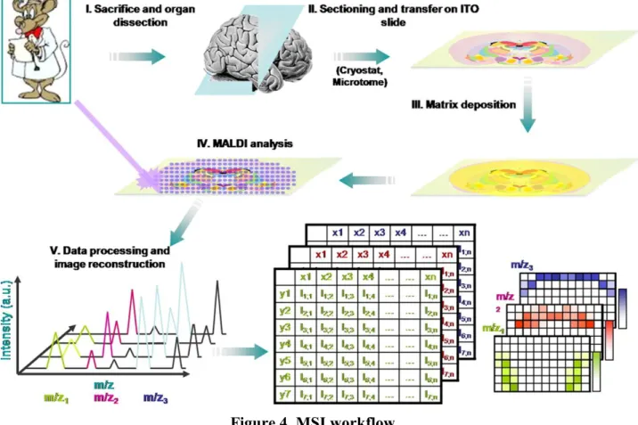
FIGURE 4.MSI WORKFLOW... 41
FIGURE 5.TOF-SIMS IMAGING OF HUMAN SERUM ALBUMIN (HSA) FILM SAMPLE. ... 46
FIGURE 6.THE CHOICE OF MATRIX DEPENDS ON THE ANALYTE TO BE DETECTED IN MALDIMSI. ... 49
FIGURE 7.N-TERMINAL DERIVATIZATION IMPROVES ON-TISSUE MS/MS IDENTIFICATION OF PEPTIDES. ... 51
FIGURE 8.TOP-DOWN LAESI-MSIMAGING OF INTACT PROTEINS IN A MOUSE LUNG SECTION. ... 53
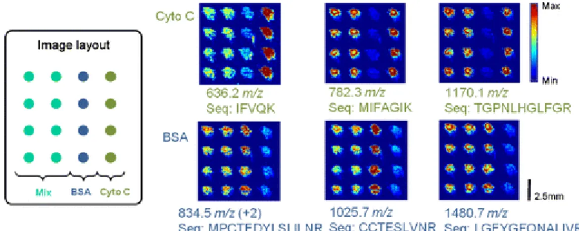
FIGURE 9.DESI-MS IMAGING OF A BINARY COMBINATION OF CYTOCHROME C AND BSA STANDARDS PRINTED ON PERMANOX SURFACES. ... 53
FIGURE 10.A TANDEM MS WORKFLOW. ... 56
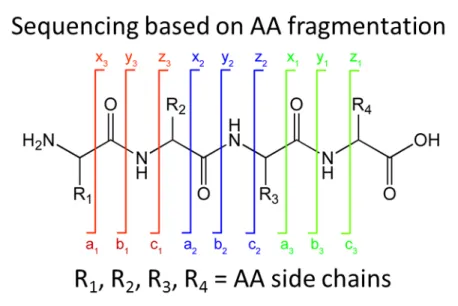
FIGURE 11.PEPTIDE ION SERIES NOMENCLATURE. ... 58
FIGURE 12.MATCHING SPECTRUM WITH A HYPOTHETICAL MODEL. ... 59
FIGURE 13.EXAMPLE OF A COMMERCIAL PIPELINE FOR INTERPRETATION OF LARGE PROTEOMIC DATASETS. ... 61
FIGURE 14.COUPLING OF THE FAMOS AUTOSAMPLER WITH THE VALVE S OF EASY NLC. ... 115
FIGURE 15.LIQUID MICROJUNCTION SIZES AS A FUNCTION OF DISPENSED VOLUME. ... 119
FIGURE 16.SAMPLE MICRODIGESTED REGIONS REDUCED IN DIAMETER UP TO 150 µM. ... 120
FIGURE 17.COMPARISON OF PROTEIN IDS IN THE DIGESTED AND UNDIGESTED REGIONS SUBJECTED TO LIQUID JUNCTION MICROEXTRACTION USING THE FAMOS AUTOSAMPLER. ... 121
FIGURE 18.PROTEIN IDS OF SUCCESSIVE EXTRACTIONS ON THE SAME SPOT USING THE FAMOS AUTOSAMPLER... 121
FIGURE 19.INCREASED CHROMATOGRAPHIC SEPARATION TIME INCREASED THE NUMBER OF PROTEIN IDS FOR THE SAME MICROEXTRACTED SAMPLE. ... 122
FIGURE 20.COMPARISON OF PROTEIN IDS OBTAINED USING TRIVERSA NANOMATE AND THE FAMOS AUTOSAMPLER. ... 124
FIGURE 21.HIERARCHICAL CLUSTERING OF PROSTATE TUMOR IMAGES OBTAINED BY MALDIMSI TO DEFINE REGIONS OF INTEREST FOR PARAFILM-ASSISTED MICRODISSECTION (PAM). ... 167
FIGURE 23.REGULATORY NETWORK PLOTTED USING BETWEENNESS CENTRALITY (NODE SIZE) AND LOG2FOLD
CHANGE (COLOR) USING THE DIFFERENTIALLY EXPRESSED PROTEIN DATASET. ... 177
FIGURE 25.MAPPING OF CANCER GENE INDEX-ANNOTATED PROTEINS USING THE TERM “NEOPLASM”(NODES IN
YELLOW) IN THE MODULES. ... 180
FIGURE 31.WORKFLOW OF THE PROJECT CALLED QUEST-MSI(QUANTITATIVE HIGHLY-SENSITIVE MASS
SPECTROMETRY IMAGING OF PROTEIN) CURRENTLY BEING PROPOSED BASED ON THE RESULTS OF THIS WORK.
7L
IST OFT
ABLESTABLE 1.AMBIENT MS METHODS AS RECENTLY REVIEWED BY BADU-TAWIAH AND COOKS,2013. ... 31
TABLE 2.PROGRAMMING SEQUENCE USED TO ENABLE LIQUID MICROJUNCTION EXTRACTION ON THE FAMOS
AUTOSAMPLER. ... 116
TABLE 3.IDENTIFICATION OF BSA STANDARD FOR EACH EXTRACTION USING THE FAMOS AUTOSAMPLER.. ... 118
TABLE 4.PARAMETERS USED FOR COMPARING MICROEXTRACTION USING THE TRIVERSA NANOMATE (LESA) AND
THE FAMOS AUTOSAMPLER. ... 123
8L
IST OFA
BBREVIATIONS1,5-DAN 1,5-Diaminonaphthalene
A2M α-2-Macroglobulin
AAT Aspartate Aminotransferase
ACN Acetonitrile
ADME Absorption, Distribution, Metabolism, and Excretion
Amu Atomic Mass Unit
ANI Aniline
AP MALDI Atmospheric Pressure Matrix-Assisted Laser Desorption/Ionization APCI Atmospheric Pressure Chemical Ionization
APEX Absolute Protein Expression
APPI Atmospheric Pressure Photoionization
APS Ammonium persulfate
AR Androgen Receptor
ASAP Atmospheric Pressure Surface Analysis Probe
AUI Advanced User Interface
BAC Benzalkonium chloride
BSA Bovine Serum Albumin
C1QBP Complement 1q-Binding Brotein
CE Capillary Electrophoresis
CGI Cancer Gene Index
CHAPS 3-[(3-Cholamidopropyl)dimethylammonio]-1-propanesulfonate
CHCl3 Chloroform
CID Collision-Induced Dissociation
CTAB Cetyl trimethylammonium bromide
CTNNA1 Catenin (Cadherin-Associated Protein), 1
DAPPI Desorption Atmospheric Pressure Photoionization
DART Direct Analysis in Real Time
DBS Dried Blood Spot
DDA Data-Dependent Analysis
DeSSI Desorption Sonic Spray Ionization
DHB 2,5-Dihydroxy benzoic acid
Mass Difference
DMF Dimethyl formamide
DMPK Drug Metabolism and Pharmacokinetics
DNA Deoxyribonucleic acid
DNA-PK or PRKDC DRE
DNA-Dependent Protein Kinase catalytic subunit Digital Rectal Exam
DTT Dithiothreitol
ECD Electron Capture Dissociation
EMT Epithelial-to-Mesenchymal Transition
ESI Electrospray Ionization
ETD Electron Transfer Dissociation
EtOH Ethanol
FA Formic acid
FASP Filter-Aided Sample Preparation
FDR False Discovery Rate
FFPE Formalin-Fixed Paraffin-Embedded
FN1 Fibronectin 1
FOLDS Fast On-Line Digestion System
FTICR Fourier Transform Ion Cyclotron Resonance FTIR Fourier Transform Infrared Spectroscopy
FWHM Full Width at Half Maximum
GDF15 Growth Differentiation Factor 15
GO Gene Ontology
GOT2 Glutamate Oxaloacetate Transferase 2
GSTM3 Glutathione S-Transferase mu 3
GWAS Genome-Wide Association Studies
HADHA Hydroxyacyl-coenzyme A (CoA) Dehydrogenase/3-ketoacyl-CoA thiolase/enoyl-CoA Hydratase (trifunctional protein), Alpha subunit
HC Hierarchical Clustering
HCD High Collision-induced Dissociation
HeLA Henrietta Lacks
HEPES 4-(2-Hydroxyethyl)-1-piperazineethanesulfonic acid
HES Hematoxylin Erythrosine Saffron
HPLC High Performance Liquid Chromatography
HR FTMS High-Resolution Fourier Transform Mass Spectrometry
HYOU1 Hypoxia Upregulated Protein 1
IAA 2-Iodoacetamide
IACUC Institutional Animal Care and Use Committee
ICP Inductively-Coupled Plasma
ID Identification
IFN- -Interferon
IR Infrared
ISD In-Source Decay
ITO Indium Tin Oxide
JeDI Jet Desorption/Ionization
KEGG Kyoto Encyclopedia of Genes and Genomes
LAESI Laser Ablation Electrospray Ionization
LCM Laser Capture Microdissection
LDTD Laser Diode Thermal Desorption
LESA Liquid Extraction Surface Analysis
LMJ-SSP Liquid Microjunction Surface Sampling Probe MALDI Matrix-Assisted Laser Desorption/Ionization
MDH2 Mitochondrial Malate Dehydrogenase 2
MeOH Methanol
MHC Major Histocompatibility Complex
MS Mass Spectrometry
MSI Mass Spectrometry Imaging
MSMB -microseminoprotein
nanoLC-MS Nano Liquid Chromatography-Mass Spectrometry NCBI National Center for Biotechnology Information
NCL Nucleolin
Nd :YAG Neodymium-Doped Yttrium Aluminium Garnet
NDEESI Neutral Desorption Extractive Electrospray Ionization
NH4HCO3 Ammonium bicarbonate
NID2 Nidogen 2
NMR Nuclear Magnetic Resonance
NSAF Normalized Spectral Abundance Factor
OCT Optimal Cutting Temperature
OGP n-Octyl--d-glycopyranoside
PA28 or PSME2 Proteosome Activator complex 28 subunit beta
PADI Plasma-Assited Desorption/Ionization
PAI Protein Abundance Index
PAM Parafilm-Assisted Microdissection
PARP1 Poly(ADP-ribose) polymerase 1
PDF Prostate-derived factor
PEP Peptide Identification Probability
PIKK Phophatidyl Inositol 3-Kinase related Kinases
PMF Peptide Mass Fingerprint
POSTN Periostin
PPP2R1A
PSA
Serine/Threonine-Protein Phosphatase 2A 65kDa Regulatory subunit A, alpha isoform
Prostate-Specific Antigen
PSD Post-Source Decay
PTM Post-Translation Modification
ROI Region Of Interest
ROs Reactive Oxygen Species
RPLC Reverse Phase Liquid Chromatography
RT Retention Time
RT-PCR Real Time-Polymerase Chain Reaction
S/N Signal-to-Noise Ratio
SA Sinapinic acid
SIMS Secondary Ion Mass Spectrometry
SRM Selected Reaction Monitoring
SSSP Sealing Surface Sampling Probe
TCEP Tris(2-carboxyethyl)phosphine
TD-APCI Thermal Desorption Atmospheric Pressure Chemical Ionization
TEC Tissue Extinction Coefficient
TEMED Tetramethylethylenediamine
TGF Transforming Growth Factor-
TFA Trofluoroacetic acid
TLC Thin Layer Chromatography
TOF Time Of Flight
TRIM28 TRUS
Tripartite motif containing 28 Transrectal Ultrasound
UV Ultraviolet
VEGF Vascular Endothelial Growth Factor
WBA Whole Body Animal
1I
NTRODUCTIONBiological processes are highly dynamic and involve millions of molecules of various physicochemical properties and functions. In order to have a relatively fine picture of their mechanisms we need to first be able to monitor the variation in terms of abundance of the molecules in time and space. This means that we need to at least first perform a large scale identification of these biomolecules, and quantify and localize them in the time course of the biological process. These first intention studies are generally performed to highlight the main features differentiating the studied physiological or physiopathological process from a normal situation. When studying physiopathological mechanisms, information about the changes in signaling pathways is expected. Omics is nowadays the related term used in general to refer to these large scale analyses which are classified according to the family of biomolecules to be studied due to the different methodologies and technologies they are relying on. Omics is thus a broad terminology including, genomics, transcriptomics, metabolomics, lipidomics and proteomics as main research fields. Omics techniques will give information on the modification of cell signalization and will help to determine markers of the physiological/physiopathological process as well as provide a better understanding of the mechanisms. These large scale analyses are generally followed by more focused studies in order to decipher precise signalization pathways which are required to obtain a more precise knowledge on the cascade of events, including specific modifications carried by the molecules, as well as their interactions and translocation into the cells requiring thus various methodologies including structural biology techniques such as NMR, X-Ray and a large panel of microscopy technologies. Various families of biomolecules act in synergy to maintain cell homeostasis and allow cell-to-cell communication in a normal function or in response to an external stimulus. Ideally, to obtain a complete picture we should need to perform systematic biology by correlating the data coming from the different Omics. Since this is a huge task, researches are generally focused on one family of biomolecules, although more and more researchers are trying to integrate several large analyses together (Weckwerth 2008; Griffiths and Wang 2009).Taking the problem from one side, proteins are one of the families of biomolecules that can be studied in order to understand physiopathological mechanisms. Large scale protein analyses are referred to as proteomics analyses as introduced in the mid 90’s by Wilkins and coll (Wilkins, Pasquali et al. 1996).
Proteomics analyses rely on a panel of technologies but among them Mass Spectrometry (MS) plays a central role. Indeed, the development of soft ionization sources such as matrix-assisted laser desorption/ionization (MALDI) and electrospray ionization (ESI) in the mid 80’s have paved the way for the establishment of MS as a tool for the analysis of molecules of biological importance (Yamashita and Fenn 1984; Karas, Bachmann et al. 1987). With these sources it is now easy to generate ions from high molecular weight compounds of high polarity (Karas and Hillenkamp 1988; Fenn, Mann et al. 1989), thus expanding the range of biological molecules that can be analyzed by MS from small metabolites, to most notably proteins. In a way, MS has revolutionized the field of proteomics by permitting large scale analysis (McDonald and Yates 2002; Yates, Ruse et al. 2009). Instrumental developments in the field of MS combined to improvements in separation methods such as liquid chromatography (LC (Zhao and Lin ; Ishihama 2005) and developments of bioinformatic tools (Swan, Mobasheri et al. ; Eng, McCormack et al. 1994; Fenyo and Beavis 2008) have greatly contributed to this achievement. Currently thousands of proteins can be identified from a single experiment using shotgun strategies (Geiger, Velic et al. 2013; Picotti, Clement-Ziza et al. 2013). In these strategies extracted proteins are altogether submitted to enzymatic digestion. After purification of the digestion peptides, they are conventionally separated using LC on-line coupled to the MS instrument. This strategy relies on the capacity of the instrument to record MS and MS/MS data from the solution eluted from the LC system. MS spectra provide access to the mass of the digestion peptides while MS/MS will give structural information on the primary sequence of these peptides. The measured masses of the proteolytic peptides and the mass collected from the structural analysis are then combined to be compared and matched with a protein or gene sequence database, and the protein identification is assigned based on the accuracy of the match. This identification process depends on the unequivocal assignment of ions, and parallel developments in instrumental design in terms of mass accuracy and resolving power led to improvements in the identification (Scigelova, Hornshaw et al. 2011). Increase in the duty cycle of instruments also greatly improves the number of identification by allowing to record more structural information (Liu, Huttenhain et al. 2013). Crucial to the development of MS-based proteomics is the parallel improvement in the classical analytical methods of sample preparation. For instance, the improvement of chromatographic separation allowed the refinement of protein purification protocols that are tailored to the demands of the protein of interest. Development of high-throughput and less cumbersome instruments permitted the interfacing of more and more methods with MS, and paved the way for incorporation of multi-dimensional preparative
procedures in an effort to decrease the complexity of samples and increase the identification potential. Currently, Shot-Gun strategies allow fast access to the identification and relative quantification through label free quantification methods to thousands of proteins (Wisniewski, Dus et al. 2013) at various levels (from whole body, organs, cells down to cell compartments or vesicles) in various biological complexes allowing now to study molecules of lower abundance with important biological functions such as chemokines or cytokines (Meissner and Mann 2014).
However, in spite of all these improvements in proteomics, one crucial dimension is lacking, which is the information about the evolution of protein abundances with respect to their localization. This information is not required when working on body fluids, but becomes crucial when studying tissues, such as solid tumors. In fact, most large scale proteomics analyses are performed on whole organs or regions of organs. This only provides access to averaged information and renders difficult the identification of proteins that are highly localized to a restricted area (even though they are in relatively high abundance) due to dilution of the total amount of proteins. Such approaches thus do not show the variation in abundance of the proteins in a specific micro-environment, which can be primordial, for example in the early development of pathologies. Various strategies were developed to circumvent these difficulties. One instance involves collection of a specific type of cells. Cell sorting can be carried out by various means including Flow Cytometry, but the most popular ones are based on Laser Capture Microdissection (LCM, (Emmert-Buck, Bonner et al. 1996)). In LCM, cells are sorted based on their morphologies, which were pre-determined by histological staining. In the beginning LCM was shown to be relatively laborious since it was difficult to collect a large number of cells sufficient for gel based strategies. The advent of shotgun strategies based on nanoflow LC systems and nanoESI ion sources, and requiring much less amount of material, has greatly helped to the improvement of these strategies (Thakur, Rejtar et al. 2011). However, even if LCM coupled to shotgun analysis is nicely working this strategy still presents certain limitations such as the number of cells to be collected (most of the time about 50,000 cells) and the morphological phenotyping-based cell selection criterion in place of molecular phenotyping. Moreover, obtaining numerous cells entails having to collect them from various tissue sections in which the micro-environment can greatly vary.
This greatly simplifies the sample preparation and stresses on the importance of analyzing proteins in the context of their localization. By doing so, MSI allows for the ability to discriminate different types of tissues in the same organ, and permits the non-subjective identification of deseased areas in contrast to normal ones. This simple and straightforward approach permitted the identification of tumor regions in otherwise normal tissues, for example, and demonstrated the potential MSI as a diagnostic tool that can be developed for future use by clinicians in the hospital setting. In this regard, MALDI MSI has shown to be the more powerful and versatile of the various MSI modalities due to the wide range of molecules accessible to the MALDI ion source (Fournier, Wisztorski et al. 2008). MALDI MSI is applicable to a large variety of endogenous molecules including proteins as well as exogenous ones such as drugs, abiotics or xenobiotics. MSI has shown to be very powerful in obtaining the localization of known compounds; one of the best illustrations is the imaging of drugs and their metabolites in whole body animals. MSI can effectively replace whole body autoradiography (WBA), which uses radiolabeled drugs and does not allow the metabolites to be imaged (Solon, Schweitzer et al. 2010). Alone or combined to multivariate statistical analysis, MSI can also be used as a tool for cell phenotyping and finding markers of physiological or physiopathological processes. This approach is often used for clinical studies of pathologies such as cancer, where imaging has shown significant applications in tissue classification and biomarker discovery (Franck, Arafah et al. 2009). In spite of its success in recent years, there are still limitations that need to be addressed in MSI. Perhaps the biggest issue is the crucial need to improve its sensitivity especially in detecting less abundant molecules. This remains especially true for proteins that are more difficult to detect due to their physico-chemical properties. In particular, higher mass proteins (Mw >30,000 Da) are clearly not detected when using conventional sample preparation and only most soluble proteins of the lowest Mw are observed (Franck, Longuespee et al. 2010). Thus, for proteins the number of compounds that can be imaged is relatively limited compared to other molecule families such as lipids. Moreover, direct imaging often results to localization of the most abundant proteins. Although this is not an issue by itself, nonetheless, abundant proteins impose a limit to the number of biomarkers that can be discerned by MSI by hindering the identification of the less abundant ones. More detailed information on physiopathological mechanisms requires the identification of these low abundance proteins, especially if they are involved in signalization pathways implicated in the disease. Identification of imaged proteins is one of the other great challenges for MALDI MSI in these untargeted approaches. These two difficulties have led to the development of On Tissue Bottom-Up strategies (Lemaire, Desmons
et al. 2007; Franck, Arafah et al. 2009). Here, proteins are in situ digested in a controlled manner to generate peptides which should be easier to detect and identify while avoiding their delocalization. Direct protein identification is a challenge since most ion activation methods do not allow fragmentation of intact species due to internal redistribution on all the degrees of freedom of the energy transferred by means of ion activation. This is circumvented by on tissue digestion of proteins. Though resolving part of the problem, on tissue digestion can increase sample complexity by increasing the number of peptide species, and again due to ion suppression effects, only digestion peptides coming from most abundant proteins are predominantly observed. Many groups, including ours, have attempted to address this issue by developing peptide derivatization to increase their propensity for fragmentation (Franck, El Ayed et al. 2009), incorporating washing and abundant protein depletion strategies into the imaging protocol, designing novel matrices to improve protein ionization from single to multi-charged species, using different methods to digest the proteins in situ, and so on. Nonetheless, the results of these approaches are limited, and the challenge to be able to detect, analyze and identify proteins of low abundance in their native environment on tissue remains a daunting task.
It is thus mandatory for the understanding of physiopathological mechanisms to elucidate prognostic and diagnostic biomarkers and their targets, and to be able to identify and image a much larger number of proteins. Clearly there is currently a need to fill the gap between the potential of conventional proteomics strategies and the potential of MS imaging to bring the localization of the molecules. This was the first goal of my PhD project. In this work, I present different strategies to demonstrate alternative ways to address the limitation of protein detection on tissue, as well as other developments I have tried to address in the course of three years of study. The first part focuses on the development of a strategy using the liquid microjunction extraction to be able to extract proteins on a localized region on a tissue section. The liquid microjunction extraction is considered as one of the ambient surface sampling techniques. In this published work, as much as 1500 proteins were identified in a region as small as 650 µm, roughly corresponding to 1900 cells. We were able to discriminate two regions on a rat brain tissue section based on these protein identifications and managed to correlate these results on the regions identified by hierarchical clustering analysis following a MALDI MSI experiment. As such, we have started to incorporate the liquid microjunction extraction strategy into our MSI platform and used it in further studies in our group. This localized extraction strategy is presented
related strategies, which could sample homogeneous cell populations but at the expense of losing localization due to limited number of cells in the sampling region, and which still also suffers from poor protein identification yield in spite of the cell population enrichment. Although the strategy presently cannot sample homogeneous cell populations given the current size of the microdigested and microextracted area attainable, nonetheless, it can be used to examine cell populations in the context of their microenvironment. The ideal situation, in retrospect, would be to sample proteins of individual cells in the context of their tissue localization, as we expect the protein expression to change in a similar manner as has been demonstrated for metabolites even for cells of the same population (Zenobi 2013). Concordance with this idea, a chapter on the recent results of the continuation of the work on this subject is presented, focusing on decreasing both the size of the microextracted and microdigested areas. This consists of the use of alternative means to create a smaller liquid microjunction and the direct coupling of this method to nanoLC-MS. Although a similar set-up has already been previously reported, the current one was designed specifically for a proteomic approach and aimed for the sampling of even smaller regions, and results of its comparison with the conventional off-line liquid microjunction nanoLC-MS approach is discussed.
In the second part, I present the parafilm-assisted microdissection method. Here, an alternative approach dealing with the limited protein identification problem is shown. This consists of mounting a tissue specimen on a parafilm-covered slide, excising regions from the tissue, and subjecting them individually to conventional shotgun proteomics MS analysis. Furthermore, the published work explores the possibility of generating molecular images from the protein identifications, using the quantitative values of the spectral counts of peptide assignments. With this approach, we were able to identify and localize high and low abundance proteins, and are currently working towards the refinement of this strategy in order to improve the resolution of the images. An application of the PAM method on prostate tumors is then presented. Here, we examined fresh frozen tumor sections, excised regions of interest, and subjected them to proteomics MS analysis. Here I demonstrate that using the PAM approach allows for the identification and quantification of protein expression levels. The importance of this in regard to prostate cancer is that the data can be used to detect changes in the regulation of proteins, and eventually biological pathways, as the prostate undergoes pathological changes.
In summary, this work is largely divided into two categories: development of MS-based methods for on-tissue protein identification, and linking these methods with quantification and use this to be able to map protein distribution across the tissue. In addition, application of on-tissue protein identification and quantification strategies in the context of prostate cancer for the discovery and development of candidate biomarkers is also presented.
Hypothesis
Current MS-based methods for the direct analysis of proteins on tissue, such as MS imaging, suffer from poor detection, identification and quantification of proteins thereby warranting the development of alternative approaches that could circumvent these inherent limitations. One way is to improve the number of protein identifications by localized microextraction on discreet regions of the tissue section. Restricting microextraction on discreet regions could improve the identification of proteins of low abundance by limiting the amount of abundant proteins co-extracted, compared for example when extraction was performed on the entire organ or tissue section. These protein IDs could then be used for back-correlation with MSI data to increase the number of protein assignments that have been localized. Another way is to perform simultaneous identification and localization of proteins in a single approach, without sacrificing the high number of protein identifications. This can be achieved by dividing a tissue section into millimeter-sized portions that can then be individually subjected to shotgun MS. The spectral counts of the proteins identified can then be used to reconstruct the image of the tissue section and map their spatial distribution.
Objectives
Develop alternative methods to ameliorate MSI protein identification strategies.
This can be achieved by developing a localized protein extraction method based on the liquid microjunction approach. The approach is similar in principle to the generation of protein ID databases that are then used for matching with peaks in MSI spectra. In contrast though, the protein database will be obtained from localized regions on the tissue section. This will be done by performing in situ tryptic digestion on those regions, followed by extracting the peptides using the liquid microjunction approach, then subjecting the extracts to shotgun nanoLC MS.
Develop quantification-based imaging strategies to directly correlate protein identification data with localization information.
Simultaneous identification, quantification and localization of proteins on tissue sections can be realized by obtaining millimeter-sized pieces of the tissue and subjecting each piece individually to shotgun nanoLC MS. Label-free quantification of the identified proteins can then be done by spectral counting, and the relative quantification data used to generate the image of the tissue by noting the positions where the tissue pieces have been excised from the section. The tissue pieces can be obtained from the tissue section by mounting the latter on a parafilm-covered slide and excising the pieces using a scalpel. Parafilm is a relatively inert material that can act as a support for the tissue during cutting of the pieces.
the sampling of charged analytes actually begins. The ionized species are then transported to the mass analyzer where their resolution in terms of m/z occurs. In tandem MS experiments, additional fragmentation of ions detected after m/z separation can be performed and the resultant MS/MS spectra can be used for analyte identification in the post-analysis step. After detection, the analysis can yield spectra for precursor or product ion scans, MS/MS spectra, as well as MS images constructed from the mapping of ion density on the selected or entire region of the sample section. Note that direct MS analysis methods are at the frontline of the scheme and thus are important in determining the outcome of the entire analysis.
The major advantage of direct on-tissue protein analysis in contrast to classical MS-based methods is the minimization or total elimination of complicated fractionation procedures that tend to introduce artifacts to the sample with each subsequent purification step (Huang, Cheng et al. 2011). By doing the analysis directly on tissue, it is possible to analyze proteins in their native state and in the presence of their interaction partners, thus providing a more realistic view of the binding of these proteins compared when their interactions are studied in solution. The point by point examination of tissue surfaces typical of direct tissue analysis methods allows the mapping of protein distribution across the whole tissue, permiting the localization of proteins.
Ambient MS
Ambient MS-based methods are those that employ direct tissue sampling under ambient conditions of temperature and pressure. Their development is mainly driven by the need for mass spectrometers that are capable of performing on the spot analysis of real-world samples thus giving access to this technology to everyday applications, such as for instance, in the determination of pesticide content in food products or, detection of substances of abuse or of stimulants in race animals and athletes, to name a few. This is possible by liberating the analysis from constraints presented by difficulties of introducing the sampling surface into the vacuum, including sample size and analyte volatility. In these methods, there is a great emphasis in the examination of samples that are preserved in their native state with minimal or no interference due to the analysis. Their use in proteomics is limited to the identification or quantification of highly abundant endogenous proteins, and proteome-wide discoveries involving these methods still have to be demonstrated.
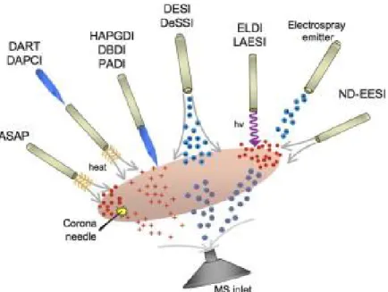
A total of 42 ambient MS-based methods are currently reported in literature (Table 1, (Badu-Tawiah, Eberlin et al. 2013)). Several reviewers have classified them into various categories based on the similarities and differences in their mode of sampling the surface (Figure 2). It is not our current objective to provide a comprehensive review of these methods, but rather to
highlight major surface sampling principles in order to emphasize our choice of approaches used in this work.
Figure 2. Overview of ambient MS-based methods.
Venter & Cooks’ classification of ambient MS-based methods based on the mode of sample extraction and transport to the mass spectrometer inlet. Reprinted from (Venter, Nefliu et al. 2008), Copyright (2008) with permission from Elsevier.
Table 1. Ambient MS methods as recently reviewed by Badu-Tawiah and Cooks, 2013.
Reproduced with permission. For references, see (Badu-Tawiah, Eberlin et al. 2013).
Method name Acronym Agent Characteristics
1 Desorption electrospray ionization DESI Sprays Imaging capabilities, reactive mode, small (<500 Da) and large molecules
2 Easy ambient sonic-spray ionization EASI Sprays Imaging capabilities, small (<500 Da) molecules 3 Desorption ionization by charge exchange DICE Sprays, chemical
ionization Small (<500 Da) molecules 4 Transmission mode desorption electrospray
ionization TM-DESI Sprays Reactive mode
5 Nanospray-desorption/electrospray ionization Nano-DESI Sprays Imaging capabilities, small (<500 Da) and large molecules
6 Probe electrospray ionization PESI Sprays Imaging capabilities, small (<500 Da) and large molecules
7 Liquid microjunction-surface sampling probe LMJ-SSP Sprays Imaging capabilities, reactive mode, small (<500 Da) molecules
8 Paper spray PS Sprays Reactive mode, small (<500 Da) and large molecules 9 Direct analysis in real time DART Plasmas Small (<500 Da) and large molecules
10 Low-temperature plasma probe LTP Plasmas Reactive mode, small (<500 Da) molecules 11 Flowing atmospheric pressure afterglow FAPA Plasmas Small (<500 Da) molecules
12 Desorption atmospheric pressure chemical
ionization DAPCI Plasmas Small (<500 Da) molecules 13 Desorption corona beam ionization DCBI Plasmas Small (<500 Da) molecules 14 Dielectric barrier discharge ionization DBDI Plasmas Small (<500 Da) molecules 15 Electrospray-assisted laser desorption
ionization ELDI Lasers, sprays
Imaging capabilities, small (<500 Da) and large molecules
16 Laser ablation electrospray ionization LAESI Lasers, sprays Imaging capabilities, small (<500 Da) molecules 17 Laser-assisted desorption electrospray
ionization LADESI Lasers, sprays
Imaging capabilities, small (<500 Da) and large molecules
18 Laser desorption electrospray ionization LDESI Lasers, sprays Imaging capabilities, small (<500 Da) and large molecules
19 Laser-induced acoustic
desorption-electrospray ionization LIAD-ESI Lasers, acoustic, sprays Small (<500 Da) and large molecules 20 Neutral desorption extractive electrospray
ionization ND-EESI Sprays Reactive mode, small (<500 Da) molecules 21 Radiofrequency acoustic desorption and
ionization RADIO Sprays, acoustic Small (<500 Da) molecules 22 Atmospheric pressure solids analysis probe ASAP Heat, plasmas Small (<500 Da) molecules 23 Infrared laser ablation metastable-induced
chemical ionization IR-LAMICI
Lasers, chemical
ionization Small (<500 Da) and large molecules 24 Rapid evaporative ionization mass
spectrometry REIMS Chemical ionization Heating, small (<500 Da) molecules 25 Desorption atmospheric pressure
photoionization DAPPI Chemical ionization Photons as proton source, small (<500 Da) molecules 26 Beta electron-assisted direct chemical
ionization BADCI Chemical ionization
Beta particles as proton source, small (<500 Da) molecules
27 Extractive electrospray ionization EESI Sprays Reactive mode, small (<500 Da) and large molecules 28 Remote analyte sampling transport and
ionization relay RASTIR Sprays Small (<500 Da) and large molecules 29 Laser ablation flowing atmospheric-pressure
afterglow LA-FAPA Plasmas Small (<500 Da) molecules 30 Surface activated chemical ionization SACI Plasmas Small (<500 Da) molecules 31 Single-particle aerosol mass spectrometry SPAMS Lasers Small (<500 Da) molecules 32 Laser diode thermal desorption LDTD Lasers, plasmas Small (<500 Da) molecules 33 Helium atmospheric pressure glow discharge
ionization HAPGDI Plasmas Small (<500 Da) molecules 34 Surface acoustic wave nebulization SAWN Acoustic Small (<500 Da) molecules 35 Ultrasonication-assisted spray ionization UASI Acoustic Small (<500 Da) and large molecules 36 Atmospheric pressure-thermal
desorption/electrospray ionization AP-TD/ESI Heat, sprays Small (<500 Da) molecules 37 Microplasma discharge ionization Microplasma Plasmas Small (<500 Da) molecules 38 Atmospheric pressure thermal desorption
ionization APTDI Heat Small (<500 Da) molecules
39 Desorption electrospray/metastable-induced
ionization DEMI Sprays, plasmas Small (<500 Da) molecules 40 Switched ferroelectric plasma ionizer SwiFerr Plasmas Small (<500 Da) molecules 41 Laser electrospray mass spectrometry LEMS Lasers, sprays Small (<500 Da) and large molecules 42 Plasma-assisted desorption ionization PADI Plasmas Small (<500 Da) molecules
Thermal Desorption/Ionization Methods
Ambient MS-based methods can be divided into several major categories based on the mechanism by which they extract samples from the surface and transport them to the inlet of the mass spectrometer (Figure 2). Thermal desorption/ionization-based methods are those that employ a heating mechanism to convert molecular species from the condensed phase in the sample surface into the vapor phase. This is traditionally followed by secondary ionization effected through the use of a corona discharge (atmospheric pressure chemical ionization, (APCI (Van Berkel, Pasilis et al. 2008)), or atmospheric pressure surface analysis probe, (ASAP (Ellis, Brown et al. 2013)). The mode by which heat is used to convert condensed phase material into gas phase, the manner of ionization, or both, can be varied thus leading to methods based on the TD-APCI principle. In laser diode thermal desorption (LDTD), the heated gas is replaced by an infrared (IR) laser that sequentially fires at the back of the sample surface, heating it until the gas phase molecules are ejected from the sample surface (Lohne, Andersen et al. 2012). In direct analysis in real-time (DART), heated He gas is converted into metastable atoms, and serves both to desorb material into gas phase and ionize them (Gross 2014). In plasma-assisted desorption/ionization (PADI), the He gas is initially made to pass between two electrodes, one of which is covered by a dielectric layer. As the gas flows, alternating voltage is applied to the two electrodes, generating plasma composed of metastable He, ions, radicals and electrons. The plasma jet is made into contact with the sample surface for desorption/ionization to occur. Molecular ionic species are generally formed using the method, although in some instances fragment ions can also be observed (Ratcliffe, Rutten et al. 2007). Lastly, in desorption atmospheric pressure photoionization (DAPPI), a microchip nebulizer delivers the heated solvent into the sample surface (Haapala, Pol et al. 2007; Luosujarvi, Arvola et al. 2008). This is then followed by ionization using an ultraviolet lamp in place of the corona discharge. The mechanism of ionization is similar with APPI using liquid sample introduction sources.
Laser Ablation Methods
Another type of ambient MS-based methods involves the use of a laser to generate an ablation plume of gas phase molecules which is then subjected to secondary ionization. Early examples of this approach used inductively-coupled plasma (ICP) as a means for secondary ionization (Van Berkel, Pasilis et al. 2008), making it useful in geology-type applications that demand elemental
example uses a CO2 laser to effect desorption of neutral molecules, followed by APCI ionization to analyze trypsinized proteins on SDS-PAGE gels (Coon, Steele et al. 2002). But perhaps the most widespread examples of this type of ambient MS-based method is when it is coupled with the ESI source. One method, termed laser ablation electrospray ionization (LAESI, (Nemes and Vertes 2007)), fundamentally accomplishes ESI-like ionization, and follows two phases. During the first phase, a strong 1µs pulse at 2940 nm excites the –OH vibration in water present in the sample, resulting to the creation of a hemispherical plume that initially expands and eventually collapses due to pressure exerted by the atmosphere during the second phase, lasting 300 µs. The explosion ejects secondary particles composed of a mixture of molecules, clusters and ablated particulate matter, which then coalesce with the charged droplets of the ESI source. A variant consists of using a 337-nm nitrogen laser operating at 20 µJ per pulse, the focused laser spot size of which is 100 µm x 150 µm with the laser beam incident angle at 45 (Shiea, Huang et al. 2005).
The main advantage of LAESI-type methods, in addition to high ionization yield, is the possibility of producing multiply-charged molecules from laser ablation modes of sampling. This limitation is inherent in laser desorption-type sampling where only singly-charged and rarely doubly-charged ions are generated. This capability stems from the use of the ESI to induce the charging of the ejected particles. As such, protein spectra having multiply-charged envelopes of ions similar to those observed in ESI-generated spectra are observed when using laser ablation-sampled spectra. Reports on top-down analysis of proteins using LAESI have recently appeared in literature (Kiss, Smith et al. 2014), and it is expected to find wider applications in proteomics, in addition to its established application in the in-depth profiling of small metabolites and in particular lipids (Shrestha, Nemes et al. 2010). Its main disadvantage though is the fact that ablation using a mid-IR range laser is dependent on the water content of the sample (Ye, Greer et al. 2011). Variations in the water content and tensile properties of the sample thus limit the replicability of this technique particularly for time-resolved imaging because they grossly affect the LAESI ion yield. The use of lower, mid-IR absorbances would also entail the use of significantly higher laser fluences in order to ablate biological samples.
Liquid and Gas Jet Desorption/Ionization Methods
Methods using liquid or gas jet, on the other hand, are those that rely on the collision of charged solvent droplets with the analyte on the surface for sample desorption, followed by charge
transfer upon impact. The mechanism of sample desorption in this case can be referred to as a liquid film-droplet sputtering process. Exemplified mainly by desorption electrospray ionization (DESI) (Takats, Wiseman et al. 2004), this method uses a pneumatically-driven electrospray that provides the charged droplets that wet the surface. The wet area is continuously impacted by the droplets, causing splashing, jetting, or creation of secondary droplets that may contribute to the ejection of the sample in addition to those that have initially been ejected upon impact of the spray (Takats, Wiseman et al. 2005). As such, no sample pre-treatments are used prior to its injection to the mass spectrometer, although it has also been demonstrated that the system can be coupled to an auxilliary chromatograhic set-up for use in more sensitive sample detection (Zhang, Yuan et al.). As in conventional ESI, a high voltage, typically 4 kV, is applied to create the charged droplets (Bereman and Muddiman 2007). By removing the voltage and increasing the nebulizing gas velocity to a sufficient level to permit ejection of analytes, a variation called desorption sonic spray ionization (DeSSI) was developed (Haddad, Sparrapan et al. 2006). Since no voltage is applied, the ESI source generates a distribution of positive and negative charged droplets. Further increasing the liquid flow up to 0.05-0.15 mL/min, among other modifications, leads to the another DESI variation called jet desorption/ionization (JeDI). In this case, the jet spray continuously erodes the sampling surface to generate gaseous analytes, making it ideal for depth profiling studies. Finally, in neutral desorption extractive electrospray ionization (NDEESI), both voltage and liquid were eliminated (Chen and Zenobi 2008). Only a moderate flow of gas at room temperature is applied on the surface to induce analyte desorption, followed by a secondary ESI ionization.
As surface sampling is done under ambient conditions, DESI can easily be applied to any type of sample surface, greatly expanding its applicability to diverse real-world problems. Its proteomics applications though are limited to the analysis of liquid (Miao and Chen 2009) or spotted protein standards (Rao, Celiz et al. 2013; Montowska, Rao et al. 2014) and imaging of the tryptic digests of these (Pasilis, Kertesz et al. 2008). It has been reported that the major limitation of DESI is its limited sensitivity; for instance, in the analysis of propanolol metabolites, it failed to detect the hydroxypropanolol glucoronide metabolite compared when using other ambient surface sampling MS methods or MALDI imaging (Kertesz, Van Berkel et al. 2008).
Liquid Extraction Surface Sampling/Ionization Methods
Surface liquid extraction methods are typically liquid-solid microextraction methods that can be coupled with any ambient ionization sources such as ESI or APCI (Van Berkel, Kertesz et al. 2008). The success of these methods is due to their greater extracting efficiency and larger surface sampling areas. It has been shown, for instance, that phase II metabolites of propranolol were detected using these methods (Van Berkel and Kertesz 2009; Kertesz and Van Berkel 2010; Kertesz and Van Berkel 2010), whereas they were unsuccessfully detected using DESI(Kertesz and Van Berkel 2010). In these techniques, the extraction of materials from the sample surface is independent of ionization by having a liquid solvent in contact with the sample surface so that extraction can occur. The solvent may then be introduced directly into the inlet of the mass spectrometer, as in continuous type liquid microjunction set-ups, or they can be collected and may be subjected to sample pre-treatment steps, as in discrete type ones. Since ionization is independent of extraction, the method can be used in conjunction with various types of ambient sources, as has been demonstrated in APCI and ESI applications. Examples of the continuous mode are the liquid microjunction surface sampling probe (LMJ-SSP, (Van Berkel, Kertesz et al. 2008)) and the sealing-surface sampling probe (SSSP, (Luftmann 2004)). This mode has been demonstrated to be useful for MS imaging, where an ESI source was used in place of the typical laser desorption/ionization source (Van Berkel, Kertesz et al. 2008). Alternatively, the solvent can be made in contact with the sample surface discretely as single droplets the formation and size of which is regulated by a syringe pump, as seen in the liquid extraction surface analysis probe (LESA, (Kertesz and Van Berkel 2010)). In this case, the solvent is carried using a conductive tip where the droplet is formed and where the subsequent extract is aspirated back to be introduced into the electrospray source. This mode is mostly applied in sample profiling, where discrete spots within the sample are extracted and the levels of analyte determined and compared for each sampling region.
Because of the independence of sample extraction from ionization, liquid extraction surface sampling techniques permit the further treatment of samples with coupled devices prior to introduction to the mass analyzer. In essence, this does not belong to the realm of ambient surface sampling anymore, however, the use of these samplers as microextraction devices is very tempting especially if the whole system is fully automated (Van Berkel, Sanchez et al. 2002; Kertesz, Ford et al. 2005). This has been shown, for instance, by coupling the LMJ-SSP device with a high performance liquid chromatography (HPLC) system (Kertesz and Van Berkel 2010).
Sealing-Surface Sampling Probe
Much of the development of the current commercially available SSSP type configuration stems from the efforts of Luftmann (Luftmann 2004 20) to design a sampling probe for the automated extraction and analysis of TLC plates. SSSP works by enclosing the sample surface using a probe with a circular cutting edge that goes through the surface deep into the TLC backing aided by a frame press. This seals the surface of interest and somehow creates a partial vacuum that aids in the extraction of the sample. Solvent flows into the probe through an inlet capillary connected to an HPLC pump that regulates the flow, and comes into contact with the surface so that extraction can take place. The solvent then exits the probe thru an outlet capillary connected to the ion source for introduction to the mass analyzer. The typical diameter of the sampling head is 2 and 4 mm. In another design, the sampling head is elliptical (2 x 4 mm) so that it adapts more to the shape of the TLC spots (Morlock and Jautz 2008). The pump can be switched from standby mode, where solvent flows directly to the ion source, to sampling mode, where the solvent enters the probe and sampling occurs, or to docking mode, where the probe can be cleaned automatically using pressurized air (Aranda and Morlock 2007; Luftmann, Aranda et al. 2007).
As previously mentioned the SSSP system is currently designed for analysis of samples spotted on TLC plates, although it is imminent that this can also be applied to analogous surfaces especially those that are porous. Thus it may also be applied to systems utilizing polymer or metallic supports. The only limitations are that it is only restricted to samples where the pressure-aided sealing mechanism is possible, and that the pressure to be used for sealing must be optimized for each surface being used to avoid leakage of the extracting solvent, although modifications have been implemented to avoid this problem (Alpmann and Morlock 2006). Liquid Microjunction Surface Sampling Probe
The LMJ-SSP system is mainly developed by Van Berkel and co-workers (Van Berkel, Sanchez et al. 2002), although prototypes of this device have been previously reported (Modestov, Srebnik et al. 2001; Wachs and Henion 2001). This probe consists of a capillary inserted inside a bigger one so that the space between them is used as an inlet connected to a solvent reservoir. Solvent enters and fills this space until the excess goes out of the capillary and comes into contact with the sampling surface. In this so-called wall-less ‘microjunction’ formed between the solid sample surface and the liquid, extraction occurs as the analytes with preferential affinity to the solvent polarity migrate to the liquid interphase and leave the sample by concentration gradient-driven
diffusion. The hollow portion of the inner capillary of the probe serves as the outlet line and is connected to the ion source. The resulting extract enters this line because of a self-aspirating mechanism driven by the pneumatic flow of the nebulizing gas towards the source.
The LMJ-SSP device has found applications in extraction of spots from hydrophobic TLC plates (Van Berkel, Sanchez et al. 2002; Ford, Deibel et al. 2005), drugs obtained by solid phase extraction cards (Wachs and Henion 2003), affinity-captured proteins (Van Berkel, Ford et al. 2006), and it has also been used for the imaging of inks on paper (Kertesz, Ford et al. 2005). In the latter case, resolution was achieved at 0.5 mm, the size of the probe. By modifying and automating the adjustment of the microjunction formation, LMJ-SSP in discrete sampling mode was used for high-throughput analyses of drugs (Van Berkel and Kertesz 2009). This also lessened the need for technical expertise in adjusting the probe-to-sample distance which is crucial to microjunction formation, and thus enables the automatic probing of a sample surface with varying thicknesses as in whole body tissue section for example. Problems in sample carry-over was observed, however, and it was suggested to perform a blank run in between samples to ensure complete cleaning of the probe prior to the next analysis. Coupling with other post-treatment devices, such as an HPLC set-up, was also recently accomplished using the LMJ-SSP (Kertesz and Van Berkel 2010). This can be very important with regard to future advances in ambient sampling because this allows clean-up of complex sample matrices that may hinder efficient sample detection when they are introduced directly to MS.
Liquid Extraction Surface Analysis
Further exploration of the discrete ambient surface sampling as an alternative to the continuous type led to the design of LESA (Kertesz and Van Berkel 2010). The principle is that the formation of the wall-less microjunction does not have to be restricted with the use of probes as in the case of LMJ-SSP. In fact, the microjunction can be formed simply by putting into contact a solvent droplet with the sampling surface, a function that can also be achieved using disposable pipette tips. After extraction, the extract is aspirated by means of a plunger mechanism using the same tip and then delivered into the electrospray head for ionization. With this system, flexibility is offered on the modulation of the size of the microjunction formed and variation of extraction time to suit a particular analysis. Currently, LESA has been demonstrated on the quantitation of drugs on dried blood spots (DBS), profiling of total drug distribution on whole body tissue sections, and extraction and quantitation on MALDI spots (Kertesz and Van Berkel 2010). It has
also recently been used for the direct and rapid identification of intact hemoglobin variants from DBS samples used for the neonatal screening of sickle cell and other hemoglobin-related diseases (Edwards, Creese et al. 2011). LESA has also been used to correlate results obtained from MS imaging experiments (Marshall, Toteu-Djomte et al. 2010). Although imaging investigations using LESA have yet to be reported, the possibility of using this technique for imaging has been implied.
Much of the developments in LESA have been obtained through the use of the TriVersa NanoMate system marketed by Advion Biosciences. Using the Advanced User Interface (AUI), Van Berkel and Kertesz (Kertesz and Van Berkel 2010) were able to use the NanoMate platform to perform LESA procedures. The TriVersa NanoMate is actually a coupling device used for automated delivery of infused samples for ESI MS analysis. It uses a robotic arm to pick up specially designed conductive tips and uses them to aspirate the sample by attaching the open end into a disposable nano-electrospray chip. The chip contains 384 electrospray emitters created using a deep reactive ion etching chemical process (Schultz, Corso et al. 2000). These nozzles provide long and stable sprays and eliminate sample-to-sample carry-over by the use of new nozzles for individual analyses. In addition, the TriVersa NanoMate platform offers portability and adaptability with a variety of compatible mass spectrometers. As such, prior to the development of the LESA mode, this device has already seen various applications in the analysis of lipids (Stahlman, Ejsing et al. 2009), metabolites (Giavalisco, Hummel et al. 2008), proteins and protein-related fields like post-translational modifications, non-covalent interactions, and top-down proteomics (Zhang, Van Pelt et al. 2003), antibodies and glycosylation (Ge, Rybakova et al. 2009), and others (Stumpo and Reinhold 2010).
The NanoMate system has three modes of operation: 1) infusion mode, 2) fraction collection and 3) LESA. In the infusion mode, the device allows for the direct introduction of samples into the ESI emitter, as previously described. The fraction collection mode is used to interface the device (and the coupled mass spectrometer) to a liquid chromatography (LC) set-up. In this mode the eluant coming from the LC system is transported to the NanoMate through a microcapillary tubing connected to a tee. The tee is connected to the mandrel and to the nanospray adapter, so that simultaneous collection of the eluant at constant time segments (through the mandrel) is possible while a portion is being electrosprayed. The resulting MS spectra can thus be correlated