i
Université de Montréal
Neural substrates and functional
connectivity associated with
sleep-dependent and insleep-dependent consolidation of
new motor skills
par
Karen Debas
Département de psychologie
Faculté des arts et des sciences
Thèse présentée à la Faculté des arts et des sciences en vue de
l’obtention du grade de PhD en neuropsychologie option recherche et
intervention
Mai, 2013
Résumé
La mémoire n’est pas un processus unitaire et est souvent divisée en deux catégories majeures: la mémoire déclarative (pour les faits) et procédurale (pour les habitudes et habiletés motrices). Pour perdurer, une trace mnésique doit passer par la consolidation, un processus par lequel elle devient plus robuste et moins susceptible à l’interférence. Le sommeil est connu comme jouant un rôle clé pour permettre le processus de consolidation, particulièrement pour la mémoire déclarative. Depuis plusieurs années cependant, son rôle est aussi reconnu pour la mémoire procédurale. Il est par contre intéressant de noter que ce ne sont pas tous les types de mémoire procédurale qui requiert le sommeil afin d’être consolidée. Entre autres, le sommeil semble nécessaire pour consolider un apprentissage de séquences motrices (s’apparentant à l’apprentissage du piano), mais pas un apprentissage d’adaptation visuomotrice (tel qu’apprendre à rouler à bicyclette). Parallèlement, l’apprentissage à long terme de ces deux types d’habiletés semble également sous-tendu par des circuits neuronaux distincts; c’est-à-dire un réseau cortico-striatal et cortico-cérébelleux respectivement. Toutefois, l’implication de ces réseaux dans le processus de consolidation comme tel demeure incertain. Le but de cette thèse est donc de mieux comprendre le rôle du sommeil, en contrôlant pour le simple passage du temps, dans la consolidation de ces deux types d’apprentissage, à l’aide de l’imagerie par résonnance magnétique fonctionnelle et d’analyses de connectivité cérébrale. Nos résultats comportementaux supportent l’idée que seul l’apprentissage séquentiel requiert le sommeil pour déclencher le processus de consolidation. Nous suggérons de plus que le putamen est fortement associé à ce processus. En revanche, les performances d’un apprentissage visuomoteur s’améliorent indépendamment du sommeil et sont de plus corrélées à une plus grande activation du cervelet. Finalement, en explorant l’effet du sommeil sur la connectivité cérébrale, nos résultats démontrent qu’en fait, un système cortico-striatal semble être plus intégré suite à la consolidation.
C’est-à-dire que l’interaction au sein des régions du système est plus forte lorsque la consolidation a eu lieu, après une nuit de sommeil. En opposition, le simple passage du temps semble nuire à l’intégration de ce réseau cortico-striatal. En somme, nous avons pu élargir les connaissances quant au rôle du sommeil pour la mémoire procédurale, notamment en démontrant que ce ne sont pas tous les types d’apprentissages qui requièrent le sommeil pour amorcer le processus de consolidation. D’ailleurs, nous avons également démontré que cette dissociation de l’effet du sommeil est également reflétée par l’implication de deux réseaux cérébraux distincts. À savoir, un réseau cortico-striatal et un réseau cortico-cérébelleux pour la consolidation respective de l’apprentissage de séquence et d’adaptation visuomotrice. Enfin, nous suggérons que la consolidation durant le sommeil permet de protéger et favoriser une meilleure cohésion au sein du réseau cortico-striatal associé à notre tâche; un phénomène qui, s’il est retrouvé avec d’autres types d’apprentissage, pourrait être considéré comme un nouveau marqueur de la consolidation.
Mots-clés :
consolidation, apprentissage moteur, sommeil, connectivité fonctionnelle, IRMf, séquence, adaptation motrice, mémoire.Abstract
Memory in humans is generally divided into two broad categories: declarative (for facts and events) and procedural (for skills and motor abilities). To persist, memories undergo a process referred to as consolidation, where a fresh, initially labile memory trace becomes more robust and stable. Sleep is known to play an important role in declarative memory consolidation, and in the past decade, there has been increasing evidence for a role of sleep in the consolidation of procedural memory as well. Interestingly, however, the beneficial effects of sleep do not seem to be homogenous. Motor sequence learning consolidation, in particular, has been found to be particularly sensitive to sleep effects, while the consolidation of motor adaptation has not. Moreover, neuroimaging research, has demonstrated that the long term retention of these two types of motor abilities rely on different neuronal networks, namely the cortico-striatal and cortico-cerebellar systems, respectively. Yet the implication of these networks in the consolidation of these two types of motor memory remains unclear. The aim of the present doctoral thesis was thus to determine the influence of sleep, while controlling for the simple passage of daytime, on the consolidation of a motor sequence learning task vs. a motor adaptation task. We further aimed to bring new insights into the underlying brain regions involved in consolidating these two forms of motor skills. Consistent with previous research, we found off-line improvements in performance for motor adaptation learning, independent of whether participants had a night of sleep or remained awake during daytime. Furthermore, these improvements were correlated with activity in the cerebellum. In contrast, we found that off-line increases in performance in motor sequence learning were evident after a night of sleep but not over the day; and the putamen was strongly associated with this sleep-dependent consolidation process. Finally, while measuring brain changes in connectivity associated with the latter process, we observed that sleep-dependent consolidation is reflected by an increased level of integration within the cortico-striatal system, but not in other functional networks. Conversely, the simple passage of daytime in the wake state
seems to result in decreased cortico-striatal integration. In sum our results highlight that not all motor memories undergo sleep-dependent consolidation. We demonstrated that these different paths to consolidation are also reflected by distinct underlying neuronal systems, namely a cortico-striatal and cortico-cerebellar network associated with the consolidation of motor sequence and motor adaptation learning respectively. Furthermore, we propose that consolidation of motor sequences during sleep protects and favors cohesion within the cortico-striatal system, a phenomenon that, if replicated in other types of memories, may be considered as a new marker of sleep-dependent consolidation.
Keywords:
consolidation, motor learning, sleep, functional connectivity, fMRI, motor sequence, motor adaptation, memory.TABLE OF CONTENTS
Chapter 1: Theoretical Background ... 1
1.1. Memory ... 1
1.1.1. Historical Perspective ... 1
1.1.2. Memory Systems Organisation ... 2
1.2. Procedural Memory ... 3
1.2.1. Motor learning ... 4
1.2.2. Behavioural Paradigms ... 4
1.2.3. Neural Correlates Mediating Motor Skill Learning ... 6
1.2.4. Motor Learning models ... 10
1.2.5. Connectivity Changes in Motor Skill Learning ... 13
1.3. Memory Consolidation ... 21
1.3.1. Levels of Consolidation ... 21
1.3.2. Memory Consolidation and Sleep ... 23
1.3.3. Motor Memory Consolidation ... 24
1.4. Sleep: Influences on the Consolidation Process ... 29
1.4.1. Sleep Architecture ... 29
1.4.2. Physiological mechanisms associated with sleep ... 30
1.4.3. Role of sleep for motor memory consolidation ... 32
1.4.4. Neuronal reorganization associated with the motor consolidation process ... 33
Aim and hypothesis ... 38
Chapter II: Research articles ... 40
2.
Article 1 “Brain plasticity related to the consolidation of
motor sequence learning and motor adaptation”... 40
2.1. Supplemental Information ... 67
3.
Article II: “Off-line consolidation of motor sequence learning
results in greater integration within the cortico-striatal functional
network” ... 71
Chapter III: General Discussion ... 99
4.1. Limits ...101
5.
The Role of the Striatum and Cerebellum in Memory
Consolidation ... 103
6.
What are the pre-requisite for sleep-dependent
consolidation? ... 106
6.1. Targeting specific brain regions as a way to understand sleep-dependent memory consolidation ...107
6.2. Different motor memory processes, different needs for consolidation ...108
6.3. Benefit of daytime for consolidation...109
6.3.1. Motor adaptation learning ...110
6.3.2. Motor sequence learning ...111
6.4. Benefit of sleep for consolidation ...115
6.4.1. Motor adaptation ...115
6.4.2. Explicit Motor sequence learning ...116
6.5. Summary - current hypothesis ...116
7.
Possible mechanisms associated with sleep-dependent
consolidation ... 119
8.
Perspective ... 121
List of Tables
Table 1. Article 1 - Effect of sleep ... 66
Table 2 . SI Article 1- Test session ... 70
Table 3. SI Article 1 - Correlation with behavioral improvement ... 70
List of Figures
Figure 1: apparatus for testing MSL and MA. ... 5
Figure 2 : Anatomy ... 8
Figure 3 : Learning model proposed by Doyon and collaborators ... 12
Figure 4 : Resting-state networks ... 16
Figure 5 : Factors influencing sleep-dependent memory consolidation – from Diecklman et al., (2009) ... 23
Figure 6 : Sleep-dependent consolidation at the behavioral level ... 26
Figure 7 : Sleep architecture – adapted from Peigneux et al. (2001) ... 30
Figure 8 : Activity of the hippocampus and striatum with learning ... 35
Figure 9 : Article 1 – Experimental design ... 62
Figure 10 : Article 1 – Behavioral Results ... 63
Figure 11 : Article 1- Functional imaging results ... 64
Figure 12 : Article 2 – Functional Large Scale Networks ... 95
Figure 13 : Article 2- Change of integration – hypothesis driven network ... 96
Figure 14 : Article 2- Change of integration – data-driven networks ... 97
List of Abbreviations
BG Basal GangliaBOLD Bold Oxygen level dependent CB Cerebellum
EEG Electroencephalogram
FDR False Discovery Rate
FEW Family Wise Error
fMRI Functional Magnetic Resonnance Imaging FTT Finger Tapping Task
HD Huntington’s Disease
Hz Hertz
ICA Independent Component Analysis LTP Long Term Potentiation
M1 Primary Motor Cortex
MA Motor adaptation
MSL Motor Sequence Learning NREM Non Rapide Eye Movement PET Positron Emission Tomography
PFC Prefrontal Cortex
PPI Psychophysiological Interaction PMv Premotor ventral area
REM Rapide Eye Movement ROI Region of Interest
SMA Supplementary Motor Area SRT Serial Reaction Time Task SVC Small volume Correction SWS Slow Wave Sleep S1 Primary Sensory Cortex
Remerciements
J’aimerai tout d’abord sincèrement remercier mon superviseur de thèse, Julien Doyon. Ce fut six années bien remplies et je te remercie de m’avoir fait confiance. Dès la première année, j’ai pu partir présenter mes premiers résultats de recherche à l’autre bout du monde et ce fut le début d’une histoire fidèle avec HBM. Grâce à ta grande générosité, j’ai pu maximiser mon apprentissage scientifique lors des congrès, mais j’ai aussi pu vivre des moments mémorables au sein de notre équipe de laboratoire! (c.f. retraites scientifiques & collègues, 2007-2011). Merci pour ton aide précieuse, ton soutien et tes encouragements et dans les moments les plus difficiles de ce long parcours.
Grâce à tous mes collègues de labo (all of you) et Francine j’ai pu cheminer dans un environnement de recherche si agréable et je vous en remercie. Le travail n’aurait pu être accompli sans l’aide notamment de Vo An, Ovidiu et Pierre, qui m’ont aidé dans les premières années. Puis Geneviève est arrivée et est tranquillement devenue ma grande sœur scientifique. Je ne sais comment t’exprimer toute ma gratitude pour ton réel accompagnement dans ce doctorat, de ton arrivée jusqu’à la fin. Merci aussi à mes collègues neuropsy, avec qui j’ai pu partager les joies de concilier les cours et la recherche. Après tous ces brunchs - soupers (et combien de showers?), je ne peux qu’être comblée de ces nouvelles amitiés qui me sont chères.
Je suis également très reconnaissante envers mes deux superviseurs d’internats, Élaine de Guise et Carole Denault. Vous m’avez offert un milieu riche d’expérience en neuropsychologie clinique et ces environnements tout aussi stimulants m’ont permis de garder un bon équilibre au cours de ma formation.
Il faut également que je souligne la grande place qu’a occupée ma famille, et surtout mes parents, tout au long de ce processus. Nos réunions familiales hebdomadaires m’ont permis de vous partager toutes les palpitantes péripéties que j’ai pu vivre durant mon doctorat. Merci pour votre écoute.
Finalement, je remercie évidemment mon amoureux Sébastien… Merci d’avoir eu la patience de m’écouter parler de striatum, d’hippocampe et de séquences de mouvements. Ta présence, ton attention et ton sens de l’humour m’ont permis de passer plus facilement au travers des moments difficiles. Tes encouragements à travailler, ou encore à me reposer, étaient tous si justes et tant appréciés. Aussi, pour la famille que nous sommes devenues et qui remplit mon cœur, MERCI.
1
Chapter 1: Theoretical Background
1.1. Memory
1.1.1.
Historical Perspective
A plethora of studies in patient populations have contributed tremendously to our understanding of memory, and have allowed the scientific community to recognize that memory is not a unitary process. For example, great insights have been gained through work with H.M, who is one of the most famous patient in the memory literature. H.M suffered from severe epilepsy and underwent a bilateral resection of the hippocampus and surrounding areas of the medial temporal lobe in order to remove epileptic foci. This surgery was successful in treating epilepsy, but resulted in what they called at that time “loss of recent memories” (Scoville and Milner, 1957). The patient could maintain some information on the very short term, but as soon as his attention was drawn away, the memory slipped from his mind. He presented an apparent loss of the ability to form new long-term memories, i.e. he suffered from complete anterograde amnesia. Hence H.M was clearly impaired on tasks requiring explicit recall and recognition of events or action. Research with H.M. and others with similar brain damage has revealed that although they had no “recent memories”, such patients could still learn some new motor tasks, and demonstrate improvement in performance from one day to the other, while having no recollection of having even seen the tasks (Milner, 1962 as cited in Cohen and Squire, 1980). For example, H.M improved on a mirror-tracing task, where the goal is to draw within two contour lines of a star, while only looking at his hand and the paper in a mirror. After 3 days of practice, H.M was able to draw within the two lines with great precision, reducing his error score and time required for completion. Yet he would always claim that it was his first time at doing this task. This patient marked a clear dissociation between the “knowing how and knowing what” (Cohen and Squire, 1980), i.e. between the ability to learn and the inability to remember the event. Since
then, multiple studies have shown that amnesic patients have memory impairments that could extend to words, digits, paragraphs, faces, names, maze routes, spatial layouts, geometric shapes, nonsense patterns, nonsense syllables, public and personal events and more (Cohen, 1984 as cited in Eichenbaum and Cohen, 2001). Nonetheless the patients’ performance did not differ from that of healthy control subjects on a variety of motor, perceptual and cognitive skills. The landmark studies on the patient H.M and other similar cases of amnesic patients have thus marked the beginning of an era, in which different forms of memory became tied to distinct brain structures (Eichenbaum and Cohen, 2001).
Apart from amnesic patients, other clinical groups with various types of lesions have also been studied on different memory tasks in order to better understand the correspondence between memory functions and brain structures. Notably, patients with Huntington’s disease (HD) have been tested. HD is a genetic neurological disorder, presenting functional abnormalities within the striatum, subcortical structures of the brain, as well as in the frontal and temporal regions. The expression of the disease is characterized by movement disorders and a decline in mental abilities. Research on these patients has revealed that, as opposed to H.M, they are still able to recognize a list of words they learned, but have difficulties learning new skills, like mirror reading (Martone et al., 1984) and motor sequences (Knopman and Nissen, 1991). Similarly, research on patients with Parkinson’s disease, characterised by insufficient formation and action of dopamine in the striatum, has also shown that they experience deficits in executing sequential movements, expressed as difficulties in switching from the first movement to the second one (Benecke et al., 1987). These findings, paired with the ones from amnesic patients, have demonstrated the existence of a double dissociation between the types of memories and the brain areas responsible for their retention in the early days of neuropsychology.
The observation of parallel memory systems in patients led to a classification of different memory types that are thought to rely on dissociated cerebral structures (Squire and Zola, 1996). The major distinction within the memory system suggested by Squire & Zola, is between the declarative and non-declarative memory. According to this nomenclature, declarative memory is defined as the capacity for conscious recollection of facts and events. It is known to depend upon the hippocampal system and related structures, as shown by the results in patient H.M and numerous studies in animals (rodents and monkeys). By contrast, non-declarative (non-conscious) memory includes procedural memory (skill and habit learning capacities) as well as other phenomenon related to performance rather than to the recollection of events. Such abilities do not need to access any conscious memory content in order to be expressed (Squire et al., 1993), and are mainly thought to be dependent upon a more extended network including the motor cortical regions, the striatum and the cerebellum, but not the hippocampus. Some investigators have reduced declarative knowledge to the term explicit knowledge (i.e., one that reaches our consciousness), and non-declarative memory to implicit memory (i.e., one that does not reach the level of consciousness). Yet, today, it is clear that habits can be learned using explicit or implicit forms of memory.
Because the present thesis project concerns motor skill learning, however, emphasis will thus be put on procedural types of memory for the rest of this essay, while other types of non-declarative memories will not be discussed.
1.2. Procedural Memory
In everyday life, we use a variety of motor skills that are acquired gradually through interactions with our environment. Skills have been defined as procedures for operating in the world, that may or may not reach consciousness (Squire et al., 1993). More specifically, Willingham (1998) refers to motor skill learning as “the increasing spatial and temporal accuracy of movements with practice.” To study the cognitive processes and the neural substrates mediating our ability to learn such skilled behaviors in the laboratory,
investigators have used multiple experimental paradigms, which could be segregated into two categories: the first measures the acquisition of distinct movements into coherent, successive series of actions (motor sequence learning), such as playing piano. This type of learning encompasses distinct motor movements executed in a specific spatially distributed sequence, which can be learned explicitly or implicitly. The second category is motor adaptation, the ability to compensate for environmental changes, such as riding a bicycle. This type of learning is naturally done implicitly and has been shown to be inefficient if done explicitly (Mazzoni and Krakauer, 2006).
1.2.1.
Motor learning
Changes in performance during motor skill learning are known to evolve slowly, requiring many repetitions for improvement to occur. Such learning process has been described as going through two steps (Karni and Sagi, 1993, Karni, 1996). First, a within-session fast learning phase in which there is a rapid and large increase in performance, followed by a slow learning phase during which more practice is needed, usually over multiple sessions, in order to see continued improvement. Above the different expression of behavioral performance with learning, it has also been shown that different brain structures are involved in the distinct learning phases (see Ungerleider, 1995, Censor et al., 2012).
1.2.2.
Behavioural Paradigms
There are several ways of testing learning for both motor sequence and motor adaptation abilities (see Figure 1). One way to measure Motor Sequence Learning (MSL) is to use the “finger opposition task” (Karni et al., 1995), in which the thumb opposes the other fingers of the same hand in a given sequence. A second approach is to use the “finger tapping task (FTT)” (e.g. Walker et al., 2002), in which subjects have to employ a response box to produce the sequence by pressing on one of the four corresponding finger/buttons of the box. Both of these methods can be used in a “speed test” (Karni, 1996, Walker et al., 2002),
i.e., by requiring subjects to do as many sequences as possible in a certain amount of time (usually blocks of 30s), hence the number of sequences executed per blocks is measured. As opposed to having fixed 30s blocks, another possibility is to employ a test where the amount of sequences per blocks of practice stays identical, and thus controlled for, from one block to the other. This approach consequently emphasizes the speed to execute one sequence and accuracy levels vary very little with a 5 element sequence. The latter approach is the one favored for the present thesis, as the task will be executed in the MR system.
The FTT can be used for testing explicitly learned motor sequences, i.e. telling the subject what the actual sequence is beforehand. In order to test for implicit sequence learning, the serial reaction time task (SRT) is usually employed (Robertson et al., 2004, Press et al., 2005). In that case, the response box is visually reproduced on a screen, and the buttons to be pressed are indicated one after the other on the screen. Unbeknownst to the participant, the button presses correspond to a specific sequence of movement. Reaction time for each button press is measured and is shown to decrease with learning, suggesting participants can anticipate the next position of the cue and that the sequential pattern is learned.
Figure 1: apparatus for testing MSL and MA.
A. when the sequence is learned explicitly, in the FTT, one simply has to execute the learned sequence on a response box. B. if the sequence is learned implicitly, like in the SRT,
screen, based on the similar spatial disposition. C. is an example of kinematic motor adaptation in which the participants uses a joystick to move a cursor on the screen. D. is an
example of dynamic motor adaptation, for which the participants uses a manipulandum, on which force is usually applied, to reach a target.
By contrast, for Motor Adaptation (MA) type of learning, investigators have used two broad categories of tests, one is called ‘dynamic adaptation’, where the use of force field disturbs the ongoing movement of the subject who is holding a robotic arm requiring some adaptation to maintain his trajectory (Shadmehr and Holcomb, 1997). The second category named ‘kinematic adaptation’ includes visuomotor adaptation, for which the visual input does not correspond to what you would expect. The latter form of learning can be tested in different ways using either the mirror tracing task described earlier, a prism adaptation paradigm in which the visual reality of the participant is distorted, or a rotation task for which the subject has to reach a target with a cursor on a screen, but the relation between the cursor and the movement is deviated by a certain angle (for example, if the rotation is of 90 degrees, moving the joystick up will cause the cursor on the screen to move right). One thus needs multiple trials in order to reach a target without making mistakes. The measures of performance typically used for this type of learning are the errors made in the movement trajectory, as well as the time taken to reach the target. Training participants on such abilities thus allows the researcher to measure the motor adaptation to a new visuomotor map.
1.2.3.
Neural Correlates Mediating Motor Skill Learning
Motor sequence learning
The advent of neuroimaging methods, like functional magnetic resonance imaging (fMRI), which allows indirect measurement of the amplitude of brain activity using the blood oxygenation level dependent (BOLD) signal, has allowed researchers to visualize the brain structures involved in the learning process of new motor skilled behaviors. Regarding motor sequence learning in particular, Karni & al. (1995) observed a within-session fast learning phase, also referred to as early learning; followed by a slow learning phase, also referred to as late learning. Interestingly, the former was accompanied by primary motor cortex (M1)
activity reduction. This was interpreted as reflecting a focus in the cells that best represented the movement executed. Yet, after multiple sessions of practice, these authors found increased activity in M1 that was specific to the learned sequence of movement. The observed change led them to think that the participants were in a different learning phase (slow learning phase), and indicated the possibility that additional cells were recruited into a critical network specific to the learned sequence of movement (Karni et al., 1995, Ungerleider, 1995).
Since then, numerous studies have been conducted in an effort to better describe the brain regions involved in early learning of motor skills. Findings regarding motor sequence learning suggest a clear role of the striatum, and the putamen in particular (Grafton et al., 1992, Jueptner et al., 1997, Doyon et al., 2002, Doyon et al., 2003, Floyer-Lea and Matthews, 2005, Lehericy et al., 2005) in the first learning episode. These structures were involved in the fast learning phase, together with the cerebellar cortex, premotor areas, anterior cingulate cortex (Jenkins et al., 1994, Doyon et al., 2002, Steele and Penhune, 2010), pre-SMA (Steele and Penhune, 2010) and the dorsal prefrontal cortex (Jenkins et al., 1994, Jueptner et al., 1997). Interestingly, some studies were even more specific and found the rostral part of the striatum to be involved in early learning, as opposed to the late learning phase. Indeed, after 5 and 14 days of practice, activity in the putamen remained, but was more prominent in the posterior part of the structure (Lehericy et al., 2005). These functional results are coherent with the well described anatomical organization of striato-cortical loops: the rostral part of the putamen is the associative compartment and receives input from the pre-SMA, an area that itself receives input from the frontal and parietal areas (Lehericy et al., 2004). On the other hand, the more caudal-posterior, sensorimotor, part of the putamen receives input from the SMA proper, an area receiving itself input from M1 and S1 (see Figure 2).
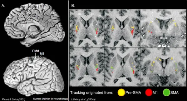
Figure 2 : Anatomy
A. Figure adapted from Picard & Strick (2001) nicely demonstrates the boarders of the different cortical motor areas of the lateral and medial wall of the brain. B. Figure adapted from Léhéricy et al., (2004a) presents the cortico-striatal tracts using diffusion tensor imaging
for an individual subject. Tracks originating from M1 and SMA were directed to the posterior part of the putamen. Tracks originating from the Pre-SMA were located rostral to SMA and
M1 tracks.
Although the activity of the putamen persists in the late learning phase, not all regions of the brain initially active remain so. Instead, dynamic changes take place over time (see Dayan and Cohen, 2011 for a review). An already learned sequence (be it a couple of hours or days of practice) is associated with activity in the putamen, SMA (Jenkins et al., 1994, Doyon et al., 2002), M1 (Karni et al., 1995, Floyer-Lea and Matthews, 2005, Steele and Penhune, 2010) and the dentate nuclei of the cerebellum (Doyon et al., 2002). The inferior parietal cortex was also involved both in early learning and late learning phases, sometimes as increased or decreased activation, suggesting the activity of that region to be more sensitive to the specific type of protocol used. For example the parietal cortex has been associated with explicit awareness of the sequence (Grafton & IVry 1995), and with a more abstract representation of the sequence, i.e. involved in the goal of the action rather than the specific movements of the sequence (Grafton et al., 1998). In addition to these motor related
regions, the hippocampus is also increasingly found to be involved in motor sequence learning (Schendan et al., 2003, Gheysen et al., 2010, Rose et al., 2011, Albouy et al., 2012a) and is thought to interact with the striatum during the early learning phase (Albouy et al., 2008). Note that the dorsal prefrontal cortex does not seem to be active in the late learning phase. This would support the idea that learning has become more automatic and that less monitoring is needed to execute the task. Others have segregated different learning components from initial learning of a sequence until the 5th day of training. The authors found that activity in the putamen, together with the hippocampus, was specifically related to the improvement in accuracy of a complex sequence; as opposed to the timing of the key presses, which was associated with lobule VIII of the cerebellum (Steele and Penhune, 2010, Penhune and Steele, 2012).
Motor adaptation learning
Learning-related modulation of brain activity has also been observed during motor adaptation. Based on studies in animals and patients, the cerebellum is now thought to be a key structure in this type of motor learning. Indeed, patients with Parkinson’s and Huntington’s diseases who suffer from damage to the cortico-striatal network, but who have an intact cerebellum, show no impairment in visuomotor adaptation learning, as tested with mirror-tracing task or prism adaptation (Agostino et al., 1996, Gabrieli et al., 1997). Yet, patients with damage to the cerebellum have difficulty or are impaired at learning such types of skills (Martin et al., 1996). Besides, in the primates, it was found that the discharge of purkinje cells of the cerebellar cortex conveyed information of both the beginning of a reaching movement, as well as the relative error at the end of the movement (Kitazawa et al., 1998). Thus these results suggest that the cerebellum is a good candidate structure for allowing the acquisition of an internal model of the body as one learns a new tool (Imamizu et al., 2000, Penhune and Steele, 2012).
The fast learning phase of motor adaptation has been studied in humans and has been associated with activity in S1, the contralateral putamen, thalamus, medial occipital gyrus and the dorsolateral PFC at the beginning of the training (Shadmehr and Holcomb, 1997).
Yet, the specific visuomotor transformation ability seems to be associated with activity in the ipsilateral posterior (Ghilardi et al., 2000, Krakauer et al., 2004) and inferior parietal regions (Ghilardi et al., 2000), as well as with the right ventral premotor cortex and lateral cerebellum (Krakauer et al., 2004). The lateral cerebellum would be responsible for learning the prediction of visual sensory consequences of the motor command executed in order to adapt to the environmental change (Miall et al., 2007, Izawa et al., 2012). Interestingly, 5.5 hours following initial learning of a motor adaptation task, the recall is associated with activity in the contralateral dorsal premotor and posterior parietal cortex, as well as the ipsilateral anterior cerebellar cortex (lobule VI). The latter pattern of results came along with a decrease of activity in the PFC and the putamen (Shadmehr and Holcomb, 1997). These results were suggested to represent a shift in the representation of the internal model developed for the skill. The authors put forward the importance of the cerebellar cortex (lobule VI) for the storage and maintenance of the motor memory. Yet it is also suggested that the cerebellum is part of a more extended motor network which would serve to store the representation of the learned skill (Penhune and Steele, 2012).
1.2.4.
Motor Learning models
Based on the numerous functional neuroimaging studies described above and in line with pre-existing neuroanatomical models demonstrating the existence of dinstict cortico-striato-cortical and cortico-cerebello-cortico-striato-cortical loops (Alexander and Crutcher, 1990, see Middleton and Strick, 2000 for a review), different theoretical models have been proposed.
Doyon and colleagues (Doyon et al., 2002, Doyon and Benali, 2005b) have proposed that representational changes within the cortico-striatal and cortico-cerebellar systems depend not only on the stage of learning, but also on the type of motor task involved, i.e. whether subjects are required to learn a new sequence of movements or to adapt to environmental perturbations (see Figure 3). They proposed that in the fast (early) learning phase, both motor sequence and motor adaptation tasks recruit similar cerebral
structures: the striatum, cerebellum, motor cortical regions (e.g., premotor cortex, SMA, pre-SMA, anterior cingulate), as well as prefrontal and parietal areas. During this phase, dynamic interactions between these structures are thought to be critical for establishing the motor routines necessary to learn the skilled motor behavior. When a task is well learned, however, the subject has achieved asymptotic performance and the execution has become more automatic. The neural representation of the new motor skill is then believed to be distributed in a network of structures that involves the cortico-cerebellar or the cortico-striatal circuit for motor adaptation and motor sequence learning respectively. Indeed, Doyon and Ungerleider (2002) suggested that for motor adaptation, at this stage, the striatum is no longer necessary for the retention and execution of the acquired skill; regions representing the skill are now involving the cerebellum and related cortical regions. By contrast, a reverse pattern of plasticity is thought to occur in motor sequence learning, such that with extended practice, the cerebellum is no longer essential, and the long-lasting retention of the skill is now believed to involve representational changes in the striatum and associated motor cortical regions.
Other models have focused on the respective role of the two cortico-striatal and cortico-cerebellar loops, based on findings from motor sequence learning studies as well as from other cognitive domains. Doya (1999) has developed learning algorithms which will not be described in details in the present thesis, but will be briefly summarised. This model suggests that the cerebellum is responsible for “supervised learning”, namely by correcting erroneous reaching motor commands. This error-based learning allowing the construction of
an internal model of the body and the environment, hence improves performance of motor control. The role of the cerebellum in MSL specifically, however, is not specified. The basal ganglia would be responsible for “reinforcement learning”, which is based on reward. Doya suggests that the basal ganglia, by means of the dopaminergic neurons, encodes and learn present rewards in order to predict future rewards, making it possible to eventually select the motor action with the highest expected reward. He proposes that in the fast learning phase, the sequence is represented at the cerebral level with visuospatial coordinates i.e. the spatial location of the elements of the sequence are learned. With practice, there is a switch to the use of motor coordinates in the slow learning phase of the learned sequence. This switch would take place from the PFC, preSMA and the anterior striatum to SMA and the body of the striatum (Doya, 2000).
Similarly, Hikosaka et al. (1999) have proposed that spatial and motor coordinates of the sequence are processed in parallel. As above, the spatial information conveys the visual location of the target, while the motor coordinates are the actual movements executed to reach the target. The authors further suggested that distinct cortico-subcortical loops are responsible for these two types of information processes. The loop responsible for the spatial aspect of the sequence would comprise association areas such as the PFC and the anterior portion of the basal ganglia (especially the head of the caudate), while the loop responsible for the motor aspect of the sequence would correspond to the premotor cortex (especially SMA), the putamen and the dentate nuclei of Cerebellum. As Doya suggested (1999), Hikosaka also proposes that the acquisition of the spatial aspect of the sequence would take place earlier than the motor property of the sequence.
1.2.5.
Connectivity Changes in Motor Skill Learning
When using conventional activation detection types of analysis, one often looks at functional segregation. Doing so, the goal is to target a particular area of the brain associated with aspecific cognitive process. Yet, analyses of neural activity that are based on functional specialization provide only a limited account of the neuronal substrate of the process investigated (Lee et al., 2003). An alternative and complementary approach is to investigate the integration of functionally specialised areas via functional connectivity, namely by quantifying the interaction amongst different brain areas. Changes in brain functioning reflected by different patterns of interactions between areas of the brain, are mediated by
functional or effective connectivity (Friston 1994, Friston, 2011). Both of these approaches
are briefly described below.
Functional Connectivity
What is Functional Connectivity
Functional connectivity is a great theoretical and methodological approach for measuring spatial relationship between brain regions as well as their temporal correlation. Functional connectivity between two regions is defined as the temporal correlation of a neurophysiological index, usually the extracted time course, measured between different brain areas (Friston et al., 1993a). Thus, this method allows to detect which areas of the brain are activated at the same time across the experimental session. These analyses are generally multivariate, such that the analysis of a voxel takes into account the activity of the nearest voxel. This is in opposition to standard activation detection type of fMRI analysis looking for the most active area of the brain using univariate analysis, namely analysing each voxel separately, as if they were independent. In functional connectivity, the assumption is that if the time courses of two brain regions covary, these two brain regions are most possibly exchanging information. Thus, an increase in functional connectivity between these structures would suggest that brain regions are interacting in a more synchronous and integrated fashion. Greater integration within a network thus means that the brain regions forming that network work together with more cohesion. Two major approaches have been used to study functional connectivity. One is hypothesis driven and the other is data-driven. In the former, one can explore the connectivity changes from one specific region of interest, with other distant areas (psychophysiological interaction, PPI) (Friston et al., 1997) or use multiple seeds to form a network. In that case, one determines a
task- specific set of regions of interest that are known to be involved in motor learning before analysing the data. In the data-driven approach, the networks are formed based on mathematical as opposed to functionally relevant criteria. In that case, no functional constraints are applied to the data and it has the advantage that no a priori knowledge is necessary; i.e. one does not need to choose specific seeds (brain regions) prior to the analysis. A data-driven mathematical approach that has been used extensively in the past years is the independent component analysis (ICA). Briefly, ICA is a “blind source separation” algorithm that decomposes the registered signal into spatial and temporal components (networks), which are statistically independent from each other (McKeown et al., 1998, see also Boly et al., 2008). This means that the activity of one spatial map (component) cannot predict the activity of another given spatial map. In that way, the relevant maps related to brain activity, are automatically separated from noise into different components. The investigator then chooses the components he is willing to discuss or use for further analysis.
There has been an emergence of studies in which researchers examined functional connectivity patterns, generally with the use of ICA, with participants remaining simply at rest in the MRI. These studies are said to explore “resting-state” because no specific cognitive task is executed. Studying resting-state gave rise to the awareness that spontaneous fluctuations in the brain are organised in a functionally coherent manner (for a review, see Fox and Raichle, 2007). These systems generally include a motor network, visual network, fronto-parietal, executive, ventral attentional networks and the default-mode, one that systematically decreases as one engages in an externally driven task (Beckmann et al., 2005, Damoiseaux et al., 2006) (see Figure 4).
These so-called resting state functional networks are, however, also known to be stable during active sate, i.e. during execution of a task (Calhoun et al., 2008, Smith et al., 2009) in addition to being stable across rest periods. Particularly, the default mode has been shown to remain during light sleep (Horovitz et al., 2008), non-rapid eye movement (NREM) sleep
(Dang-Vu et al., 2008) and coma (Boly et al., 2008). Apart from this impressive stability across studies, the degree of connectivity within networks, however, seems to vary with age (Damoiseaux et al., 2008), neurodegenerative diseases (Filippini et al., 2009, Wu et al., 2009) and interestingly so, with learning (Albert et al., 2009b, Lewis et al., 2009b).
Figure 4 : Resting-state networks
Figure adapted from Beckmann et al., (2005) demonstrating the different resting state networks found using principal independent component analysis. A. medial visual cortical
areas. B. Lateral visual cortical areas. C. Auditory system. D. sensory-motor system. E. Visuo-spatial system F. Executive control. G. and H. Dorsal visual stream each one
lateralized
Functional connectivity with motor sequence learning
With the availability of the different motor learning models, as well as the studies characterizing the neural correlates of motor learning, methodological tools using functional connectivity allowed to better understand the brain plasticity taking place with MSL. Some studies using standard activation detection types of analysis also used PPI as a way to quantify changes in connectivity between specific ROIs. It is suggested that across multiple
days of MSL, connectivity between M1 and the cerebellum lobule VII-VIII increases as synchronization of performance on the sequence also increases. During training, the hippocampus also shows competitive interaction (negative correlation) with the putamen (Albouy et al., 2008) or caudate nucleus (Albouy et al., 2013b), which, in the latter case predicted gains in performance following a night of sleep. Quantifying connectivity changes within or between networks, as opposed to between one specific brain region and the rest of the brain, has the advantage of uncovering brain communication at the systemic level. For example, Sun et al., (2007) demonstrated that when practicing a new sequence, greater inter- and intra-hemispheric integration takes place within a motor network during the early as compared to the late learning phases of learning (Sun et al., 2007). The results also revealed greater connectivity between frontal and motor cortical regions for the early vs. late phases of learning. In contrast, when executing an already learned sequence of movements, no change in functional connectivity was observed across the session. These results thus suggested enhanced inter-hemispheric coupling within a motor network only during the early stages of learning. Another study using a similar model free approach with motor sequence vs random trials, found that two independent components correlated with the task (Tamas et al., 2008). One network, comprising fronto-parietal and cerebellar regions, correlated with both types of trials; whereas a second network, including posterior parietal and premotor regions, was exclusively present during the sequence trials. Moreover activity in the latter network correlated with the amount of learning across the session. These results demonstrated how a data-driven approach was successful in identifying a motor network specific to sequence learning, as opposed to random trials, during a first learning phase. Furthermore, it suggests that premotor and posterior parietal brain regions interact with each other during early sequence learning.
Others have explored the dynamic changes occurring with motor sequence learning using a hypothesis-driven approach. Coynel et al., 2010 selected multiple seeds, based on previous fMRI results (Lehericy et al., 2005), to form a sensorimotor and an associative motor network, aiming to measure the interaction between these two motor systems as sequence
learning progresses. The findings first revealed greater overall integration, i.e. greater cohesion, within and between networks during the first learning episode of a new sequence as compared to an overlearned and automatized one. Second, across 28 days of training, an overlearned sequence was associated with a lower level of integration, mainly because of a decrease in integration within the associative network. A relatively high level of integration between the two motor systems remained with learning (Coynel et al., 2010). These results thus suggest that with learning, there is a decrease of integration between higher order brain regions, which are part of the associative network; yet the interaction between this system and the sensorimotor network remains necessary.
In sum, it seems that during initial training sessions on motor sequence learning, there is a global increase or maintenance in connectivity observable between motor brain regions. This is followed, after multiple training sessions, by a decreased connectivity within a network comprising higher order brain regions such as the premotor cortex, Pre-SMA and the parietal cortex (Coynel et al., 2010).
Few studies examined functional connectivity changes with long term motor adaptation learning. In one of them, the authors used regions of the brain for which activity changed across learning, as seeds for their functional connectivity analysis (Della-Maggiore and McIntosh, 2005). They reported an increase in functional connectivity after 7 days of practice on a kinematic motor adaptation task between the bilateral anterior cerebellum, left (contralateral) middle temporal gyrus, cingulate gyrus and right putamen. Dynamic cerebral changes associated with motor adaptation was also studied using the initial post-learning rest period. Using a seed based approach; Vahdat et al (2011) dissociated changes in connectivity associated with the perceptual vs. motor aspect of motor adaptation. Changes associated with the perceptual function included increased connectivity between the second sensory cortex and frontal motor areas (PMv, SMA) as well as between the prefrontal cortex, cerebellar anterior cortex (lobule VI) and superior parietal lobule. With motor learning
specifically, they found increased connectivity between cerebellar cortex adjacent to posterior-superior fissure (lobule VI, Crus I) and left M1 and SMA, as well as between cerebellar cortex and the superior parietal lobule (Vahdat et al., 2011). Increased connectivity was also observed in the post-learning resting state period, using a data-driven approach (Albert et al., 2009b). An experimental group was assigned to a visuomotor adaptation tracking task and a control group was assigned to the same task with no rotations. They found increased connectivity strength in the frontoparietal network only in the group that learned the motor adaptation, while no change in connectivity was apparent in the control group. Furthermore, a cerebellar network, which was only present in the experimental group, also showed increased connectivity following learning. These results suggest that changes in resting state activity were induced by MA learning. Furthermore, they propose that increased connectivity in a fronto-parietal circuit as well as the cerebellum might reflect the on-going “off-line” processing of information gained from earlier learning. These results bring insights to a hypothesis suggesting that these intrinsic networks observable at rest might contribute to the off-line processing and consolidation of memory (Miall and Robertson, 2006, Albert et al., 2009a).
Effective connectivity
Another mathematical approach used in fMRI studies is effective connectivity, which allows to examine the actual influence a neuronal system exerts over another. Two major methods are often used: Dynamic causal modelling (DCM) and Structural equation modelling (SEM) (Friston, 1994). The major advantage of these methods is that in contrast to correlational relationship between two brain regions, as measured in functional connectivity, these methods offer causal relationship. Yet, these types of approaches are also limited by the need of specific and strong functional and anatomical a priori knowledge. Furthermore, the consequence is that the findings will highly depend on the definition of the model (e.g. the choice of ROIs) chosen.
Some authors have explored effective connectivity during the course of MSL while looking at 6 regions of interest: M1, cerebellum, dorsal premotor cortex, basal ganglia, SMA, and the
PFC. The inter-regional connectivity were measured from day 1, to 2 weeks and 4 weeks of motor learning. Importantly, the findings revealed that connectivity from the cerebellum to M1 decreased across training, while connectivity from the BG to M1 increased. This is in accord with Doyon et al., 2002’s model which predicts less involvement of the CB (but more of the BG) as the sequence becomes more automatic. Other interesting findings included a gradual reduction of the connection from PFC to M1, as well as strengthening of the connections from the BG to SMA and from SMA to the premotor cortex. These results were interpreted as being the reflection of a decreased need for attentional resources, an increased effectiveness in sequence control and in motor planning respectively (Ma et al., 2010).
The implication of an extended cortico-striatal network has also been studied during initial learning of a motor adaptation task. The findings suggest increases in effective connectivity as learning progresses, between the calcarine fissure and the middle temporal gyrus; and from there to the anterior striatum and the dorsal precentral gyrus. Afferents from the inferior frontal sulcus to the anterior striatum also showed increased connectivity with learning. In contrast, the authors reported significant decreases between the frontal cortical regions (Toni et al., 2002). These findings corroborate previously described imaging studies suggesting a role of the striatum in early learning of associative visuomotor learning. Yet, the findings are limited by the approach used (effective connectivity) in the sense that only the regions included by the authors in the model can be discussed. For example, it would have been interesting to explore also the regions of the cerebellum and compare its involvement at this point in learning with other motor brain regions.
These changes in the recruitment of the motor network, in terms of activation or connectivity, are thought to support learning. Yet in order to better understand long-term retention and learning, one has to understand the process of consolidation.
1.3. Memory Consolidation
For memories to be stored, be it declarative or procedural, consolidation is required. Consolidation is a process of brain plasticity whereby a fragile memory trace becomes more robust and less susceptible to interference. It was primarily noticed with declarative memory and a first theory was brought up by Müller & Pilzecker (1900) who observed that the ability to recall recently acquired verbal information deteriorated as a function of the interpolation of other tasks (John, 1967). Time-dependent effects of consolidation were also observed with the introduction of electroconvulsive shock (ECS) used to treat depression. It was demonstrated that ECS abolishes retention of a list of words paired associate learned prior to therapy, and that the severity of impairments was an inverse function of the time lapse between initial learning and ECS (Zubin and Barrea, 1941; as cited in John, 1967). These studies revealed that after a certain laps of time, the memory is stored and less susceptible to interference, i.e. consolidated. How that process occurred was unknown, but today the mechanisms responsible for consolidation can be viewed at a cellular level as well as at a system’s level (Dudai, 2004).
1.3.1.
Levels of Consolidation
Synaptic Consolidation
Consolidation at the cellular level (synaptic consolidation) can be seen in two forms: short-term and long short-term. In the short run, the memory is thought to be formed through little stimulation and to remain in a labile state. On the contrary, long term memory is believed to be formed after multiple repetitions of a stimulus, which result in the synthesis of new proteins, as reflected, for example, by synaptic growth or synaptic remodelling (for a review, see Kandel and Squire, 2000, Dudai, 2004). This process occurs mostly at the local level, i.e. in the same structure that was used for the encoding of the memory.
Systemic Consolidation
Consolidation at the systems level comes from the indication, among others, that H.M could no longer consolidate new declarative memories, yet he still had some memories from his
childhood, indicating that the memory once encoded in the hippocampus, must have “migrated” somewhere else in the cortex. Thus the first model of system consolidation involved the hippocampal formation. According to this view, the stabilization of the memory trace is assumed to involve synaptic consolidation locally, achieved in minutes to hours. In parallel, or as a consequence of it, the process of system consolidation is initiated and is characterized by a shift of the representation of the information retained, from the medial temporal lobe to the neocortex (McClelland et al., 1995, Dudai, 2002, 2004). Although it is not clear how the change in neural representation occurs, the memory transformation theory emphasises the dynamic nature of the memory trace (Winocur and Moscovitch, 2011). The authors suggest that the hippocampus is the storing site of a memory as long as it is context-dependent. For each memory retrieval, a new trace is added and serves to reinforce and strengthen the memory. With time, an abstract representation of the memory is formed (schema) and is thought to be represented neocortically.
In the procedural memory domain, it has been shown that functional reorganization, particularly a shift in the motor representation, takes place among a motor cortical and subcortical network (Karni, 1995, Ungerleider et al., 2002, Doyon et al., 2003, Doyon and Benali, 2005b, Floyer-Lea and Matthews, 2005, Lehericy et al., 2005), but as noted by Dudai (2004), the protocol used in the majority of the studies involve learning across time, thus changes observed across the multiple testing sessions could be the reflection of practice, and not necessarily consolidation per se. Dissociating the learning process from the consolidation process, if possible, is quite a challenge as they certainly co-occur. Nevertheless, as it will be discussed in the next sections, it is known that consolidation continues offline following an initial training session, without further practice. This offline period leads sometimes to the stabilisation of memory, which reduces its susceptibility to interference. Alternatively, consolidation is reflected by offline gains in performance observable in a subsequent training session (see Dayan and Cohen, 2011, Censor et al., 2012).
1.3.2.
Memory Consolidation and Sleep
Sleep is an active process despite the absence of consciousness and multiple studies have linked sleep to memory. Indeed, it has been shown that sleep deprivation affects behavior, that post-learning sleep architecture changes following learning and that one can observe behavioral benefits following post-learning sleep, as opposed to post-learning wakefulness (Maquet, 2001, Peigneux et al., 2001). There is also ample work showing that sleep facilitates the consolidation and long-term retention of new memories (see Diekelmann et al., 2009 for a review). Yet, the involvement of sleep, and the specific sleep characteristics found to optimize the memory consolidation process, is not yet well understood because it is thought to depend on multiple factors (see Figure 5); for example the type of learning and material used.
Figure 5 : Factors influencing sleep-dependent memory consolidation – from Diecklman et al., (2009)
Both declarative and procedural memories have been shown to benefit from sleep. In declarative memory, for example, a better retention of pairs of words at cued recall has been found after sleep as opposed to the same time spent awake. Similar gains in being able to recall greater information following sleep has been found with the learning of object
locations, short stories and wordlists (see Diekelmann et al., 2009). More recently it has been suggested that the particular role of sleep could be in the form of a triage of relevant vs. irrelevant information to be retained. Namely, sleep would produce specific enhancement of memory for declarative information that was cued to be remembered, but not for others cued to be forgotten (see Stickgold and Walker, 2013). In procedural memory, enhanced performance on different adaptations of the serial reaction time task, finger tapping task and visual discrimination tasks has been observed specifically following sleep.
1.3.3.
Motor Memory Consolidation
Generally speaking, consolidation is thought to occur between the fast and slow learning phases of motor learning and to last several hours (Karni and Bertini, 1997). Several investigators have reported that sleep plays a critical role in the consolidation of motor sequence learning in particular (Karni et al., 1998, Fischer et al., 2002, Walker et al., 2002). The important concept being that time, and sometimes sleep, is needed for the memory to become “fixed” and eventually resistant to interference, even in the long term after several weeks without practicing (Nezafat et al., 2001, Penhune and Doyon, 2002, Della-Maggiore and McIntosh, 2005). Nevertheless it is now acknowledged that consolidation is not a one-time process and that reactivation of the memory trace might render it de novo labile, and again susceptible to interference until reconsolidation of the trace has occurred (Nader et al., 2000, Nader, 2003, Marin et al., 2010), findings that have inspired Hollywood movies like “Eternal Sunshine of a Spotless Mind”.
Paradigms for testing Motor Memory Consolidation
In the field of skill learning, there are 2 ways that are often used to test for the presence of consolidation at a behavioural level. The first type of protocol described here is called “interference”, in which subjects are tested using an A1-B-A2 paradigm, where A is the first task learned and B is a similar, but different task. Retrograde interference can be measured by looking at the disruptive effect of task B on consolidation of A (i.e., the motor task
originally learned) by manipulating the time interval between A1 and B. Consolidation processes are measured by retesting performance of A (A2) after the interfering episode. Using this procedure, it is possible to identify the critical time window that enables (or not) consolidation to take place. Differently, anterograde interference can be examined through the effect of A on the learning of the interfering task B. Although this type of protocol has been used to measure consolidation in motor sequence learning (Korman et al., 2007) it has more often been applied to measure consolidation of adaptation learning (see Shadmehr and Holcomb, 1997, Caithness et al., 2004, Krakauer et al., 2005). A second type of protocol to test for the occurrence of consolidation is to use a parametric (test-retest) design. This is done by using multiple testing sessions separated by different laps of time, while quantifying differences in behavioural performance as time goes by. This paradigm has often been used to test for the role of sleep, this so with one of two approaches: 1) the effect of post-training sleep deprivation vs. regular sleep or (2) the effects of sleep vs. the simple passage of daytime on the expression of memory consolidation by measuring performance on a delayed retest session. Using a test-retest paradigm, two phenomena have been observed when sufficient time is given for consolidation to occur: savings, defined as a faster rate of relearning at retest (mostly seen in motor adaptation tasks), and gains in performance, described as a sudden increase in performance, following a latent period of time without further practice.
Consolidation as reflected at the behavioral level
In a pioneering study, Walker et al. (2002) used a computerized version of the finger opposition task to demonstrate that spontaneous gains in performance occurred specifically after sleep, but not after the simple passage of time. There were 17-20% improvements in speed after a night of sleep (with no changes in accuracy), but only 2% improvement after an equivalent latent period during daytime (see Figure 6). Interestingly, they verified that the absence of gain during daytime was not due to interference, as subjects were asked to wear mittens during the whole day, hence minimizing hand movements during the day. Complementary findings from Fischer et al. (2002) revealed that gains in performance are
similar after both night-sleep and day-sleep, on both speed and accuracy measures, suggesting no significant circadian, or time of day, influence on the consolidation of motor sequences. These first studies thus suggest an important role of sleep in the off-line consolidation process of motor sequence learning, which is reflected as off-line gains in performance.
Figure 6 : Sleep-dependent consolidation at the behavioral level
Figure adapted from Walker et al., (2002). The Y axis represents the number of sequences executed within a 30 second period. The graph on the left demonstrates that when starting
the training in the morning, only a period of sleep allows significant improvements in performance. The graph on the right demonstrates the same phenomenon but having
participants trained in the evening.
Since then, our group has replicated the findings suggesting a benefit of sleep specifically for MSL (Doyon et al., 2009b). Furthermore it is now known that daytime naps are also sufficient to elicit similar gains in performance (Nishida and Walker, 2007). Yet, different factors are found to influence the contribution of sleep to consolidation. For example, the benefits of sleep are thought to be more important if the sequence is more complex (Kuriyama et al., 2004) or if the subject is a fast learner as opposed to a slow one (Albouy et al., 2008). Expectation to be retested is also known to favor greater sleep-dependent consolidation. Indeed, a group that learned the MSL task while expecting to be retested the following morning, performed better than a group which did not expect to be retested (Wilhelm et al., 2011).
Yet, post-training sleep does not appear to be necessary for the consolidation of all types of motor sequences, as daytime alone is sufficient to elicit off-line gains in performance when learning occurs implicitly, i.e. without being aware of the sequence (Robertson et al., 2004). These results suggest that consolidation of implicit and explicit learning of MSL seem to rely on distinct mechanisms. Yet, it is also possible to segregate MSL based on the different memory processes involved, as opposed to a segregation based on the level of consciousness (Cohen et al., 2005). Findings reveal that the spatial representation of the sequence, namely what is effector independent, is the specific process which consolidates during sleep. In opposition, the effector-dependent or motoric representation aspect of the learned sequence requires only time for consolidation to occur (Cohen et al., 2005, Witt et al., 2010, Albouy et al., 2013a).
As mentioned earlier, some researchers have explored consolidation as reflected by a resistance to interference. In these studies, consolidation is also often expressed as stabilisation, before giving rise to gains in performance (enhancement). This has been shown when a learned sequence is no longer susceptible to interference. Korman and colleagues (2007) have demonstrated that training on a second interfering sequence at 2 but not 8 hours, following the initial learning, did prevent the expression of delayed gains observed at 24-hours after training (Korman et al., 2007). These results suggested that 8 hours were necessary to stabilise the memory trace for it to undergo sleep-dependent consolidation. Similarly, Walker et al., (2003) also suggested that if a second sequence (B) was learned right after the original one (A), retest the following day showed significant gains in performance speed, but not accuracy. In contrast, when the B sequence was learned 6 hours after A, post-sleep gains were observed in both speed and accuracy measures, suggesting that 6 hours were sufficient to stabilise the memory. Yet, it is not clear why the interference was only reflected on the accuracy measure as this characteristic has not consistently been shown to change with sleep (Walker et al., 2002, Morin et al., 2008, Doyon et al., 2009b).
The effect of interference on motor adaptation is also very well documented (Brashers-Krug et al., 1996, Shadmehr and Holcomb, 1997, Krakauer et al., 2000, Krakauer et al., 2005, Krakauer and Shadmehr, 2006) (but see Caithness et al., 2004). The results of these studies suggest that, when using an ABA paradigm, learning task B prevents savings (a faster relearning rate) of task A if learned after 5 minutes, but not 24 hours (Krakauer et al., 2005). Similarly, others have shown that if a task B is learned right after A, no gains in performance are apparent the following day, whereas if 4 hours is allowed between A and B, the participants demonstrate better performance on the retest of A the following day (Brashers-Krug et al., 1996). These results thus suggest that 4 hours would be sufficient for the memory trace to be consolidated, as shown by no susceptibility to interference.
Importantly, because savings or gains in performance have been observed multiple times following 4-6 hours of daytime, the off-line consolidation of motor adaptation does not seem to be dependent on sleep. Yet Huber and colleagues have found otherwise. They used a visuomotor adaptation task where subjects learned anti-clockwise rotations of 15 degrees, then 30, 45 and 60 degrees. When two groups were retested after a night of sleep or the passage of daytime, increase on the accuracy measure was only observed after a night of sleep (Huber et al., 2004). Yet, more recently Doyon and colleagues have corroborated studies suggesting no specific effect of sleep (Doyon et al., 2009). They used 3 groups of participants executing the MA task: the first one napped during daytime, the second one slept during night time and the last one remained awake during daytime. The results between the test and retest sessions revealed no interaction between the groups, but significant delayed gains in performance in all groups, suggesting that sleep did not play a significant role. The apparent discrepancy in the results might be due to methodological differences. For example, motor adaptation in Doyon et al’s study was assessed using a joystick and not a robotic arm, and only one single rotation was learned (180°), as opposed to Huber’s study in which multiple rotations were learned. In addition, the behavioral measures used in these two studies were different, which could also account for the divergence of the results. Indeed, Albouy et al. (2012) examined the effect of sleep on 7
different behavioral parameters. They found a protective effect of sleep, as reflected by a stabilisation of performance on most behavioral measures, except for the directional error in reaching a target. The error in the trajectory executed was found to decrease following both sleep and the simple passage of time, suggesting that time was sufficient for at least some consolidation to occur. Thus the role of daytime vs. sleep in the consolidation process for motor adaptation certainly differs from the one of motor sequence learning. The differential pattern of results between the two types of motor learning points out to the heterogeneity of the effect of sleep on motor memory consolidation. For this reason it is an opportunity to compare, using the same protocol, the effect of sleep vs. the simple passage of day time on the consolidation of both of these types of learning.
In order to better appreciate neuroscientific findings on the neural correlates associated with the behavioral changes described above, I will first describe some of the basic characteristics of sleep and the mechanisms susceptible to play a role in consolidation.
1.4. Sleep: Influences on the Consolidation Process
1.4.1.
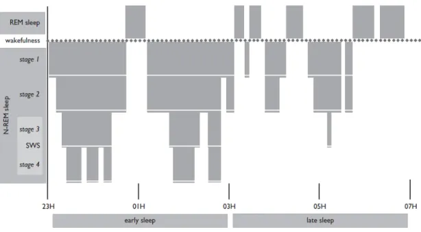
Sleep Architecture
Sleep can be divided into two states, REM and NREM (Figure 7). NREM sleep comprises 4 stages characterized by different levels of neural synchronization, from lower synchronization in lighter sleep stages (1 and 2) to higher synchronization in deeper stages 3 and 4 (slow-wave sleep; SWS). Multiple electroencephalogram (EEG) rhythms define NREM sleep: the transition of alpha waves, present during wakefulness (8 to 13 Hz), to theta waves (4-7 Hz) in stage 1; sleep spindles (12-15 Hz) and “K-complex” in stage 2; and finally SWS is characterized by delta waves (1-4 Hz), that are prominent in stages 3 and 4. REM sleep, by contrast, is characterized by EEG activation that is unsynchronized, closer to what is found during wake states. Muscle atonia and episodic burst of rapid eye movements are also characteristics of REM sleep. Although the organisation of these stages is quite stable across individuals and across one night of sleep, SWS sleep is more prominent at the