Tissue-specific induction of ADAMTS2 in monocytes and macrophages by
glucocorticoids
Thomas P. J. Hofer1,*, Marion Frankenberger1, Jörg Mages2, Roland Lang2, Reinhard Hoffmann2, Alain Colige3,
Löms Ziegler-Heitbrock1,4,†
1 Clinical Cooperation Group Inflammatory Lung Diseases (GSF National Research Center for Environment and Health and Asklepios Fachkliniken München-Gauting), Robert-Koch-Allee 29, D-82131 Gauting, Germany
2 Institute for Medical Microbiology, Immunology, and Hygiene. Technical University of Munich, Munich, Germany 3 Laboratory of Connective Tissues Biology, CBIG Research Center, University of Liège, Liège, Belgium
4 Department of Infection, Immunity and Inflammation, University of Leicester, Leicester, UK
Abstract
The regulated expression of ADAMTS2 (a disintegrin and metalloproteinase with thrombospondin motifs), a secreted metalloproteinase involved in the processing of procollagen to collagen, was studied in peripheral blood mononuclear cells (PBMC). Stimulation with glucocorticoids (GC) resulted in a pronounced dose-and time-dependent increase of ADAMTS2 mRNA levels in PBMC. The increase of ADAMTS2 expression was specific for CD14++ monocytes (440-fold) and alveolar macrophages (200-fold), whereas CD3+ (T lymphocytes), phytohemagglutinin-activated CD3+ (T lymphocytes), and CD19+ (B lymphocytes) showed no significant changes in ADAMTS2 mRNA after GC treatment. Treatment of monocyte-derived macrophages (MDM) with GC also resulted in an increase of ADAMTS2 protein in the culture tissue media. Using the GC analog RU486, mediated induction of ADAMTS2 mRNA was blocked, implicating that GC acts specifically via the GC-receptor. In agreement with findings in blood monocytes, cell lines of the monocytic lineage (MM6, THP-1) showed significant GC-induced significant increases in ADAMTS2 mRNA, while in epithelial cells (A549, Calu-3, Colo320, BT-20) and fibroblast (MRC-5, WI-38, and two NHDF-c cell types from adult cheek and upper arm), they showed no or little responsiveness to GC. As macrophages have important functions in immune defense and tissue homeostasis, these findings suggest that GC-mediated specific induction of ADAMTS2 in these cells may play a crucial role in the resolution of inflammation and wound repair.
Keywords : ADAMTS2 ; Glucocorticoids ; Inflammation ; Monocytes ; Macrophages ; Mono Mac6 Abbreviations
ADAMTS2 A disintegrin and metalloproteinase with thrombospondin motifs-2
MM6 Mono Mac 6 cell line
MDM monocyte-derived macrophages
MP methylprednisolone
PBMC peripheral blood mononuclear cells h hour
GC glucocorticoid
GCR glucocorticoid receptor
* THOMAS P. J. HOFER received his PhD in cell biology at the Technical University of Munich, Germany. He is presently a postdoctoral
fellow with the U.S. Environmental Protection Agency Human Studies Division, through an appointment with the Center for Environmental Medicine, Asthma, and Lung Biology at the University of North Carolina at Chapel Hill. His research interests include the pathogenesis of COPD and the molecular mechanisms of particulate matter-induced injury in the lung.
† LOEMS ZIEGLER-HEITBROCK trained as a physician at the University of Hamburg, Germany and there received his MD degree in molecular
biology of leuke-mias. Until 2006, he has been Chair in Clinical Immunology at the University of Leicester, UK and now is Head of Clinical Trials at the GSF-National Research Center for Environment and Health. His research interest revolves around monocytes and macrophages, monocyte subpo-pulations in human blood, and their gene expression.
Introduction
The major functions of monocytic cells within the immune system are to regulate local inflammatory reactions by releasing pro- and anti-inflammatory molecules, to support a primary defense mechanism via phagocytosis and respiratory burst, and to mediate immune responses by antigen-processing, antigen-presentation, and cytokine production [1]. Based on these properties, macrophages play a key role in inflammation and immune activation. In addition, macrophages are central to tissue homeostasis in that they are involved in wound repair and tissue remodeling, secreting critical mediators like TGFβ and metalloproteinases [2, 3], The properties of the macrophages are strongly influenced by GC. The effects of GC on immunological functions involves potent anti-inflammatory and immunosuppressive properties [4]. The anti-anti-inflammatory effect of GC is mediated through an inhibitory effect on proinflammatory transcription factors such as NF-KB[5], which results in reduced cytokine expression. In cell mediated immunity, reduced IL-2 levels lead to decreased T cell proliferation and activation. Humoral immunity is suppressed by inhibiting IL-2 and IL-2 receptor expression which, in turn, reduces B cell expansion and subsequent antibody production [6]. In macrophages, GC upregulate annexin-1 expression which suppresses phospholipase A2 activity and, thereby, limits the availability of arachidonic acid for the synthesis of eicosanoids like prostaglandin and leukotriene, which are crucial for acute inflammatory responses[7].
Besides these well-known mechanisms, monocytes/macrophages also play a major role in modifying the structure of the surrounding tissue. Recent findings indicate that the action of GC is also mediated by suppression of another pro-inflammatory transcription factor, activator protein-1 (AP-1). Activation of AP-1 induces the transcription of several pro-inflammatory genes, including that of the metalloproteinases MMP-2 and MMP-9. These MMPs hydrolyze extracellular matrix proteins such as collagen, which normally function to localize inflammatory reactions within tissues [8, 9, 10]. Suppression of AP-1 activity by GC reduces expression of MMP-2 and MMP-9 and, thus, may limit the extent of inflammation.
Another important family of enzymes that modify extracellular matrix proteins is the ADAMTS (a disintegrin
and metalloproteinase with thrombospondin motifs) protease family. In humans, this enzyme family is
comprised of 19 members, the domain structure of these enzymes is complex and consists of an N-terminal reprolysin-type pro-metalloproteinase domain attached to a C-terminal ancillary domain, which contains at least one thrombospondin type 1 repeat [11]. After secretion, ADAMTS enzymes undergo processing, including cleavage of the N-terminal signal peptide and pro-domain and of the C-terminal region, which affects substrate specificity and localization of the enzymes [12]. Their known functions include collagen processing (ADAMTS2, 3, 14), cleavage of matrix proteoglycans (ADAMTS4, 5), inhibition of angiogenesis (ADAMTS1, 8), and blood coagulation homoeostasis (ADAMTS13) [11].
ADAMTS2 is a procollagen N-proteinase that cleaves procollagen I, II, and III to the corresponding collagens and is required to generate collagen monomers that are able to assemble into elongated and cylindrical collagen fibrils [13]. Collagen is a major constituent of the extracellular matrix. In humans, mutation in the ADAMTS2 gene causes the dermatosparactic type of Ehlers-Danlos syndrome (previously known as Ehlers-Danlos syndrome type VIIC), a disease mainly characterized by an extreme skin fragility. Besides this, a properly processed collagen is crucial for a functional assembly of the extracellular matrix (ECM), which is, in turn, responsible for ECM-cell interactions necessary not only for providing cells with mechanical stability, but also for physiological events like lineage decisions during embryogenesis, differentiation, cell migration, and wound repair [14].
We show herein that the expression of ADAMTS2, a protease involved in collagen synthesis, is strongly induced by GC in a tissue-specific fashion only for cells of the monocyte/macrophage lineage but marginal in many other cell types including fibroblasts and epithelial cells.
Materials and methods
Donors, isolation of peripheral blood mononuclear cells, and alveolar macrophages
Human PBMC were isolated from heparinized (10 U/ml) blood from healthy human volunteers by density gradient centrifugation (Lymphoprep, 1.077 g/ml, no. 1053980, Axis-Shield PoC AD, Oslo, Norway). Cells were directly used for subsequent isolation of monocytes or cultured under LPS-free conditions. Written informed consent was obtained from each individual. The protocol was approved by the Ethics Committee of the Medical School of the Ludwig-Maximilians-University (Munich, Germany).
with sarcoidosis or fibrosis. After informed consent, lavage was performed during fiberoptic bronchoscopy by instilling 160 ml of 0.9% saline solution in 20 ml aliquots into the lingula or middle lobe and withdrawing the fluid immediately. All patients underwent the lavage procedure for routine diagnostic purpose and leftover material used herein.
Bovine heparinized (10 U/ml) blood from apparently healthy cows was obtained from the animal clinic, department "Innere Medizin und Chirurgie der Wiederkäuer" of the Ludwig-Maximilians-University (Munich-Oberschleissheim, Germany) and PBMC were isolated as mentioned above.
Cells and culture medium
Primary human and bovine cells, the monocytic cell lines Mono Mac 6 [15], THP-1 [16], BT-20 [17], Colo320
[18], and NHDF-c adult from cheek and upper arm (PromoCell, Heidelberg, Germany) were cultured in RPMI
1640 medium (no. F1415, Biochrom, Berlin, Germany), containing OPI (oxaloacetate, pyruvate, insulin; no. O-5003, Sigma, Taufkirchen, Germany), L-glutamine 2 mM (no. 15140-114, Invitrogen, Karlsruhe, Germany), penicillin 200 U/ml/streptomycin 200 µg/ml (no. 15140-114, Invitrogen, Karlsruhe, Germany), and 1× nonessential amino acids (no. 11140-35, Invitrogen, Karlsruhe, Germany). After the addition of supplements, the medium was ultra-filtered through a Gambro 2000 column (Gambro, Hechingen, Germany) to remove any inadvertent LPS contamination, followed by the addition of 10% FCS (no. S0115, Biochrom, Berlin, Germany). The cell lines MRC-5 [19], WI-38 [20], A549 [21], and Calu-3 [22] were cultured in Dulbecco's minimal essential medium NUT mix F12 (no. 21331-020, Invitrogen, Karlsruhe, Germany), containing 10% FCS (no. S0115, Biochrom, Berlin, Germany), L-glutamine 2 mM (no. 15140-114, Invitrogen, Karlsruhe, Germany), and penicillin 200 U/ml/streptomycin 200 µg/ml (no. 15140-114, Invitrogen, Karlsruhe, Germany).
Enrichment of CD14++ monocytes and generation of monocyte-derived macrophages
For cell enrichment, the MACS magnetic separation technique was used (all columns and reagents from Miltenyi Biotec, Bergisch-Gladbach, Germany). For isolation of CD14++ monocytes, PBMC were in a first step depleted of CD16-positive cells. For this, a total of 20 × 10 cells were resuspended in 80 µl of phosphate-buffered saline (PBS) containing 25 µl of Anti-CD 16 microbeads (no. 130-045-701). After incubation for 30 min at 4°C, cells were washed and resuspended in 1.5 ml PBS, and this was loaded onto a LD column (no. 120-000-497) that was positioned in a MidiMACS magnet (no. 130-042-302). Nonadherent cells were recovered and used for enrichment of CD14++ cells. For this, anti-CD14 microbeads (no. 130-050-201) was diluted 1:5 in PBS and added to the cells to a final volume of 100 µl. After incubation for 30 min at 4°C, cells were washed and resuspended in 1.5 ml PBS, and this was loaded onto a LS column (no. 120-000-475). The column was washed five times with 2 ml PBS each. Cells were recovered from the column by pressing 2 ml PBS through the column for five times. CD14++ cells were washed and resuspended in supplemented RPMI 1640 medium (mentioned above).
To determine purity of the CD14++ monocytes, a sample was stained with FITC-labeled anti-CD14 antibody (My4-FITC, no. 6603511, Coulter, Krefeld, Germany) and PE-labeled anti-CD16 antibody (Leu11c-PE no. 332779, Becton-Dickinson, Heidelberg, Germany) and measured by FACS. CD14++ monocytes with a purity of 96% or higher were used.
Enrichment of CD3+ T lymphocytes and CD19+ B lymphocytes
PBMC were used for enrichment of CD3+ T lymphocytes and CD19+ B lymphocytes. For this, either anti-CD3 microbeads (no. 130-050-101) or anti-CD19 microbeads (no. 130-050-301) were diluted 1:5 in PBS and added to 10 × 106 freshly isolated PBMC to a final volume of 100 µl. After incubation for 30 min at 4°C, cells were
washed and resuspended in 1.5 ml PBS and this was loaded onto a LS column (no. 120-000-475). The column was washed five times with 2 ml PBS each. Cells were recovered from the column by pressing 2 ml PBS through the column for five times. CD3+ and CD 19+ cells were washed and resuspended in supplemented RPMI 1640 medium (mentioned above).
To determine purity of the CD3+ and CD19+ lymphocytes, a sample of each was stained with FITC-labeled anti-CD3 antibody (no. 1281, Coulter, Krefeld, Germany), or FITC-labeled anti-CD19 antibody (no. F0768, Dako, Hamburg, Germany), respectively, and measured by FACS. CD3+ and CD19+ lymphocytes with a purity of 96% or higher were used.
Experimental setup
For gene expression analysis, cells were cultured in a final concentration of 2 × 10⁶ in 2 ml culture medium per
well in ultralow attachment tissue plates (no. 3473, Corning Costar, Wiesbaden, Germany) at 37°C and 5% CO2.
For stimulation with methylprednisolone (MP; no. M-0639, Sigma, Taufkirchen, Germany), cells were incubated for 5 days with 1 µM MP or remained untreated as control. For activation of CD3+, T lymphocytes were additionally incubated with phytohemagglutinin (PHA; no. 0528-56, Difco Laboratories, Heidelberg, Germany) with a final concentration of 5 µg/ml for 5 days.
For blocking studies of MP action, the glucocorticoid analog RU-486 was used (no. M-8046, Sigma, Taufkirchen, Germany) in a final concentration of 2 µM and added to cell culture medium 15 min before MP. For Western blot analysis, bovine PBMC were cultured in a final concentration of 1.7 × 10⁷ in 4 ml RPMI 1640
medium (as detailed above) per well in tissue culture plates (no. 3506, Corning Costar, Wiesbaden, Germany) at 37°C and 5% CO2 for 3 days, supplemented with 100 ng/ml M-CSF (rh M-CSF, lot no. Ex3-001, kindly
provided by Genetics Institute, Cambridge Massachusetts, USA) and in the absence or presence of MP (1 (µM). On day 3, the medium was changed and replaced with serum-free medium, supplemented with 100 ng/ml M-CSF and in the absence or presence of MP (1 µM) and cultured for additional 2 days. On day 5, conditioned medium and cells were collected separately.
Total RNA isolation and RT-PCR
Polymerase chain reaction (PCR) was performed according to the method of Wang and colleagues [23]. Total RNA was extracted from MM6 by using TRI Reagent (no. T-9424, Sigma, Taufkrichen, Germany) according to the manufacturer's instruction. In brief, cells were lysed in 200 µl TRI Reagent, and 15 µg tRNA as carrier were added per sample. After isolation, the RNA was reverse transcribed with oligo(dT) as primer.
Using the LightCycler system (Roche Diagnostics, Mannheim, Germany) according to the manufacturer's instruction, quantitative PCR were performed using the following primers:
ADATMTS1 [24] 5' primer: 5'-CAG CCC AAG GTT GTA GAT GGT A-3'
3' primer: 5'-TTC ACT TCG ATG TTG GTG GCT C-3'
ADATMTS2 [25] 5' primer: 5'-CTG GCA AGC ATT GTT TTA AAG GA-3'
3' primer: 5'-GGA GCC AAA CGG ACT CCA AG-3'
ADATMTS3 [26] 5' primer: 5'-TCA GTG GGA GGT CCA AAT GCA-3'
3' primer: 5'-GCA AAG AAG GAA GCA GCA GCC-3'
ADATMTS9 [27] 5' primer: 5'-GGA CAA GCG AAG GAC ATC C-3'
3' primer: 5'-ATC CAT CCA TAA TGG CTT CC-3'
ADATMTS14 [28] 5' primer: 5'-ACA CCT GTG TGA CCA CAA GAA GAG-3' 3' primer: 5'-GCA TGA CCT TGT GGG TTC CAT TGG-3'
ADATMTS20 [27] 5' primer: 5'-GGT GGC ATG TTA TTG GCA AAA-3' 3' primer: 5'-CAC AGT TAC CAT GGC ATA GTT CTT-3'
α-enolase 5' primer: 5'-GTT AGC AAG AAA CTG AAC GTC ACA-3'
3' primer: 5'-TGA AGG ACT TGT ACA GGT CAG-3'
Of cDNA, 3 µl was used for amplification in the SYBR Green format using the LightCycler-FastStart DNA Master SYBR Green I kit from Roche (no. 2239264, Mannheim, Germany). For quantitative PCR, the LightCycler system offers the advantage of fast and real-time measurement of fluorescent signals during amplification. The SYBR Green dye binds specifically to the minor groove of double stranded DNA. Fluorescence intensity is measured after each amplification cycle. During PCR, a doubling of template molecules occurs in each cycle only during the log-linear phase. Melting curves have been performed after each amplification to ensure that primer dimers did not contribute to the fluorescence intensity of the specific PCR product. Amplificates were run out on a gel, and bands were observed on the expected molecular weight. As an internal control, the housekeeping gene α -enolase was amplified.
Western blot
Conditioned medium (20 ml) was extensively dialyzed in 0.3 M ammonium acetate, lyophilized, and recovered in 0.3 ml H2O. Samples (60 µl ) were denatured in Laemmli denaturation buffer containing 0.1 M DTT,
submitted to electrophoresis (7.5% SDS-PAGE) and analyzed by Western blotting using a monoclonal antibody specific for the fourth TSPI repeat of ADAMTS2 as described before [13],
Four different products were detected, two of them (150 and 104 kDa) displaying aminoprocollagen peptidase activity.
Oligonucleotide array
RNA was isolated with TRIzol RNA isolation reagent (Invitrogen, Karlsruhe, Germany), labeled, and hybridized to Affymetrx HG U133plus2 arrays (Affymetrix, High Wycombe, UK) according to the manufacturer's recommendations. Data were normalized, and transcript-specific gene expression levels were calculated, using
rma [29], [30] as implemented in R [31] and Bioconductor [32]. Differentially expressed genes were identified
using the permutation-based method of Tusher et al. [33] (sam) as implemented in the "samr" R package, using the median false discovery rate over 100 data permutations as a measure for statistical significance. Genes were considered to be differentially expressed when their fluorescence readings were greater than 100 arbitrary units and genes showing differences greater than twofold or more. The array data can be accessed at
http://www.ncbi.nlm. nih.gov/geo/ with the accession number GSE8608.
Statistics
For statistical analysis of the data, we used the Student's t test. Results were considered significant if p<0.05. Oligonucleotide array data were statistically analyzed as mentioned above.
Results
Effect of MP exposure on ADAMTS2 mRNA expression
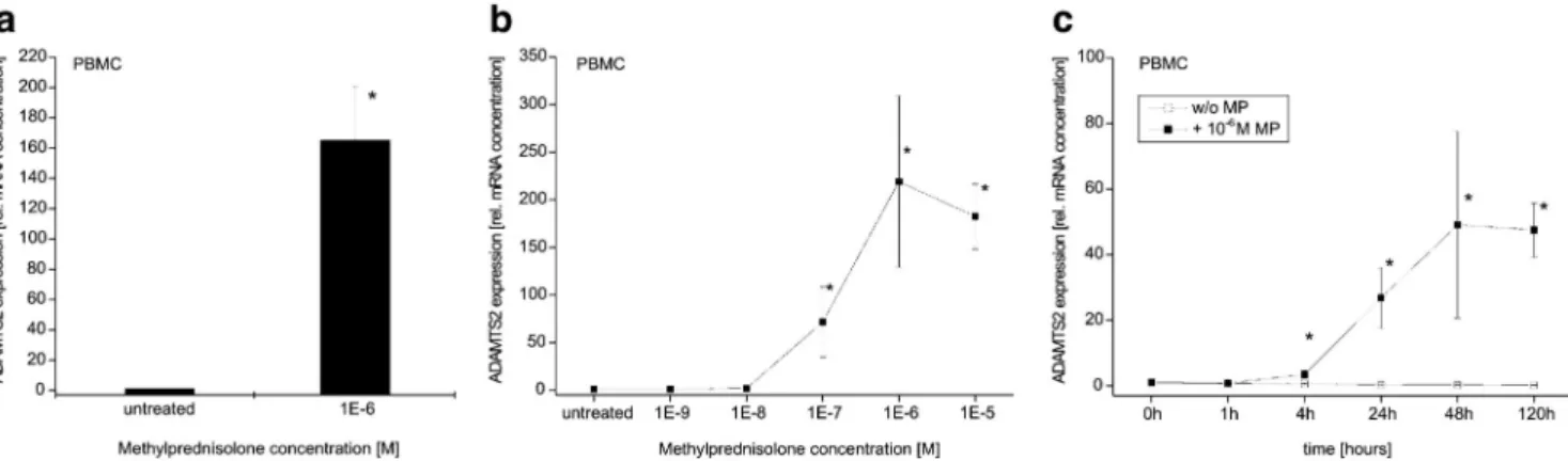
In initial studies, we analyzed the effect of glucocorticoids (GC) on PBMC by incubating these cells in the presence or absence of 1 µM methylprednisolone (MP) continuously for 5 days. To quantitate ADAMTS2 mRNA levels, we used LightCycler analysis allowing real-time monitoring of cDNA amplification. As shown in Fig. la, expression of ADAMTS2 mRNA was induced 165-fold in PBMC treated with MP relative to controls.
We next examined the dose- and time-dependence of ADAMTS2 expression in MP-stimulated PBMC. MP-con-centrations of 1 nM and 0.01 µM do not have any effect on ADAMTS2 mRNA expression (Fig. 1b). In contrast, MP concentrations of 0.1 µM (71.6-fold ± 36.5) and 1 µM (220-fold ± 90) MP induced a marked increase in
ADAMTS2 mRNA levels in PBMC. A higher concentration (10 µM) did not induce further increases in ADAMTS2 transcriptional expression.
Time course experiment showed that moderate increases (3.45-fold ± 1) in ADAMTS2 transcript levels can be detected as early as 4 h after treatment with MP (Fig. 1c). The increase is more pronounced after 24 and 48 h (27-fold ± 9, and 49-fold ± 28, respectively). After 120 h incubation with MP, ADAMTS2 was still highly expressed (47.5-fold ± 1). Hence, maximun induction of ADAMTS2 mRNA by GC requires 2 days of culture with the hormone.
MP-induced expression of ADAMTS2 is restricted to blood monocytes
To determine which of the various leukocytes in PBMC expresses ADAMTS2 upon MP treatment, CD 14++ monocytes, CD3+ T lymphocytes, PHA-activated CD3+ lymphocytes, and CD 19+ B lymphocytes were first isolated from PBMC using MACS magnetic bead separation. Cells were then stimulated for 5 days with 1 µM MP and PHA at 5 mg/ml, respectively, and ADAMTS2 mRNA expression levels were analyzed. During such culture, monocytes mature and develop into macrophages as evidenced by increases in size, granularity, and upregulation of the CD68 marker (data not shown).
ADAMTS2 expression in T lymphocytes (1.6-fold ± 0.95) and in B lymphocytes (0.52 ± 0.36) showed no effect
after MP treatment, in PHA-activated T lymphocytes (32.5 ± 49.6), a moderate but also not significant effect after MP treatment was seen (Fig. 2). In contrast, MP induced a marked increase in ADAMTS2 levels in macrophages derived from CD14++ monocytes (440-fold ± 180). These data show that among PBMC, it is the monocytes which respond to MP with a pronounced upregulation of ADAMTS2 mRNA. Monocytes go from blood into tissue and develop into macrophages. We therefore have asked whether GC treatment will also induce
ADAMTS2 mRNA in macrophages taken from the lung. As shown in Fig. 2, MP led to a substantial 200-fold induction of ADAMTS2 gene expression in alveolar macrophages.
Fig. 1 Effect of methylprednisolone (MP) on ADAMTS2 mRNA levels in blood mononuclear cells (PBMC).
PBMC were isolated from blood of healthy donors by density gradient separation, a Cells were incubated for 5 days with 1 µM MP or remained untreated. Baseline is untreated cells and was set as 1; n = 5; mean±SD; * p < 0.05. b For dose-response effect of methylprednisolone (MP) on ADAMTS2.mRNA levels, PBMC was incubated for 5 days with different concentrations of MP. Baseline is untreated cells and was set as 1; n = 3; mean±SD; *p < 0.05. c For time-dependence effect of methylprednisolone (MP) on ADAMTS2 mRNA levels, PBMC was incubated with or without 1 µM MP for different periods of time. Baseline is untreated cells and was
set as 1; n = 4; mean±SD; *p < 0.05
Fig. 2 Effect of methylprednisolone (MP) on ADAMTS2 mRNA levels in CD14++ monocytes (n = 4), CD3+ T
lymphocytes (n = 3), PHA-activated CD3+ T lymphocytes (n = 6) and CD19+ B lymphocytes (n = 3), respectively. Cells were isolated from blood of healthy donors by density gradient separation and subsequent enrichment via MACS separation. Cells were incubated for 5 days with 1 µM MP, or 5 µg/ml PHA plus 1µM MP, respectively, or remained untreated. Baseline is untreated cells and was set as 1; mean±SD; *p < 0.05
Glucocorticoid-receptor mediated action of MP
To determine whether MP acts specifically via the glucocorticoid (GC)-receptor to induce ADAMTS2, the inactive GC-analog RU486 was used (Fig. 3). In PBMC, MP increased ADAMTS2 mRNA expression in a dose-dependent manner, with 0.1 µM MP increasing expression 270-fold (± 132) and 1 µM MP inducing levels that
were 575-fold (± 125) above controls. RU486 alone (2 µM) had no effect on ADAMTS2 mRNA (1.35-fold ± 0.33). However, the effect of MP on ADAMTS2 expression levels was blocked completely by RU486 at both doses of MP (0.1 µM MP 2.2-fold ± 1.2, and 1 µM MP 2.3-fold ± 0.7). These findings indicate that ADAMTS2 induction by GC is mediated by the glucocorticoid receptor (GCR).
Fig. 3 Effect of RU486 and methylprednisolone (MP) on ADAMTS2 mRNA levels in blood mononuclear cells
(PBMC). Cells were incubated for 5 days with RU486 (2 µM), MP (as indicated), and a combination of both, or remained untreated. Baseline is untreated cells and was set as 1; n=3; mean±SD; *p<0.05
Western blot analysis of ADAMTS2 protein
Next, we analyzed the effect of MP on ADAMTS2 protein using PBMC isolated from bovine blood. The bovine system was used because of the availability of a well-established antibody against bovine ADAMTS2 suitable for Western blotting. After incubation with MP, elevated concentrations of ADAMTS2 protein were detected. As can be seen in Fig. 4, there was a substantial increase of the 150 kDa form of ADAMTS2, a form that displays amino-procollagen peptidase activity. Densitometric analysis showed an induction of 148-, 48-, and 2.2-fold for animals 1, 2, and 3, respectively. A similar pattern was seen for the other active form of ADAMTS2 (104 kDa band) except for animal 3, where we found a faint band in untreated cells and no induction by MP (Fig. 4).
Fig. 4 Western blot analysis of ADAMTS2 in bovine macrophages. Cells were incubated for 5 days with 1 µM
MP or remained untreated. Four different ADATMS2 products are detected, two of them (150 and 104 kDa) displaying aminoprocollagen peptidase activity are shown [13]
MP-induced expression of ADAMTS genes
closely related or more distantly related ADAMTS mRNAs. Compared to ADAMTS2, with an average 150-fold induction (Fig. 5), the closely related ADAMTS3 and 14 mRNAs showed only a 3- and 13-fold induction, respectively. Also, the distantly related ADAMTS9, 20, and 1 genes gave only a 4- to 11-fold induction in PBMC treated with MP (Fig. 5). Hence, while GC treatment strongly induces ADAMTS2, there was only a weak induction of these selected ADAMTS mRNAs.
Fig. 5 Effect of methylprednisolone (MP) on mRNA levels of ADAMTS2, 3, 14, 9, 20, 1 in PBMC (n = 3, except
ADAMTS1 n = 4). Cells were incubated for 5 days with 1 µM MP or remained untreated. Baseline is untreated cells and was set as 1; mean±SD
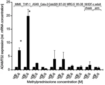
Fig. 6 Effect of methylprednisolone (MP) on ADAMTS2 mRNA levels in cell lines of the monocyte macrophage
lineage MM6 (n = 3), and THP-1 (n = 4), in epithelial cell lines A549 (n = 3), Calu-3 (n = 3), Colo320 (n = 4), and BT-20 (n = 4) and in fibroblast cell lines MRC-5, WI-38, NHDF-c adult cheek, and NHDF-c adult upper arm (all n = 3), respectively. Cells were incubated for 5 days with 1 µM MP or remained untreated. Baseline is untreated cells and was set as 1; mean ±S.D.; *p < 0.05
MP-induced ADAMTS2 mRNA expression in cell lines representing different tissues
epithelial origin and fibroblast lines, the latter being a typical source of ADAMTS2. In line with the results obtained using blood monocytes, ADAMTS2 mRNA expression was increased after stimulation with 1 µM MP for 5 days in cell lines of the monocyte lineage Mono Mac 6, and THP-1, both established from acute monocytic leukemias. In Mono Mac 6, induction of mRNA level was 7.2-fold±l, in THP-1 cells, 19.9-fold ± 2.5 (Fig. 6).
By contrast, epithelial cell lines did not show a statistically significant GC-inducible ADAMTS2 mRNA. In the epithelial cell line A549, ADAMTS2 mRNA increased after MP-treatment 2.45-fold ± 0.08, in the Calu-3 cell line, a 0.45-fold ± 0.04 decrease could be detected, while Colo320 and BT-20 cell line showed an increase of 3.96-fold ± 3.20 and 3.93-fold ± 2.76, respectively (Fig. 6). Also, in the fibroblast cell lines MRC-5 (1.44-fold ± 0.3), WI-38 (1.13-fold ± 0.4), NHDF-c adult cheek (1.75-fold ± 0.7), and NHDF-c adult upper arm (1.24-fold ± 0.3), a marginal and statistically not significant increase in ADAMTS2 mRNA expression could be detected.
Discussion
In earlier experiments, using gene expression arrays, we noted a strong induction of ADAMTS2 mRNA expression in MDM derived from patients who received oral treatment with GC
(http://www.ncbi.nlm.nih.gov/geo/, accession number GSE8608). We verified this effect of GC treatment on
ADAMTS2 gene expression in vitro using cells of the monocyte/macrophage lineage (Fig. 1, 2). Although the impact of GC on global gene expression has been studied [34-37], this is a novel finding not reported in the literature before. This effect may have been overlooked because it takes 48 h to reach its full magnitude, with a plateau at 120 h (Fig. 1c).
Dose-response analysis shows effects on ADAMTS2 mRNA expression with GC concentrations that are in the physiological range (0.1 µM), with more pronounced effects at levels of 1 µM and beyond, as seen with GC therapy [38] (Fig. 1b). Therefore, it is plausible that this effect would be observed in vivo in patients treated with GC for several days.
We observed such a strong MP-mediated ADAMTS2 gene expression among leukocytes of the monocytic lineage, but not in cultured T and B cells (Fig. 2). In phytohemag-glutinin (PHA)-activated T lymphocytes, a moderate (32.5 ± 49.6) effect of MP treatment was detected. In any event, co-stimulation of macrophages by, for instance, LPS is not required for GC to induce ADAMTS2 in these cells, as we grow our cultures under stringent exclusion of LPS contamination. When looking at macrophages from tissue, we could also see induction in cells from the lung (alveolar macrophages), and this induction was also about 200-fold (Fig. 2). In comparison to CD14++ monocyte-derived macrophages, the lower induction of ADAMTS2 mRNA may be due to a different maturation process in alveolar macrophages. While the induction of ADAMTS2 by glucocorticoids in alveolar macrophages was done on samples obtained during diagnostic procedures from patients with inflammatory lung disease, we assume that the same type of response will be seen in macrophages from healthy individuals, but this will have to be formally demonstrated. As ADAMTS2 can be induced by GC in primary alveolar macrophages and in monocyte-derived macrophages, the latter cells are a useful model for tissue macrophages, and we suggest that induction of ADAMTS2 by GCs will also be seen in primary macrophages from other tissues.
We then asked whether the strong induction of ADAMTS2 in macrophages would also lead to a similar induction of protein. For this, we turned to the bovine system, where adequate monoclonal antibodies for detection of ADAMTS2 by Western blots are available [13]. We could show a pronounced induction of the ADAMTS2 protein in monocyte-derived macrophages generated from three of four animals (Fig. 4). We do not know why induction did not occur in animal no. 4, but there may be interindividual variation. Using RT-PCR, we found a higher basal level of ADAMTS2 mRNA expression in cow compared to human samples (data not shown). Of note, we did see induction of bovine ADAMTS2 mRNA in all animals including animal no. 4, suggesting that the failure to produce protein might be at the posttranscriptional level. There also may be enhanced degradation of the protein by other enzymes in animal no. 4, as we analyzed samples without addition of proteinase inhibitors. We also performed an ADAMTS2 activity assay using monocytes/macrophages conditioned culture medium and aminoprocollagen type I, a substrate of ADAMTS2 (data not shown). However, due to massive production of various proteases by monocytes/macrophages, aminoprocollagen type I was rapidly degraded preventing the specific evaluation of ADAMTS2 activity. Moreover, as most of these secreted proteases belong to the metalloproteinase family, their inhibition would also alter the ADAMTS2 activity, explaining why we were unable to directly measure the aminoprocollagen peptidase activity in the various conditioned media. However, we were able to demonstrate the presence of ADAMTS2 polypeptides with apparent sizes of 150 and 104 kDa, which previously have been shown to be catalytically active [13],
Upon testing for GC induction of other ADAMTS mRNAs, we did not find such a pronounced effect as seen for
ADAMTS2 (Fig. 5). The additional genes were selected based on the evolutionary relationship as reviewed by Apte [11]. We have tested the closely related ADAMTS3 and 14 genes and the more distant genes ADAMTS9, 20, and 1, which, in contrast to ADAMTS2, belong to a proteoglycan-processing superclade. All of the selected genes showed only a moderate induction between 4- and 13-fold, which is low compared to ADAMTS2 which was induced 150-fold in the same experiments. In the original description, ADAMTS1 was shown to be induced by interleukin-1 and lipopolysaccharide suggesting a role in inflammatory processes [39]. Therefore, a modulation by GC treatment could have been expected, but the effect seen was only a 4-fold induction in GC-treated MDM (Fig. 5). Taken together, the strong induction by GC treatment in MDM of ADAMTS2 appears to be selective and does not extend to a series of other representative ADAMTS genes.
ADAMTS2 is known to be produced by various mesenchymal cells such as adipocytes, smooth muscle cells, and fibroblasts [25]. Therefore, we speculated that MP treatment would up-regulate this gene in these cells as well. Surprisingly there was no or only a modest change in ADAMTS2 mRNA levels in the cell lines of fibroblast and epithelial origin (Fig. 6). Of note, we did see ready induction of the gene in two cell lines of monocytic origin. This argues in favor of a tissue-specific induction of ADAMTS2 by GC. We do not see a direct link between monocytes/macrophages and mesenchymal cells. Expression of ADAMTS2 in our culture was indeed a surprise. Two hypothesis can be made: Treatment of macrophages by GC induce their differentiation as "alternatively activated macrophages". These alternatively activated macrophages are anti-inflammatory and pro-fibrotic, and induce wound repair [40]. In this context, production of an enzyme by alternatively activated macrophages, which is critical for collagen maturation, but quite limiting and poorly inducible in fibroblasts, makes sense and would favor collagen and extracellular matrix accumulation. As an alternative hypothesis, ADAMTS2 may be directly involved in the antiinflammatory process by cleavage of a so far unidentified substrate (e.g., cytokine, receptor, inflammation mediator) leading to its activation or inhibition. Further investigations will be required to answer these questions.
When looking at the mechanism involved in induction, we were interested to see whether this would involve the glucocorticoid receptor. For this, we treated macrophages with the steroid-receptor blocker RU486 [41]. These data did show a potent and complete inhibition of GC action even at equimolar concentrations of the GC and the inhibitor (Fig. 3). This finding suggests that MP acts via the GCR to induce ADAMTS2. The GCR may act on transcription either indirectly or by binding to other transcription factors in an enhanceosome [42]. An enhan-ceosome is a complex consisting of transcription activators that promote the interaction and cooperative binding to the DNA and allows additive and synergistic effect on gene transcription [43]. When inspecting the 5' region of the human ADAMTS2 gene, we noted two potential GCR binding sites (data not shown) suggesting that the GCR may act by directly binding to the promoter.
What could the role of the induction of ADAMTS2 by GC be? ADAMTS2 is required for maturation and assembly of collagen fibers and therefore for production of the extracellular matrix [13]. A defect in ADAMTS2 function leads to the dermatosparactic type of Ehlers-Danlos syndrome [44]. Therefore, induction of the gene by GC will favor generation of proper ECM. This may be important in late-phase inflammation, where lesions undergo repair. The question is whether this will occur under physiological conditions. In fact, the concentration of 0.1 µM reflects a level that is seen for steroids like Cortisol where an average 8 A.M.serum level is 0.3 µM. The data suggest that GC treatment of patients may be sufficient to induce ADAMTS2 expression in macrophages. Analysis of individual samples from our initial array pool using RT-PCR demonstrated that two patients without systemic GC treatment showed a low level of ADAMTS2 transcripts (2.5-fold higher than in cells from healthy controls). By contrast, macrophages from three cases treated with oral methylprednisolone had high levels of ADAMTS2 mRNA (35.5-fold higher than in cells from healthy controls). These data show that therapy with GC can induce ADAMTS2 transcriptional expression in macrophages in vivo.
There certainly is the possibility that ADAMTS2 has additional properties. Potential targets are those containing thrombospondin repeats, and here, one candidate molecule would be properdin. As properdin is an enhancer of complement activation [45, 46], a digestion of properdin might lead to reduced complement activity and, therefore, inflammation.
Taken together, we demonstrate herein a strong induction of ADAMTS2 mRNA and protein by glucocorticoids in a macrophage-restricted manner. The biological consequences of this induction remain elusive at this point. Given the fact that GCs are among the most widely used immunosuppressive drugs, elucidation of the action of macrophage-produced ADAMTS2 will be an important topic.
Acknowledgement We thank the Animal Clinic, Department "Innere Medizin und Chirurgie der Wiederkäuer" of the Ludwig-Maximilians-University (Munich-Oberschleissheim, Germany) for providing us with bovine blood, Claudia Unterberger for help with CD19 MACS separation, and Dr. Thomas Werner (Genomatix, Munich) for advice in bioinformatics. This work was supported in part by DFG (Zi 288).
References
1. Nathan C (2002) Points of control in inflammation. Nature 420:846-852 2. Nicod LP (1999) Pulmonary defence mechanisms. Respiration 66:2-11
3. Rennard SI, Bitterman PB, Crystal RG (1983) Response of the lower respiratory tract to injury. Mechanisms of repair of the parenchymal cells of the alveolar wall. Chest 84:735-739
4. Rhen T, Cidlowski JA (2005) Antiinflammatory action of glucocorticoids—new mechanisms for old drugs. N Engl J Med 353:1711-1723 5. Glass CK, Ogawa S (2006) Combinatorial roles of nuclear receptors in inflammation and immunity. Nat Rev Immunol 6:44-55
6. Angeli A, Masera RG, Sartori ML, Fortunati N, Racca S, Dovio A, Staurenghi A, Frairia R (1999) Modulation by cytokines of glucocorticoid action. Ann N Y Acad Sci 876:210-220
7. Kamal AM, Flower RJ, Perretti M (2005) An overview of the effects of annexin 1 on cells involved in the inflammatory process. Mem Inst Oswaldo Cruz 100:39-48
8. Jonat C, Rahmsdorf HJ, Park KK, Cato AC, Gebel S, Ponta H, Herrlich P (1990) Antitumor promotion and antiinflammation: down-modulation of AP-1 (Fos/Jun) activity by glucocorticoid hormone. Cell 62:1189-1204
9. Aljada A, Ghanim H, Assian E, Mohanty P, Hamouda W, Garg R, Dandona P (1999) Increased IkappaB expression and diminished nuclear NF-kappaB in human mononuclear cells following hydrocortisone injection. J Clin Endocrinol Metab 84:3386-3389
10. Saadat F, Khorramizadeh MR, Mirshafiey A (2005) Apoptotic efficacy and inhibitory effect of dexamethasone on matrix metal-loproteinase. Med Sci Monit 11:BR253-257
11. Apte SS (2004) A disintegrin-like and metalloproteinase (repro-lysin type) with thrombospondin type 1 motifs: the ADAMTS family, hit J Biochem Cell Biol 36:981-985
12. Porter S, Clark IM, Kevorkian L, Edwards DR (2005) The ADAMTS metalloproteinases. Biochem J 386:15-27
13. Colige A, Ruggiero F, Vandenberghe I, Dubail J, Kesteloot F, van Beeumen J, Beschin A, Brys L, Lapiere CM, Nusgens B (2005) Domains and maturation process that regulate the activity of ADAMTS-2, a metalloproteinase cleaving the aminopropeptide of fibrillar procollagens types I-III and V. J Biol Chem 280:34397-34408
14. Tang BL (2001) ADAMTS: a novel family of extracellular matrix proteases. Int J Biochem Cell Biol 33:33-44
15. Ziegler-Heitbrock HWL, Thiel E, Fütterer A, Herzog V, Wirtz A, Riethmüller G (1988) Establishment of a human cell line (Mono Mac 6) with characteristics of mature monocytes. Int J Cancer 41:456-461
16. Tsuchiya S, Kibayashi Y, Goto Y, Okumura H, Nakae S, Konno T, Tada K (1982) Induction of maturation in cultured human monocytic leukemia cells by a phorbol diester. Cancer Res 42:1530-1536
17. Lasfargues EY, Ozzello L (1958) Cultivation of human breast carcinomas. J Natl Cancer Inst 21:1131-1147
18. Quinn LA, Moore GE, Morgan RT, Woods LK (1979) Cell lines from human colon carcinoma with unusual cell products, double minutes, and homogeneously staining regions. Cancer Res 39:4914-4924
19. Jacobs JP, Jones CM, Bailie JP (1970) Characteristics of a human diploid cell designated MRC-5. Nature 227:168-170 20. Hayflick L, Moorhead PS (1961) The serial cultivation of human diploid cell strains. Exp Cell Res 25:585-621
21. Giard DJ, Aaronson SA, Todaro GJ, Arnstein P, Kersey JH, Dosik H, Parks WP (1973) In vitro cultivation of human tumors: establishment of cell lines derived from a series of solid tumors. J Natl Cancer Inst 51:1417-1423
22. Fogh J, Wright WC, Loveless JD (1977) Absence of HeLa cell contamination in 169 cell lines derived from human tumors. J Natl Cancer Inst 58:209-214
23. Wang AM, Doyle MV, Mark DF (1989) Quantitation of mRNA by the polymerase chain reaction. Proc Natl Acad Sci USA 86:9717-9721
24. Paulissen G, Rocks N, Quesada-Calvo F, Gosset P, Foidart JM, Noel A, Louis R, Cataldo DD (2006) Expression of ADAMs and their inhibitors in sputum from patients with asthma. Mol Med (Camb Mass) 12:171-179
25. Porter S, Scott SD, Sassoon EM, Williams MR, Jones JL, Girling AC, Ball RY, Edwards DR (2004) Dysregulated expression of adamalysin-thrombospondin genes in human breast carcinoma. Clin Cancer Res 10:2429-2440
26. Fernandes RJ, Hirohata S, Engle JM, Colige A, Cohn DH, Eyre DR, Apte SS (2001) Procollagen II amino propeptide processing by ADAMTS-3. Insights on dermatosparaxis. J Biol Chem 276:31502-31509
27. Somerville RP, Longpre JM, Jungers KA, Engle JM, Ross M, Evanko S, Wight TN, Leduc R, Apte SS (2003) Characterization of ADAMTS-9 and ADAMTS-20 as a distinct ADAMTS subfamily related to Caenorhabditis elegans GON-1. J Biol Chem 278:9503-9513 28. Bolz H, Ramirez A, von Brederlow B, Kubisch C (2001) Characterization of ADAMTS14, a novel member of the ADAMTS metalloproteinase family. Biochim Biophys Acta 1522:221-225
29. Bolstad BM, Irizarry RA, Astrand M, Speed TP (2003) A comparison of normalization methods for high density oligonucleotide array data based on variance and bias. Bioinformatics (Oxford, England) 19:185-193
30. Irizarry RA, Bolstad BM, Collin F, Cope LM, Hobbs B, Speed TP (2003) Summaries of Affymetrix GeneChip probe level data. Nucleic Acids Res 31:e15
31. R Development Core Team (2007) R: a language and environment for statistical computing. R foundation for statistical computing, Vienna, Austria
32. Gentleman RC, Carey VJ, Bates DM, Bolstad B, Dettling M, Dudoit S, Ellis B, Gautier L, Ge Y, Gentry J, Hornik K, Hothorn T, Huber W, Iacus S, Irizarry R, Leisch F, Li C, Maechler M, Rossini AJ, Sawitzki G, Smith C, Smyth G, Tierney L, Yang JY, Zhang J (2004) Bioconductor: open software development for computational biology and bioinformatics. Genome biology 5 :R80
33. Tusher VG, Tibshirani R, Chu G (2001) Significance analysis of microarrays applied to the ionizing radiation response. Proc Natl Acad Sci USA 98:5116-5121
34. Schmidt S, Rainer J, Riml S, Ploner C, Jesacher S, Achmuller C, Presul E, Skvortsov S, Crazzolara R, Fiegl M, Raivio T, Janne OA, Geley S, Meister B, Kofler R (2006) Identification of glucocorticoid-response genes in children with acute lymphoblastic leukemia. Blood 107:2061-2069
35. Almon RR, Dubois DC, Jin JY, Jusko WJ (2005) Pharmacoge-nomic responses of rat liver to methylprednisolone: an approach to mining a rich microarray time series. AAPS J 7:E156-194
36. Gupta V, Galante A, Soteropoulos P, Guo S, Wagner BJ (2005) Global gene profiling reveals novel glucocorticoid induced changes in gene expression of human lens epithelial cells. Mol Vis 11:1018-1040
37. Nakamura R, Okunuki H, Ishida S, Saito Y, Teshima R, Sawada J (2005) Gene expression profiling of dexamethasone-treated RBL-2H3 cells: induction of anti-inflammatory molecules. Immunol Lett 98:272-279
38. Derendorf H, Mollmann H, Krieg M, Tunn S, Mollmann C, Barth J, Rothig HJ (1991) Pharmacodynamics of methylprednisolone phosphate after single intravenous administration to healthy volunteers. Pharm Res 8:263-268
39. Kuno K, Kanada N, Nakashima E, Fujiki F, Ichimura F, Matsushima K (1997) Molecular cloning of a gene encoding a new type of metalloproteinase-disintegrin family protein with thrombospondin motifs as an inflammation associated gene. J biol chem 272:556-562 40. Kreider T, Anthony RM, Urban JF Jr., Gause WC (2007) Alternatively activated macrophages in helminth infections. Curr Opin Immunol 19:448-453
41. Jung-Testas I, Baulieu EE (1983) Inhibition of glucocorticosteroid action in cultured L-929 mouse fibroblasts by RU 486, a new anti-glucocorticosteroid of high affinity for the anti-glucocorticosteroid receptor. Exp Cell Res 147:177-182
42. Lerner L, Henriksen MA, Zhang X, Darnell JE Jr (2003) STAT3-dependent enhanceosome assembly and disassembly: synergy with GR for full transcriptional increase of the alpha 2-macroglobulin gene. Genes Dev 17:2564-2577
43. Carey M (1998) The enhanceosome and transcriptional synergy. Cell 92:5-8
44. Colige A, Sieron AL, Li SW, Schwarze U, Petty E, Wertelecki W, Wilcox W, Krakow D, Cohn DH, Reardon W, Byers PH, Lapiere CM, Prockop DJ, Nusgens BV (1999) Human Ehlers-Danlos syndrome type VII C and bovine dermatosparaxis are caused by mutations in the procollagen I N-proteinase gene. Am J Hum Genet 65:308-317
45. Schwaeble W, Huemer HP, Most J, Dierich MP, Strobel M, Claus C, Reid KB, Ziegler-Heitbrock HW (1994) Expression of properdin in human monocytes. Eur J Biochem 219:759-764
Properdin, a positive regulator of complement activation, is released from secondary granules of stimulated peripheral blood neutrophils. J Immunol 158:4444-4451