HAL Id: dumas-02451323
https://dumas.ccsd.cnrs.fr/dumas-02451323
Submitted on 23 Jan 2020HAL is a multi-disciplinary open access
archive for the deposit and dissemination of sci-entific research documents, whether they are pub-lished or not. The documents may come from teaching and research institutions in France or abroad, or from public or private research centers.
L’archive ouverte pluridisciplinaire HAL, est destinée au dépôt et à la diffusion de documents scientifiques de niveau recherche, publiés ou non, émanant des établissements d’enseignement et de recherche français ou étrangers, des laboratoires publics ou privés.
La latéralité dans le cancer colorectal métastatique :
quels facteurs pour expliquer les différences de
pronostic ?
Maria Kogay
To cite this version:
Maria Kogay. La latéralité dans le cancer colorectal métastatique : quels facteurs pour expliquer les différences de pronostic ?. Human health and pathology. 2019. �dumas-02451323�
1
UNIVERSITÉ DE NICE SOPHIA-ANTIPOLIS
FACULTÉ DE MEDECINE DE NICE
Année 2018-2019
THÈSE
Présentée et soutenue publiquement le 12 avril 2019 par
KOGAY Maria
Née le 16 Septembre 1990, à Khodjili (Ouzbékistan)
Pour l’obtention du diplôme d’état de Docteur en Médecine
La latéralité dans le cancer colorectal métastatique:
quels facteurs pour expliquer les différences de
pronostic?
Jury de thèse :
M. le Professeur J-M. FERRERO, Président du jury
M. le Professeur P. CHEVALLIER, Assesseur
M. le Professeur E. BENIZRI, Assesseur
M. le Docteur J. DOYEN, Assesseur
9 A mes parents à qui je dois tout
A mes amis à qui je dois le bonheur A mes maîtres, à qui je dois le savoir A mes patients, à qui je dois l’humilité
10 Remerciements
A mon Président de Jury, Monsieur Jean Marc FERRERO
Je suis très heureuse que vous accepté de parachever mes années d’étude en me faisant l’honneur de présider le jury de ma thèse et de juger mon travail.
Que ce travail soit le témoignage de ma reconnaissance.
A mon Directeur de Thèse, Monsieur Eric FRANCOIS
Vous avez accepté de me soutenir et de m’accompagner tout au long de ce travail. Je vous remercie pour vos conseils. J’ai eu la chance de travailler à vos côtés lors de mes premiers pas en tant qu’interne et j’ai grandement apprécié vos qualités pédagogiques et professionnelles. Votre savoir et votre disponibilité m’ont été très précieux. Recevez ma sincère gratitude.
A Monsieur le Professeur Emmanuel BENIZRI
Vous me faites l’honneur d’apporter votre expérience à la critique de ce travail en siégeant dans mon jury de thèse. Je n’ai pas eu la chance de travailler avec vous mais j’ai pu apprécier vos qualités d’enseignant lors de multiples cours que vous avez dirigé et vos qualités humaines d’écoute et d’expertise, lors de la soutenance de thèse de mes amis.
Je vous prie de bien vouloir accepter ma respectueuse considération.
A Monsieur le Professeur Patrick CHEVALLIER
Je vous remercie d’avoir accepté de siéger dans mon jury de thèse et me faire l’honneur de juger mon travail. J’ai eu la chance de travailler avec vous lors de mon semestre en gastro à l’Archet et j’ai grandement apprécié vos qualités humaines, pédagogiques et professionnelles.
Veuillez croire en l’expression de ma respectueuse considération.
A Monsieur le Docteur Jérome DOYEN
Jérome, merci d’avoir accepté d’être membre de mon jury et merci de l’intérêt que tu as porté à mon travail. Radiothérapeute brillant, à ta compétence s’ajoute aussi la gentillesse et l’humour. Reçois ma sincère gratitude.
11 A mes parents, à qui je dois tout. Les mots manquent pour apprécier à leurs justes valeurs l’éducation que vous m’avez inculqués. Vous m’avez offert toutes les chances pour réussir. Merci pour vos sacrifices et votre soutien inconditionnel tout au long du chemin parcouru. Que la réalisation de ce travail, soit également l’aboutissement de vos efforts ainsi que l’expression de votre réussite.
A Louba la grande sœur qui a toujours veillé sur moi. Merci de m’avoir supporté et soutenu pendant ces longues années de médecine. Notre lien est plus fort que tout et je serais toujours là pour toi.
A Johan le beau-frère parfait! Merci de prendre soin de ma soeur, je crois que mes parents pouvaient pas rêver mieux que toi.
A Nour, ma sœur de cœur, les mots manquent pour décrire notre amitié. J’ai une immense chance de t’avoir comme amie. Merci pour toutes ces années où tu m’as soutenue sans faille, pour ta présence et ton écoute dans des moments de doute. A tous ces beaux moments et tous les voyages qu’on a partagé et qui nous attendent. Tu es l’épaule sur laquelle je m’appuie, ton amitié m’est si précieuse.
A Farah, je pense que si je devais te décrire en 3 mots je dirais : détermination, honnêteté et force de caractère. J’admire tes valeurs et notamment celle de ne pas tolérer l’injustice et pour ça tu n’as pas peur d’affronter le plus grand PU-PH de France ..tu m’impressionne. Quand tu deviendra un grand Professeur, reconnu au niveau mondial, je pourrais me vanter de faire partie de tes amis. Je suis fière de toi. Merci pour ton amitié.
A la famille Cadour ..Toutouka, Fouad et Jade, merci pour votre gentillesse et l’accueil toujours chaleureux dans votre maison. Merci pour votre soutien et vos précieux conseils. Grace à vous je sais que l’argent ne se récolte pas sur les arbres.
A Anais et toute la famille Lepinette. L’internat nous a éloignée physiquement mais notre amitié est resté intacte. Je n’oublierais jamais notre tour de France post ECN et les 2 années de colocs que nous avons partagé ensemble, qui sont parmi les plus beaux souvenirs.
12 A mes amis :
A Anissa, à tes boulettes, à ta spontanéité et à ta gentillesse! Surtout reste comme tu es! A Catoch et à Florencia et à “the place to be” merci pour votre amitié pendant toutes ces années d’internat.
Marie et Ben, comment parler de l’un sans l’autre, votre amour est à tout épreuve et votre force est un exemple pour nous tous.
A Lila qui s’est expatrié à Paris, on ne t’oublie pas pour autant!
A Borja et Denis et aux gins toniques de la revue que je ne pourrais plus jamais oublier! A Maria et PH, la famille parfaite. Maria, interne et maman parfaite, toujours de bonne humeur, tu nous épate tous.
A la belle Camille, pétillante, intelligente et toujours pleine d’énergie.
A Claire Wadouxxx, à notre rencontre au CAL qui s’est ensuite changée en amitié. A ta bonne humeur, ta douceur et ta gentillesse.
A Boris et à nos belles soirées ensemble.
A Maud ma co interne, ma co voitureuse, ma confidente pendant ses 6 mois à Cannes.. et au décours. Tu es devenue plus qu’une co-interne éphémère .. une amie. Tu m’as toujours impressionnée par ta générosité, par ta gentillesse et ta simplicité. Surtout ne change pas! A Johanne et à Sonia et à notre voyage à Budapest inoubliable..même sous la pluie! J’espère que plein d’autre s’en suivront!
A mes amis de la fac, qui, dès les premières années ont été à mes côtés:
A Sam et Ludo les inséparables! Merci pour votre amitié, c’est en regardant tout ces années d’amitié que je m’aperçois à quel point on a grandi.
A Zoé qui prend soin de notre Sam maintenant!
A Morli, Raph, Micka, Valentin, Pépito et à nos pauses à la machine à café et à toutes ces heures passés à la BU.
A Charlotte et Anais et à notre trio pendant l’externat!
A Violaine, la plus gentille des radiologues. Quel soulagement de tomber sur toi de garde. Merci pour ton amitié depuis tant d’années.
A Rémy et Marjorie .. merci pour votre bonne humeur, pour votre humour et votre gentillesse. Rémy à toutes ces années d’aventure depuis la P1 jusqu’à maintenant et à Marjorie qui a su si facilement intégrer le cercle fermé de la médecine.
13 A Magali .. sans “e” .. l’internat nous a rapproché et j’ai découvert en toi une personne que j’apprécie énormément: droite, honnête et brillante. Merci pour ton soutien et ton aide pendant la rédaction de ce travail. A nos RCP téléphoniques actuelles et futures..ainsi que les sessions potins (un véritable détective).
A mes co interne de spécialité; à notre entraide, solidarité et notre future collaboration! On a vécu le pire mais le meilleur aussi.
A Victoria, Lucile, Caroline, Marc, Maud, Delphine, David
A Thibault, Benjamin et Nicolas qui sont maintenant devenus chefs
A Emilien alias Mimi. L’internat est fait de belles rencontrer et tu en fait partie. Toujours de bonne humeur, toujours calme et prêt à aider les autres… c’est un plaisir de travailler avec toi. A Claire alias sexy Jaraudy .. je crois que la relève est définitivement assurée. Bosseuse, petillante et surtout toujours partante pour aller picoler avec moi..surtout ne change pas haha! A Angélique, merci pour ta bonne humeur, ton dynamisme et ton énergie si communicatives. A Rémy et Magalie (encore).. à notre parcours, à notre trio de la promo oncologie-radiothérapie 2014.
Aux internes dont j’ai croisé la route, si chacun d’entre nous à pris des directions diverses, j’espère que le temps réserve le meilleur à chacun d’entre vous :
A Caroline Maurel la pharmacienne la plus cool du CAL A Hamza et ton calme légendaire
A Sylvain le Montpelliérain, bientôt ca sera à ton tour de m’accueillir sur ta terre natale. Romain, David, Navid, Anne Capucine : les futurs PETologues!
A Vincent, toi qui m’a fait tellement rire au B3 avec tes Pongerardises “l’aiguille est dans le poumon”, “il est jaune comme un mignon”. Merci de m’avoir accompagné dans mes premiers pas de conduite!
A Diane et à nos premiers pas en HDS.. inoubliables et douloureux A Anne .. alias grosse boule, à ces 6 mois au B5
A Ariane, quelle belle rencontre grâce à ton travail de thèse.. comme quoi ça ne sert pas uniquement à galérer.
14 A Emilie, merci pour ta gentillesse, merci d’avoir supporté moi et ma mauvaise humeur en début des gardes ;)
A Sandra à ta belle personnalité et à ton énergie..et aux soirées apéro!!!
A toutes les infirmières et aides-soignantes que j’ai rencontré pendant ma formation (B3, B4, B5, HDJ, HDS, Cannes, Gastro, Monaco) ces héros de tous les jours, vous faites un travail remarquable. J’étais souvent impressionné par vos compétences et votre humanité. Sans vous notre travail n’aurait pas de sens.
A toute l’équipe de oncologie - médecine interne de Cannes:
A vous tous qui m’avez accueilli au sein de votre équipe, je vous adresse un grand merci! Florence, Stéphanie, Jennifer, Elsa, Andréa, Zack, Amandine, Coco, Aurélie, Sandrine. A Aurélie: merci pour le soutien, pour tes conseils .. depuis le premier jour jusqu’à maintenant. A Elea : cet être incroyable, tellement forte, tellement humaine; merci pour ces soirée bar à vins ou on a tant appris à se connaître.
A Régis qui je ne remercierais jamais assez. Merci pour ta disponibilité et ton humanité sans limite. Merci pour la confiance que tu m’as accordé. Tu m’as appris énormément autant en médecine que dans la vie en général. Tu es un médecin brillant et travailler à côté de toi m’a redonné gout à la médecine pendant cette période houleuse de ma vie.
A Nico et ces références cinématographiques que je n’ai jamais compris. Tu m’as toujours impressionné par ta culture et ton humour.
A Nathalie Montagne, Laurence Saudes et Matteo Vassallo : merci pour les connaissances que vous m’avez transmis.
A toute l’équipe de gastro de l’Archet :
Avec des gens comme vous, malgré le travail et des gardes parfois stressantes, ces 6 mois en gastro ont été un réel plaisir.
A Anne Claire et Delphine merci pour tout ce que vous m’avez appris. Anne Claire, à ta spontanéité et ton rire légendaire, tu es une pépite.
Delphine, tu m’as connue toute petite (lors de mes premiers pas en tant que FFI au CAL) et tu m’as appris à être persévérante et rigoureuse. Merci!
15 A Audrey et Gauthier, le plus beau couple de la promo! Audrey, la co interne parfaite. Merci pour ces 6 mois de rire et d’amitié.
Audrey, Clémént, Dorsa, Maude, Edouard merci pour votre accueil en gastro, honnêtement je me suis sentie comme chez moi! Votre solidarité entre internes est exemplaire.
Anne Claire, Delphine, Audrey et Dorsa à nos soirées Bachelor mémorables!
A l’équipe de B3:
Virginie, Cathy, Béa, Audrey, Jonathan, Quentin, Floriane, Valériane, Michel, Dalenda, Serena, Laurent, Réné, Sindy, Nathalie et les autres.
A Ludovic Evesque, merci pour la relecture de mon travail et tes remarques toujours pertinentes. Ton exigence m’a appris à approfondir davantage ma réflexion.
A Caroline Astier, grâce à ta gentillesse et à ta douceur, j’ai pu commencer mes premiers pas dans le monde des soins palliatifs en douceur. Un grand merci pour ce semestre et nos moments partagés au décours.
A l’équipe de B4/HDS : j’ai passé 6 mois de fou rires avec vous.
La palme revient à sa majesté sérénissime le Dr Anthony Muggeo, une vraie découverte du B4, une personnalité hors du commun et un excellent médecin. Avec ta bonne humeur et ton grain de folie les 6 mois ont filé de toute vitesse. Toujours de bon conseil tu es devenu mon confident. Merci pour ton amitié.
A Ilanite, la nouvelle Muggeo du B4. Pétillante et pleine de vie .. le B4 n’a plus qu’à bien se tenir!
A Audrey alias Baguette, Andrea, Olivia, Cynthia, CamCam, Cathy, à Michmich, Mezni, Oriane, Marianne , SoukiJo.
A Audrey Carrere et à Coralie les secrétaires de folie!
A Christelle, à la calinothérapie, merci pour ton écoute et pour ton soutien.
A Agnès, la chti du groupe! Avec ta bonne humeur tu as su rapidement trouver ta place. Merci pour le savoir que tu m’as transmis!
16 A Mr THYSS, quand j’étais externe dans votre service j’ai été impressionné par votre charisme et votre savoir et vous m’avez donnée envie de faire de l’oncologie mon métier. Merci pour votre soutien, votre bienveillance et l’implication que vous avez eu dans ma formation. Vous avez pris soin de moi pendant les six mois au B5 et au décours et je ne l’oublierai jamais. Permettez-moi d’exprimer mon plus profond respect et ma gratitude.
A notre maman Annick merci de m’avoir supporté ces 6 mois au B5. Merci de prendre soins de tous les internes, qui viennent souvent pleurer dans ton bureau! Tu es une personne incroyable et on ne te le dit pas assez. Longue vie aux “reines de l’hémato” et à toutes les soirées alcoolisées qui en découlent.
A Lauris le ROI des reines de l’hémato ;) A ta gentillesse et ton charme irrésistible! Merci pour tout le savoir que tu m’as transmis chaque jour et pour tes conseils toujours bienveillants.
A Mr Peyrade, merci pour l’implication que vous avez eu dans ma formation. Avec votre bonne humeur à toute épreuve, même dans les journées les plus tristes on retrouve le sourire.
A toutes les infirmières et aides-soignantes du B5.. quelle équipe! Merci d’avoir pris soin de moi, le jour.. la nuit.. les week-end d’astreinte! A Lydia (un jour je vais dominer le monde mais pour l’instant je me fais dominer), Carole, Emmanuelle alias Nini, Heloise, Julia, Stéphanie, Marina (encore), Eva, Margot, Cécilia, Aurélia, Elodie, Julie, Sylvana, Sabine, Ophélie, Marion, Helene, Sousou, Audrey et les autres. Je ne vous oublierais pas!
A Sylvie et Malika, le duo de choc! Et à Audrey, travailler avec vous a été un réel plaisir! A l’équipe de l’HDJ:
Aux infirmières : Annie, Virginie, Camille, Cassandre, Laurie, Alicia, Agnès Aux secrétaires : à Manon alias Manouchka, Coralie, Sonia, Johanna, Murielle A Ophélie merci pour tes conseils précieux, pour ta confiance et pour ton amitié. A l’équipe de Radiothérapie du CAL:
17 A Lorraine un jour on dominera le monde, à Dani la terreur de la Radiothérapie qui s’adoucit à la vue de la nourriture, à Karen et Audrey la touche féminine de la radiothérapie merci pour votre gentillesse, à Alexander alias Falkinator.
A Romain, merci pour ta gentillesse et pour ta disponibilité. Tu as été le premier à me former lors de mes premiers pas d’internat.. celui que Natacha appelait quand on était en galère (le fameux coup de fil à un ami).
A Shakeel alias Shakeeloulou, merci pour ces 6 mois dans le monde de la radiothérapie. A tous les physiciens, manip et secrétaires pour m’avoir aidé et guidé dans mes débuts de rayons!
Aux cadres du CAL :
Magalie, Mumu, Natachix, Monsieur Stephane, Marilou .. vous m’avez connu, pour beaucoup, toute petite et vous m’avez accompagné dans mes premiers pas. Merci pour votre soutien, votre aide et votre confiance.
A toute l’équipe de oncologie - médecine interne du CHPG:
Merci de m’avoir accueilli aussi chaleureusement, merci à toute l’équipe soignante et aux secrétaires qui font un travail extraordinaire et toujours dans la bonne humeur. Maire Laure, Sabine, Lydie, Sidonie, Sylvie, Marjorie, Kelly, Justine, Carole, Virginie, Marina, Alexandrine, Mélodie, Claire, Valérie, Karine, Marine, Celia et les autres.
A Georges et Willy ma reconnaissance est telle que je ne saurais par où commencer! Je suis honorée de venir travailler à vos côtés.
A Willy, cela a été un réel bonheur de travailler à coté de toi. Merci pour ta gentillesse, ta patience et ton calme à toute épreuve. Merci d’avoir pris le temps de partager avec moi tes réflexions et tes connaissances qui ont été si instructives, merci pour la motivation que tu as su m’insuffler dans des moments de doute. Je suis honoré de la confiance et la liberté d’action que tu m’as accordé dès le début. Permets moi d’exprimer toute mon estime et mon admiration pour la personne et le médecin que tu es.
18 A Georges, chef de service et médecin de “compétition”. Comme dirait Charles Ferrari “tu es une encyclopédie, toujours ouverte à la bonne page”. Malgré ça tu as su rester simple et accessible avec toujours beaucoup d’humour! Merci pour ta disponibilité, ta gentillesse et ton savoir que tu m’as transmis.
A Stephane Ciucca pour nos galères mutuelles à chaque hospitalisation d’un patient, Philippe Heudier le seul qui vient en vélo de Nice et Bruno Taillan.
A l’équipe des Biostatisticiens:
Sans vous ce travail n’aurait pas vu le jour.
Un énorme merci à Renaud, toujours plein d’idées novatrices et partant pour tous les projets, ta motivation est communicative. Merci pour ton aide si précieuses pour ce travail.
A Antoine et à Emmanuel Chamorey: merci pour votre gentillesse, votre implication et votre expertise dans ce travail.
A tous les médecins qui ont participé de près ou de loin à ma formation : Delphine Borchiellini, Esma Saada, Christophe Hebert, Josiane Otto, Michel Poudenx, Hannah Ghalloussi, Gérard Cavaglionne, Catherine Ciais, Anne Fogliarini merci pour votre accompagnement.
19 TABLE DES MATIERES
Première partie Résumé de l’article ………...20 Deuxième partie Article soumis Abstract……….21 Introduction………...23 Matériel et méthode………...24 Résultats..………..25 Discussion..………...28 Conclusion………31 Références……….32 Tableaux et Figures………...37 Publications………...42 Serment d’Hippocrate………...44
20 Résumé de l’article
Introduction: Des études récentes ont mis en évidence l’impact prédictif et pronostic de la localisation de la tumeur primitive dans le cancer colorectal métastatique. L’objectif de notre étude était d’analyser les différences de présentation clinique et biologique au diagnostic pour expliquer l’impact de la latéralité.
Matériel et méthodes: Tous les patients présentant un cancer colorectal métastatique (à l’exclusion des tumeurs du côlon transverse) traités entre le 1/12/2007 et le 1/12/2016 dans notre établissement, avec un dossier complet, ont été étudiés. Des analyses uni et multivariés ont été effectuées pour identifier les variables pronostiques de la survie.
Résultats: 284 patients (pts) ont été inclus: 83pts (29%) avec un cancer du côlon droit (CCD), 123pts (43%) avec un cancer du côlon gauche (CCG) et 78 (28%) avec un cancer du rectum (CR). Les métastases hépatiques, pulmonaires, ganglionnaires et péritonéales étaient respectivement retrouvées dans 63%, 36%, 23% et 20% de la population. L'incidence ou le nombre de métastases hépatiques n'étaient pas influencés par la latéralité (p = 0.06), toutefois les CCG présentaient plus souvent une atteinte bilobaire que les CCD ou CR (p=0.017). L’atteinte péritonéale était significativement plus fréquente dans les cancers coliques (p=0.002); l’atteinte ganglionnaire était plus fréquente dans les CCD (p = 0.008), alors que les métastases pulmonaires étaient plus fréquentes dans les CR (p<0.001).
Le statut RAS était connu pour 230pts (81%); avec plus de mutations du gene RAS dans les CCD et les CR (p = 0.009). Le statut BRAF était connu pour 171pts (60%); la mutation BRAF était plus fréquente dans les CCD (p = 0.007). Le statut MSI (p = 0.079) était également plus fréquent à droite, sans toutefois atteindre la significativité, probablement en raison de la taille de l’échantillon.
En analyse multivariée : la résection de la tumeur primitive (RTP) et la réponse complète après le premier traitement étaient significativement associées à une meilleure SG. Il existait une tendance non statistiquement significative pour les CCG et CR. Alors que l’atteinte pulmonaire, péritonéale et ganglionnaire étaient significativement associés à une moins bonne SG.
Conclusion: Ces résultats suggèrent que les CCRm ont une présentation clinique différente au diagnostic. En association avec des caractéristiques moléculaires ces différences peuvent expliquer la différence de pronostic en fonction de la latéralité tumorale.
21 Sidedness in metastatic colorectal carcinoma: Which are the factors influencing the prognosis?
Kogay M1, Falcoz2,Evesque L1, Milano G4, Saint A1, Cavaglione G1, Baudin G3, Schiappa
R2, Chamorey E2, Francois E1.
1 Digestive Oncology Unit, Department of medical oncology, Centre Antoine-Lacassagne, 33 Avenue de Valombrose, 06189 Nice France.
2 Unit of epidemiology and biostatistics, Centre Antoine-Lacassagne, 33 Avenue de Valombrose, 06189 Nice France.
3 Department of Radiology, Centre Antoine-Lacassagne, 33 Avenue de Valombrose, 06189 Nice France.
4 Oncopharmacology laboraroty, Centre Antoine-Lacassagne, 33 Avenue de Valombrose, 06189 Nice France.
Recent reports demonstrate the importance of sidedness in metastatic colorectal cancer. Our study aimed to determine the influence of sidedness on metastatic distribution and patterns. 284 patients with metastatic colorectal cancer treated from 1/12/2007 to 1/12/2016 in our institution were analyzed: 83 with right sided colon cancer, 123 with left sided colon cancer and 78 with rectal cancer. Hepatic, lung, lymph nodes and peritoneal metastases were respectively found in 63%, 36%, 23% and 20%. The incidence or number of liver metastases were not influenced by sidedness (p=0.06). Peritoneal carcinomatosis was significantly correlated to colon cancer (p=0.002), whereas lung metastases were more common in rectal cancer (p< 0.001). Patients with right sided colon cancer more often presented distal lymph node involvement (p = 0.008).
RAS mutation status was more frequent in right sided colon cancer and rectal cancer (p=0.009). BRAF mutation status (p=0.007) was more common in right sided colon cancer.
On a multivariable analysis: primary tumor resection and complete response after first line therapy remained significant to improve OS. While pulmonary, lymph node and peritoneal metastases remained significant for worse OS.
This suggests that metastatic colorectal cancer had different clinical and molecular presentation that may explain the independent prognostic marker for OS of the sidedness.
22 Conflict of interest statement
EF has consulted for Roche, Merck, Amgen, Servier and Sanofi. All other authors declare that they have no conflict of interest.
23 INTRODUCTION
Colorectal cancer is one of the most common malignancies, with an estimated 140 250 new cases and 50 630 deaths in 2018 in the United States (1). A large proportion of patients presenting with metastatic disease or developing a recurrence (2). A recent retrospective analysis of the pivotal CALGB/SWOG study (3), presented at the 2016 ASCO Annual Meeting, suggest that primary tumour location might be both predictive and prognostic for clinical outcome.
This analysis included 1137 patients with KRAS wild type metastatic Colorectal Cancer (mCRC). Patients with left sided mCRC had a better prognosis than right sided mCRC, with median overall survival (OS) 33.3 versus 19.4 months respectively (HR=1.55 CI95%=1.32-1.82 p<0.0001). Among patients who received cetuximab, OS was 36 months for patients with left sided mCRC but only 16.7 months for patients with right sided mCRC (HR=1.87 CI95%=1.48-2.32 p<0.0001). Similar trends were observed among patients treated with bevacizumab: OS was 31.4 months for patients with left sided mCRC and 24.2 months for those with right sided mCRC (HR = 1.32 CI95%= 1.05-1.65 p=0.01).
Similary, additional retrospective analysis data from other trials (PRIME, PEAK, CRYSTAL, FIRE3) (4,5,6,7) and different meta-analysis (8,9) including this studies have reported concordant results. The pejorative character of right sided mCRC is no longer debated.
These results raised interest of laterality in mCRC. Several hypotheses have been suggested to explain these results and the differences in embryologic, anatomic, histologic, molecular features and fecal exposure of the bowel have been reported between the left and right sided mCRC. (10,11,12)
However many studies show that sidedness remained an important prognostic factor for Overall Survival (OS) when adjusted for these features. (13,14) The other hypothesis could be the difference in time at detection with right sided tumors generally diagnosed at a more advanced stage. (15,16)
Tumors arising from right side tend to have a different symptomatology, often presenting with subtle signs and symptoms such as microcytic anemia, as opposed to rectal bleeding and alteration in bowel habit, which are more evident in left sided mCRC. (17)
It has been speculated that the delay in the diagnosis for right sided tumors results in more extensive metastatic disease at diagnosis that could explain a worse survival. (18)
24 Our study aimed to describe our institutional experience with mCRC to determine the drivers of disparities at diagnosis which may explain the sidedness independent prognostic marker for OS.
METHODS
Study population and data collection
All patients diagnosed with mCRC and treated in our institution Centre Antoine Lacassagne from 1/12/2007 to 1/12/2016 were included in a computerized database and were retrospectively analyzed. Demographic data, treatment-related, outcome-related data were collected. Initial imaging, colonoscopy, operative reports, pathology reports for each patients were reviewed to determine the location of the primary tumor and the number and distribution of metastasis. RAS, BRAF and MSI status were also documented if available. Patients with transverse colon cancer, overlapping position, unknown location, recurrent cancer, concomitant malignant tumors, multiple primary cancer, incomplete clinical data or missing follow up and different histological subtype than adenocarcinoma were excluded. Patients were also not considered for this study if initial imaging was not available and patients with no first line treatment (chemotherapy or surgery/ablative local therapy) possible due to deteriorated general status or organ dysfunction. In the most of trials colon and rectal cancer patients have been grouped together. In our analysis, we separated the right sided and left sided mCRC which included left sided colon cancer and rectal cancer together, then we analyzed the differences between right sided colon cancer, left sided colon cancer and rectal cancer separately.
This retrospective study was approved by National Ethics Committee.
Definition of terms
For the purposes of this study, right sided mCRC will consist of cancers of the caecum, ascending colon and right flexure. We use the term of left sided mCRC for cancers of the left flexure, descending colon, sigmoid colon, rectosigmoid junction and rectal cancer. Left sided Colon Cancer (LCC) will consist of cancers of the left flexure, descending colon, sigmoid colon and rectosigmoid junction excluding rectal cancer.
25 Synchronously detected metastases were defined as metastases detected prior to or concurrently with the primary tumor. Overall Survival (OS) was defined as time from diagnosis of metastatic disease to death from any cause. Progression Free Survival (PFS) was defined as time from diagnosis of metastatic disease to either distant or local progression as defined by the RECIST version 1.1 criteria or death. Patients showing no event (death or progression) or lost to follow-up were censored at the date of their last contact. In our study Complete Response (CR) was defined as disappearance of all target lesions after chemotherapy alone or multimodal treatment including local ablative therapy and/or surgery.
Statistical analysis
Univariate log-rank test and multivariate Cox regression models were used to analyze relationship between tumor location and OS or PFS. All variables associated with p<0.05 on univariate analysis were included in a multivariate model in order to avoid all confounding factors that could modify OS and PFS. No colinearity was identified between all variables included in the tested multivariate models. Proportional hazards were tested for all entered variables using graphical Schoenfeld residuals. Statistical analyses were two-sided and were performed using R-3.2.3 for Windows.
RESULTS FOR LEFT VERSUS RIGHT SIDED mCRC Demographic and clinico-pathological features
A total of 284 patients (pts) with available data were analyzed: 83 (29%) of them showed right sided mCRC, 201 (71%) left sided mCRC.
Table 1 shows patients characteristics for demographic, pathological, oncological data of the 284 patients depending on the location of the primary tumor.
122 (43%) were >70 years old, 138 (49%) were between 50-70 years old and 24 (9%) were <50 years old. Patients with right sided mCRC had a non significant tendancy to be older at diagnosis (p=0.06).
One hundred fifty three pts (54%) had synchronous metastasis versus 131 (46%) metachronous metastasis. RAS mutation status was known for 230 (81%), of those 101 (36%) had a mutated RAS or KRAS status with no significant differences between right and left sided mCRC (p=0.49). BRAF status was known for 171 pts (60%). Thirteen pts (4,6%) had a mutated BRAF
26 status which were more common in right sided mCRC (p=0.002). Mismatch repair deficient (dMMR) status was not different between right and left sided mCRC (p=0.107).
Intra and extra hepatic patterns
One hundred and sixty seven (58.8%) had only one metastatic site, with no difference between right and left sided mCRC (p=0.85). One hundred and twenty (63.4%) presented synchronous liver metastases at diagnosis. Of them: 35 (12.9%) presented a single liver metastases, 61 (22.5%) presented between 2 to 5 lesions and 68 (25.4%) had 5 or more liver metastasis. Sidedness was not influenced by neither incidence nor number of metastasis. Left sided mCRC presented more frequent bilobar involvement compared to right sided mCRC (p=0.037). The most common extra hepatic metastatic site was lung (36%), followed by distant lymph node metastasis (23%) and peritoneal metastases (20%).
Seventy one (66%) of 107 patients with pulmonary metastases also had metastases to extrathoracic sites; only 36 pts (34%) had lesions confined to the lung only. Lung metastases often occured together with liver metastases, in 54 of 107 pts (50%).
Right sided mCRC tend to have more peritoneal carcinomatosis than the left sided mCRC (p=0.09). Lung metastases were more common in left sided mCRC (p<0.002). Patients with right sided mCRC presented more often distal lymph node involvement (p=0.003).
Treatments
Among the 284 treated pts, 217 (76%) underwent surgery for their primary tumor. Two hundred and fifty one (88%) received a first-line chemotherapy, 33 (12%) were treated with metastasectomy or local ablative treatment alone and 105 pts (39%) were treated by combined treatment with curative intent (chemotherapy and/or metastasectomy/ablative local therapy). Patients with right sided mCRC tended to underwent more intervention with curative intent than patients with left sided mCRC (49.3% versus 36,9% p=0,09). One hundred and seventy five (62%) received targeted treatment in first-line including 66 (23%) treated with anti EGFR and 109 (39%) treated with anti VEGF. One hundred and ninety four (68%) received a second-line and 103 (36%) a third second-line chemotherapy. No difference was observed between right and left sided mCRC and number of administered treatment line (p=0.66).
27 The median follow up was 46 months [IC95% 32-71]. RECIST 1.1 response rate were assessed: 87 pts (32%) showed a complete response (CR), 80 pts (30%) a partial response, 44 pts (16%) stable disease and 46 pts (17%) progressive disease after first line treatment including chemotherapy and/or local treatment. CR rates were significantly higher for right sided mCRC with 44% of CR versus 29% in left sided mCRC (p=0.028). Median OS for pts with right sided mCRC was 45.3 months [IC95% 30.4-NA] and 40.9 [IC95% 33.5-46.1] for left sided. Median PFS was 13.6 [IC95% 10.4-20.3] and 14.0 [IC95% 13.2-16.2] months for right and left sided mCRC respectively. OS was significantly worse for right sided mCRC comparatively to left sided mCRC (HR=1.49 p= 0.01) for unresected primary tumor. Fig 1.
Predictors of outcomes : Univariable and multivariable analyses
On univariable analysis concerning PFS, primary tumor resection (PTR) (p<0.0001 HR=0.47 IC95% 0.35-0.64), metastasectomy or local ablative therapy of metastases (p<0.0001 HR=0.43 IC95% 0.17-0.38) and complete response after first line therapy (L1) (p<0.0001 HR=0.43 IC95% 0.33-0.57) were a significant prognostic factor for better PFS. Whereas peritoneal carcinomatosis (p<0.02 HR=1.5 IC95% 1.1-2) and number of metastatic sites (>1) (p<0.001 HR=1.6 IC95% 1.2-2) were associated with poor PFS.
As could be expected metastatic stage at time of diagnosis (p=0.41 HR=1.4 IC95% 1-2), number of liver metastasis (>5) (p<0.0001 HR=3.3 IC95% 2.1-5.1), liver bilobar involvement (p<0.0001 HR=2.6 IC95% 1.7-4), pulmonary metastases (p=0.008 HR=1.6 IC95% 1.1-2.2), lymph node metastases (p<0.01 HR=1.9 IC95% 1.3-2.7), peritoneal carcinomatosis (p<0.001 HR=1.9 IC95% 1.3-2.7) and number of metastatic sites (>1) (p<0.0001 HR=2.5 IC95% 1.8-3.4) were all associated with a worse OS on a univariable analysis. Whereas PTR (p<0.0001 HR=0.28 IC95% 0.2-0.4), metastasectomy or local ablative therapy (p<0.0001 HR=0.26 IC95% 0.17-0.38) and L1 complete response (p<0.0001 HR=0.22 IC95% 0.14-0.34) were associated with a better OS on a univariable analysis.
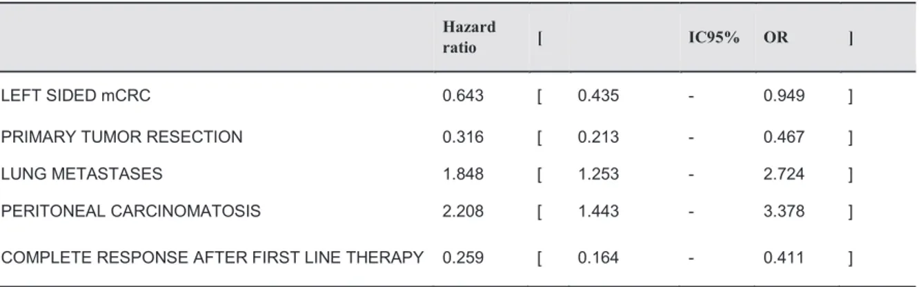
On multivariable adjusted analysis, left sided mCRC (p=0.03, HR=0.64 IC95% 0.44-0.95), PTR (p=0.0000001, HR=0.32 IC95% 0.21-0.47), and L1 complete response (p=0.0000001, HR=0.26 IC95% 0.16-0.41) remained significant good prognostic factors for OS. While pulmonary metastases (p=0.002, HR=1.85 IC95% 1.25-2.72) and peritoneal carcinomatosis
28 (p=0.0003, HR=1.85 IC95% 1.25-2.72) remained significant poor prognostic factors for OS (Table 2).
RESULTS FOR LCC, RCC and RC SEPARATELY
In the sub-group analysis of 201 pts with left sided tumors: 123 pts (43%) presented LCC and 78 pts (28%) presented RC. RCC presented significantly more synchronous metastases at diagnosis than LCC or RC (p=0.038). There was significantly more RAS mutant tumors in RC and RCC versus LCC (p=0.009). Whereas BRAF mutation were significantly more common in RCC (p=0.007) and dMMR status were also quite more frequent in RCC (p=0.07). Liver metastases seemed to be more often diagnosed in colon cancer compared to rectal cancer (p = 0.064).
When analyzed separately LCC, RCC and RC peritoneal carcinomatosis were significantly correlated to colon cancer (p=0.02) with respectively 27%, 24% and 6% of peritoneal carcinomatosis at diagnosis. Lung metastases were more common in LCC and RC with 22% of lung metastasis in RCC, 36% in LCC and 51% in RC (p<0.001). Patients with RCC presented more frequently distal lymph node involvement (p=0.008). Regarding the treatment: RC received significantly more neo-adjuvant radio or chemotherapy (p <0.001). Otherwise there was no significant difference between the treatments received by the RCC, LCC or RC. Concerning the outcomes: median OS for RCC was 45.3 months [IC95% 30.4-NA], 39.2 months [IC95% 33-65] for LCC and 43.6 months [IC95% 31-54.4] for RC.
On multivariable analysis: LCC (p=0.05, HR=0.66 IC95% 0.43-1.00), primary tumor resection (p=0.0000001 HR=0.31 IC95% 0.21-0.46) and L1 complete response (p=0.0000001 HR=0.28 IC 95% 0.18-0.44) were significantly associated with better OS. Pulmonary metastasis (p=0.002 HR=1.82 IC95% 1.24-2.69), distant lymph node metastasis (p=0.04 HR=1.52 IC95% 1.02-2.26) and peritoneal carcinomatosis (p=0.0001 HR=2.34 IC95% 1.51-3.64) were significantly associated with worse OS (Table 3).
DISCUSSION
Different outcomes between right and left sided mCRC have been clearly demonstrated in recent studies (3,8,9). Our study confirms this reports showing that primary tumor site remains a statistically significant prognostic factor. However, the main objective of our study was to
29 evaluate the influence of primary site on metastatic distribution and patterns and to assess if these disparities could explained differences of prognosis between right and left sided mCRC. We find that lung metastases were significantly more often detected in patients with RC. These results agrees with previous reports and can be explained by the anatomical difference with different dissemination processes between colon and rectum cancers. Cancers from the lower two-thirds of the rectum can give pulmonary metastases directly by the inferior rectal veins and the inferior vena cava while cancers from the colon or upper third of the rectum are drained through the portal system to liver. (19,20)
Peritoneal carcinomatosis was more often seen in right sided mCRC. However this difference did not reach statistical significance, possibly due to the study power. In our study only 5 pts (6%) with metastatic RC presented a peritoneal carcinomatosis at diagnosis. Nevertheless cautious interpretation of data is required due to limited sample size. This findings agree with previous studies. (21,22,41) Table 4 shows results of different studies which compared clinico-pathological features and prognosis between left and right sided colon cancer. A Swedish study reported different patterns of metastases between colon and rectal cancer. Like in our study, rectal cancer were more frequently metastatics on thoracic organs (OR=2.4) and less frequently in the peritoneum (OR=0.3) (22). Another Dutch retrospective study including 904 cases of primary colorectal cancer with synchronous peritoneal carcinomatosis has shown that the risk of peritoneal carcinomatosis was increased in case of right sided tumors (HR 1.4 (1.1-1.7) (23). A possible explanation may be a longer asymptomatic period, with a more advanced stage for right sided mCRC with serosal infiltration of the primary tumor and shedding of malignant cells into the peritoneal cavity. But previous data also suggested that BRAF mutation is associated with an increased risk of peritoneal carcinomatosis and nodal metastases (24). The more frequent peritoneal involvement in the right sided mCRC may be due to a more frequent BRAF mutation in the RCC (25,26,41). The location of extra hepatic disease is known to affect survival, with lung metastases showing better outcome (27). In our study, contradicting results were observed with lung metastases significantly associated with worse OS in the multivariate analysis.
The possible explanation is that lung metastases are often occurred together with another metastatic site. In our study 71 (66%) of 107 patients with pulmonary metastases also had metastases to extrathoracic sites; only 36 pts (34%) had lesions confined to the lung only.
30 We showed that peritoneum metastasis are involved with poor prognosis compared with other visceral organs metastatic disease. This data is comparable with previous reports in precedent studies (28). Our study confirmed the known molecular differences between right and left sided mCRC with more often RAS, BRAF mutated status and MMR deficient tumors on right sided mCRC (29). Although we found no significant differences between left and right sided mCRC for RAS status probably due to low sample size. Some shortcomings of the present study need to be acknowledged. First, patients included in our study may imply a selection bias. Indeed patients with the oligometastatic disease have been frequently referred to our institution to benefit local ablative therapy. Whereas patients with extensive disease and with a deteriorated general status that couldn’t receive a systemic treatment were excluded. This could explain the high number of oligometastatic disease and those who underwent a treatment with a curative intent.
In fact 39% of pts were treated by a multimodal strategy including surgery, radiofrequency ablation, hepatic arterial infusion chemotherapy, cytoreduction with hyperthermic intraperitoneal chemopersuion (HIPEC) surgery that may achieve long term survival and even cure. Few studies have previously reported the long term outcome of mCRC patients who underwent R0 resection with multimodal strategy (30-33). Nowadays, aggressive surgical treatment of isolated liver metastases, combined with chemotherapy, provides 5-year survivals of 35 to 60% depending on the series. (31,32) The existence of extrahepatic metastases is no longer considered as contraindication to surgery, only if R0 resection can be carried out. Robinson et al. (33) were the first to demonstrate the survival benefit of resection of pulmonary and hepatic metastases compared to patients treated with chemotherapy alone.
In our population the R0 resection rate was particularly high. The patients who received an aggressive metastatic treatment showed the best OS and PFS compared with those who received chemotherapy alone.
In addition, our findings suggest a survival benefit among those who underwent surgical resection for their primary tumor. The role of surgery of the primary tumor in mCRC is still controversial. For mCRC patients with resectable metastasis, the NCCN and ESMO guidelines recommend the resection of the primary tumor to achieve the goal of cure (34,35). When for mCRC with unresectable metastases presenting no symptoms the resection of primary tumor is controversed. Our data showed that patients who underwent surgery of the primary tumor had better prognosis than those who didn’t. Recent studies from Ghiringhelli et al. (36), Bennouna et al. (37) and Lim et al. (38) suggested that the addition of bevacizumab to chemotherapy is
31 only beneficial in patients who have undergone primary tumor resection. Ghiringhelli showed that there was a significant improvement of OS for patients treated with bevacizumab only for patients who had previously benefit of PTR (41.9 vs 25.3 months, HR 0.61, p=0.0003). There was no benefit when the primitive tumor was still in place (18.5 vs 17.1 months, HR 0.98, p=0.6).
PTR appears to promote the efficacy of bevacizumab, a humanized monoclonal antibody that targets the VEGF-A ligand and prevents its binding to VEGFR 1 and 2 expressed by both endothelial and tumor cells. In metastatic tissues, the regulation of neoangiogenesis depends mainly on VEGF-A whereas the mechanisms of neoangiogenesis appear to be more complex in the primary tumor and more difficult to inhibit with bevacizumab alone (39,40).
In our study patients more often received treatment by bevacizumab and 76% of patients underwent primary tumor resection, this could invariably improve the prognosis of RCC.
CONCLUSION
This suggests that mCRC had different clinical presentation at diagnosis. In association with molecular features that may explain the independent prognostic marker for OS of the sidedness. RCC may benefit from more intensive systemic and local therapy to improve their worse prognosis.
32 REFERENCES
1. Siegel RL, Miller KD, Jemal A. Cancer statistics, 2018. CA Cancer J Clin. 2018;68:7Ǧ30.
2. Ferlay J, Colombet M, Soerjomataram I et al. Estimating the global cancer incidence and mortality in 2018: GLOBOCAN sources and methods. Int J Cancer. 2019 Apr 15;144(8):1941-1953.
3. Venook AP, Niedzwiecki D, Lenz HJ et al. Effect of First-Line Chemotherapy Combined With Cetuximab or Bevacizumab on Overall Survival in Patients With KRAS Wild-Type Advanced or Metastatic Colorectal Cancer: A Randomized Clinical Trial. JAMA. 2017 Jun 20;317(23):2392-2401.
4. Douillard JY, Siena S, Cassidy J et al. Final results from PRIME: randomized phase III study of panitumumab with FOLFOX4 for first-line treatment of metastatic colorectal cancer. Ann Oncol. 2014 Jul;25(7):1346-55.
5. Rivera F, Karthaus M, Hecht JR et al. Final analysis of the randomised PEAK trial: overall survival and tumour responses during first-line treatment with mFOLFOX6 plus either panitumumab or bevacizumab in patients with metastatic colorectal carcinoma. Int J Colorectal Dis. 2017 Aug;32(8):1179-1190
6. Van Cutsem E, Köhne CH, Hitre E et al. Cetuximab and chemotherapy as initial treatment for metastatic colorectal cancer. N Engl J Med 360: 1408– 1417,2009.
7. Modest DP, Stintzing S, Von Weikersthal LF et al. Exploring the effect of primary tumor sidedness on therapeutic efficacy across treatment lines in patients with metastatic colorectal cancer: analysis of FIRE-3 (AIOKRK0306). Oncotarget. 2017 Nov 11;8(62):105749-105760.
8. Holch JW, Ricard I, Stintzing S et al. The relevance of primary tumour location in patients with metastatic colorectal cancer: A meta-analysis of first-line clinical trials. Eur J Cancer. 2017 Jan;70:87-98.
33 9. Arnold D, Lueza B, Douillard JY et al. Prognostic and predictive value of primary tumour side in patients with RAS wild-type metastatic colorectal cancer treated with chemotherapy and EGFR directed antibodies in six randomized trials. Ann Oncol. 2017 Aug 1;28(8):1713-1729.
10. Yang SY, Cho MS, Kim NK et al. Difference between right-sided and left-sided colorectal cancers: from embryology to molecular subtype. Expert Rev Anticancer Ther. 2018 Apr;18(4):351-358.
11. Baran B, Mert Ozupek N, Yerli Tetik N et al. Difference Between Left-Sided and Right-Sided Colorectal Cancer: A Focused Review of Literature. Gastroenterology Res. 2018 Aug;11(4):264-273.
12. Merlano MC, Granetto C, Fea E et al. Heterogeneity of colon cancer: from bench to bedside. ESMO Open. 2017 Aug 22;2(3):e000218.
13. Venook AP, Ou F, Lenz H et al: Primary tumor location as an independent prognostic marker from molecular features for overall survival in patients with metastatic colorectal cancer: Analysis of CALGB/SWOG 80405. 2017 ASCO Annual Meeting. Abstract 3503. Presented June 5, 2017.
14. Kamran SC, Clark JW, Zheng H et al. Primary tumor sidedness is an independent prognostic marker for survival in metastatic colorectal cancer: Results from a large retrospective cohort with mutational analysis. Cancer Med. 2018 May 17.
15. Weiss JM, Pfau PR, O’Connor ES et al. Mortality by stage for right versus left-sided colon cancer: analysis of surveillance, epidemiology, and end results-medicare data. J Clin Oncol. 2011 Nov 20;29(33):4401-9.
16. Ishihara S, Nishikawa T, Tanaka T et al. Prognostic impact of tumor location in stage IV colon cancer: a propensity score analysis in a multicenter study. Int J Surg. 2014;12(9):925-30.
34 17. Snaebjornsson P, Jonasson L, Jonsson T et al. Colon cancer in Icelande a nationwide comparative study on various pathology parameters with respect to right and left tumor location and patients age. Int Journal Cancer Journal international du cancer 2010;127:2645–53
18. Price TJ, Beeke C, Ullah S et al. Does the primary site of colorectal cancer impact outcomes of patients with metastatic disease? Cancer. 2015 Mar 15; 121(6):830-5.
19. Viadana E, Bross, ID, Pickren JW et al. The metastatic spread of cancers of the digestive system in man. Oncology. 1978;35(3):114-26.
20. Riihimaki M, Hemminki A, Sundquist J et al. Patterns of metastasis in colon and rectal cancer. Sci Rep. 2016 Jul 15;6:29765.
21. Mendis S, Beck S, Lee B et al. Right versus left sided metastatic colorectal cancer: Teasing out clinicopathologic drivers of disparity in survival. Asia Pac J Clin Oncol. 2019 Feb 13.
22. Engstrand J, Nilsson H, Strömberg C et al. Colorectal cancer liver metastases – a population-based study on incidence, management and survival. BMC Cancer. 2018 18:78
23. Lemmens VE, Klaver YL, Verwaal VJ et al. Predictors and survival of synchronous peritoneal carcinomatosis of colorectal origin: a population-based study. Int J Cancer. 2011 Jun 1;128(11):2717-25.
24. Jang MH, Kim S, Hwang DY et al. BRAF-Mutated Colorectal Cancer Exhibits Distinct Clinicopathological Features from Wild-Type BRAF-Expressing Cancer Independent of the Microsatellite Instability Status. J Korean Med Sci. 2017 Jan;32(1):38-46.
25. Atreya CE, Greene C, McWhirter RM et al. Differential Radiographic Appearance of BRAF V600E-Mutant Metastatic Colorectal Cancer in Patients Matched by Primary Tumor Location. J Natl Compr Canc Netw. 2016 Dec;14(12):1536-1543.
26. Guinney J, Dienstmann R, Wang X et al. The consensus molecular subtypes of colorectal cancer. Nat Med. 2015;21:1350–6.
35 27. Luo D, Liu Q, Yu W et al. Prognostic value of distant metastasis sites and surgery in stage
IV colorectal cancer: a population-based study. Int J Colorectal Dis. 2018 Jun 21.
28. Franko J, Shi Q, Meyers JP et al. Prognosis of patients with peritoneal metastatic colorectal cancer given systemic therapy: an analysis of individual patient data from prospective randomised trials from Analysis and Research in Cancers of the Digestive System (ARCAD) database. Lancet Oncol. 2016Dec;17(12):1709-1719.
29. Salem ME, Weinberg BA, Xiu J et al. Comparative molecular analyses of left-sided colon, right-sided colon, and rectal cancers. Oncotarget. 2017 Sep 21;8(49):86356-86368.
30. Inoue M, Ohta M, Iuchi K et al. Benefits of surgery for patients with pulmonary metastases from colorectal carcinoma. Ann Thorac Surg. 2004 Jul;78(1):238-44.
31. Tomlinson JS, Jarnagin WR, DeMatteo RP et al. Actual 10 year survival after resection of colorectal liver metastases defines cure. J Clin Oncol 2007;25:4575-80.
32. De Jong MC, Pulitano C, Ribero D et al. Rates and patterns of recurrence following curative intent surgery for colorectal liver metastasis: an international multi-institutional analysis of 1669 patients. Ann Surg 2009;250:440-8.
33. Robinson BJ, Rice TW, Strong SA et al. Is resection of pulmonary and hepatic metastases warranted in patients with colorectal cancer? J Thorac Cardiovasc Surg 1999;117:66-75.
34. Benson AB 3rd, Venook AP, Al-Hawary MM et al. NCCN Guidelines Insights: Colon Cancer, Version 2.2018. J Natl Compr Canc Netw. 2018 Apr;16(4):359-369.
35. Schmoll HJ, Van Cutsem E, Stein A et al. ESMO Consensus Guidelines for management of patients with colon and rectal cancer. a personalized approach to clinical decision making. Ann Oncol. 2012 Oct;23(10):2479-516.
36 36. Ghiringhelli F, Bichard D, Limat S et al. Bevacizumab efficacy in metastatic colorectal cancer is dependent on primary tumor resection. Ann Surg Oncol. 2014 May;21(5):1632-40.
37. Cabart M, Frenel JS, Campion L et al. Bevacizumab Efficacy Is Influenced by Primary Tumor Resection in First-Line Treatment of Metastatic Colorectal Cancer in a Retrospective Multicenter Study. Clin Colorectal Cancer. 2016 Dec;15(4):e165-e174.
38. Lim C, Doussot A, Osseis M et al. Bevacizumab improves survival in patients with synchronous colorectal liver metastases provided the primary tumor is resected first. Clin Transl Oncol. 2018 Oct;20(10):1274-1279.
39. Abajo A, Bitarte N, Zarate R et al. Identification of colorectal cancer metastasis markers by an angiogenesis-related cytokine-antibody array. World J Gastroenterol. 2012;18:637–45.
40. Kim YW, Ko YT, Kim NK et al. A comparative study of protein expression in primary colorectal cancer and synchronous hepatic metastases: the significance of matrix metalloproteinase-1 expression as a predictor of liver metastasis. Scand J Gastroenterol. 2010;45:217–25.
41. Mendis S, Beck S, Lee B et al. Left versus right side metastatic colorectal cancer. Teasing out clinicopathologic drivers of disparity in survival. ASCO GI 2019. Abstract 623. Presented Saturday, January 19.
37 Fig 1 Influence of the PTR as function of the laterality of primary tumor
38
Table 1 Characteristics of 284 patients depending on the location of the primary tumor
Right Sided mCRC Left Sided mCRC p
SEX 0.4904 MALE 46 (55.42%) 122 (60.7%) - FEMALE 37 (44.58%) 79 (39.3%) - AGE CATEGORY 0.0612 <50 4 (4.82%) 20 (9.95%) 50-70 35 (42.17%) 103 (51.24%) >70 44 (53.01%) 78 (38.81%) SYNCHRONOUS METASTASIS 0.7505 43 (51.81%) 110 (54.73%) -
PRIMARY TUMOR RESECTION 0.5225
66 (79.52%) 151 (75.12%) -
HISTOLOGICAL TYPE 0.1500
WELL DIFFERENTIATED 18 (26.09%) 64 (36.78%) - MODERATELY/ POORLY DIFFERENTIATED 51 (73.91%) 110 (63.22%) - NEO ADJUVANT CHEMOTHERAPY <0.0001
0 (0%) 44 (21.89%) - NEO ADJUVANT RADIOTHERAPY <0.0001
0 (0%) 54 (26.87%) -
ADJUVANT CHEMOTHERAPY 0.1633
33 (39.76%) 61 (30.35%) -
LIVER METASTASIS 0.2916
57 (68.67%) 123 (61.19%) -
NUMBER OF LIVER METASTASES 0.6613
<5 34 (61.82%) 62 (56.88%) - >5 21 (38.18%) 47 (43.12%) -
BILOBAR INVOLVEMENT 0.0374
39
Right Sided mCRC Left Sided mCRC p
LUNG METASTASES 0.0021
18 (21.69%) 84 (41.79%) - DISTAL LYMPH NODE METASTASES 0.0032
29 (34.94%) 36 (17.91%) -
PERITONEAL CARCINOMATOSIS 0.0923
22 (26.51%) 34 (16.92%) -
NUMBER OF METASTATIC SITES 0.8541
1 50 (60.24%) 117 (58.21%) - 2-3-4 33 (39.76%) 84 (41.79%) - RAS 0.4909 WILD TYPE 33 (51.56%) 96 (57.83%) - MUTATED 31 (48.44%) 70 (42.17%) - BRAF 0.0029 WILD TYPE 41 (82%) 117 (96.69%) - MUTATED 9 (18%) 4 (3.31%) - MSI STATUS 0.1071 MSS 11 (78.57%) 49 (96.08%) - MSI - high 3 (21.43%) 2 (3.92%) - LOCAL TREATMENT OF METASTASES 0.0905
36 (49.32%) 69 (36.90%) - COMPLETE RESPONSE AFTER FIRST LINE
THERAPY 0.0279
34 (44.16%) 53 (29.12%) -
SECOND LINE THERAPY 0.145
51 (61.45%) 143 (71.14%) -
THIRD LINE THERAPY 0.6637
40 Table 2 Multivariate survival analysis in patients with right or left sided mCRC
Hazard
ratio [ IC95% OR ]
LEFT SIDED mCRC 0.643 [ 0.435 - 0.949 ] PRIMARY TUMOR RESECTION 0.316 [ 0.213 - 0.467 ] LUNG METASTASES 1.848 [ 1.253 - 2.724 ] PERITONEAL CARCINOMATOSIS 2.208 [ 1.443 - 3.378 ] COMPLETE RESPONSE AFTER FIRST LINE THERAPY 0.259 [ 0.164 - 0.411 ]
Table 3 Multivariate survival analysis in patients with RCC, LCC and RC
Hazard
ratio [ IC95% OR ]
LCC 0.661 [ 0.434 - 1.007 ]
RC 0.788 [ 0.481 - 1.292 ]
PRIMARY TUMOR RESECTION 0.312 [ 0.211 - 0.461 ] LUNG METASTASES 1.822 [ 1.236 - 2.686 ] LYMPH NODE METASTASES 1.519 [ 1.02 - 2.261 ] PERITONEAL CARCINOMATOSIS 2.344 [ 1.511 - 3.637 ] COMPLETE RESPONSE AFTER FIRST LINE THERAPY 0.278 [ 0.175 - 0.443 ]
41 Table 4 Results of different studies who compared clinicopathological features between left and right sided colon cancer
Year n (pts)
Right sided mCRC Left sided mCRC Outcome data Engstrand J et
al. (22)
2018 1026 Peritoneal carcinomatosis Liver metastases Lung metastases Inferior OS for RCC Mendis S et al. (41) 2019 2306 Peritoneal carcinomatosis KRAS mutated, BRAF mutated, dMMR status Liver metastases Lung metastases Inferior PFS, OS for RCC
Our study 2019 284 Peritoneal carcinomatosis Nodal involvement BRAF mutated
Lung metastases Inferior OS for RCC