HAL Id: dumas-02443466
https://dumas.ccsd.cnrs.fr/dumas-02443466
Submitted on 17 Jan 2020
HAL is a multi-disciplinary open access archive for the deposit and dissemination of sci-entific research documents, whether they are pub-lished or not. The documents may come from teaching and research institutions in France or abroad, or from public or private research centers.
L’archive ouverte pluridisciplinaire HAL, est destinée au dépôt et à la diffusion de documents scientifiques de niveau recherche, publiés ou non, émanant des établissements d’enseignement et de recherche français ou étrangers, des laboratoires publics ou privés.
Le thrombus intra-ventriculaire gauche dans
l’insuffisance cardiaque
Anne-Iris Lemaitre
To cite this version:
Anne-Iris Lemaitre. Le thrombus intra-ventriculaire gauche dans l’insuffisance cardiaque. Sciences du Vivant [q-bio]. 2019. �dumas-02443466�
Université de Bordeaux
U.F.R. DES SCIENCES MEDICALES
Année 2019 - Thèse N° 3102 Thèse pour l’obtention du DIPLOME D’ETAT DE DOCTEUR EN MEDECINE Discipline: Cardiologie et Maladies vasculaires Présentée et soutenue publiquement Le 1er Octobre 2019 Par Madame Anne-Iris LEMAITRE Née le 26 Juillet 1988 à Caen Le thrombus intra-ventriculaire gauche dans l'insuffisance cardiaque Directeur de thèse Monsieur le Docteur François PICARD Rapporteur de thèse Monsieur le Professeur Nicolas GIRERD Composition du jury : Monsieur le Professeur Pierre DOMINGUES DOS SANTOS, Président Monsieur le Professeur Laurent BARANDON, Juge Monsieur le Docteur Nicolas DELARCHE, Juge Monsieur le Professeur Stéphane LAFITTE, Juge Monsieur le Docteur Vincent MAURIN, Juge Monsieur le Docteur François PICARD, Directeur2
Remerciements
Monsieur le Professeur Pierre DOMINGUES DOS SANTOS, Président Je vous remercie de me faire l’honneur d’accepter de présider le jury de cette thèse. Merci pour l'intérêt que vous avez montré envers ce travail et pour vos conseils. Recevez ici toute ma reconnaissance et l'expression de mon plus profond respect. Sachez que je suis extrêmement honorée de pouvoir travailler dans votre équipe par la suite. Monsieur le Professeur Nicolas GIRERD, Rapporteur Je suis honorée que vous ayez accepté d’évaluer l’intérêt scientifique de ce travail. Je vous suis sincèrement reconnaissante pour le temps que vous y avez accordé, pour vos précieux conseils et encouragements. Monsieur le Professeur Laurent BARANDON, JugeJe vous remercie pour l’honneur que vous me faites en acceptant d’être membre de mon jury de thèse. J'admire sincèrement votre travail quotidien et votre implication auprès des patients.
Monsieur le Docteur Nicolas DELARCHE, Juge
Merci infiniment de me faire l'honneur de participer à ce jury de thèse. Je souhaiterais pouvoir vous exprimer ma sincère gratitude pour ce semestre passé dans votre service, pour votre accueil chalheureux, pour vos échanges, et pour les portes que vous avez bien voulu m'ouvrir. Monsieur le Professeur Stéphane LAFITTE, Juge Je suis très honorée de vous compter parmi les membres du jury de cette thèse. Merci pour votre disponibilité pour nous aiguiller dans notre parcours au long de l'internat. Monsieur le Docteur Vincent MAURIN, Juge Je te remercie d'avoir accepté de faire partie du jury de cette thèse. C'est une belle chose de pouvoir s'entourer des personnes qui ont compté pendant l'internat à cette occasion et je suis très heureuse de te trouver parmi elles. Merci pour ces deux semestres, pour ton enseignement, ta compétence, pour ces moments spontanés et joyeux.
Monsieur le Docteur François PICARD, Directeur
Je te remercie de m'avoir accordé ta confiance, en me faisant l'honneur de diriger cette thèse. Tu es pour moi un exemple, par ton extrême compétence, ta bienveillance envers les patients et tes confrères, ta disponibilité. Je mesure la chance que j'ai de pouvoir continuer à apprendre et travailler avec toi. Merci.
3
Remerciements
A Dorian. Merci pour ta joie au quotidien, ton soutien en toutes circonstances, pour tout ce que tu es.A Agathe sans qui je n'aurais pas pu avancer bien loin, je te souhaite d'être la plus heureuse. A mes parents, Poupoune et Mamie Co, Mamie Edith, merci pour votre amour et votre soutien dans les entreprises diverses et variées qui ont pu m'animer. A Didi, Néné, Siboubou, Nina, Igro et Léon, vous avez été d'un sacré soutien au cours de l'internat, vivre, rire et chanter à vos cotés est un véritable bonheur, qui j'espère n'est pas prêt de s'arrêter.
A Charlotte et Valentin, Paupiette, Steak et LC, Stéphane, Marie et Nell, Claire et Rolland, Laure, Sabdoule. Je suis sincèrement heureuse à chaque fois que je pense à vous, à nos prochaines retrouvailles, et à l'amitié dont vous me faites l'honneur. A Cécile, Didou et Fanny, Greg, Marie, Maxime-kiwi, Svet, Claire M. pour ces 4 années BGB en votre compagnie et toutes les suivantes je l'espère. A mes bufflonnes de la faculté, à Noémie, Karim. A Berengère et les enfants, Matthieu, Emma, Jonas, Marine, Carlos, Delphine, Nono et toutes ces belles personnes rencontrées à Bordeaux et au 3SF.
A Lucas, Quentin, Thomas, Edouard, Romain P., Claire, Hugo, Maud, Clément, Sophie, Nicolas, Alexandre, Romain T., Alexis, Louis, Juliette, Marine, Clara, Baptiste, Julien, Camille, Aude & Marie et Eric, Pierre-Antoine, Momo, Paulochon, Marie DC, merci pour ces bons moments passés en stage, en collocation, en cours, en soirée ou ailleurs tout au long l'internat. Merci à Maxime, pour ton aide à la réalisation de cette thèse, je te dois toujours un gâteau, et je me réjouis à l'idée de travailler avec toi prochainement.
Merci aux docteurs Romain Boulestreau, Prune Gaillard, et toute l'équipe de saint-André, Damien Barcat, Marie Lebas, Jean-Marie Perron et toute l'équipe de Libourne, Simon Vitte, Grégoire De-Saint-Trivier, Jean-François Rivière, Hugues Bader, Maxime De-Guillebon, Raphaël Lasserre, Philippe Ritter, Sylvain Ploux, Nicolas Welte, Rémi Chauvel et toute l'équipe du 3ième Est, Jérôme Pillot, Emmanuel Muller, Séverin Cabasson, Sebastian Pease, William Marie, Chloé Gisbert-Mora et sa panthère, Alexandre Gros, Eric Abergel, Marc Simon, Christophe Chauvel, Emmanuel Bogino et Maria Gimenez.
Merci à Charlotte, Mathilde, Maëlys, François-Xavier, Paulo, Arnaud, Khaled, Laura, Benjamin, Edouard, M Coste et à toute l'équipe paramédicale des soins intensifs avec qui je partage ce dernier semestre de l'internat.
4
Le thrombus intra-ventriculaire gauche dans
l'insuffisance cardiaque
Résumé
Contexte: Les patients avec insuffisance cardiaque (IC) à fraction d'éjection ventriculaire gauche altérée (FEVG<40%) constituent une source croissante de thrombus intra-ventriculaire gauche (TIVG). Pourtant, les cardiomyopathies ischémiques (CMI) ou dilatées non-ischémiques (CMD) avec TIVG sont peu décrites dans la littérature.
Objectif: Cette étude rétrospective monocentrique cherche à décrire ces deux populations, leur pronostic et complications.
Méthodes: Ont été inclues toutes les CMD ou CMI avec TIVG hospitalisées dans notre institution de 2005 à 2018. Ont été exclus: sténoses valvulaires, prothèses mécaniques, assistance VG, transplantés cardiaques, cardiopathies congénitales, infarctus du myocarde (IDM) récents. Le critère de jugement principal composite était: mortalité toute cause, évènement embolique symptomatique (AVC, IDM, embol périphérique) la 1ère année. Les critères secondaires étaient: évènement embolique transitoire/suspecté, hémorragie majeure. Le remodelage inverse était une augmentation de FEVG >10%, ou baisse de diamètre/volume >10%. Résultats: Sur 38 CMD et 67 CMI incluses, l'âge moyen était 56±13 ans, avec 85% d'hommes. Les CMD avaient un NYHA et BNP plus élevés, une FEVG plus basse. Le critère de jugement principal est survenu dans 20% des cas. La mortalité toute cause était de 5.7% (6 patients), la mortalité cardio-vasculaire de 4.8% (5 pts). 15% des patients ont présenté un évènement embolique symptomatique sans différence entre les groupes, 13% un évènement suspecté/transitoire (29% des CMD, 4.5% des CMI, p<0.01). 5 patients ont été opérés du TIVG, tous furent anticoagulés sauf 1 CMD (hémorragie majeure) et 2 CMI (risque hémorragique important). 86% des CMD ont présenté un remodelage inverse à 1 an, 54% des CMI (p=0.02). Les CMD avec TIVG avaient un BNP plus élevé que celles sans TIVG.
Conclusion: Les TIVG dans l'IC sont à risque d'évènement embolique mais ne semblent pas impacter la mortalité ni empêcher un remodelage inverse.
Mots-clé: thrombus intra-ventriculaire gauche, insuffisance cardiaque, cardiopathie dilatée, risque thrombo-embolique, accident vasculaire cérébral.
5
Left ventricular thrombus in heart failure
Abstract
Background: Heart failure with reduced ejection fraction (EF<40%) is a growing source of left ventricular thrombus (LVT) but LVT complicating ischemic (ICM) or non-ischemic dilated cardiomyopathy (DCM) are barely described.
Purpose: Through a retrospective monocentric study, we aimed at describing DCM and ICM with LVT, their prognosis and complications.
Methods: We studied all DCM and ICM patients with LVT in our institution from 2005 to 2018. Stenosing or prosthetic valves, heart transplant, LVAD, congenital diseases, acute myocardial infarction (AMI) were excluded. Our primary endpoint was a composite of all-cause mortality (ACM), symptomatic embolic event (stroke, AMI, symptomatic peripheral embolic event) during the 1st year. Secondary endpoints were suspected/transient embolic events, major bleeding and LV reverse remodelling (LVRR) described as an increase of LVEF >10% or a decrease in LV volume/diameter >10%. We compared our DCM patients to DCM without LVT for case-control analysis.
Results: 38 DCM and 67 ICM were included. Mean age was 56±13, 85% were males. DCM had higher NYHA, BNP level, lower LVEF. The primary endpoint occurred in 20% without difference between groups. ACM the 1st year was 5.7% (6 patients) and cardio-vascular mortality 4.8% (5 pts). Symptomatic embolic events occurred in 15% without difference between groups. We noted suspected/transient embolic events in 13% of cases: 29% of DCM (11 pts), 4.5% of ICM (3 pts, p<0.01). 5 patients had a LVT surgery, 1 DCM didn't receive anticoagulant treatment because of a major bleeding, 2 ICM because of a high bleeding risk. Major bleedings weren't different (4.8%). LVRR was observed in 65% of cases after 1 year and more frequently in DCM (86%) than ICM (54%, p=0.02). DCM patients with a LVT had a significantly higher BNP level compared to DCM without LVT.
Conclusion: LVT is associated with a significant rate of embolic events but doesn't seem to impact mortality and doesn't preclude reverse remodelling.
Key-words: left ventricular thrombus, heart failure, dilated cardiomyopathy, thrombo-embolic risk, stroke.
6
Sommaire
Introduction ... 7 Methods ... 9 Study population and design ... 9 Collected data and follow-up ... 10 Study outcomes ... 10 Statistical analysis ... 11 Results ... 12 Description of the study population ... 12 Diagnosis mode of left ventricular thrombus ... 15 Impact of left ventricular thrombus on prognosis: primary endpoint ... 15 Secondary endpoints ... 18 Evolution and reverse remodelling ... 19 Risk factors and impact on prognosis of embolic events ... 20 Management of left ventricular thrombus ... 22 Risk factors of left ventricular thrombus for dilated cardiomyopathies ... 23 Discussion ... 24 Conclusions ... 26 References ... 27 Abbreviations ... 29
7
Left ventricular thrombus in heart failure
Introduction
Left ventricular thrombus (LVT) is a feared complication of left
ventricular dysfunction or myocardial infarction. After a cardiac ischemic event, LVT are commonly searched for, mainly during the following week after an anterior myocardial infarction, most of times by trans-thoracic echocardiography (TTE). If the incidence of LVT after a myocardial infarction seems to have declined in the era of primary percutaneous intervention and the great improvement of peri-procedural care, its reported occurrence is still variable, ranging from 4 to 15% after an ST-elevation myocardial infarction, but reaching up to 25% after an anterior myocardial infarction [1].
Besides, a growing source of LVT is emerging, represented by patients with heart failure with reduced left ventricular ejection fraction (LVEF), due to an ischemic cardiomyopathy (ICM) or due to a non-ischemic dilated cardiomyopathy (DCM) [2]. The incidence of LVT in these patients, their characteristics, circumstances of diagnosis and prognosis are unknown, as well as their optimal diagnosis modality and treatment. It has been shown that heart failure is in itself associated with a pro-thrombotic state prone to systemic embolic events. The haemostatic system may trigger inflammation, endothelial dysfunction, and thrombosis, all of which may play a role in the progression of heart failure and in the pathogenesis of clinical events [3]. Indeed, heart failure is associated with an increased risk of stroke and systemic embolism, even in the absence of atrial fibrillation. Despite this fact, recent studies failed to show any benefit from anticoagulation treatment [4–8] and the latest recommendations do not advocate any preventive anticoagulation in patients with heart failure for lack of evidence, unless they have another indication such as atrial fibrillation or history of previous embolic events [9].
The natural history of LVT remains unclear. It is suggested they emerge
from the presence of one or several elements of Virchow's triad: blood stasis (in touch with an akinetic myocardial wall or due to poor ventricular contraction),
8 endothelium damage (necrosis or inflamed myocardium, shear stress related to a cardiac overload), hypercoagulable state (systemic inflammation, thrombophilia, prothrombotic factors' activation). The evolution of LVT is not well characterised either and, so far, their embolic potential is quite unpredictable but highly feared. In a study of 62 patients, Lee et al reported an occurrence of systemic embolic events as high as 29%, with a higher risk in non-ischemic DCM [10]. Having a notion of embolization risk factors could help orientate treatment strategies. Despite a significant burden of morbidity and mortality associated with systemic embolism, there is still no clear recommendation regarding a systematic diagnosis modality, prevention and treatment of LVT.
Through this descriptive study, we aimed at better characterizing patients with a LVT in the context of heart failure, bearing an ICM or a non-ischemic DCM, with an altered LVEF < 40%. We attempted to analyse LVT's influence on prognosis with regard to mortality and embolic events, and looked for embolic risk factors. We described the treatment strategies encountered, since there are no clear recommendations on this subject. Finally, we tried to highlight potential LVT risk factors.
9
Methods
The data, analytic methods, and study materials will be made available to other researchers for purposes of reproducing the results. Study population and designThe current observational retrospective study was conducted in Bordeaux teaching hospital (France).
We searched for hospital electronic medical records of any patient whose file was labelled with the diagnosis "intra-cardiac thrombosis", "intra-ventricular thrombosis", excluding the ones concerning exclusively the atria and right cavities, from January 2005 to July 2018. This database query extracted a list of 1223 hospital stays matching 658 patients whose electronic medical record was consulted. From these files, we discriminated two populations of patients with LVT: patients with a non-ischemic DCM; and patients with a chronic ICM (dating back more than 1 month at the time of their first admission, previously known or not), both with LVEF of 40% or less. Patients with a ST or non-ST elevation myocardial infarction < 1 month, patients that were diagnosed a LVT in another hospital, mislabelled patients (atrial thrombus, right ventricular thrombus, or pulmonary embolism without LVT), any uncertain situation (unclear diagnosis that was rejected with contrast-TTE or magnetic resonance imaging (MRI) were excluded. Patients who did not benefit from exams such as TTE, MRI or computed tomography (CT) scan to confirm the diagnosis of LVT), patients with concomitant severe valvular stenosis or mechanic valves, patients that were already heart transplanted or with a left-ventricular-assist-device (LVAD) at the diagnosis of LVT, patients with complex congenital heart diseases, Tako-tsubo, and when no information was available in the electronic medical record (some files before 2009 were not computer recorded) were also excluded. In order to look for risk factors of LVT in DCM, we used a previous cohort of DCM without LVT, composed of all patients with heart failure with reduced EF (LVEF < 40%), managed for the first time in our heart failure unit between May 2011 and April 2015. Patients with and without LVT were matched for age, sex, New
10 York Heart Association (NYHA) stage and LVEF in TTE and MRI.
Collected data and follow-up
The date of inclusion was the date of LVT diagnosis. Electronic medical records of all included patients were carefully consulted to collect case history, demographic, clinical, biological, echocardiographic, MRI, and catheterization data at the date of diagnosis, date of last contact and major events like all-cause mortality, cardio-vascular (CV) mortality, symptomatic, transient, or suspected embolic events. The investigator checked every TTE and MRI images. Administered treatments: medical treatment, implanted devices, LVAD implantation, heart transplantation, and associated surgery were collected. All clinical, biological, and imaging data were collected at 6 and 12 months after LVT diagnosis.
For the DCM cohort without LVT, the available data were: baseline characteristics, BNP, NYHA stage, echocardiographic data at 0 and 6 months, initial MRI data, all-cause mortality.
Study outcomes
The composite primary endpoint was the occurrence during the first year after diagnosis of LVT of any event among: all-cause mortality, CV mortality and symptomatic embolic event.
Symptomatic embolic events were defined as persistent clinical symptoms of an emboli (neurological deficit, limb pain and ischemic symptoms, abdominal pain, chest pain), leading to an imaging exam confirming the emboli in the organ (brain, limb, kidney, spleen, myocardium).
Secondary endpoints were: suspected or transient embolic events, transient ischemic attacks, major bleeding,
Suspected embolic events were defined as an accidental discovery of an emboli (ischemic lesions on a CT scan) after diagnosis of LVT, without any reported symptom.
Transient embolic events are, in the case of transient ischemic attacks, defined as a transient neurological deficit, without ischemic lesion on a cerebral imaging.
11 Major bleeding were defined as bleeding events requiring hospitalization and clinically overt major bleeding events as defined by the International Society on Thrombosis and Haemostasis i.e., associated with a decrease in haemoglobin level of ≥2g/dl, transfusion of 2 or more units of packed red cells or whole blood, concerning a critical site (intracranial, intra-spinal, intraocular, pericardial, intra-articular, intramuscular with compartment syndrome, or retro- peritoneal), or with a fatal outcome. Other studied outcomes were: reverse remodelling (defined as increase of LVEF of more than 10% or decrease of indexed LV end diastolic diameter (LVEDD) or volume (LVEDV) of more than 10%), re-hospitalization for heart failure (HF) one year after diagnosis of LVT, or during the whole follow-up. Statistical analysis A descriptive analysis of DCM and ICM with LVT was performed. We compared the two populations on diverse clinical, biological and imaging data at the diagnosis of LVT.
All statistical analysis were univariate and performed with Medistica. pvalue.io, a graphic user interface to the R statistical analysis software. Continuous variables are presented as mean ± standard deviation (SD) or 1st and 3rd quartile, then compared between groups using Welch's t-test or Mann-Whitney test when appropriate. Categorical variables are presented as percentage and number (between brackets) of patients, and are compared between groups using the Chi square test or Fisher test when appropriate. Statistical significance was defined as a 2-sided P value < 0.05.
We also described the occurrence of all-cause mortality and symptomatic embolic events in the two populations with Kaplan-Meier comparison and Log-rank test.
The same analysis was performed to compare patients with or without symptomatic embolic events, and DCM with and without LVT.
12
Results
Description of the study population
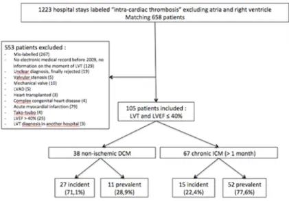
The exhaustive analysis of our data base allowed to identify 1223 hospital stays whose file was labelled with the diagnosis cardiac thrombosis", "intra-ventricular thrombosis", corresponding to 658 patients. Among these 658 patients 105 met the inclusion criteria and were included (figure 1).
Among the 105 included patients with a LVT and LVEF of 40% or less, 38 (36.2%) had a non-ischemic DCM. Among these 38 patients, 27 (71.1%) were "incident", meaning their cardiomyopathy was diagnosed less than a month ago, 11 (28.9%) were "prevalent". Labelled causes of DCM were: idiopathic (19 patients), alcoholic (8 patients), post-chemotherapy (4 patients), non-compaction (2 patients), hereditary with identified mutation (1 patient), Friedreich ataxia (1 patient), Becker myopathy (1 patient), rhythmic (1 patient), post-myocarditis (1 patient). Sixty-seven patients (63.8%) were at a chronic phase of an ischemic cardiomyopathy (ICM). Among these 67 patients, 15 (22.4%) were “incident” (meaning an ancient but unknown coronary disease was diagnosed at the same time as the LVT), 52 (77.6%) were "prevalent". Mean delay between diagnosis of the coronary disease and LVT was 2284 days, i.e 6.3 years) for prevalent patients. Figure 1: Study flow-chart. DCM: Dilated cardiomyopathy. ICM: Ischemic cardiomyopathy. LVT: Left ventricular thrombus. Incident: cardiopathy was diagnosed in the previous month. Prevalent: cardiopathy was diagnosed more than a month before LVT diagnosis.
13
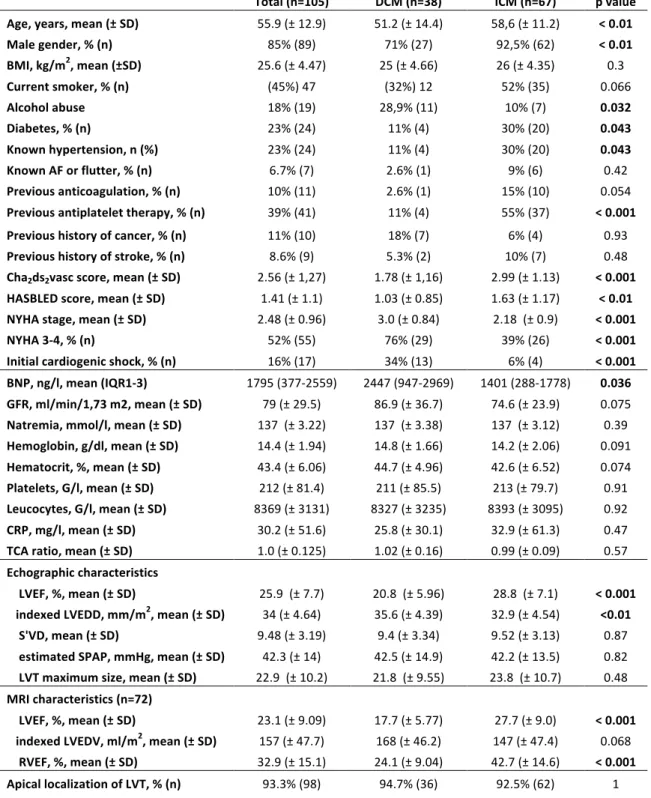
Mean age at diagnosis of LVT was 55.9 (± 12.9). DCM patients were significantly younger (51.2 ± 14.4 years old) than ICM patients (58.6 ± 11.2 years old, p < 0.01, table 1). The majority of patients were males (89 patients, 85%), with a significant difference between DCM (27 patients, 71% males) and ICM (62 patients, 92.5%, p < 0.01, table 1). Diabete and hypertension were more frequent in ICM population, and alcohol abuse was more frequent in DCM population. Unsurprisingly, ICM patients had a higher HASBLED score, in relation with a higher rate of treatment with anti-platelet agents, and a higher Cha2ds2vasc score because of their coronary disease. NYHA stage was higher in DCM patients (3 ± 0.84, and 76% NYHA 3-4) than in ICM population (2.18 ± 0,9 and 39% NYHA 3-4, p < 0.001, table 1). Indeed, initial severity is different among groups with 34% of DCM patients (13 patients) with a cardiogenic shock compared to 6% (4 patients) in ICM population (p < 0.001, table1), and with a higher BNP value for DCM patients (2447 ng/l, IQR 947-2969) than for ICM (1401 ng/l, IQR 188-1778). The two populations had similar blood levels of sodium, haemoglobin, haematocrit, platelets, leucocytes and TCA. DCM patients had a significantly lower LVEF at TTE (20.8 ± 5.96 % versus 28.9 ±7.54 %, p < 0.001, table 1) as well as with MRI (MRI data were available for 32 DCM and 40 ICM, table 1), and higher left ventricular diastolic diameter (35.6 ± 4.39 mm/m2 for DCM versus 32.9 ± 4.54 mm/m2 for ICM, p < 0.01, table1). In addition, DCM patients had a significantly lower right ventricular ejection fraction (RVEF) at MRI (24.1 ± 9.04 % versus 42.7 ± 14.6 %, p < 0.001). The LVT had an apical localization in a vast majority of cases (98 patients, 93.3% of cases).
14
Total (n=105) DCM (n=38) ICM (n=67) p value
Age, years, mean (± SD) 55.9 (± 12.9) 51.2 (± 14.4) 58,6 (± 11.2) < 0.01 Male gender, % (n) 85% (89) 71% (27) 92,5% (62) < 0.01 BMI, kg/m2, mean (±SD) 25.6 (± 4.47) 25 (± 4.66) 26 (± 4.35) 0.3 Current smoker, % (n) (45%) 47 (32%) 12 52% (35) 0.066 Alcohol abuse 18% (19) 28,9% (11) 10% (7) 0.032 Diabetes, % (n) 23% (24) 11% (4) 30% (20) 0.043 Known hypertension, n (%) 23% (24) 11% (4) 30% (20) 0.043 Known AF or flutter, % (n) 6.7% (7) 2.6% (1) 9% (6) 0.42 Previous anticoagulation, % (n) 10% (11) 2.6% (1) 15% (10) 0.054 Previous antiplatelet therapy, % (n) 39% (41) 11% (4) 55% (37) < 0.001 Previous history of cancer, % (n) 11% (10) 18% (7) 6% (4) 0.93 Previous history of stroke, % (n) 8.6% (9) 5.3% (2) 10% (7) 0.48 Cha2ds2vasc score, mean (± SD) 2.56 (± 1,27) 1.78 (± 1,16) 2.99 (± 1.13) < 0.001 HASBLED score, mean (± SD) 1.41 (± 1.1) 1.03 (± 0.85) 1.63 (± 1.17) < 0.01 NYHA stage, mean (± SD) 2.48 (± 0.96) 3.0 (± 0.84) 2.18 (± 0.9) < 0.001 NYHA 3-4, % (n) 52% (55) 76% (29) 39% (26) < 0.001 Initial cardiogenic shock, % (n) 16% (17) 34% (13) 6% (4) < 0.001 BNP, ng/l, mean (IQR1-3) 1795 (377-2559) 2447 (947-2969) 1401 (288-1778) 0.036 GFR, ml/min/1,73 m2, mean (± SD) 79 (± 29.5) 86.9 (± 36.7) 74.6 (± 23.9) 0.075 Natremia, mmol/l, mean (± SD) 137 (± 3.22) 137 (± 3.38) 137 (± 3.12) 0.39 Hemoglobin, g/dl, mean (± SD) 14.4 (± 1.94) 14.8 (± 1.66) 14.2 (± 2.06) 0.091 Hematocrit, %, mean (± SD) 43.4 (± 6.06) 44.7 (± 4.96) 42.6 (± 6.52) 0.074 Platelets, G/l, mean (± SD) 212 (± 81.4) 211 (± 85.5) 213 (± 79.7) 0.91 Leucocytes, G/l, mean (± SD) 8369 (± 3131) 8327 (± 3235) 8393 (± 3095) 0.92 CRP, mg/l, mean (± SD) 30.2 (± 51.6) 25.8 (± 30.1) 32.9 (± 61.3) 0.47 TCA ratio, mean (± SD) 1.0 (± 0.125) 1.02 (± 0.16) 0.99 (± 0.09) 0.57 Echographic characteristics LVEF, %, mean (± SD) 25.9 (± 7.7) 20.8 (± 5.96) 28.8 (± 7.1) < 0.001 indexed LVEDD, mm/m2, mean (± SD) 34 (± 4.64) 35.6 (± 4.39) 32.9 (± 4.54) <0.01 S'VD, mean (± SD) 9.48 (± 3.19) 9.4 (± 3.34) 9.52 (± 3.13) 0.87 estimated SPAP, mmHg, mean (± SD) 42.3 (± 14) 42.5 (± 14.9) 42.2 (± 13.5) 0.82 LVT maximum size, mean (± SD) 22.9 (± 10.2) 21.8 (± 9.55) 23.8 (± 10.7) 0.48 MRI characteristics (n=72) LVEF, %, mean (± SD) 23.1 (± 9.09) 17.7 (± 5.77) 27.7 (± 9.0) < 0.001 indexed LVEDV, ml/m2, mean (± SD) 157 (± 47.7) 168 (± 46.2) 147 (± 47.4) 0.068 RVEF, %, mean (± SD) 32.9 (± 15.1) 24.1 (± 9.04) 42.7 (± 14.6) < 0.001 Apical localization of LVT, % (n) 93.3% (98) 94.7% (36) 92.5% (62) 1
Table 1: Baseline characteristics of the two groups. DCM: Dilated cardiomyopathy. ICM: Ischemic cardiomyopathy. SD: Standard deviation. BMI: Body mass index. AF: Atrial fibrillation. BNP: Brain natriuretic peptide. IQR: Inter-quartile range. GFR: Glomerular filtration rate. TTE: Trans-thoracic echocardiography. MRI: Magnetic resonance imaging. LVEF: left ventricular ejection fraction. LVEDD: Left ventricular end diastolic diameter. LVT: Left ventricular thrombus. LVEDV: Left ventricular end diastolic volume. RVEF: Right ventricular ejection fraction.
15 Diagnosis mode of left ventricular thrombus LVT were mainly diagnosed at first by TTE (56% of patients), followed by MRI (37% of patients), and CT-scan (6,7% of patients, table 2). The diagnosis mode was not statistically different among groups (p = 0.3). If the LVT was initially suspected at TTE, then confirmed with another imaging modality, TTE was considered as the diagnosis mode.
Table 2: Diagnosis mode of left ventricular thrombus among groups. DCM: Dilated cardiomyopathy. ICM: Ischemic cardiomyopathy. TTE: Trans-thoracic echocardiography. MRI: Magnetic resonance imaging. CT-scan: Computed tomography scanner.
Impact of left ventricular thrombus on prognosis: primary endpoint
Median follow-up was of 8.7 years. During the first year, the primary endpoint occurred for 20% of patients (21 patients). This occurrence is not statistically different among groups (18% of DCM patients, 21% of ICM patients, p=0.96, table 3). All cause mortality during the first year was 5.7% (6 patients): 2.6% (1 patient) for DCM, 7.5% (5 patients) for ICM (p=0.41, table 3) and cardio-vascular (CV) mortality was 4.8% (5 patients). CV mortality was due to a cardiogenic shock for 4 patients, and a massive ischemic stroke for 1 patient. The other patient who died from a non-CV death during the first year died from a septic shock.
Symptomatic embolic events occurred in 15% of cases (16 patients): 16% (6 patients) in the DCM group, 15% (10 patients) in the ICM group (table 3, figure 2C). They were: stroke (7 patients), acute ischemia of a limb (5 patients), acute Total (n=105) DCM (n=38) ICM (n=67) TTE 56% (59) 53% (20) 58% (39) MRI 37% (39) 45% (17) 33% (22) CT-scan 6.7% (7) 2.6% (1) 9% (6)
16 myocardial infarction (2 patients), acute kidney ischemia (1 patient), and acute spleen ischemia (1 patient). No DCM patient had disabling stroke (2 had a complete recovery, one had a slight dysarthria left, none had thrombolysis therapy). Among ICM, one ischemia of a limb underwent surgical amputation, one had only toe necrosis (all other had Fogarty without sequel), and two strokes had disabling consequences.
During the whole follow-up, all cause mortality was 10% (11 patients) and wasn't different among groups (p=0.63, figure 2B). Six patients died from cardiogenic shock, 1 from ventricular arrhythmia, 1 from ischemic stroke, 1 from intracerebral bleeding, 2 from septic shock. All symptomatic embolic events occurred during the first 30 days following the diagnosis of LVT.
No significant difference was seen when comparing the incident versus prevalent subgroups (data not shown).
17
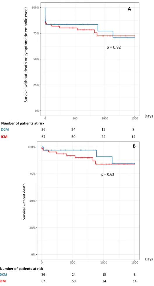
Figure 2: Kaplan-Meier for non-ischemic dilated cardiomyopathy (DCM) and ischemic cardiomyopathy (ICM) for all-cause mortality and symptomatic embolic event (A), All-cause mortality (B). Su rv iv al w ith ou t d eath o r s ym pto m ati c em bo lic e ve nt Days A p = 0.92 Number of patients at risk DCM 36 24 15 8 ICM 67 50 24 14 DCM 36 24 15 8 ICM 67 50 24 14 Su rv iv al w ith ou t d eath Number of patients at risk Days p = 0.63 B
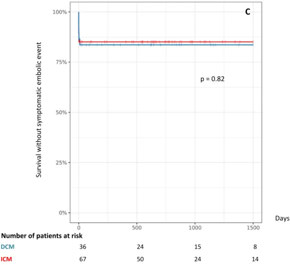
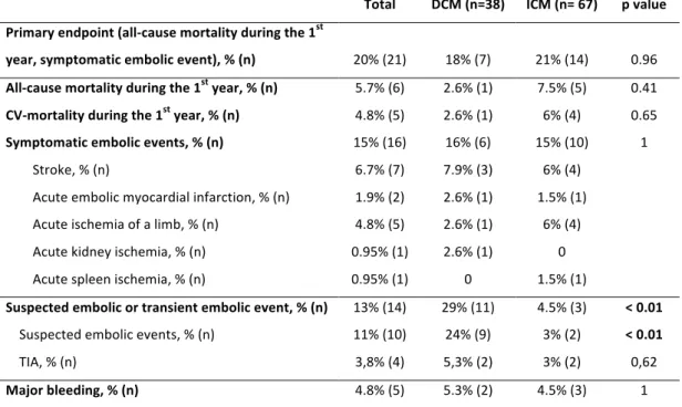
18 Secondary endpoints We noted a suspected or transient embolic event in 13% of all patients, with a significant difference between DCM (29%, 11 patients), and ICM (4.5%, 3 patients, p < 0.01, table 3). Major bleeding events were not different among groups (4.8% of all patients, 5 patients, table 3). Su rv iv al w ith ou t s ym pto m ati c em bo lic e ve nt Days p = 0.82 DCM 36 24 15 8 ICM 67 50 24 14 Number of patients at risk C Figure 2: Kaplan-Meier for non-ischemic dilated cardiomyopathy (DCM) and ischemic cardiomyopathy (ICM) for all-cause mortality and symptomatic embolic event (A), All-cause mortality (B), symptomatic embolic event (C).
19
Total DCM (n=38) ICM (n= 67) p value Primary endpoint (all-cause mortality during the 1st year, symptomatic embolic event), % (n) 20% (21) 18% (7) 21% (14) 0.96 All-cause mortality during the 1st year, % (n) 5.7% (6) 2.6% (1) 7.5% (5) 0.41 CV-mortality during the 1st year, % (n) 4.8% (5) 2.6% (1) 6% (4) 0.65 Symptomatic embolic events, % (n) 15% (16) 16% (6) 15% (10) 1 Stroke, % (n) 6.7% (7) 7.9% (3) 6% (4) Acute embolic myocardial infarction, % (n) 1.9% (2) 2.6% (1) 1.5% (1) Acute ischemia of a limb, % (n) 4.8% (5) 2.6% (1) 6% (4) Acute kidney ischemia, % (n) 0.95% (1) 2.6% (1) 0 Acute spleen ischemia, % (n) 0.95% (1) 0 1.5% (1) Suspected embolic or transient embolic event, % (n) 13% (14) 29% (11) 4.5% (3) < 0.01 Suspected embolic events, % (n) 11% (10) 24% (9) 3% (2) < 0.01 TIA, % (n) 3,8% (4) 5,3% (2) 3% (2) 0,62 Major bleeding, % (n) 4.8% (5) 5.3% (2) 4.5% (3) 1 Table 3: Primary and secondary endpoints among groups. DCM: dilated cardiomyopathy. ICM: Ischemic cardiomyopathy. CV: Cardio-vascular. TIA: transient ischemic attack. Evolution and reverse remodelling Reverse remodelling was observed in 65% of cases after 12 months of follow up, and was significantly more frequent in DCM patients (86%, 19 patients, available data for 22 patients) than ICM patients (54%, 25 patients, available data for 46 patients, p=0.021, table 4). One year after diagnosis of LVT, LVEF was significantly higher in DCM patients (40.2% ± 13.5) compared to ICM patients (33.9% ± 9.3, p = 0.039), BNP was lower (195 ng/l versus 596 ng/l, p < 0.01, table 4). Hospitalization rate within the first year or during follow-up was not statistically different among groups.
Total (n=105) DCM (n=38) ICM (n=67) p value
LV reverse remodelling (n=68), % (n) 65% (44) 86% (19) 54% (25) 0.021 TTE LVEF at 12 months, %, mean (±SD) 35.9 (± 11.1) 40.2 (± 13.5) 33.9 (± 9.3) 0.039 Change in LVEF, %, mean (±SD) S'VD at 12 months, cm, mean (±SD) 9.62 (± 13.5) 11.2 (± 2.91) 20.7 (± 15.5) 11.8 (± 3.4) 4.19 (± 8.15) 10.9 (± 2.56) < 0.001 0.3 BNP at 12 months, pg/ml, mean (IQR1-3) 460 (68-528) 195 (25-204) 597 (101-797) < 0.01 Hospitalization for HF within the 1st year 9.4% (8) 3.3% (1) 13% (7) 0.25 Hospitalization for HF during follow-up 21% (18) 21% (6) 22% (12) 1 Table 4: Evolution of echographic, biological, parameters, and hospitalization for heart failure. DCM: dilated cardiomyopathy. ICM: ischemic cardiomyopathy. LV: left ventricle. TTE: Trans-thoracic echocardiography. LVEF: Left ventricular ejection fraction. SD: standard deviation. BNP: brain natriuretic peptide. IQR: inter-quartile range. HF: heart failure.
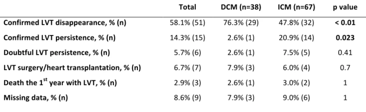
20 As for the thrombus' evolution, LVT disappearance was more frequently confirmed during the first year after diagnosis for DCM (76.3% of LVT disappearance) compared to ICM (47.8% of LVT disappearance, p < 0.01, table 5), whereas it was significantly more frequently persistent one year after diagnosis for ICM (20.9% versus 2.6% for DCM, p=0.023, table 5). Among all patients, 7 had LVT surgery or heart transplantation within the first year after diagnosis, 3 died with their LVT, and LVT's persistence or disappearance was doubtful for 6 patients. This data was missing for 9 patients (lost of follow-up, no imaging exam available at 1 year ± 3 months or when the only exam available is a poor quality TTE that can not exclude LVT's persistence according to the operator).
Total DCM (n=38) ICM (n=67) p value
Confirmed LVT disappearance, % (n) 58.1% (51) 76.3% (29) 47.8% (32) < 0.01 Confirmed LVT persistence, % (n) 14.3% (15) 2.6% (1) 20.9% (14) 0.023 Doubtful LVT persistence, % (n) 5.7% (6) 2.6% (1) 7.5% (5) 0.41 LVT surgery/heart transplantation, % (n) 6.7% (7) 7.9% (3) 6.0% (4) 0.7 Death the 1st year with LVT, % (n) 2.9% (3) 2.6% (1) 3.0% (2) 1 Missing data, % (n) 8.6% (9) 7.9% (3) 9.0% (6) 1 Table 5: Left ventricular thrombus disappearance or persistence according to groups, within the first year after diagnosis. DCM: dilated cardiomyopathy, ICM: ischemic cardiopathy, LVT: left ventricular thrombus Risk factors and impact on prognosis of embolic events
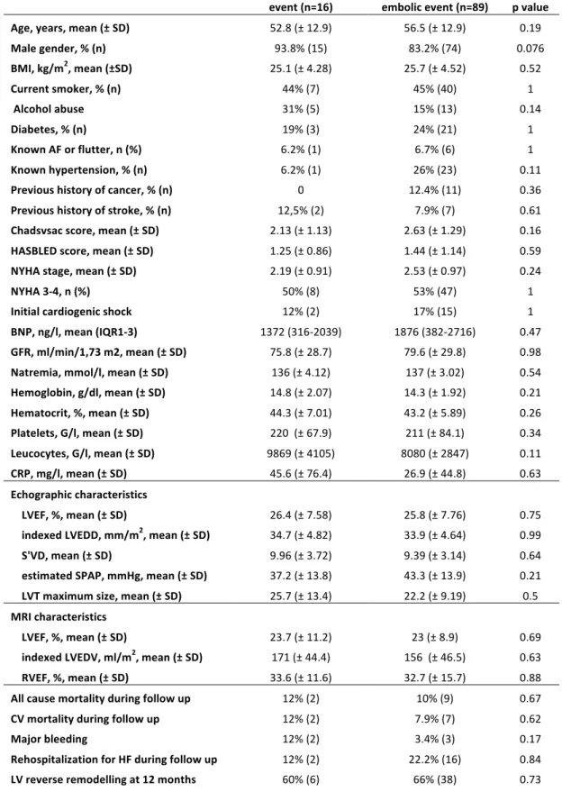
We could not find any significant difference as for baseline characteristics between patients with a symptomatic embolic event (16 patients) and without symptomatic embolic event (89 patients), including Cha2ds2vasc score. Mortality during follow-up, hospitalization rate for heart failure, and LV remodelling rate were not significantly different among groups (table 6).
21
Symtomatic embolic
event (n=16) embolic event (n=89) No symptomatic p value
Age, years, mean (± SD) 52.8 (± 12.9) 56.5 (± 12.9) 0.19 Male gender, % (n) 93.8% (15) 83.2% (74) 0.076 BMI, kg/m2, mean (±SD) 25.1 (± 4.28) 25.7 (± 4.52) 0.52 Current smoker, % (n) 44% (7) 45% (40) 1 Alcohol abuse 31% (5) 15% (13) 0.14 Diabetes, % (n) 19% (3) 24% (21) 1 Known AF or flutter, n (%) 6.2% (1) 6.7% (6) 1 Known hypertension, % (n) 6.2% (1) 26% (23) 0.11 Previous history of cancer, % (n) 0 12.4% (11) 0.36 Previous history of stroke, % (n) 12,5% (2) 7.9% (7) 0.61 Chadsvsac score, mean (± SD) 2.13 (± 1.13) 2.63 (± 1.29) 0.16 HASBLED score, mean (± SD) 1.25 (± 0.86) 1.44 (± 1.14) 0.59 NYHA stage, mean (± SD) 2.19 (± 0.91) 2.53 (± 0.97) 0.24 NYHA 3-4, n (%) 50% (8) 53% (47) 1 Initial cardiogenic shock 12% (2) 17% (15) 1 BNP, ng/l, mean (IQR1-3) 1372 (316-2039) 1876 (382-2716) 0.47 GFR, ml/min/1,73 m2, mean (± SD) 75.8 (± 28.7) 79.6 (± 29.8) 0.98 Natremia, mmol/l, mean (± SD) 136 (± 4.12) 137 (± 3.02) 0.54 Hemoglobin, g/dl, mean (± SD) 14.8 (± 2.07) 14.3 (± 1.92) 0.21 Hematocrit, %, mean (± SD) 44.3 (± 7.01) 43.2 (± 5.89) 0.26 Platelets, G/l, mean (± SD) 220 (± 67.9) 211 (± 84.1) 0.34 Leucocytes, G/l, mean (± SD) 9869 (± 4105) 8080 (± 2847) 0.11 CRP, mg/l, mean (± SD) 45.6 (± 76.4) 26.9 (± 44.8) 0.63 Echographic characteristics LVEF, %, mean (± SD) 26.4 (± 7.58) 25.8 (± 7.76) 0.75 indexed LVEDD, mm/m2, mean (± SD) 34.7 (± 4.82) 33.9 (± 4.64) 0.99 S'VD, mean (± SD) 9.96 (± 3.72) 9.39 (± 3.14) 0.64 estimated SPAP, mmHg, mean (± SD) 37.2 (± 13.8) 43.3 (± 13.9) 0.21 LVT maximum size, mean (± SD) 25.7 (± 13.4) 22.2 (± 9.19) 0.5 MRI characteristics LVEF, %, mean (± SD) 23.7 (± 11.2) 23 (± 8.9) 0.69 indexed LVEDV, ml/m2, mean (± SD) 171 (± 44.4) 156 (± 46.5) 0.63 RVEF, %, mean (± SD) 33.6 (± 11.6) 32.7 (± 15.7) 0.88 All cause mortality during follow up 12% (2) 10% (9) 0.67 CV mortality during follow up 12% (2) 7.9% (7) 0.62 Major bleeding 12% (2) 3.4% (3) 0.17 Rehospitalization for HF during follow up 12% (2) 22.2% (16) 0.84 LV reverse remodelling at 12 months 60% (6) 66% (38) 0.73
Table 6: Comparison of baseline characteristics and follow-up data for patients with and without a symptomatic embolic event. SD: Standard deviation. BMI: Body mass index. AF: Atrial fibrillation. BNP: Brain natriuretic peptide. IQR: inter-quartile range. GFR: Glomerular filtration rate. LVEF: left ventricular ejection fraction. LVEDD: left ventricular end diastolic diameter. LVEDV: left ventricular end-diastolic volume. MRI: Magnetic resonance imaging. RVEF: Right ventricular ejection fraction. CV: Cardio-vascular. HF: Heart failure. LV: Left ventricle.
22
Management of left ventricular thrombus
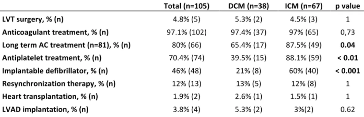
5 patients (4.8%) had a surgery of LVT: 2 DCM patients had no size reduction of a mobile LVT in spite of anticoagulant treatment during at least 5 days, with suspected cerebral embolic events (asymptomatic ischemic lesions on a CT scan) 1 ICM had a symptomatic ischemia of a kidney and a voluminous and mobile residual LVT, 1 ICM had a very voluminous LVT (maximum size 44 mm) and had a TIA, one ICM had coronary artery bypass and left ventricle aneurysm resection at the same time. 97.1% (102 patients) received anticoagulant treatment (table 6). All given anticoagulant drugs were vitamin K antagonists. One DCM patient didn't receive anticoagulant treatment because of a major bleeding, 2 ICM patients didn't receive it because of double anti-platelet therapy and high estimated bleeding risk.
Long-term anticoagulant treatment was maintained in 80% of cases (66/ 81 available data) and was significantly more frequent in ICM population (87.5%, 49 patients), than DCM (with still 65.4%, 17 patients, p=0.04, table 7).
Unsurprisingly, ICM patients received anti-platelet therapy and implantable defibrillator more frequently. Resynchronization therapy, heart transplantation, and left-ventricular-assist-device implantation were not different among groups (table 7).
Total (n=105) DCM (n=38) ICM (n=67) p value
LVT surgery, % (n) 4.8% (5) 5.3% (2) 4.5% (3) 1 Anticoagulant treatment, % (n) 97.1% (102) 97.4% (37) 97% (65) 0,73 Long term AC treatment (n=81), % (n) 80% (66) 65.4% (17) 87.5% (49) 0.04 Antiplatelet treatment, % (n) 70.4% (74) 39.5% (15) 88.1% (59) < 0.01 Implantable defibrillator, % (n) 46% (48) 21% (8) 60% (40) < 0.001 Resynchronization therapy, % (n) 12% (13) 13% (5) 12% (8) 1 Heart transplantation, % (n) 1.9% (2) 2.6% (1) 1.5% (1) 1 LVAD implantation, % (n) 3.8% (4) 5.3% (2) 3%(2) 0.62
Table 7: Management of LVT (left ventricular thrombus). DCM: dilated cardiomyopathy. ICM: ischemic cardiomyopathy. AC: Anticoagulant. LVAD: Left ventricular assistance device
23
Risk factors of left ventricular thrombus for dilated cardiomyopathies
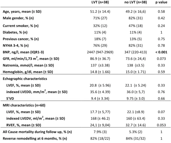
When comparing DCM with LVT with a cohort of 38 DCM without LVT, matched for age, sex, NYHA stage and LVEF in TTE and MRI (MRI was available for 60 patients), we didn't find any difference in terms of diabetes rate, smoking rate, biological profile except BNP, TTE and MRI characteristics, all cause mortality during follow-up, and reverse remodelling at 6 months. However, we found a significantly higher BNP level in patients with LVT. LVT (n=38) no LVT (n=38) p value Age, years, mean (± SD) 51.2 (± 14.4) 49.2 (± 16,6) 0.58 Male gender, % (n) 71% (27) 82% (31) 0.42 Current smoker, % (n) 32% (12) 47% (18) 0.24 Diabetes, % (n) 11% (4) 11% (4) 1 Previous cancer, % (n) 18% (7) 13% (5) 0.75 NYHA 3-4, % (n) 76% (29) 82% (31) 0.78 BNP, ng/l, mean (IQR1-3) 2447 (947-2969) 347 (220-413) < 0.001 GFR, ml/min/1,73 m2, mean (± SD) 86.9 (± 36.7) 73.6 (± 24,4) 0.073 Natremia, mmol/l, mean (± SD) 137 (±3.38) 138 (±3.5) 0.33 Hemoglobin, g/dl, mean (± SD) 14.8 (± 1.66) 15.0 (± 1.71) 0.59 Echographic characteristics LVEF, %, mean (± SD) 20.8 (± 5.96) 22.1 (± 5.24) 0.33 indexed LVEDD, mm/m2, mean (± SD) 35.6 (± 4.39) 36.0 (± 5,7) 0.76 S'VD 9.4 (± 3.34) 9.75 (± 3.0) 0.66 MRI characteristics (n=60) LVEF, %, mean (± SD) 17.7 (± 5,77) 22.1 (±8.9) 0.07 indexed LVEDV, ml/m2, mean (± SD) 168 (± 46.2) 160 (± 63.4) 0.33 RVEF, %, mean (± SD) 24,1 (± 9,04) 32.7 (± 14.6) 0.053 All Cause mortality during follow up, % (n) 7.9% (3) 5.3% (2) 1 Reverse remodelling at 6 months, % (n) 82% (18/22) 84% (31/32) 1
Table 7: Baseline and evolution characteristics of a dilated cardiomyopathy, with and without left ventricular thrombus (LVT). SD: Standard deviation. NYHA: New York Heart Association. BNP: Brain natriuretic peptide. IQR: inter-quartile range. GFR: Glomerular filtration rate LVEF: left ventricular ejection fraction. LVEDD: left ventricular end diastolic diameter. LVEDV: left ventricular end-diastolic volume. MRI:
24
Discussion
This retrospective study aimed at better characterizing LVT in a context of non-ischemic DCM or ICM and heart failure, excluding any recent myocardial infarction, providing new data on this unknown population. We found a relatively high rate of embolic events (15%), but a low CV mortality rate of 4.8% within the first year and a high proportion of patients with DCM experienced a reverse remodelling (86% at 12 months). Despite the fact that DCM patients had more severe heart failure (higher BNP level and NYHA stage, lower LVEF), we found no difference in the rate of embolic events between DCM and ICM.
Nowadays, patients with heart failure keep on having a poor prognosis despite medical management improvements, and a significant rate of embolic events despite the absence of detected atrial fibrillation. Therefore, we keep on looking for a potential interest of an anticoagulation therapy for patients with heart failure in order to prevent embolic events and mortality [3]. Several trials tested a potential benefit of anticoagulation toward antiplatelet therapy in heart failure, with mitigated conclusions. A meta-analysis by Hopper et al [11], of four relevant prospective randomized controlled trials (WASH [7], HELAS [6], WATCH [5] and WARCEF [8]) including patients with HF and reduced ejection fraction in sinus rhythm, showed no difference in all-cause mortality when aspirin was compared with warfarin, but a significant reduction in all strokes (fatal and non-fatal ischaemic and haemorrhagic strokes) was seen with warfarin compared with aspirin (relative risk of 0.59) allowing to hope for anticoagulants to be beneficial against no treatment at all. However, the recent large-scale COMMANDER-HF trial was inconclusive [4]. This major placebo-controlled trial including 5022 patients with heart failure due to a ICM, LVEF < 40%, in sinus rhythm, tested the hypothesis of a benefit of the direct factor Xa inhibitor rivaroxaban for death, myocardial infarction, and stroke. It did not show any significant difference between the two groups, supporting the position that oral anticoagulation is not indicated for patients with heart failure and reduced ejection fraction in the absence of atrial fibrillation, in accordance to previous studies. However, in a post hoc analysis of the COMMANDER-HF trial, rivaroxaban significantly reduced the occurrence of all-cause strokes or TIA
25 compared with placebo by 32%, in the vulnerable early and late phase after a recent episode of worsening of heart failure [12] suggesting some patients could still benefit from an anticoagulant therapy. In this context, we have good reasons to wonder about the interest of anticoagulant treatment in patients with heart failure in sinus rhythm.
If left ventricular thrombus is one remaining indication of anticoagulant
treatment in heart failure, its natural history, risk factors, and embolic potential are to be clarified. To our knowledge, our study is the largest recent retrospective cohort focusing on a population of patients with heart failure and reduced ejection fraction, distinguishing non-ischemic DCM from ICM, with a LVT. Several studies before the MRI era have tried to determine prevalence and embolic potential of LVT in a context of heart failure and dilated cardiomyopathy, but with mixed etiologies [13], with very few patients with idiopathic DCM [14] and, interestingly, couldn't find any correlation between the presence of a LVT and embolic events [15,16]. One major interest of our study remains in the fact that patients with DCM and a LVT are barely described in literature, as a distinct group with specificities. We have found that non-ischemic DCM with a LVT are initially especially severe, that they have a relatively high rate of embolic events, but with few long term consequences, a good recovery potential with 86% of reverse remodelling at 12 months, and a low mortality < 3% the first year. Risk factors of LVT were hard to highlight in this retrospective study: a high BNP value was associated with LVT.
The low impact of LVT on mortality put into question the role of LVT surgery, and would rather suggest a wait-and-see attitude. Indeed, heart failure patients who undergo surgical thrombectomy are exposed to significant perioperative complications and high mortality, or may be precipitated toward LVAD implantation or heart transplant after cardioplegia, with a risk of worsening organs’ ischemic injury. If no other surgical gesture is done in the same time, as coronary bypass or valve repair, the risk-benefit balance of a LVT surgery could be in favour of abstention. However, vitamin K antagonists remain the reference treatment. No non-vitamin K oral anticoagulant (NOAC) was used in this study, but they will probably emerge for this indication in the future since
26 there is no study showing a higher efficacy of vitamin K antagonists over NOAC in the resorption of LVT, and they have already been used in some cases [17,18].
This retrospective study has several limitations, starting with the recruiting mode with a high rate of patients excluded for mislabelling, reflecting the failing classification system of our electronic medical record device. Without any doubt, we missed as many left ventricular thrombi that were not correctly labelled, which is why this study cannot determine their prevalence. However it should not introduce bias in patients' characteristics.
We were not able to highlight any embolic risk factor and many other paths need to be further explored. For example we were not able to report the mobility of thrombi in a proper way, and didn't have access to a systematic imaging to look for systemic embolism in the absence of related symptoms, showing the absence of any defined approach of LVT complications, compared to endocarditis for example.
This study is single-centred, the number of patients is low (38 DCM, 67 ICM) and the retrospective nature of the study caused missing data. However, DCM patients with a LVT are a rare population, for which it is difficult to find series.
Conclusions
Left ventricular thrombus is associated with a significant rate of embolic events, but the presence of LVT or embolic events does not seem to impact mortality. Moreover, the presence of LVT does not preclude reverse remodelling under medical treatment. The occurrence of LVT may be linked to the initial severity. Further investigation is needed to identify risk factors of LVT and of embolic events. Meanwhile, a wait-and-see attitude under cover of anticoagulation could be the best management of LVT in heart failure.
27
References
[1] McCarthy CP, Vaduganathan M, McCarthy KJ, Januzzi JL, Bhatt DL, McEvoy JW. Left Ventricular Thrombus After Acute Myocardial Infarction: Screening, Prevention, and Treatment. JAMA Cardiology 2018;3:642. doi:10.1001/jamacardio.2018.1086.
[2] McCarthy CP, Murphy S, Venkateswaran RV, Singh A, Chang LL, Joice MG, et al. Left Ventricular Thrombus. Journal of the American College of Cardiology 2019;73:2007–9. doi:10.1016/j.jacc.2019.01.031.
[3] Zannad F, Stough WG, Regnault V, Gheorghiade M, Deliargyris E, Gibson CM, et al. Is thrombosis a contributor to heart failure pathophysiology? Possible mechanisms, therapeutic opportunities, and clinical investigation challenges. International Journal of Cardiology 2013;167:1772–82. doi:10.1016/j.ijcard.2012.12.018.
[4] Zannad F, Anker SD, Byra WM, Cleland JGF, Fu M, Gheorghiade M, et al. Rivaroxaban in Patients with Heart Failure, Sinus Rhythm, and Coronary Disease. New England Journal of Medicine 2018;379:1332–42. doi:10.1056/NEJMoa1808848.
[5] Massie BM, Collins JF, Ammon SE, Armstrong PW, Cleland JGF, Ezekowitz M, et al. Randomized Trial of Warfarin, Aspirin, and Clopidogrel in Patients With Chronic Heart Failure: The Warfarin and Antiplatelet Therapy in Chronic Heart Failure (WATCH) Trial. Circulation 2009;119:1616–24. doi:10.1161/CIRCULATIONAHA.108.801753.
[6] Cokkinos DV, Haralabopoulos GC, Kostis JB, Toutouzas PK, for the HELAS investigators. Efficacy of antithrombotic therapy in chronic heart failure: The HELAS study. European Journal of Heart Failure 2006;8:428–32. doi:10.1016/j.ejheart.2006.02.012.
[7] Cleland JGF, Findlay I, Jafri S, Sutton G, Falk R, Bulpitt C, et al. The Warfarin/Aspirin study in heart failure (WASH): a randomized trial comparing antithrombotic strategies for patients with heart failure. American Heart Journal 2004;148:157–64. doi:10.1016/j.ahj.2004.03.010.
[8] Homma S, Thompson JLP, Pullicino PM, Levin B, Freudenberger RS, Teerlink JR, et al. Warfarin and Aspirin in Patients with Heart Failure and Sinus Rhythm. New England Journal of Medicine 2012;366:1859–69. doi:10.1056/NEJMoa1202299.
[9] Ponikowski P, Voors AA, Anker SD, Bueno H, Cleland JGF, Coats AJS, et al. 2016 ESC Guidelines for the diagnosis and treatment of acute and chronic heart failure: The Task Force for the diagnosis and treatment of acute and chronic heart failure of the European Society of Cardiology (ESC)Developed with the special contribution of the Heart Failure Association (HFA) of the ESC. European Heart Journal 2016;37:2129–200. doi:10.1093/eurheartj/ehw128.
[10] Lee JM, Park JJ, Jung HW, Cho Y-S, Oh I-Y, Yoon C-H, et al. Left Ventricular Thrombus and Subsequent Thromboembolism, Comparison of Anticoagulation, Surgical Removal, and Antiplatelet Agents. Journal of Atherosclerosis and Thrombosis 2013;20:73–93. doi:10.5551/jat.13540.
[11] Hopper I, Skiba M, Krum H. Updated meta-analysis on antithrombotic therapy in patients with heart failure and sinus rhythm. European Journal of Heart Failure 2013;15:69–78. doi:10.1093/eurjhf/hfs171.
[12] Mehra MR, Vaduganathan M, Fu M, Ferreira JP, Anker SD, Cleland JGF, et al. A comprehensive analysis of the effects of rivaroxaban on stroke or transient ischaemic
28
attack in patients with heart failure, coronary artery disease, and sinus rhythm: the COMMANDER HF trial. European Heart Journal 2019. doi:10.1093/eurheartj/ehz427.
[13] Gottdiener JS, Gay JA, VanVoorhees L, DiBianco R, Fletcher RD. Frequency and embolic potential of left ventricular thrombus in dilated cardiomyopathy: Assessment by 2-dimensional echocardiography. The American Journal of Cardiology 1983;52:1281–5. doi:10.1016/0002-9149(83)90588-X.
[14] Ciaccheri M, Castelli G, Cecchi F, Nannini M, Santoro G, Troiani V, et al. Lack of correlation between intracavitary thrombosis detected by cross sectional echocardiography and systemic emboli in patients with dilated cardiomyopathy. Heart 1989;62:26–9. doi:10.1136/hrt.62.1.26. [15] Katz SD. Left ventricular thrombus and the incidence of thromboembolism in patients with congestive heart failure: can clinical factors identify patients at increased risk? Journal of Cardiovascular Risk 1995;2:6. doi:10.1177/174182679500200203.
[16] Cioffi G, Pozzoli M, Forni G, Franchini M, Opasich C, Cobelli F, et al. Systemic thromboembolism in chronic heart failure: A prospective study in 406 patients. European Heart Journal 1996;17:1381–9. doi:10.1093/oxfordjournals.eurheartj.a015073.
[17] Nakasuka K, Ito S, Noda T, Hasuo T, Sekimoto S, Ohmori H, et al. Resolution of Left Ventricular Thrombus Secondary to Tachycardia-Induced Heart Failure with Rivaroxaban. Case Reports in Medicine 2014;2014:1–5. doi:10.1155/2014/814524.
[18] Kolekar S, Munjewar C, Sharma S. Dabigatran for left ventricular thrombus. Indian Heart Journal 2015;67:495–6. doi:10.1016/j.ihj.2015.06.010.
29
Abbreviations
AC: Anticoagulant BNP: Brain natriuretic peptide CT-Scan: Computed tomography scanner CV: Cardio-vascular DCM: Dilated cardiomyopathy GFR: Glomerular filtration rate HF: Heart failure ICM: Ischemic cardiomyopathy LV: Left ventricle LVEF: Left ventricular ejection fraction LVT: Left ventricular thrombus LVEDD: Left ventricular end-diastolic diameter LVEDV: Left ventricular end-diastolic volume MRI: Magnetic resonance imaging NOAC: Non-vitamin K oral anticoagulant NYHA: New York Heart Association RVEF: Right ventricular ejection fraction SD: Standard deviation SPAP: Systolic pulmonary arterial pressure TIA: Transient ischemic attack TTE: Trans-Thoracic Echocardiography
30
Serment médical
Au moment d’être admise à exercer la médecine, je promets et je jure d’être fidèle aux lois de l’honneur et de la probité.
Mon premier souci sera de rétablir, de préserver ou de promouvoir la santé dans tous ses éléments, physiques et mentaux, individuels et sociaux.
Je respecterai toutes les personnes, leur autonomie et leur volonté, sans
aucune discrimination selon leur état ou leurs convictions. J’interviendrai pour les protéger si elles sont affaiblies, vulnérables ou menacées dans leur intégrité ou leur dignité. Même sous la contrainte, je ne ferai pas usage de mes connaissances contre les lois de l’humanité. J’informerai les patients des décisions envisagées, de leurs raisons et de leurs conséquences. Je ne tromperai jamais leur confiance et n’exploiterai pas le pouvoir hérité des circonstances pour forcer les consciences. Je donnerai mes soins à l’indigent et à quiconque me les demandera. Je ne me laisserai pas influencer par la soif du gain ou la recherche de la gloire. Admise dans l’intimité des personnes, je tairai les secrets qui me seront confiés. Reçue à l’intérieur des maisons, je respecterai les secrets des foyers et ma conduite ne servira pas à corrompre les mœurs.
Je ferai tout pour soulager les souffrances. Je ne prolongerai pas abusivement les agonies. Je ne provoquerai jamais la mort délibérément.
Je préserverai l’indépendance nécessaire à l’accomplissement de ma
mission. Je n’entreprendrai rien qui dépasse mes compétences. Je les entretiendrai et les perfectionnerai pour assurer au mieux les services qui me seront demandés.
J’apporterai mon aide à mes confrères ainsi qu’à leurs familles dans l’adversité.
Que les hommes et mes confrères m’accordent leur estime si je suis fidèle
à mes promesses ; que je sois déshonorée et méprisée si j’y manque.
31
Le thrombus intra-ventriculaire gauche dans l'insuffisance cardiaque
Contexte: Les patients souffrant d'insuffisance cardiaque (IC) à fraction d'éjection ventriculaire
gauche altérée (FEVG<40%) constitue une source croissante de thrombus intra-ventriculaire gauche (TIVG). Pourtant, les cardiomyopathies ischémiques (CMI) ou dilatées non-ischémiques (CMD) avec TIVG sont peu décrites dans la littérature. Objectif: Cette étude rétrospective monocentrique cherche à décrire ces deux populations, leur pronostic et complications.
Méthodes: Ont été inclues toutes les CMD ou CMI avec TIVG hospitalisées dans notre institution
de 2005 à 2018. Ont été exclus: sténoses valvulaires, prothèses mécaniques, assistance VG, transplantés cardiaques, cardiopathies congénitales, infarctus du myocarde (IDM) récents. Le critère de jugement principal composite était: mortalité toute cause, évènement embolique symptomatique (AVC, IDM, embol périphérique) la 1ère année. Les critères secondaires étaient: évènement embolique transitoire/suspecté, hémorragie majeure. Le remodelage inverse était une augmentation de FEVG >10%, ou baisse de diamètre/volume >10%. Résultats: Sur 38 CMD et 67 CMI incluses, l'âge moyen était 56±13 ans, avec 85% d'hommes. Les CMD avaient un NYHA et BNP plus élevés, une FEVG plus basse. Le critère de jugement principal est survenu dans 20% des cas. La mortalité toute cause était de 5.7% (6 patients), la mortalité cardio-vasculaire de 4.8% (5 pts). 15% des patients ont présenté un évènement embolique symptomatique sans différence entre les groupes, 13% un évènement suspecté/transitoire (29% des CMD, 4.5% des CMI, p<0.01). 5 patients se sont fait opérés du TIVG, tous furent anticoagulés sauf 1 CMD (hémorragie majeure) et 2 CMI (risque hémorragique important). 86% des CMD ont présenté un remodelage inverse à 1 an, 54% des CMI (p=0.02). Les CMD avec TIVG avaient un BNP plus élevé que celles sans TIVG. Conclusion: Les TIVG dans l'IC sont à risque d'évènement embolique mais ne semblent pas impacter la mortalité ni empêcher un remodelage inverse. Left ventricular thrombus in heart failure Background: Heart failure with reduced ejection fraction (EF<40%) is a growing source of left
ventricular thrombus (LVT) but LVT complicating ischemic (ICM) or non-ischemic dilated cardiomyopathy (DCM) are barely described. Purpose: Through a retrospective monocentric study, we aimed at describing DCM and ICM with LVT, their prognosis and complications.
Methods: We studied all DCM and ICM patients with LVT in our institution from 2005 to 2018.
Stenosing or prosthetic valves, heart transplant, LVAD, congenital diseases, acute myocardial infarction (AMI) were excluded. Our primary endpoint was a composite of all-cause mortality (ACM), symptomatic embolic event (stroke, AMI, symptomatic peripheral embolic events) during the 1st year. Secondary endpoints were suspected/transient embolic events, major bleeding and LV reverse remodelling (LVRR) described as an increase of LVEF >10% or a decrease in LV volume/diameter >10%. We compared our DCM patients to DCM without LVT for case-control analysis. Results: 38 DCM and 67 ICM were included. Mean age was 56±13, 85% were males. DCM had higher NYHA, BNP level, lower LVEF. The primary endpoint occurred in 20% without difference between groups. ACM the 1st year was 5.7% (6 patients) and cardio-vascular mortality 4.8% (5 pts). Symptomatic embolic events occurred in 15% without difference between groups. We noted suspected/transient embolic events in 13% of cases: 29% of DCM (11 pts), 4.5% of ICM (3 pts, p<0.01). 5 patients had a LVT surgery, 1 DCM didn't receive anticoagulant treatment because of a major bleeding, 2 ICM because of a high bleeding risk. Major bleedings weren't different (4.8%). LVRR was observed in 65% of cases after 1 year and more frequently in DCM (86%) than ICM (54%, p=0.02). DCM patients with a LVT had a significantly higher BNP level compared to DCM without LVT. Conclusion: LVT is associated with a significant rate of embolic events but doesn't seem to impact mortality and doesn't preclude reverse remodelling.
Key-words: left ventricular thrombus, heart failure, dilated cardiomyopathy, thrombo-embolic
risk, stroke.
Université de Bordeaux U.F.R. DES SCIENCES MEDICALES Discipline: Cardiologie et Maladies vasculaires