1
Université de Montréal
Comparison of vitrification protocols in
immature equine oocytes
Par
Karla Elena Herrera Hidalgo
Département de biomédecine vétérinaire, Faculté de médecine vétérinaire
Mémoire présenté à la Faculté de médecine vétérinaire en vue de l’obtention du grade de Maîtrise ès sciences (M. Sc.)
en sciences vétérinaires, option reproduction
Décembre 2019
2
Université de Montréal
Unité académique : Département de biomédecine vétérinaire, Faculté de médecine vétérinaire
Ce mémoire intitulé
Comparison of vitrification protocols in immature equine oocytes
Présenté par
Karla Elena Herrera Hidalgo
A été évalué par un jury composé des personnes suivantes Mila Freire Présidente-rapporteuse Mouhamadou Diaw Directeur de recherche Derek Boerboom Codirecteur Christopher Price Codirecteur Jocelyn Dubuc Membre du jury
3
Résumé
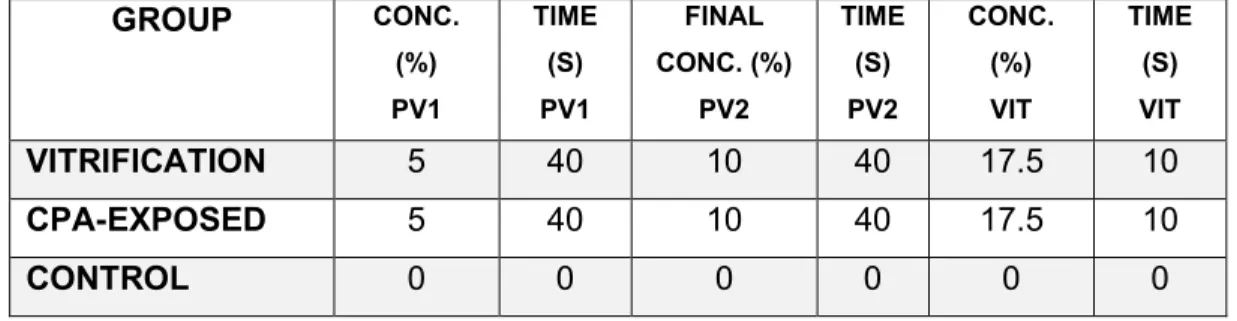
La cryoconservation d'ovocytes est une méthode qui faciliterait la conservation du potentiel génétique chez la femelle et permettrait plus de flexibilité dans l'application des techniques de reproduction assistée chez les animaux domestiques et les espèces en voie de disparition. Chez le cheval, le taux de réussite de cette technique est faible comparée à celui obtenu chez d’autres espèces animales. Par conséquent, plus d’études seront nécessaires pour élucider les mécanismes spécifiques responsables du faible taux de succès après la cryopréservation. Le but de cette étude était d'évaluer l'effet de la vitrification d'ovocytes équins immatures sur leur taux de maturation, de clivage et le développement de blastocystes en utilisant un protocole de vitrification en trois étapes avec de l’ethylène glycol (EG) et du diméthylsulfoxyde (DMSO), ainsi que comparer l'effet des milieux hors congélation. Le protocole de vitrification utilisé dans la présente étude a été conçu en fonction des résultats obtenus au cours d’études préliminaires. Des ovocytes provenant de follicules immatures de juments ont été conservés pendant une nuit (14-18 heures) à température ambiante (~22⁰C) dans un milieu de maintien. Le lendemain, les ovocytes ont été dénudés et placés dans une solution de base (BS) composée de 20% de sérum de veau fœtal (FBS) + M199/Hanks’ salts. Les ovocytes ont ensuite été répartis au hasard dans différents groupes : contrôle, vitrification et exposés aux agents cryoprotecteurs (CPA)-. Les ovocytes du groupe contrôle ont été immédiatement mis en maturation in vitro (IVM). Trois ovocytes ont été exposés à un protocole de vitrification en trois étapes décomposées en (1) solution de pré-vitrification (PVS) 1 (5% EG / 5 DMSO) 40s. (2) PVS 2 (10% EG / 10% DMSO) 40s et enfin, (3) solution de vitrification (VIT) (17,5% EG / 17,5% DMSO / 3 M saccharose) 10s. Le groupe vitrification est plongé dans l'azote liquide alors que les groupes CPA-exposés ont été exposés aux cryoprotecteurs mais n’ont pas été congelés. Les ovocytes ont ensuite été transférés sur un maillage en acier inoxydable stérile puis réchauffés à 42 ° C dans un BS pendant 5 min. Les ovocytes ont ensuite été soumis à l’IVM, fécondés par injection intracytoplasmique d’un spermatozoïde puis mis en culture dans le but de produire des embryons. Les différences en termes de maturation, de clivage et de taux de blastocystes entre les groupes ont été analysées par le test exact de Fisher. Le taux de maturation des deux groupes vitrification et CPA-exposés ne différait pas significativement avec le groupe contrôle. Aucun blastocyste n'a cependant été obtenu des groupes vitrification et CPA-exposés. Ces résultats ont montré que les ovocytes
4
équins immatures peuvent maintenir une viabilité et une compétence méiotique après vitrification similaires à celles du groupe contrôle; de plus, l'exposition aux cryoprotecteurs n'a pas abouti à la formation de blastocystes en comparaison avec le groupe contrôle. Une étude plus approfondie sur la physiologie des ovocytes équins est nécessaire afin de pouvoir optimiser la production d’embryons.
Mots-clés : cryoconservation, vitrification, équidés, ovocytes immatures, cryoprotecteurs.
5
Abstract
Oocyte cryopreservation would facilitate the conservation of female genetic material and allow more flexibility in the application of assisted reproductive techniques in domestic animals and endangered species. The overall success rate of this technique in the horse is low compared with other species. Therefore, further research is required to elucidate the species-specific mechanisms responsible for poor survivability following vitrification. This study aimed to evaluate the effect on maturation rate, cleavage and blastocyst development of vitrified immature equine oocytes, using a three-step vitrification protocol with ethylene glycol (EG) and dimethyl sulfoxide (DMSO); and comparing the effect of media without freezing. The vitrification protocol was designed based on the results of preliminary experiments. Oocytes were recovered from immature follicles of live mares. Oocytes were held overnight at room temperature (14-24 hrs) in a holding medium. Oocytes were then denuded and placed in a base solution (BS) composed of 20% fetal bovine serum (FBS) + M199/Hanks’ salts. Oocytes were randomly allotted to control, vitrification, and cryoprotectant agents (CPAs)-exposed groups. Control oocytes were cultured directly for in-vitro maturation (IVM). Three oocytes were exposed to a three-step vitrification protocol composed of a pre-vitrification solution (PVS) 1 (5% EG/ 5% DMSO); PVS 2 (10% EG/ 10% DMSO) during 40s each; and finally vitrification solution (VS) (17.5% EG/ 17.5% DMSO/ 3 M sucrose), during 10s. All media were diluted in M199/Hanks’ salts + 20% FBS. Oocytes were then transferred to a 75-μm sterile stainless steel mesh. The oocytes were warmed at 42°C in the BS for 5 minutes. Oocytes from the vitrified group were plunged into liquid nitrogen, while oocytes from CPA-exposed groups were only CPA-exposed to cryoprotectants. Oocytes were then subjected to IVM, fertilization and embryo culture. Fisher's Exact Test analyzed differences in maturation, cleavage and blastocyst rates between groups. The maturation rate of vitrified and CPA-exposed groups did not differ significantly from control oocytes. However, no blastocysts were obtained from CPA-exposed and vitrified groups. Vitrification and control groups showed that immature equine oocytes could maintain viability and meiotic competence; moreover, cryoprotectant exposure did not show any blastocyst formation as compared to control. Further investigation is necessary to understand the overall physiology of equine oocytes in order to optimize the developmental capacity of embryos. Keywords: cryopreservation, vitrification, equine, immature oocytes, cryoprotectants.
6
Table of content
Résumé ... 3 Abstract ... 5 Table of content ... 6 List of tables ... 8 List of figures ... 9 List of abbreviations ... 10 Dedication ………... 12 Acknowledgment ... 13 1. Introduction ... 15 2. Literature review ... 172.1. Ovarian anatomy of the mare ... 17
2.2. Assisted reproductive techniques in the horse ... 19
2.3. Principles of cryopreservation ... 21
2.4. Oocyte damage caused by vitrification ... 27
2.5. Vitrification protocols ... 34
3. Hypothesis and objectives ... 43
4. Methodology ... 44
4.1. Ovary and oocyte collection ... 44
4.2. Experimental design ... 47 4.3. Vitrification ... 49 4.4. Warming ... 50 4.5. In-vitro maturation ... 50 4.6. Assessment ... 51 4.7. Statistical analysis... 54 5. Results ... 55
5.1. Vitrification study 1. Effect of different vitrification protocols on maturation rate. . 55
5.2. Vitrification study 2. Effect of a vitrification protocol with freezing and without freezing on maturation, cleavage, and blastocyst rates. ... 56
6. Discussion ... 58
6.1. Three-step vitrification protocol ... 58
6.2. Potential causes for experimental failure ... 61
7
8. Conclusion ... 65
9. Bibliography ... 66
10. Appendix Preliminary studies ... 82
10.1. Methodology ... 82
10.2. Results ... 85
8
List of tables
Table 1. Overview of vitrification treatments reported in equine oocytes. ... 42
Table 2. Treatments of vitrification study 1. ... 48
Table 3. Treatments of vitrification study 2. ... 49
Table 4. Rates of in-vitro maturation to MII with polar body extrusion, cleavage, and blastocyst formation after ICSI of vitrified-warmed and CPA-exposed immature equine COCs. ... 57
Table 5. Treatments of preliminary study 4. ... 84
Table 6. Treatments of preliminary study 5. ... 85
9
List of figures
Figure 1. Hormone Patterns during estrus in mares ... 19 Figure 2. Comparison of ice crystal formation between slow-freezing and vitrification. . 23 Figure 3. Schematic representation of the potential mechanisms of damage that can occur during cryopreservation. ... 27 Figure 4. Lipid droplets of stained immature oocytes (dark zones in the cytoplasm). A) murine, B) bovine, and C) porcine. ... 31 Figure 5. Schematic representation of the major steps in the progression of nuclear maturation in mammalian oocytes. ... 33 Figure 6. Schematic presentation of the vitrification procedure. ... 37 Figure 7. Recovery of COCs through a vacuum pump procedure. ... 45 Figure 8. COCs before IVM with stereomicroscopy 50X. (A) Expanded oocyte; (B) Compacted oocytes; (C) Viewed oocytes with four to five layers of cumulus cells. ... 45 Figure 9. Transvaginal placement of probe and rectal palpation. ... 47 Figure 10. Schematic representation of the IVM plate preparation. ... 51 Figure 11. Chromatin configuration classification after Hoechst staining 40x. (A) GV; (B) MI: Metaphase I; (C) MII: Metaphase II; and (D) Degenerating. ... 52 Figure 12. Maturation rate comparison between PG and DMSO, evaluating different times in the VS solution for each treatment ... 56 Figure 13. In-vitro matured equine oocytes. A) Control, B) CPA-exposed, C) Vitrified. . 57 Figure 14. Maturation rate comparison between two different non-permeable
cryoprotectants (sucrose and trehalose). ... 86 Figure 15. Maturation rate comparison between levels of concentration with sucrose exposure and different time exposures. ... 87 Figure 16. Maturation rates comparison between two different vitrification treatments, evaluating different time exposure in the VS of each treatment ... 89 Figure 17. Maturation rate comparison between two different treatments (0h and 2h) with either DMSO or PG vitrification protocols. ... 90
10
List of abbreviations
AI: artificial insemination
ART: assisted reproductive technology ATP: adenosine triphosphate
BS: base solution
BSA: bovine serum albumin C: carbon
Ca++: Intracellular calcium CAT: catalase
COCs: cumulus oocyte complexes CPAs: cryoprotectant agents DMSO: dimethyl sulfoxide DNA: Deoxyribonucleic acid EG: ethylene glycol
FBS: fetal bovine serum
FSH: follicle-stimulating hormone GSH: glutathione
GV: germinal vesicle H: hydrogen
ICSI: intracytoplasmic sperm injection IV: intravenous
IVF: in vitro fertilization IVM: in vitro maturation LH: luteinizing hormone
MAPK: mitogen activated protein MI: metaphase I
MII: metaphase II Mt: mitochondria O: oxygen
OPS: open pulled straws PG: propylene glycol
PV: pre-vitrification Solution ROS: reactive oxygen species
11 SOD: superoxide dismutase
TVA: transvaginal ultrasound-guided follicular aspiration TZPs: transzonal projections
12
Con todo cariño dedico este logro a mis padres, hermanos y Hemant porque siempre han creído en mí y me han impulsado para lograr mis metas.
13
Acknowledgment
I feel very fortunate to have been part of this Master’s program. The success of this project is due to the guidance and assistance from many people. I wish to express my sincere gratitude and respect to everybody who accompanied me on this journey.
Firstly, I would like to express my deep gratitude to my supervisor Dr. Diaw for his support, patience and guidance. His enthusiasm for academic, research and clinical practices always motivated me to be actively engaged in my degree program.
I would also like to thank my co-directors, Dr. Price and Dr.Boerboom, for their patience and guidance. It was a pleasure working under their monitoring; their constructive advice always encouraged me to do a better job. Furthermore, a sincere thanks for providing me with a clean, friendly and scientific lab environment to conduct my studies.
A warm thanks to Dr. Freire for being the president of my jury and member of my master’s committee. Her dynamism and motivation always inspired me to overcome all obstacles in my research project. Her valuable advice was very helpful in completing this project. A sincere thanks to Dr. Dubuc for being part of my jury. His patience, advice and knowledge were very helpful.
To Dr. Zamberlam, thank you for being part of my master’s committee, for his spirit and his encouraging advice.
I express my special thanks to Dr. Hinrichs for her help, cooperation, and advice throughout my research project. And for giving me an internship opportunity to develop a concrete understanding of my research work.
I had the distinct pleasure of interacting with two excellent professors, Dr. Bruce Murphy and Dr. Beauchamp Guy. I would like to thank them for their support, encouragement and for all their valuable advice to complete this project.
I express many thanks to the faculty and staff at the University of Texas A&M (TAMU), USA fortheir help and advice. Special thanks to Joao Luna and Luisa Ramírez for their help in the Laboratory of TAMU and their unconditional friendship. I also express my sincere thanks to all students who worked with meat the Université de Montréal: Daniela, Rodrigo, Vincent, Flore, Marion and my colleague and dear friend Margaux.
14
I am profoundly grateful to Dr. Guerrero for introducing me to the field of animal reproduction; her advice was very helpful to determine my path.
I would like to express my gratitude to every member of the RQR and the Université de Montréal who contributed towards the accomplishment of this project. I have met wonderful people over the last two years, who provided me with strength, friendship, encouragement, and humor. I am particularly appreciative of all of those with whom I have worked directly and assisted me with this work; this includes Olivia, Lauriane, Karine, Esdras, Nour, Manue, Marie-Charlotte, Anthony, Guillaume, Philippe, Luis, Vicky, Fanny,Jacinthe, Alysson, Julieta, Mareva, Lorelai, Servane, Daryna, Lau, Marina, Valérie, Joane and all my friends that are not here but I still have them in my heart.
Finally, my heartfelt gratitude to my loving family for the moral support and encouragement.
15
1. Introduction
The equine industry has experienced a slow but steady development of several assisted reproductive techniques (ART) to support the management of biological aspects of equine reproduction (1) ranging from simple methods such as artificial insemination (AI) (2) to more advanced techniques as nuclear transfer (3). The transfer of an embryo to a recipient mare to produce more than one foal per year or obtain foals from mares with fertility problems can be pursued by either oocyte recovery, intracytoplasmic sperm injection (ICSI) and embryo culture or direct embryo recovery (4). Although there is still a long process to follow for the improvement of ART in horses, the equine industry's increasing demand justifies the labor and expenses needed to obtain valuable horses (5, 6).
Equine oocyte cryopreservation is an important step in ART for preserving genetic material from domestic and endangered breeds by maximizing the availability of gametes and making the application of ART independent of time and geographic location (7, 8)
.
For instance, in a clinical application, oocytes from valuable mares could be recovered and cryopreserved in areas where the availability of ART is limited and then shipped to an assisted reproduction laboratory for embryo production, making it possible to decide in the future the male parent. The research field can provide a reliable source of immature equine oocytes in countries without access to equine slaughterhouses, such as the United States (9). Finally, oocyte cryopreservation allows for preserving genetic material from valuable horses and endangered breeds (8).Over the last few decades, cryopreservation has progressed rapidly in many fields. At present, this technique is routinely used for the preservation of oocytes (10, 11), sperm (12), and embryos (13) in both animals (14) and humans for ART (15). Although equine-assisted reproductive technologies, such as embryo (13) and semen cryopreservation (16), have evolved rapidly and relatively new techniques have further advanced the process, oocyte cryopreservation remains in its infancy (9, 17-20). Ever since the method of conventional “slow freezing” preservation proved to be less effective in the embryonic development of oocytes from humans (21) and animals (22), vitrification has been the most commonly used cryopreservation technique. Vitrification involves high concentrations of cryoprotectant agents (CPAs) and a fast cooling rate (23). Many conditions in the vitrification process can profoundly affect the survival rate of oocytes.
16
Among these, the type and concentration of the cryoprotectant (24) and the exposure to the cryoprotectant solution (25) are important factors for improving survival rates for further embryo development.
In humans, various approaches have been utilized to cryopreserve oocytes, such as slow freezing (21) and, more recently, the technique of vitrification (15, 26). Chen (27) reported for the first time the success of freezing human oocytes using a slow-freezing technique. In that study, only one of 12 human oocytes survived. Even though oocyte vitrification's clinical application started only a decade ago, thousands of children have been born with this technique (28). Moreover, in domestic animals, the most extensive research has been conducted in cattle. Calves have been born from vitrified oocytes following in-vitro fertilization (IVF) and embryo culture (14). On the other hand, a limited number of studies have been done on the vitrification of small ruminant oocytes, especially in sheep, for which poor developmental rates were obtained following oocyte vitrification (29). The application of vitrification techniques to mature porcine oocytes has resulted in limited embryo development rates after IVF (30) or ICSI (31). In recent years, a protocol for the vitrification of immature porcine oocytes has been developed and resulted in the production of 18 piglets (32). After certain modifications, the vitrification protocol has become more efficient (33). Also, in horses, immature oocytes undergo significant damage during controlled freezing. Only five studies have reported embryo development after the vitrification of immature oocytes (9, 17-20), and the efficiency of blastocyst production was low. From these reports, the highest rate of blastocyst development from equine oocytes is 15% (17), and only two reports have had offspring from vitrified oocytes (18, 19).
This thesis presents the strategies and protocols used for oocyte vitrification in different species to explore their outcomes and to optimize the vitrification protocol of equine oocytes by improving the embryo development rate of vitrified equine oocytes. We also evaluated a designed three-step vitrification protocol through the resumption of metaphase II (MII) after IVM and subsequent blastocyst development
17
2. Literature review
2.1. Ovarian anatomy of the mare
2.1.1. External structure
The equine ovary is kidney-shaped and has two surfaces (lateral and medial), two borders (attached and free), and two poles or extremities (cranial or tubal and caudal or uterine). On the free border, there is a very prominent depression (ovulation fossa); this is the only area from which ovulation occurs, interfering with multiple ovulations and limiting the number of embryos to just one or sometimes two and rarely three (34). The ovary’s poles are rounded, and its cranial or tubal pole is attached to a portion of the fimbriae of the oviduct. In contrast, the caudal or uterine pole is attached to a point just caudal to the end of the uterine horn by the proper ligament of the ovary (35). Equine ovaries are larger compared to other domestic species (36). The average size is 50 x 30 x 30 mm, but considerable variations occur with changes in follicular activity and stage of the reproductive cycle, including preantral follicles and corpora lutea (37).
2.1.2. Internal structure
The relationship between cortical and noncortical areas of the ovary is peculiar in the mare (35). By definition, the cortex of an organ is the outer portion or external layer, while the medulla is the softer, vascularized area in the center (36). In the mare, the medulla or vascular zone is superficial, and the cortical region with follicles is in the interior of the organ. The cortex reaches the surface only at the ovulation fossa on the free border and the corpus luteum does not project from the greater surface of the ovary as in other species. In mares, a projection (ovulation papilla) may be seen in the ovulation fossa, especially in the newly-forming corpus luteum (35).
2.1.3. Function
The ovaries have gametogenic (development of gametes) and endocrine (production of hormones) functions. The two major endocrine structures on the equine ovaries are follicles and the corpus luteum. Follicles are fluid-filled structures that play a dual role
18
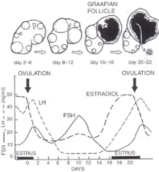
(production of oocytes, estrogens, and inhibin), whereas the function of the corpus luteum is endocrine only (production of progesterone) (36). Ovarian activity in the mare is very dynamic, involving changes from the development of a large pre-ovulatory follicle to the formation of a corpus hemorragicum then a corpus luteum during the estrous cycle (38); to no follicular activity during anestrus in which around 85% of mares will cease ovarian cyclicity (39). During this period of hormonal transition between the estrous cycle and anestrus periods, ovaries may have multiple and large follicles. Follicles are classified into primary, secondary, and tertiary structures and during the estrous cycle, the dominant follicle will approximately grow at a rate of 3 mm per day (37). The developing dominant follicle becomes responsive to follicle-stimulating hormone (FSH), reaching a size at the ovulation of around 40 mm in diameter (40). As ovulation approaches, the preovulatory follicle becomes soft and sensitive (41). Luteinizing hormone (LH) is responsible for the final maturation of the follicle and the induction of ovulation. The increase of LH is observed 24-48 h before ovulation, resulting in the peak of LH after ovulation, as shown in figure 1 (35). About 50% of ovulations result in the formation of an immediate corpus luteum and 50% of ovulations result in the formation of a corpus hemorrhagicum (41). The corpus luteum is located centrally in the equine ovary and, in non-pregnant mare, will be lysed at the end of diestrus (14-15 days post ovulation), in association with a growing population of follicles (34).
19
Figure 1. Hormone Patterns during estrus in mares (42).
2.2. Assisted reproductive techniques in the horse
In Thoroughbred artificial reproductive techniques are forbidden when the progeny is to be registered in the Weatherbys General Stud Book (the original register of Thoroughbred). On the other hand, ART in non-thoroughbred horses has been increasingly used in the equine industry; currently, most mares are inseminated successfully with fresh, cooled, or frozen semen, while a few are covered naturally (43). Frozen semen is shipped worldwide, expanding the choice of stallions available to mare owners (7). Furthermore, other technologies have developed in recent years; these include embryo transfer, with a pregnancy rate after transfer of about 75% (44), ICSI of in-vitro matured oocytes recovered from live or post-mortem mares with blastocyst rates up to 54% (45), nuclear transfer (3), and the vitrification of small blastocysts, associated with good pregnancy and foaling rates (76 and 71%, respectively) (46).
The use of the different ART for in-vitro embryo production by ICSI to obtain more than one foal per year has gained acceptance in commercial equine breeding programs (47). ART can be used to overcome reproductive problems in mares; for example, when the owner does not want the mare to be pregnant during physical activity like sports, the mare
20
is too valuable to risk the potential dangers during pregnancy or foaling, or the mare has lameness, pelvic damage, or maternal behavior problems (7). Moreover, in-vitro embryo production allows the reproduction and storage of valuable genetic material outside of the breeding season or to schedule a breeding program, rendering the technologies commercially attractive (5).
The collection of oocytes, followed by in-vitro oocyte maturation, ICSI, and in-vitro embryo culture, is an effective method for producing foals in mares that cannot become pregnant or provide an embryo under standard reproductive management (47). There are two main approaches to collect oocytes: one from live mares and the other from postmortem mares (9). The first approach is the aspiration of all the immature follicles from the ovaries from donor mares without ovarian stimulation with a recovery rate of 50% (48). The main disadvantage of immature follicle aspiration is that it is complicated, and the practitioner needs extensive training. The second approach is the aspiration of oocytes from excised ovaries from post-mortem mares (49). Some reports have shown a high recovery rate of oocytes (mean of 18 oocytes/mare; range 0–35) by scraping the wall of follicles with a bone curette(4); the disadvantage of this method is that it is a time-consuming process, but it is feasible to perform for valuable mares (6). Maturation is one of the final steps in the development of an oocyte and it is defined as the resumption of meiosis that occurs just before ovulation and subsequent fertilization (50). It has been reported that in-vitro maturation protocols in horses can achieve rates of 42% of mature oocytes (49). The optimum duration of maturation for blastocyst production varies according to the period that the oocyte spends within the ovary before it is recovered. Oocytes collected from the ovary within 1 hour after death require a more extended culture period to reach an optimal blastocyst formation rate (48).
In the few published IVF reports in horses, there has been a low success in fertilization rates ranging from 2% to 33% (51, 52). The failure is related to the inability of the spermatozoa to penetrate the zona pellucida in-vitro. On the other hand, methods for ICSI have been well developed in horses, as described above. Although ICSI has many drawbacks, including the need for expensive equipment, expertise in micromanipulation, oocyte and embryo handling and culture expertise, it also has significant advantages because it may be used with semen having low sperm numbers or quality (53). Currently, equine ICSI using frozen semen has become a standard procedure for oocyte fertilization.
21
In stallions, freezing semen is a commonly used technique to ensure the preservation of valuable male genetic material indefinitely. Semen can be collected from stallions as ejaculates (54), from the epididymis after castration (55), or post-mortem (16). In mares, however, there is currently no efficient method for gamete preservation (7). Oocyte cryopreservation is still in its infancy, and only two publications are available reporting the birth of a foal from previously-cryopreserved oocytes (18, 19). Oocyte cryopreservation is a promising technique that would allow more flexibility in the application of ART in clinical practice, in the research field and for the conservation of female genetic material.
2.3. Principles of cryopreservation
Nowadays, animal breeding has reflected an increased interest in cryopreserving oocytes and embryos of valuable animals, such as pigs, cows, and sheep, to increase meat and milk production (56). The United Nations suggests that the world population is predicted to grow to 9.15 billion by 2050, increasing as well the consumption of meat up to 20% per capita calorie intake (57). Additionally, the transportation of valuable animals is both expensive and logistically challenging to perform. Hence, the prospect of transporting frozen gametes instead of living animals is attractive (58).
Besides the facilitation and cost-effectiveness of transporting gametes instead of live animals, cryopreservation would also minimize the risk of most disease transmission within and between species (59). Additionally, ART, such as pre-implantation genetic diagnosis and sex determination techniques, could be adopted effectively (60). This would be economically favorable to agricultural breeding companies that own genetically valuable individuals. Cryopreservation also can save certain endangered species and native animal breeds (61, 62)
.
Cryopreservation involves cells or whole tissue preservation exposed to a sub-zero temperature in liquid nitrogen (LN2) (-196°C). At such temperature, biological activity is effectively stopped, and the functional status of the cells may be preserved indefinitely (63). However, several physical stresses damage the cells at these low temperatures. For this reason, freezing protocols use a combination of dehydration, freezing point depression, supercooling, and cryoprotectants in an attempt to prevent cell damage (64, 65); such as spermatozoa (66), embryos (67), and more recently, oocytes (27). While the
22
cryopreservation of spermatozoa (12, 68) and embryos (46, 69) has undergone significant advances these last years, oocyte cryopreservation has only recently seen consistent success in some species (22, 33).
The cryopreservation protocols for oocytes can be divided into two categories: (1) slow freezing/rapid thawing and, (2) rapid cooling/warming or vitrification (Figure 2). Initial attempts to freeze oocytes employed the same slow-freezing methods considered the gold standard for embryo cryopreservation (27, 63). However, the slow-freezing oocyte protocols have resulted in relatively low survival and pregnancy rates (70, 71). With the recent advances in the vitrification technique, the efficacy of oocyte cryopreservation has significantly improved. Several groups worldwide have reported high survival and pregnancy rates using vitrified oocytes from different species (14, 33, 72).
23
Figure 2. Comparison of ice crystal formation between slow-freezing and vitrification (56).
2.3.1. Slow freezing
Cryopreservation by slow freezing is a process in which extracellular water crystallizes (73). This method allows cells to be cooled to relatively low temperatures while minimizing intracellular ice crystal formation and simultaneously attempting to reduce the detrimental influences of increased solute concentrations and osmotic stress (74). Freezing induces water transformation into ice, leading to the separation of water from dissolved substances (64). The presence of intracellular ice crystals can be lethal to the oocytes during slow freezing (70). The method has given acceptable results for oocytes of species that are not
24
sensitive to freezing, such as humans (75) and mice (76). However, the oocytes of cows (77), pigs (78), and horses (19) are more sensitive to slow freezing techniques and have resulted in poor results.
In slow freezing techniques, oocytes are gradually exposed to relatively low concentrations of permeating CPAs such as glycerol (79), DMSO (24), EG (70), and propylene glycol (PG) (80) and non-permeating CPAs such as sucrose, glucose, trehalose, or fructose (81). During freezing, CPAs' effect increases the extracellular space, leading to a hypertonic medium due to the increment of solutes. Therefore, water leaves the cell, resulting in cell shrinkage (82). Cells are then loaded in small volumes into 2-mL straws, with cooling rates of 1°C/min from -5 to -9°C and they are kept in the straws for several minutes to equilibrate. After equilibration, ice crystallization is induced by a process known as “seeding,” which results in heterogeneous ice nucleation (63). After seeding, the cooling rate is reduced to 0.3–0.5°C/min until a lower temperature is reached (usually between -30 and -150°C). These procedures typically take several hours. Once the desired temperature is reached, the straws are plunged into LN2 for storage (27).
2.3.2. Vitrification
This process's physical definition is the glass-like solidification of solutions at low temperatures without the formation of intracellular ice crystals (56). Vitrification protocol in embryos was first described in 1985 (83); this protocol was achieved with a solution containing DMSO, PG, and EG; but the solution was toxic to embryos and oocytes (83, 84). With the rapidly growing success of oocyte vitrification in human ART, IVF clinics around the world have changed from the traditional slow-freezing technique to the vitrification method as routine (15). Vitrification has been described as inexpensive because it does not require special equipment compared with slow freezing (26), making it an effective alternative technique to use in various fields. Once vitrified, cells can be stored for extended periods with no noticeable deterioration (85). In cryopreservation by vitrification, both intra- and extracellular compartments vitrify after cellular dehydration (73). While this approach improves the cells' viability, a high concentration of cryoprotectant is required to prevent ice crystal formation (13). Therefore, cryopreservation strategies are based on two main principles that determine vitrification's success to avoid cell injury and death: viscosity and cooling-warming rates (86).
25
The intracellular medium's viscosity is defined by the concentration and behavior of various CPAs and other additives during vitrification (87), and it plays an essential role during dehydration of the cell. A way to prevent ice crystal formation is to remove as much of the intracellular water as possible. However, removing the water results in cell injury and death due to osmotic shock (88). Therefore, the viscosity of high concentrations of CPAs will make water solidify without the formation of ice crystals (80). The high viscosity encountered during freezing in vitrification solutions, due to the high concentration of CPAs, and the glass transition temperature, reduce the chance of ice nucleation and crystallization (89). A successful vitrification protocol considers proper dehydration of the oocytes to reduce intracellular ice formation (90).
High cooling rates is the second principle to decrease detrimental physiological effects in vitrification protocols (91). Such rates could be achieved using minimum volume methods, facilitating the vitrification with less concentrated cryoprotectants (92). It has been reported that a small droplet of vitrification solution (around 1µL) can prevent ice crystal formation and fracture injury (27). Different techniques have been used to minimize the volume of vitrification solutions and submerge samples swiftly into LN2, achieving cooling rates of approximately 2500°C/min (93); moreover, there are reports of suitable carrier systems, such as the open pulled straw, which reached cooling rates of 20,000°C/min (22). A 0.25mL conventional straw was initially used for the vitrification of oocytes. The cooling rate was around 2,500ºC/minute, and the warming rate was 1,300ºC/minute (93). Vajta et al. (22) developed open pulled straws (OPS) to hold bovine oocytes with a small amount of vitrification solution (1μL), reaching cooling and warming rates of 16,700°C/minute and 13,900°C/minute, respectively. This technique has been widely used, achieving successful rates of several species (94). Other cryo-devices have been reported, including the cryoloop called the Cryotop™ (95), and special carriers, such as the micro drops (96), mall nylon coils (97), and nylon mesh (9), which have achieved higher cooling rates, permitting the use of less concentrated solutions with less toxicity to the cell. Moreover, it is essential to note that when cells are immersed directly in LN2, the process results in extensive boiling because cells are warmer objects. Evaporation occurs, and coated vapor surrounds the cells, creating an insulating layer that decreases the cooling rate. Minimizing the volume surrounding the cell, avoiding LN2 vapor formation,
26
and establishing direct contact between the cryoprotectant and the LN2 increase the cooling and warming rates during cell vitrification (98).
Ultra-rapid cooling rates have led to decreased CPA concentrations and have made vitrification a competitive alternative to conventional slow freezing. However, the solution's viscosity and composition, rather than the cooling or warming rate, has been the most critical factor determining the success of cryopreservation (99). Today, vitrification is a popular method for the cryopreservation of many different cell types, tissues, and organs. But the extent of cryoinjury and developmental rates are highly variable, depending on the species (9, 15, 33, 100).
2.3.3. Warming
There are different terms employed in cryopreservation: thawing is specifically the melting of ice, commonly used for vitrification, but inaccurate Thawing can occur rapidly, with temperature changes that exceed 360°C/min. Detrimental effects in the cell result from the recrystallization during thawing or osmotic stresses (63, 74). Warming is a more accurate description than thawing in the context of vitrified systems (80). Another concept is the critical warming rate that suppresses ice formation during warming, which depends strongly on the total solute content of the system and the chemical nature of the solute (23). During warming, full ice development is much more rapid. Hence, warming rates required to avoid significant devitrification are far higher than the cooling rates initially required to achieve vitrification(90). The solutes used for vitrification are generally the same as for warming. Warming solutions based on sugars help todisplace more water and have a more substantial effect on solution viscosity, thus reducing the critical warming rate (101).
The use of a high extracellular concentration of sucrose (e.g., 1 mol/l) counterbalances the high concentration of the cryoprotectant agents in the cell, reducing the difference in osmolarity between the intra and extracellular compartments (33, 99). Canesin et al. (17) found that in equine oocytes, warming solutions without sugar are as effective as the standard high concentrations of sucrose in warming solutions that had been reported earlier (80). This approach has a practical advantage during the manipulation of oocytes
27
because it simplifies finding and transferring oocytes from the warming solution to the maturation medium.
2.4. Oocyte damage caused by vitrification
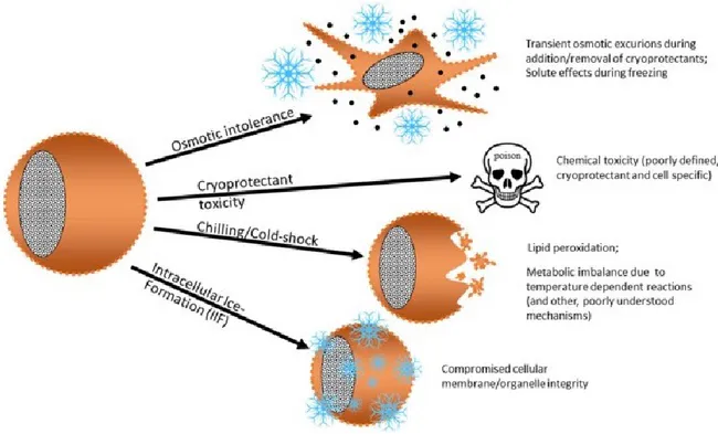
During cryopreservation, cells are exposed to mechanical, thermal, and chemical stressors, which can compromise cell function and cell death because of disturbances in the homeostatic state (56). The two major causes of cellular damage in cells are mechanical damage resulting from the formation of ice crystals (83) and chemical damage due to cryoprotectants (102). To summarize, we can classify the detrimental effects to oocytes during vitrification as follows: osmotic intolerance due to osmotic swelling or shrinking; toxicity due to permeable cryoprotectants, cold-shock injury due to the sensitivity of oocytes to the reduction of temperature; and injury associated with intracellular ice crystal formation as shown in figure 3 (103).
Figure 3. Schematic representation of the potential mechanisms of damage that can occur during cryopreservation (103).
28 2.4.1. Osmotic stress
The exposure of cells to solutions containing CPAs before cooling can be damaging due to an osmotic effect. Most of the commonly used permeating CPAs have lower plasma membrane permeability coefficients than water (104). This relationship results in cells experiencing osmotically-driven volume variations during cryoprotectant addition and removal from the cell (105). The cell membrane’s water permeability dictates the rate at which water flows out of a cell (106). The permeability of cells to water depends on several factors, including temperature, CPAs, and cell type (82). Oocytes are quite different from sperm cells and embryos. In general, it has been demonstrated that oocytes are more sensitive to cryodamage (22) than embryos (107). Oocytes have several characteristics that reduce their membrane permeability to both water and cryoprotective agents compared to other cells, making them sensitive to osmotic effects; one of these characteristics is the low permeability coefficient of the plasma membrane. Mouse oocytes do not express aquaporins at a functional level, while bovine oocytes only express a small number of aquaporins to move water molecules. Therefore, water moves through the plasma membrane of oocytes predominantly by simple diffusion, in contrast to mouse morulae and blastocysts, in which the water mainly moves by facilitated diffusion via water channels (108) . The aquaporins are a family of water channels consisted of proteins and expressed at the plasma membranes of cells involved in fluid transport (109). Cryoprotective agents move through the oocytes and early embryos, similar to the water movement (104). The permeability to DMSO, EG, and PG of oocytes is low comparing with blastocyst permeability (82). Furthermore, the permeability value of PG is higher than other cryoprotectants in the oocytes. This might be related to its higher hydrophobicity compared to other cryoprotectants (104).
Another characteristic that makes the plasma membrane of oocytes different from that of embryos is the increment of intracellular free calcium produced following fertilization, facilitating the permeation of water and CPAs (110). Additionally, the concentration of sub-membranous polymerized filamentous actin of embryos increases and its conformation changes, which facilitates the penetration of water and CPAs (111). Finally, the higher strength of the cell membrane increases embryos’ osmotic tolerance during warming, allowing them to resist osmotic changes better than oocytes, while in oocytes, the
29
presence of zona pellucida and cumulus cells can present obstacles to the penetration of CPAs (112).
Finally, the effects of cumulus cells on oocyte survival after freezing and warming are controversial. Nevertheless, the presence of granulosa cells may offer some protection against osmotic changes and stresses induced during the process of addition and removal of the cryoprotectant during the vitrification process (113); the optimal times for equilibration are different for the oocyte than for the cumulus cells (114). Additionally, the immersion of oocytes in hypertonic CPAs causes cell shrinkage. This shrinking may disrupt the granulosa cell processes and gap junction communication with the oocyte so that physiological interactions between the oocyte and the granulosa cells cannot be preserved (113).
2.4.2. Chilling injury
One of the principal consequences of chilling injury is the intracellular ice formation during cryopreservation when there is not enough time for water to exit the cell. As a result, large ice crystals form within the intracellular compartments. These crystals cause breaks in the cell membranes and damage cell organelles (80). Therefore, the cell dies due to intracellular ice formation, caused by the interaction of cooling velocity and water loss (115). If the cell can dehydrate before intracellular nucleation, intracellular ice crystals do form, but these crystals are much smaller (116). Vitrification reduces damage caused due to ice crystal formation during the cooling process because of a substantial rise in viscosity, which results in the formation of a solid glasslike form. This solid “glassy” layer is amorphous, meaning that it can readjust and take the shape of the cell, enabling the cell to maintain its structure and remain intact (83). Moreover, the use of permeating CPAs is helpful for the replacement of intracellular liquid and decreasing ice formation (102).
The oocyte needs to maintain the integrity of several structural features to undergo fertilization and further development. These structures include the plasma membrane, the zona pellucida, the cortical granules, the microtubular spindle, microfilaments, and condensed chromosomes (20). Post-warmed oocytes can show zona pellucida or cytoplasmic membrane fractures, resulting in various cytoskeletal and chromosomal modifications (22). A common problem in the cryopreservation of oocytes, which makes
30
them particularly susceptible to intracellular ice formation, is their large size and spherical shape that gives a smaller surface area relative to their large internal volume (117). Previous experiences with the cryopreservation of various cell types have led to the understanding that as cell size increases, difficulty in cryopreservation also increases (101); moreover, due to their large size and spherical shape, it is more likely for a large volume of water to be trapped inside the cells during cooling (118). This makes the dehydration and penetration of cryoprotectants challenging to achieve. On the other hand, it has been reported that some oocytes, depending on the species, can fully or partially repair themselves (27, 119, 120).
Another feature that determines the chilling sensitivity during cryopreservation is the intracellular lipid composition (56). There is a threshold level below which cell membrane function is weakened due to a phase transition in membrane fats. The phase transition is the change from a liquid state to a solid-state at a specific freezing point. The temperature at which this lipid phase transition occurs is inversely proportional to the amount of unsaturated fatty acids within the membrane (121). Thus, by altering their lipid membranes' composition, different organisms can adjust this threshold temperature (122). Therefore, cells' ability to survive at low temperatures is partly due to the increase in the ratio of unsaturated fatty acids within the cell membrane and lipid content, and low cryotolerance during vitrification has been linked to high lipid content (9, 30, 32, 56).
Mammalian oocytes have high lipid content, which gives them a dark appearance. Differences in the color of the cytoplasm have been correlated to the amounts of lipids within the cytoplasm (dark color and opaqueness of cytoplasm) (Figure 4) (123). In mammals, primary lipid storage consists of saturated and monosaturated fatty acids, which are part of the cytoplasmic membrane and organelles (124). Lipids are thought to interact with oocyte cytoskeletons, disrupting the arrangement of microtubules and microfilaments that form the cytoskeleton. These interactions are irreversibly affected during cryopreservation (123). Lipid content varies from species to species and also varies
according to the level of maturity. For instance, porcine and equine oocytes (9) present
a darker appearance than mouse and bovine oocytes (124). Mouse and bovine oocytes
31
Figure 4. Lipid droplets of stained immature oocytes (dark zones in the cytoplasm). A) murine, B) bovine, and C) porcine (124).
Effect on development
The maturation of oocytes is a highly energy-intensive process. Adenosine triphosphate (ATP) is essential for the ability of motor proteins to capture and transport chromosomes along the spindle (125). During oocyte maturation, mitochondria accumulate around the nucleus before the germinal vesicle (GV) breakdown (126). Vitrification reduces mitochondrial function by altering the integrity of mitochondria (mt) deoxyribonucleic acid (DNA), and the ability to develop into the blastocyst stage in embryos and oocytes (127). The availability of functional mitochondria has been recognized as a critical determinant of embryo development competence since defects at the structural and mtDNA levels have been identified in compromised oocytes/embryos (128).
Free radicals are formed during the cryopreservation of tissues, resulting from changes in temperature and osmotic damage in cells (129). The enzymatic system is composed of superoxide dismutase (SOD), catalase (CAT), glutathione (GSH), and peroxiredoxins, which detoxify reactive oxygen species (ROS) directly. Non-enzymatic systems contain mainly vitamins A, C, and E (130). The activation of cellular oxidative defense systems occurs in response to damage produced upon vitrification. They can cause structural and functional alterations of cells due to their oxidative property (129). Oocytes are incredibly vulnerable to oxidative stress, not only from the cellular metabolism but also from external influences, such as light, rapid change of oxygen concentration, and temperature changes (40). With the oxidative propriety of ROS, it could oxidize proteins, lipids, and DNA, causing structural and functional alteration of the cell (129). When the oxidative stress reaches a certain level, it may induce the release of cytochrome C and other apoptogenic factors from mitochondria, which eventually activate programmed cell death (131).
32 2.4.3. Toxicity of CPAs
In addition to the injury of osmotic shock and chilling injury, oocytes must also tolerate toxic levels of CPAs generally used in vitrification protocols. PG and DMSO were found to be the most toxic CPAs, whereas glycerol and ethylene glycol were much less toxic (99, 104). The detrimental effects of CPAs depend on three factors: their concentration, the temperature at which the cells are exposed, and the length of time for which the cells are held (82). Survival also depends on the cell type and its ability to overcome physicochemical changes during the process (108). As a result, oocytes can only be exposed to a minimal vitrification media volume for a short period (less than a minute) (132). All the CPAs and other additives have different toxicities, penetration rates, and transition temperatures (99). The combination of varying CPAs is often used to increase viscosity, transition temperature, and reduce the level of toxicity (92).
Effect on the metaphase II spindle
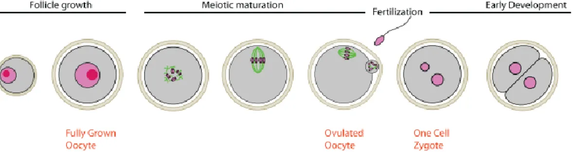
In vitrified oocytes, the chemicals used during cryopreservation, CPAs, increase abnormalities in spindle and chromosome configuration, resulting from the depolymerization of meiotic spindles and the disappearance of microtubule-organizing centers. Therefore, the fertilization of oocytes with disrupted spindles can lead to aneuploidy, disgeny, and arrested cleavage (113, 133). The meiotic spindles of oocytes consist of microtubules constructed by polymerization of tubulin dimers of α- and β-tubulin. Microtubules start from microtubular organizing centers at both poles and anchor chromosomes at the kinetochores. The synapsed chromosome pairs in metaphase I (MI), then the sister chromatids in MII have to be pulled to the opposite ends of the dividing oocytes along the mitotic spindle, which is guided by the extending and shortening of the meiotic spindles (Figure 5) (134). The tubulin dimer polymerizes and depolymerizes at various stages of a cell cycle. The meiotic spindles are crucial for the events following fertilization during the completion of meiosis, second polar body formation, migration of the pronuclei, and formation of the first mitotic spindle (135).
33
Figure 5. Schematic representation of the major steps in the progression of nuclear maturation in mammalian oocytes (134).
Various meiotic stages exhibit different sensitivity to freezing (136). Oocytes may be cryopreserved at the immature GV stage (11) or at the mature MII stage (100), depending on the species. Some reports have demonstrated that freezing immature oocytes is better because the meiotic spindle has not being formed and the DNA is confined within the nucleus (137, 138); however, immature oocytes are thought to be more sensitive to anastomotic stress and feature lower cell membrane stability than MII stage oocytes (114, 139).
In many species, studies have used matured oocytes at the MII stage (93, 100, 140). In species most susceptible to freezing, such as pigs and horses, vitrified MII stage oocytes generally demonstrate low developmental competence in terms of blastocyst formation rates and cell numbers in blastocysts (20, 141). Oocytes at the MII stage have undergone both cytoplasmic and nuclear maturation, including extrusion of the first polar body and alignment of chromosomes on a meiotic spindle. In contrast, the chromatin of GV oocytes is still in the diplotene phase of prophase I without a meiotic spindle (134). At the MII stage, the oocyte DNA is condensed into chromosomes that are aligned along the equatorial region of the meiotic spindle and are susceptible to disruption (140).
Researchers have demonstrated that oocyte exposure to cooling and CPAs can cause depolymerization and the disorganization of spindle microtubules (20, 107). Spindle damage in oocytes can have a fatal consequence, preventing the extrusion of the second polar body and resulting in abnormal chromosomal numbers in embryos (27, 123, 140). On the other hand, numerous reports have demonstrated that the metaphase spindle microtubules that depolymerize during oocyte cryopreservation by vitrification can
34
repolymerize, forming normal structural and functional spindles post-cryopreservation given time and the proper temperature (133, 140, 142).
Effect on cortical granule exocytosis
Intracellular calcium (Ca++) has been implicated as a second messenger in somatic cells and in the events that follow sperm penetration of the oocyte. Upon fusion, the sperm induces a series of calcium transients through a sperm-specific phospholipase C (143). Calcium is responsible for initiating cortical granule fusion with the oolemma, resulting in the release of their contents into the surrounding zona pellucida (144).
The exposure of the CPAs may increase intracellular Ca++ and trigger premature exocytosis of cortical granular material, leading to zona pellucida hardening and making sperm penetration and fertilization impossible (145). The use of ICSI overcomes the effects of zona hardening, resulting in higher fertilization rates (146). Although this potential problem has been alleviated from a practical perspective, zona hardening reflects that the oocyte has started to undergo activation has not been addressed (110). This might explain in part why oocytes do not develop well following cryopreservation (145).
Three commonly used cryoprotectants (EG, PG, and DMSO) induce an immediate increase in Ca++, but the magnitude and duration of the Ca++ transient are different depending on the cryoprotectant, with ethylene glycol resulting in the smallest and shortest increase and propanediol causing the most protracted elevation (147). The literature also says that DMSO affects Ca++ stores directly (145).
2.5. Vitrification protocols
It was discovered in the 1940s (66) that the addition of glycerol for protection against cryodamage greatly enhanced the survival of cryopreserved cells. This gave rise to the investigational concept of cryoprotectants. Some authors have mixed EG with other permeating agents, such as DMSO or PG, to reduce the concentration of a single cryoprotectant and to decrease the individual specific toxicity (27, 148). Additionally, the strategy for lowering toxicity by applying rapid exposure to a high CPA concentration has been widely used (149, 150). Vitrification media are currently prepared from buffered
35
media (e.g., TCM-199) with a stable pH between 7.2 and 7.4 and CPAs that prevent ice crystal formation after freezing by increasing cellular dehydration and viscosity (63). Typically, combinations of permeating and non-permeating CPAs are used (148, 151). Permeating CPAs are of low molecular weight, such as glycerol, EG, PG and DMSO. They penetrate the cell and form hydrogen bonds with intracellular water molecules, preventing crystallization (102, 152)
.
The non-permeating CPAs are of low molecular weight and include sucrose, glucose, trehalose, and fructose. They remain extracellular and draw free water out of the cell by osmosis, resulting in intracellular dehydration (153). The last component of vitrification media is a high molecular weight, non-permeating polymer used to reduce the concentration of CPAs necessary for vitrification, reducing the toxicity of the solution and protecting the zona pellucida against cracking. Standard solutions are FBS or bovine serum albumin, polyethylene glycol, polyvinylpyrrolidone, Ficoll, and polyvinyl alcohol (25, 116, 154).Ethylene glycol, a mono-ethylene glycol with the molecular formula C2H2(OH)2, is an important component of vitrification solutions, which forms from ethylene oxide hydrolysis, characterized by low toxicity and rapid permeation of the cell (99). DMSO is a polar compound (C2H6SO2) widely used as a cryoprotectant in different species (29, 151, 155). The direct hydrolysis of propylene oxide produces propylene glycol (C3H8O2). 1,2-propylene glycol and 1,3-1,2-propylene glycol are formed simultaneously through the sequential addition of propylene oxide (104). Since water is not viscous, it can only be vitrified by quite rapid cooling rates or by using high concentrations of permeable CPAs, which are small molecules that penetrate cells and limit the amount of intracellular and extracellular water that converts into ice during cooling (99, 150). But, the use of the lowest possible concentration of permeable CPAs to minimize problems with toxicity is effective by replacing penetrating CPAs with carbohydrates (90). Trehalose and sucrose are the most common carbohydrates used as non-permeating CPAs in the cryopreservation protocols of murine, bovine, equine, porcine, and ovine oocytes and/or embryos (32, 99, 156, 157). Other sugars are not commonly used as components of vitrification solutions in oocytes but are widely used to preserve organs, tissues, and cells or as components of a culture media (158). For instance, raffinose, fructose, and glucose have been used for sperm cryopreservation (159).
36
The addition of carbohydrates has facilitated the dehydration of embryos and works as an essential component of osmotic buffers (160). These solutions are used to increase the effective osmolality of the extracellular medium because sugars are unable to cross the cell membrane naturally, enhancing cellular dehydration by osmotic pressure, thus limiting the extent of swelling (158). It has been suggested that sugars can preserve the structural and functional integrity of membranes at low levels of water activity (99, 101). And it has further been reported that the critical cooling rate required to avoid ice crystallization in solutions with penetrating cryoprotectants was altered following the addition of sugar (101). Thus, the different types of sugars should be analyzed during the design of vitrification solutions.
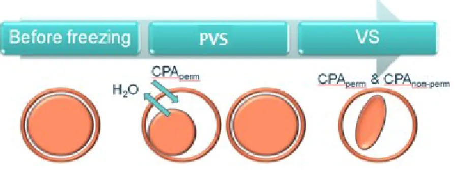
Since vitrification is a method that requires a relatively high concentration of cryoprotectants, stepwise addition of cryoprotectants is used to decrease the toxic effects and minimize damage due to osmotic shock, resulting in higher maturation, fertilization and blastocyst formation rates (161, 162). Some strategies can be applied; the most common is the two-step protocol in which oocyte is exposed to a PVS as a first step, containing DMSO, EG, or PG (permeating CPAs); depending on the protocol, the oocyte may or may not remain for a relatively brief exposure in this first step (9, 19). The low concentration of cryoprotectants in the first solution is less toxic than the VS. Oocytes in the first solution shrink initially and gradually re-expand to their original volume. The second step is a relatively prolonged incubation in VS, containing higher concentrations of permeating CPAs and non-permeating CPAs (sucrose or trehalose) to dehydrate the cells (Figure 6) (9, 80, 163). The oocyte is then loaded onto a cryo-device with a small volume of vitrification medium before being plunged into liquid nitrogen (80).
37
Figure 6. Schematic presentation of the vitrification procedure (164).
The procedure employed to load the CPAs into the oocytes will vary between the authors. Some are time-specific (19), some require that the processes be conducted at room temperature (17), and some are performed at higher temperatures (25). One-step may, because of the possibility of poor permeation of the cryoprotectants, result in intracellular ice formation during cooling or warming (27, 161, 162). Extensive discussions are unfolding about which vitrification container is the best, but it has been also claimed that the vitrification method thoroughly depends upon the technical skill of the one who performs the vitrification (142).
2.5.1. Vitrification protocols in species other than horses
The first report of oocyte cryopreservation with a viable pregnancy in humans originated from slow-rate freezing-thawing, containing phosphate-buffered saline, FBS and dimethyl sulphoxide (27). Several studies have compared slow-freezing techniques (156, 165, 166) and vitrification (167-169) in human embryology. Vitrification has been suggested to be more suitable for the cryopreservation of human oocytes by applying a higher concentration of cryoprotectants and a rapid cooling speed to prevent ice crystal formation (21). Some of the first reports using vitrification protocols with ethylene glycol and sucrose had a survival rate from 65% to 90% and a blastocyst formation rate of around 30% (170).
Several modifications in protocols for vitrification were proposed to improve the survival of frozen–warmed oocytes, employing an array of different vitrification systems, including electron microscope grids, open pulled straws, open hemistraws and cryoloops (167, 168, 171, 172). Significantly increased success rates were reported with the cryotop method performed with MII oocytes. This cryotop method consisting of 15% ethylene glycol, 15%
38
dimethyl sulfoxide, and 0.5 M sucrose was highly reproducible, reporting 91% survival, 81% cleavage, 50% blastocyst and 41.9% pregnancy rates (168). Significant points of consideration among the different protocols are the time and temperature of oocyte exposure to the vitrification solution, the concentration of the solution surrounding the oocytes, the carrier to hold the oocyte during vitrification and the temperature and media using during warming (101).
Data on mouse oocytes has demonstrated the superiority of vitrification over slow freezing in every parameter measured. A two-step vitrification protocol with a CPA solution composed of DMSO, EG, and sucrose achieved blastocyst rates of 67%, when the resultant mouse blastocysts were transferred, embryos derived from vitrified oocytes implanted at the same rate as blastocysts from non-cryopreserved oocytes (88% and 87%, respectively) (173). Recently, the viability of oocytes after a two-step vitrification protocol, with a CPA solution composed of EG, DMSO, trehalose and FBS, was reported to be more than 90%, and no significant difference of blastocyst formation was observed between the fresh and vitrified oocytes after IVF (174).
On the other hand, oocytes from cattle are extremely sensitive to cold temperatures, and slow freezing dramatically affects blastocyst formation (4.5%) (71). Although the cryopreservation of bovine oocytes remains a challenge, some of the highest blastocyst formation rates among domestic animals have been obtained from this species. A 25% blastocyst rate formation of vitrified matured oocytes has been reported using a two-step protocol with an open pulled straw and a solution composed of EG, DMSO and sucrose (22). Methods of vitrification of bovine embryos have significantly progressed in recent years. Currently, live offspring have been born from vitrified oocytes using a three-step protocol, achieving a maturation rate of 50% and a pregnancy rate of 36%. The protocol was as follows: oocytes were suspended in a PVS1 composed of 3% EG for 5–10 min for initial oocyte cryoprotectant exposure; groups of 5-6 oocytes were placed in a PV2 composed of 10% EG + 10% DMSO for 30 s; subsequently, oocytes were suspended in a VS composed of 20% EG + 20% DMSO + 17.1% sucrose, supplemented with 0.1% polyvinyl alcohol; finally, oocytes were transferred to a 2 μl VS droplet (the total exposure time to the VS was 25 s) and oocytes were loaded by capillarity into open‐pulled glass micropipettes (14).
39
In efforts to improve development competence during oocyte vitrification in bovine, modification of the devices, such as an electron microscopy grid (10), has been approved to facilitate the holding of the cells. Moreover, it has been reported that with bovine oocytes, the survival, as well as the cleavage and blastocyst development rate, was not significantly different following vitrification and warming with or without cumulus cells (175). Additionally, the three-step protocol with EG/ sucrose and ficoll reported fewer abnormalities in the oocytes than the single-step exposure; this method was used to enhance the feasibility of nylon-mesh holder for vitrification of immature oocytes (176).
Currently, a limited number of studies are available on vitrification of small ruminant oocytes (177-179). In sheep, a two-step vitrification protocol using EG, DMSO and trehalose with a nylon loop resulted in a blastocyst development rate of 29.4% after several trials where low developmental rates were obtained following immature and mature oocyte vitrification (29). Poor success in ovine oocyte cryopreservation has been attributed to damage to enzymes such as mitogen-activated protein (MAPK) kinase, critical for oocyte maturation and subsequent embryo development (114).
Regarding the porcine species, oocytes are particularly susceptible to cellular damage by freezing because the meiotic spindle of porcine oocytes is quite sensitive to cryopreservation, resulting in impaired development at MII (107). Because porcine oocytes contain more lipid droplets than other species, a recent study combined removing cytoplasmic lipid droplets with microtubule stabilization and found that vitrified porcine immature oocytes could develop to the blastocyst stage and maintain the ability to develop into fetuses (180). After the improvement of a vitrification protocol with a reasonable survival rate (approximately 50%) and a blastocyst rate of 2% (30); the first study of successful piglet production from vitrified immature oocytes was reported with a two-step vitrification method with EG, PG, cytochalasin B, bovine serum albumin (BSA) and trehalose. However, the blastocyst rate was still very low (5.2%) (32). Improving the survival and development of oocytes after cryopreservation has also been based on optimizing the conditions of in vitro culture with granulosa cells in a two-step vitrification protocol with EG and sucrose, achieving a blastocyst rate of 43% after parthenogenetic activation (181).