Direction des bibliothèques
AVIS
Ce document a été numérisé par la Division de la gestion des documents et des archives de l’Université de Montréal.
L’auteur a autorisé l’Université de Montréal à reproduire et diffuser, en totalité ou en partie, par quelque moyen que ce soit et sur quelque support que ce soit, et exclusivement à des fins non lucratives d’enseignement et de recherche, des copies de ce mémoire ou de cette thèse.
L’auteur et les coauteurs le cas échéant conservent la propriété du droit d’auteur et des droits moraux qui protègent ce document. Ni la thèse ou le mémoire, ni des extraits substantiels de ce document, ne doivent être imprimés ou autrement reproduits sans l’autorisation de l’auteur.
Afin de se conformer à la Loi canadienne sur la protection des renseignements personnels, quelques formulaires secondaires, coordonnées ou signatures intégrées au texte ont pu être enlevés de ce document. Bien que cela ait pu affecter la pagination, il n’y a aucun contenu manquant.
NOTICE
This document was digitized by the Records Management & Archives Division of Université de Montréal.
The author of this thesis or dissertation has granted a nonexclusive license allowing Université de Montréal to reproduce and publish the document, in part or in whole, and in any format, solely for noncommercial educational and research purposes.
The author and co-authors if applicable retain copyright ownership and moral rights in this document. Neither the whole thesis or dissertation, nor substantial extracts from it, may be printed or otherwise reproduced without the author’s permission.
In compliance with the Canadian Privacy Act some supporting forms, contact information or signatures may have been removed from the document. While this may affect the document page count, it does not represent any loss of content from the document.
Phosphoproteome profiling approaches for comprehensive monitoring
of cell signaling events in interferon-y stimulated macrophages
par
Maria Marcantonio
Département de Biochimie, Université de Montréal Faculté de Médecine
Mémoire présenté à la Faculté des études supérieures en vue de l'obtentîon du grade de Maîtrise ès sciences (M.Sc.)
en Biochimie
Avril 2008
Faculté des études supérieures
Ce mémoire intitulé:
Phosphoproteome profiling approaches for comprehensive monitoring of cell signaling events in interferon-y stimulated macrophages
présenté par: Maria Marcantonio
a été évalué par un jury composé de personnes suivantes :
Dr. Michel Bouvier président-rapporteur Dr. Pierre Thibault directeur de recherche Dr. Sylvain Meloche membre du jury
La phosphorylation des protéines joue un role primordial dans les voies de signalisation cellulaire. De récents progrès en spectrométrie de masse (MS) permettent l'identification de milliers de sites de phosphorylation. Cependant, l'inefficacité d'ionisation des phosphopeptides limite leur détection. Ce travail présente une méthode d'identification des phosphopeptides à partir d'extraits cellulaires enrichis avec Ti02 et déphosphorylés par la phosphatase alcaline. La corrélation des temps de rétention de plus de 750 paires de phospho- et déphosphopeptides indique que les changements d'hydrophobicité sont partiellement expliqués par l'interaction électrostatique avec des
/
acides aminés basiques. Une augmentation d'au moins un facteur de 2 dans le nombre de phosphopeptide détectables a été observée suite à la déphosphorylation. Cette méthodologie a été appliquée à des extraits cellulaires de macrophage 1774 traités ou non à l'interféron-gamma (IFN-y) et a permis l'identification de phosphoprotéines impliquées dans différentes voies de signalisation.
Le profil cinétique du phosphoprotéome des cellules 1774 stimulées par IFN-y (jusqu'à 180 min) et enrichis avec Ti02 a été obtenu pour mieux comprendre l'effet biologique de l'IFN-y sur les macrophages. Des changements significatifs de phosphorylation ont été observés à 5 et 30 min après stimulation. L'analyse par 2D-LC-MS/MS des macrophages stimulés pendant 5 et 30 min par l'IFN-y a permis l'identification de 2831 phosphopeptides incluant 1673 nouveaux sites de phosphorylation. L'analyse du réseau d'Întéraction des phosphoprotéines a permis d'identifier des protéines impliquées dans les voies de signalisation du complexe NADPH oxydase (p67phox), des phospholipides (SHIP-2), de la traduction de ARNm (eIF4B) et de l'apoptose (PEA-15, NF-KB).
Mots-clés: Phosphorylation, spectromètre de masse, profil d'expression, enrichissement par Ti02, IFN-y, macrophages, phosphatase alcaline, réseau d'interactions protéine-protéine, voies de signalisation, prédiction de kinases.
Abstract
In biological systems, signal transduction pathways are primarily guided through various prote in phosphorylation and dephosphorylation events. Major analytical advances in mass spectrometry have enabled the direct identification of protein phosphorylation sites in complex mixtures. Yet, phosphoproteome analyses are still quite challenging due to the poor ionization efficiency of phosphopeptides. This work presents a combined enzymatic and data mining approach enabling more than 2-fold increase of phosphopeptides detection following alkaline phosphatase digestion of TiOz-enriched J774 cell ex tracts. The retenti on time of more than 750 phosphopeptides was shifted toward higher and lower values compared to their dephosphory lated counterpart. The loss in hydrophobicity is partially explained by the electrostatic interaction of the phosphate group with basic amino groups. This approach was applied to 5 min interferon-gamma (IFN-y) treated mouse macrophage cells and led to the identification of key IFN-y signaling events.
Furthermore, activated signaling transductions were more clearly defined to understand IFN-y biological effects on macrophages. We compared the temporal changes in the phosphoproteome of TiOz-enriched J774 macrophages following IFN-y stimulation (up to 180 min). Amongst the different trends observed, most significant changes in phosphorylation occurred to the same extent 5 and 30 min after IFN-y induction. The 2D-LC-MS/MS analysis on TiOz-enriched 5 and 30 min IFN-y treated macrophages identified a total of 2831 phosphopeptides including 1673 novel phosphorylation sites. Network analyses of differentially regulated phosphopeptides led to the identification of early signaling and regulatory events including members of the NADPH complex (p67Phox), phospholipids (SHIP-2), protein synthesis (eIF4B) and apoptotic (PEA-IS, NFK:B) pathways.
Key words: Phosphorylation, mass spectrometry, expression profiling, TiOz
enrichment, IFN-y, macrophages, alkaline phosphatase, protein network, signaling pathways, kinase prediction.
Résumé ... .i
Abstract. ... .ii
Table of contents ... .iii
List of tables ... vi
List of figures ... vii
List of abbreviations ... " ... .ix
Acknow ledgements ... xiii
1. General introduction ... ... 1
1.1 Project' s goal. ... 2
1.2 Innate immunity ... 3
1.2.1 Macrophages and inflammation ... 3
1.2.2 IFN-y signa1ing pathways ... A 1.2.2.1 Transcriptiona1 regulation ... 5
1.2.2.2 Cell growth and apoptosis ... 6
1.2.2.3 mRNA trans1ation ... 8
1.2.204 Phospho1ipid signa1ing ... 9
1.2.2.5 Production of cytotoxic molecules ... 1 0 1.3 Phosphoproteomics ... 13
1.3.1 Mass spectrometry approaches for phosphopeptide detection ... 14
1.3.1.1 Immunoprecipitation assay ... 15
1.3.1.2 Phosphopeptide enrichment methods ... 16
1.3.1.3 Chemical modification methods ... 17
1.3.104 Alkaline phosphatase treatment. ... 19
1.3.1.5 Separation methods ... 19
1.4 Mass spectrometry ... .- ... 21
1.4.1 Electrospray ionization ... 22
1.4.2 LTQ-Orbitrap mass spectrometer ... 23
1.4.3 Tandem MS ... 25
1.4.4 Data processing ... 27
1.4.4.1 MS/MS ion search using Mascot ... 27
2. Combined enzymatic and data mining approaches for comprehensive
phosphoproteome analyses; application to cell signaling events of interferon-y
stimulated macrophages ... 30
2.1 Abstract. ... : ... 31
2.2 Introduction ... 32
2.3 Experimental procedures ... 35
2.3.1 Materials ... 35
2.3.2 Preparation of standard proteins ... 35
2.3.3 Cell cultures ... 35
2.3.4 Protein extraction and immobilized trypsin digestion ... 36
2.3.5 Phosphopeptide isolation ... .36
2.3.6 Alkaline phosphatase treatment. ... 3 7 2.3.7 LC-MS/MS analysis ... 37
2.3.8 Database searching with mass spectrometry data ... .38
2.3.9 Peptide detection and c1ustering ... .39
2.4 Results ... 40
2.4.1 Evaluation of phosphopeptide enrichment methods ... .40
2.4.2 Evaluation of alkaline phosphatase treatment. ... .44
2.4.3 Phosphopeptide behaviour in LC-MS ... .49
2.4.4 Application of alkaline phosphatase treatment for large-scale phosphoproteomic analyses ... 55
2.5 Discussion ... 63
2.6 References ... 69
3. Molecular dissection of early interferon-y cell signaling events in macrophages via quantitative phosphoproteomics ... 72
3.1 Abstract. ... 73
3.2 Introduction ... 74
3.3 Experimental procedures ... 76
3.3.1 Materials ... ~. 76
3.3.2 Cell cultures, protein extraction and trypsin digestion ... 76
3.3.3 Phosphopeptide isolation ... 76
3.3.4 LC-MSIMS analyses and Mascot database searching ... 77
3.3.6 Fuzzy c-means clustering ... 78
3.3.7 Kinase and motif prediction tools ... 78
3.3.8 Phosphoprotein networking ... 79
3.4 Results ... 80
3.4.1 Phosphorylation dynamics of IFN-y treated 1774 phosphopeptides ... 80 1 3.4.2 Statistical analysis of macrophage phosphoproteome ... 82
3.4.3 Network analysis in signaling pathways of activated macrophages .... 88
3.5 Discussion ... 96
3.6 References ... 102
4. General conclusion ... 106
List of Tables
Table 2.1: Comparison ofTi02 and IMAC (Ga3+) phosphopeptide enrichment methods
using 250 J.!g 1774 celI extract ... .41
Table 2.2: Statistics on overall distribution of correlated phosphopeptides and their dephosphorylated counterparts from J774 AP study ... 50
Table 2.3: Summary of identification obtained from different approaches ... 57
Table 2.4: Putative phosphopeptides showing differential abundance in 5 min IFN-y stimulated 1774 cytosol extracts upon AP treatment. ... 62
Table 3.1: Statistics on overall distribution ofphosphopeptides from IFN-y stimulated 1774 kinetic study ... 83
Table 3.2: Phosphopeptides showing differential abundance in 5 and 30 min IFN-y stimulated 1774 cytosol extracts ... 92
Figure 1.1: The classical JAK-STAT pathway following IFN-y stimulation of
macrophages ... 5
Figure 1.2: The classical apoptotic pathway during inflammatory response ... 7
Figure 1.3: IFN-y activation of Ras-MAPK and PI3K1AktimTOR signaling pathways to initiate mRNA translation ... 9
Figure 1.4: Microscopic examination of J774 macrophage ceUs highlighting
morphological changes taking place upon IFN-y stimulation ... l 0
Figure 1.5: Upon IFN-y administration, induction of cytosolic factors (p47ph0X, p40Phox and p67Phox) recruitment to the membrane to fonn the NADPH oxidase
complex through phosphorylation and interaction with arachidonic acid .. ll
Figure 1.6: Chemical structure of phosphorylated serine, threonine and tyrosine
residues ... 13
Figure 1.7: (a) ~-elimination reaction ofphosphoserine and phosphothreonine. (b) Addition of a thiol group and biotin tag on phosphoserine for further enrichment on avidin columns ... o • • • o • • • • • • o • • • • • o • • • • • • • • • • • • • • • • • • 18
Figure 1.8: General mass spectrometry-based proteomics ... 21
Figure 1.9: Electrospray ionization chamber ... .22 Figure 1.10: LTQ-Orbitrap hybrid mass spectrometer (Thenno Electron) coupled to a
nano-flow LC (Eksigent) with a Spark-Holland autosampler (Thermo Electron) ... 24
Figure 1.11: A schematic representation of the LTQ-Orbitrap mass spectrometer ... 24
Figure 1.12: Peptide fragmentation in low collisional energy illustrating b and y
ions ... 26
Figure 1.13: Mascot search engine page showing all parameters for a MS/MS ion search ... 27
Figure 1.14: Contour plot generated by Mass Sense ... 29
Figure 2.1: Comparison of IMAC (Ga3+) and Ti02 enrichment procedures ... .40
Figure 2.2: Capacity test of 1.25 mg Ti02 micro-tips using spiked phosphopeptides from J774 cytosolic cell extract (n=3) ... .42
Figure 2.3: Loading capacity and linear dynamic range of Ti02 enrichment
Figure 2.4: Overview of the analytical strategy combining enzymatic and data mining approaches ... 45
Figure 2.5: Changes in peptide abundance of positive and negative control following AP treatment. ... 47
Figure 2.6: Comparison of non-treated and AP-treated a-casein contour
plots ... 48
Figure 2.7: Phosphopeptide behavior in LC-MS following alkaline phosphatase treatment (n=3) ... ... 51
Figure 2.8: Distribution of matched phospho- and dephosphorylated peptides with different ~ retenti on time according to the phosphate group position from both C and N-terminus ... 53
Figure 2.9: Comparison ofretention time shifts of a-casein phosphopeptides and their N-acetylated counterpart following AP treatment. ... 54
Figure 2.10: Volcano plot distribution of (a) non-treated and (b) AP-treated cytosolic extracts of 1774 control and IFN-y treated (5 min) ... 56
Figure 2.11: Identification ofphosphopeptide candidates using alkaline
phosphatase ... 59
Figure 2.12: Identification of differentially expressed phosphoproteins from J774 following 5 min IFN-y stimulation ... 60
Figure 3.1: Overview of the analytical strategy and data mining approaches for time-resolved phosphoproteomics ... 80
Figure 3.2: Clustered profiles of dynamic phosphorylation ... 82
Figure 3.3: Volcano plot distribution of(a) 5 and (b) 30 min IFN-y treated cytosolic extracts of J774 cells ... 84
Figure 3.4: NetworKlN kinase prediction of identified phosphopeptides ... 86
Figure 3.5: Over-represented motif-x patterns of aIl identified phosphopeptides ... 87
Figure 3.6: Over-represented motif-x patterns of differentially expressed
phosphopeptides ... 88
Figure 3.7: Phosphoprotein interaction networks ofJ774 macrophages ... 89
Figure 3.8: Classical apoptotic signaling pathways highlighting phosphoproteins identified upon 5 and 30 min IFN-y ... 91
AA ACN AP ATP Ba(OH)2 BCA CDK2 CE CIS CK2 cPLA2 C-trap CV 2D DHB DMEM DTT 4EBPl EDT eEF eIF ERK1/2 ESI FA FADD Arachidonic Acid Acetonitrile Alkaline Phosphatase Adenosine Triphosphate Barium hydroxide Bicinchoninic Acid
Cell Division Prote in Kinase 2
Capillary Electrophoresis
Cytokine-Inducible SH2 protein
Casein Kinase 2
Cytosolic Phospholipase A2
C-shaped storage trap
Coefficient of Variation
Two-Dimensional
2,5-Dihydroxybenzoic Acid
Dulbecco's Modified Eagle's Medium
D L-Dithiolthreitol
eIF4E-Binding Prote in 1
1,2-Ethanedithiol
eukaryotic Elongation Factor
eukaryotic Translation Initiation Factor
Extracellular signal-Regulated Kinases 1/2
Electrospray Ionization
Formic Acid
FBS FMC FTIRC GSK-3 GTP HPLC IFN-y IFNGR 1/2 IKK IL IMAC IPI IRFs JAK1I2 LC-MS LC-MS/MS LiOH LPS LTQ
Fetal Bovine Serum
Fuzzy c-Means Clustering
Fourier Transform Ion Cyclotron Resonance
Glycogen Synthase Kinase-3
Guanosine-5'-Triphosphate
High Performance Liquid Chromatography
Interferon-gamma
IFN-y Receptor 1/2
IKB Kinase
Interleukin
Immobilized Metal Ion Chromatography
International Prote in Index
IFN Regulatory Factors
Janus Activated Kinase 1/2
Liquid Chromatographic-Mass Spectrometry
Liquid Chromatography-Tandem Mass Spectrometry
Lithium hydroxide
Lipopolysaccharide
Linear Trap Quadrupole
MALDI-TOF Matrix Assisted Laser Desorption Ionization-Time-of-Flight
MAPKl4 MHC MNK1/2 MS MSKI mTOR
Mitogen-Activated Protein Kinase 14
Major Histocompatibility Complex
MAP Kinase Signal-Interacting Kinase 1/2
Mass Spectrometer
Mitogen- and Stress-Activated Protein Kinase 1
NF-KB NO O2 -PBS PDKI PI3K PIM2 Pl(4,5)P2 PMSF PPSP p70 S6K PTEN PTMs Q-TOF RAPI RF RIP RNS ROS RSD RSK-2 RT SCX SDS-PAGE SHIP-2 Nuclear Factor-Kappa B Nitric Oxide Superoxide
Phosphate Buffer Saline
Pyruvate Dehydrogenase Kinase isozyme 1
Phosphatidylinositol 3-Kinase
Serine/Threonine-Protein Kinase Pim-2
Phosphatidylinositol 4,5-biphosphate
Phenylmethyl Sulphonyl Fluoride
Prediction of PK -Specific Phosphorylation Site
Ribosomal protein S6 kinase beta-l
Phosphatase and Tensin Homologue Deleted on Chromosome Ten
Post-Translational Modifications
Hybrid Quadrupole-Time-of-Flight Hybrid
Repressor Activator Prote in 1
Radio Frequency
Receptor-Interacting Protein
Reactive Nitrogen Species
Reactive Oxygen Species
Relative Standard Deviation
Ribosomal Protein S6 Kinase alpha-3
Retention Time
Strong Cation Exchange
Sodium Dodecyl Sulfate Polyacrylamide Gel Electrophoresis
SOCS SODD STATl/2 STRING TCEP TFA TGFBR2 Ti02 TLRs TNF-a TNFRI TPCK TRADD TRAF2 z
Suppressors of Cytokine Signalling Silencer of Death Domain
Signal Transducers and Activators of Transcription 1/2 Search Tool for the Retrieval of Interacting Genes/Proteins Tris(2-carboxyethyl)Phosphine
Trifluoroacetic Acid TGF-Beta Receptor type-2 Titanium Dioxide
Toll-Like Receptors
Tumor Necrosis Factor-alpha Tumour Necrosis Factor Receptor 1
L-I-Tosylamido-2-Phenylethyl Chloromethyl Ketone TNF-R-Associated Death Domain Protein
Tl\,JF -Receptor -Associated Factor 2 Charge
First, 1 would like to thank my research director, Dr. Pierre Thibault, for giving me the opportunity to conduct my graduate studies in his laboratory. 1 realize that my Master's experience would not have been so successful and rewarding without his continuing assistance in every aspect of my project. His superior teaching abilities and knowledge in mass spectrometry broadened my horizons in the field of phosphoproteomics. 1 really appreciate his immense help and effort he gave me despite his very busy schedule and demanding role. 1 will always be grateful for the great opportunities he has given me to advance in my field.
1 would also like to acknowledge Dr. Matthias Trost, who was always able to provide me with valuable information conceming my project. 1 thank him for playing such an important role in the completion of my thesis. His friendship and guidance has been very helpful throughout my years of study.
A special thanks to Dr. Eric Bonneil, whose excellent expertise III mass spectrometry helped immensely in the success of my experiments. Furthermore, 1 would like to thank Gagandeep Jaitly and Kevin Eng for their great assistance in data mining analyses. 1 also wish to express my appreciation to Mathieu Courcelles for the great input that he provided regarding the different bioinformatics' applications used in phosphoproteomics. 1 would also like to acknowledge Christelle Pomiès for her technical assistance. Given that their aid was needed quite frequently, 1 very much appreciate their patience and genuine willingness to devote time for my project.
Thanks to ail Dr. Thibault's research team for their friendship, constant moral support and assistance. Whenever Pierre was not available, the y took it upon themselves to show me the ropes. 1 am especially grateful to Gaëlle Bridon for her contribution in the second part of my thesis.
As weil, the generous graduate scholarship from Natural Sciences and engineering research Council (NSERC) is greatly acknowledged.
Finally, this work would have never been possible without the support and patience from my husband, family and friends, who always encouraged me to strive and reach my goals in life.
1.1 Project's goal
Upon IFN-y stimulation, macrophages are primed to prote ct the organism against infectious diseases [1]. With the exception of the classical anti-inflammatory JAK-STAT pathway [2], no other important IFN-y stimulated pathways are completely characterized to fully understand the cell defence mechanism against invading pathogens [3]. The JAK-ST AT pathway was also one of the first pathways demonstrating that phosphorylation plays a central role during IFN-y activated cell signaling. Since only a small fraction of phosphoproteins is phosphorylated at any given time, powerful and sensitive tools like mass spectrometry are required for their detection [4].
In this context, the goal of this thesis consists of evaluating the phosphoproteome of intracellular signa1ing pathways in IFN-y treated macrophages using mass spectrometry approaches. Phosphopeptide analysis by mass spectrometry is still limited since phosphorylated peptides do not efficiently ionize in positive ion mode due to their negative charge contribution [5]. Hence, the first part of this project will focus on the development and optimization of a novel technique for a higher phosphopeptide identification level. The method consists of treating ha If of the Ti02-enriched phosphopeptide samples with alkaline phosphatase. The treated and non-treated chromatograms will be subsequently compared using in-house bioinformatic tools. Removal of the phosphate group is expected to not only enhance phosphopeptides detection but may also provide interesting feedback on phosphopeptide behaviour in MS. Following validation of the enzymatic approach, the method will be applied on 1774 mouse macrophages to determine changes in phosphorylation occurring upon IFN-y stimulation.
The second part of the study covers the evaluation of the phosphoproteome dynamics of IFN-y treated macrophages. High-throughput phosphoproteomic strategies su ch as strong-cation exchange (SCX) chromatography combined with Ti02 enrichment will be used to analyse the phosphoproteome changes of macrophages treated with the optimal cytokine incubation time. Furthermore, differentially expressed phosphoproteins will be identified using in-house softwares su ch as Mass Sense and peptide c1ustering. The last part of the analysis will combine kinase and motif prediction assays with protein networking to determine activated signaling pathways mediating important IFN-y biological effects.
3
1.2 Innate immunity
Infectious diseases are the major cause of death and health problems in our society [6]. Innate and adaptive immune systems evolved to prevent and control the formation of inflammation diseases [7]. The innate immune system is the first line of defence against invading organisms with receptors recognizing the molecular structure of pathogens. For instance, Toll-like receptors (TLRs) identify the bacterial cell wall components and pathogen nucleic acids to subsequently activate intracellular signaling [8]. The innate immune system will also trigger the adaptive immune system by inducing pro-inflammatory cytokines, which will activate specialized cells and antibody production [8].
Macrophages are specialized cells responsible for the elimination of pathogens and cell debris from the system. The presence of inflammatory signaIs warn macrophages of viral andmicrobial infection [9]. The most important cytokine activation signal is interferon-gamma (IFN-y). IFN-y induces transcriptional regulation and activates signal transduction pathways involved in cytoskeletal reorganization, phospholipids composition, cell viability and protein synthesis, enabling the cell to fight invading micro-organisms [3, 10, Il].
1.2.1 Macrophages and inflammation
Inflammation response consists of the release of cytokines by T cells upon detection of micro-organisms and the recruitment of specialized cells such as macrophages that will in tum also further release mediators [10]. The name "macrophage" actually relates to "large eater" entailing that thesecells clear invading micro-organisms. Macrophages can be stimulated by different types of innate and acquired immune activation systems su ch as the tumor necrosis factor-alpha (TNF-a) and IFN-y. The classical activation of macrophages is mediated by IFN-y and can be enhanced by a microbial trigger such as bacterial lipopolysaccharide (LPS) [12]. Binding of microbialligands to phagocytic receptors on the macrophage activates and promotes changes in gene transcription and protein expression [2]. On the other hand, suppressors of cytokine signaling (SOCS) and cytokine-inducible
SH2 protein (CrS) negatively regulate cytokine activation to prevent over reaction of cytokines [13].
During inflammation, primed macrophages are versatile cells that play three important roles. Initially, these activated cells will engulf the foreign particle and form organelles called phagosomes. In these phagosomes, macrophages kill microbes by the production of nitric oxide and superoxide, the reduction of pH by the v-ATPase and the acquisition of hydrolases and proteases by fusion with endosomes and lysosomes [12].
IFN-y aiso enhances the transcription of major histocompatibility complex (MHC) class 1
and II molecules, that will present microbial peptides, sugars and lipids to the cell surface [14]. Finally, several pro-inflammatory cytokines, enzymes and regulatory factors including IL-l and IL-6 are secreted by activated macrophages to regulate the immune response and the development of inflammation [12]. This whole process results into activation of the adaptive and innate immune response [7].
Due to their major role in defending the organism by phagocytosis, macrophages are implicated in many infectious diseases of the immune system. For instance, macrophages are involved in atherosc1erosis and targets of specific pathogens such as Mycobacterium tuberculosis or trypanosomes of the Leishmania genus [14-16]. Since cytokines are responsible to form the n€twork of communication signaIs between the cells of the immune system, the se molecules are often used as therapeutic agents [16, 17].
1.2.2 IFN-y signaling pathways
IFNs are widely expressed pleiotropic cytokines classified into two categories, type 1 and II. IFN-y is the only type II IFN and is structurally unrelated to type L IFN-y is mostly produced by CD4+ T cells and has a strong anti-viral effect [9, 18]. Macrophages are capable to overcome inflammation through a multitude of activated signaling pathways responsible to mediate the IFN-y biological effects.
5
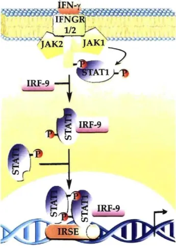
1.2.2.1 Transcriptional regulationThe first signaling pathway shown to be activated by lFNs was the JAK-STA T pathway. IFN-y receptor 1/2 (IFNGR1/2) binds to IFN-y molecules which then triggers the recruitment of Janus activated kinase 1/2 (Jak1l2) for the initiation of signal transduction [2]. Activated Jakl phosphorylates signal transducers and activators of transcription 1 (Statl) proteins which will in turn dimerize and translocate to the nucleus (Fig. 1.1) [19]. Stat 1 phosphorylation is necessary for full transcriptional activation [3
J
.
This cascade of tyrosine and serine phosphorylation is known to occur very promptly, in less than 1 min[9]. ÎFN-y rIFNGRj-~-
--11/2
~
_~
J
AK2J
AKI ~"",~-'-".J'''''#_''''''''''-:;
,
IRF-9
,
+
:ATIIRF-9
Figure 1.1: The classical JAK-STA T pathway following IFN-y stimulation of
macrophages. Ligand binding induces confonnational changes of IFNGR1I2, which will translocate Jak1l2 to the membrane. Stimulated Jakl will phosphorylate tyrosine residues on the lFNGR 1/2 receptor to allow recruitment ofStatl. Following tyrosine phosphorylation and lRF-9 binding in the cytoplasm, Statl fonns a homodimer. Dimerized Statl translocates to the nucleus and binds the lFN-stimulated response element (ISRE) to initiate or suppress transcription of lFN-y-regulated genes.
Statl can even function with IFN regulatory factors (IRFs) to establish the delayed response entailing the assurance of proper transcription control for the antiviral state (Fig. 1.1) [20]. On the other hand, Jak kinase activity is negatively regulated by SOCS-l and SOCS-3 proteins [9]. These Jak kinase inhibitor proteins bind to the phosphorylated tyrosine residue on IFNGRI and mask Statl docking site [10].
In addition to Jak-Stat pathway, other IFN-y signaling pathways involving key molecules such as NF-KB and phosphatidylinositol 3-kinase (PI3K), lead to changes in transcription control as part of the inflammation response [21, 22].
1.2.2.2 Cell growth and apoptosis
The effect of IFN-y on apoptosis in macrophages is still subject to controversy. Several studies show that apoptosis is induced by IFN-y, whereas others report that it serves as an anti-apoptotic signal [9, 23]. IFNs can activate the PI3K-Akt signaling pathway, and generate either pro-survival or apoptotic signaIs depending on the cellular context [3, 24]. In most cases, survival of the cell is crucial for the generation of IFN-y induced proteins necessary for the destruction of the invading organism [3]. On the other hand, apoptosis might also be the right choice for the destruction of tumor cells. For instance, IFN-y induces up-regulation of Fas protein which is responsible for the activation of cell death [25].
The anti-apoptotic transcription factor, NF-KB, is stimulated by proinflammatory cytokines and plays a crucial role in macrophages [26]. NF-kB regulates genes responsible for both the innate and the adaptive immune response [21]. In resting cells, NF-KB is bound to IKB protein and remains inactive in the cytoplasmic compartment. Extracellular stimulation induces signaling pathways that converge to free NF-KB from IKB [10]. NF-KB, then translocates to the nucleus to regulate the expression of multiple target genes to respond to the inflammation, the immune response, inflammatory response, cell adhesion, cell growth, and apoptosis (Fig. 1.2) [21].
Pro-inflammatory cytokines
~
~ 1 " ! 1 JSODD/ '
u~
P) .P uu
~u~~1j
h:Ba 265 proteasome ) } KB siteFigure 1.2: The c1assical NF-KR signaling pathway during inflammatory response. Pro-inflammatory cytokines su ch as IFN-y and TNF-a activate tumour necrosis factor receptor 1 (TNFR 1) which triggers silencer of death domain (SODD) release and recruitment of TNF-receptor-associated death domain prote in (TRADD), receptor-interacting protein (RIP) and TNF-receptor-associated factor 2 (TRAF2) to the membrane. Subsequently, hcB kinase (lKK)-complex is recruited to the TNFR 1 receptor by TRAF2 [21]. Activated IKK phosphorylates hcB, which indu ces confonnational change of hcB and exposes lysine residues for ubiquination. Thus, IKB becomes target for degradation by 26S proteasome and releases NF-KB dimers (p50 & p65) from the cytoplasmic NF-KB-IKB complex [10]. Free NF-KB translocates to the nucleus and binds to DNA for the regulation of gene transcription.
1.2.2.3 mRNA translation
IFN-y can also block cell growth and inhibit viral replication by deactivating the mRNA translation protein machinery. Most invading organisms su ch as bacteria and parasites have their own metabolism and only rely on the host for nutrients. On the other hand, viruses take over the host's protein synthesis machinery for their own repli cation [27]. For example, poliovirus and influenza virus specifically shut down the transcription and protein synthesis machinery respectively of the host cell so that the host cell replication, transcription and translation machinery will be dedicated for the synthesis of viral proteins [7].
IFN-y activation will mai nI y inhibit the translation of viral mRNAs during virus replication. The dsRNA will activate IFN-inducible RNA-dependent protein kinase (PKR), which will in tum phosphorylate eIF-2a subunit to inactivate viral prote in synthesis [28].
On the other hand, IFN is also known to induce protein expression of IFN-sensitive genes that act as direct or indirect mediators of their biological effect. Since prote in synthesis requires considerable amounts of metabolic energy (A TP & GTP) and amine acids, this event is highly controlled [3]. The initiation step corresponds to the decisive point during mRNA translation to deterrnine the fate of protein synthesis. Hence, extracellular stimuli mediate signal transduction oftwo different pathways (Ras-MAPK and PI3K1 AktimTOR), which converge and rigorously regulate the specific activity of key initiation translation factors (Fig. 1.3) [22, 29, and 30].
In summary, IFN-y stimulation was demonstrated to be directly involved in blocking virus repli cation and in the synthesis of critical proteins for the inflammation response. Yet, similarly to apoptosis, the precise role of IFN-y effect on mRNA translation is still not c1early defined [3].
,
\,
,
"
.N-...
...
...
r -___ ... _ _ _~3K
-
-
-
dependent translation Initiation of capFigure 1.3: IFN-y activation of Ras-MAPK and PI3KJAkt/mTOR signaling pathways to initiate mRNA translation. IFN-y induction leads to the activation of the PI3K and Ras pathways [29]. Consequently, PDKI and ERK1/2 phosphorylate and stimulate ribosomal protein S6 kinases (RSKs) [22]. AIso, activated mammalian target of rapamycin (mTOR) phosphorylates 4EBP 1, eIF4G and p70 S6K. Induced p70 S6K kinase and RSKs phosphorylate eIF4B resulting in initiation of mRNA translation [31]. In contrast, phosphorylation of 4EBP 1 disrupts its interaction with eIF4E. In the free state, eIF4E gets phosphorylated by MAP kinase signal-interacting kinase 1/2 (MNK 1/2), which activates its binding to the cap for translation initiation [29].
1.2.2.4 Phospholipid signaling
Another major pathway activated in response to extracellular stimuli consists of PLCs, phosphoinositide kinases and phosphatases, which are responsible for the change in phosphoinositide concentration. As discussed in the mRNA translation signaling pathways, PI3K was shown to play a major role upon IFN-y activation. Nonetheless, PI3K also produces a potent messenger phospholipid Pl(3,4,5)P3 from PI(4,5)P2. Phosphatase and tensin homologue deleted on chromosome ten (PTEN) prote in and inositol polyphosphate
PI(4,5)P2 and PI(3,4)P2, respectively [32].
The phosphoinositide, PI(4,5)P2, is involved in large number of cellular processes
such as cell survival, membrane transport and bacterial uptake and transport. For example, PI( 4,5)P2 participates in the cytoskeleton remodelling by promoting actin filaments
polymerization to form pseudopods. Activated macrophages develop membrane protus ions for locomotion and final capture of the prey [33]. Figure lA clearly illustrates the cell morphology change upon 24 h IFN-y stimulation.
.\Iacrophage relIs
IFl\-,'
~
24 h 100 li/mlActivated
.\Iacrophage relIs
Figure 1.4: Microscopie examination of J774 macrophage cells highlighting morphological changes taking place upon IFN-y stimulation.
Furthermore, hydrolysis of PI(4,5)P2 leads to the production of arachidonic acid (AA),
which also plays an important role in inflammatory response. Once AA is released, it interacts with the cytosolic factor, p47phox, further involved in the activation of the NAPDH
oxidase complex [34]. Interestingly, the change in phospholipid composition within macrophages in response to IFN-y leads not only to cytoskeleton rearrangements but also the induction of cytotoxic molecules.
1.2.2.5 Production of cytotoxic mo/ecu/es
To completely eliminate the invading organlsm, macrophages need to produce specific hannful metabolites. To do so, IFN-y-activated macrophages produce two types of strong cytotoxic molecules, reactive oxidative species (ROS) and reactive nitrogen species
(RNS) by induction of the NADPH-dependent phagocyte oxidase [9]. The NADPH complex is localized in the plasma membrane of the phagocytic cup and is composed of cytochrome b558 (gp91 phox and gp22phOX), p47Pho
X,
p40Phox and p67phox
, Rac and Rap l.
Superoxide (02-) is produced in the phagosome by accepting electrons from the NADPH oxidase complex from the cytosolic side of the membrane [35].
The NADPH oxidase complex is only complete and active during macrophage stimulation. In resting cells, the components of the complex are located in the cytosolic compartment of the cell [35]. Upon IFN-y activation, many different signaling pathways lead to its formation. For instance, neutrophil cytosolic factors, p47phox, p40PhOX & p67phox,
are recruited to the membrane by phosphorylation [35]. p47phox can also be translocated to the plasma membrane through the interaction with activated arachidonic acid (Fig. 1.5) [36]. Two different signaling pathways originating from the phospholipids and the phox proteins converge to synergistically stimulate NADPH oxidase complex.
Synergistic activation of p47 phox
by AA and phosphorylation
Active NAPDH oxidase enzyme complex
~GDI
Figure 1.5: Upon IFN-y administration, induction of cytosolic factors (p47pho
X,
p40PhOl and p67PhOl) recruitment to the membrane to form the NADPH oxidaseln summary, important pathways su ch as JAK-STAT, P13K-Akt-mTOR, NF-KB, and NAPDH oxidase, were shown to be affected by lFN-y activation. These changes in the gene and protein regulation, cell survival and phospholipid composition are important to prepare the macrophage against viral infection and finally lead to phagocytosis and pathogen destruction.
1.3 Phosphoproteomics
Upon extracellular stimuli activation, a cascade of signaling events is triggered to transmit the message from the membrane to the nucleus and induce changes in different cell function such as cytoskeletal structure, protein expression and gene transcription [37]. The proteins engaged in these stimulated pathways may undergo post-translational modifications (PTMs) such as ubiquitination, phosphorylation, acetylation and methylation to regulate their function, localization, binding specificity and stability [38]. Prote in phosphorylation is the most abundant PTM and it is involved in various essential cell functions such as cell division, proliferation, apoptosis and protein translocation [39].
In
eukaryotic cells, the most common phosphorylation occurs on hydroxyamino acids such as serine, threonine and tyrosine residues with a ratio of about 1000: 100: 1, respectively (Fig. 1.6) [40].
o
0 Il Il HO- P-O-CHrCH-C-OH 1 1 OH NH2o
Il HO-P-OH 1o
0 1 \1 phospho-serine H3C-r-TH-C-OH H NH2 phospho-threonineo
0Il
-IQ\-~
Il
HO-l-O~H2iH-C-OH OH NH2 phospho-tyrosineFigure 1.6: Chemical structure of phosphorylated serine, threonine and tyrosine residues [41).
Detection of phosphoproteins is rather difficult since only 1-2% of proteins are found in the phosphorylated form at any given time during signaling pathways [42]. The phosphorylation· status of a particular protein depends upon the equilibrium between the activities of the corresponding kinases and phosphatases. Since the phosphatase reaction dominates and the phosphate group is very labile, the chance to observe a phosphoprotein is
very low [5]. Aiso experimental analysis ofphosphoproteins is not an easy task because the phosphoamino acid only corresponds to a small fraction of the whole prote in sequence [40].
To counteract these problems, cell biologists use three main techniques for phosphoprotein detection: 32 p radioactive labeling, Western blotting and 2D-gels. The most popular method is 32p radioactive labeling since enrichment of the sample is not required and the detection signal is very sensitive (fmol) and quantifiable [42]. Yet, working with radioactive substances is not very desirable due to their toxic nature. AIso, 32p labeling can be tedious and sometimes even impossible since phosphate turnover rate is slow and only small amounts of radioactive phosphate are incorporated and may thereby escape detection [37].
As an alternative, Western blotting is another very sensitive (fmol) technique, which uses mono/poly-clonal antibodies directed against phosphorylated amino acids in order to de te ct phosphoproteins transferred from a ID or 2D gel to a membrane [40]. However, serine and threonine residues possess a very small and structural1y similar group, which makes it harder to obtain selective affinity interaction of the antibody to phosphoserines and phosphothreonines and the applicability ofthis method is reduced [37].
The last classical technique, 2D-gels, allows high-resolution separation of proteins from a complex sample according to their size and isoelectric point. However, 2D-gels analysis contains many drawbacks such as low-abundance prote in (ng) detection and solubility problems of membrane proteins [43]. These molecular biology tools can provide quick information on the presence of one relatively abundant phosphoprotein but cannot identify the specific phosphorylation site. As an alternative, mass spectrometry has shown to be an excellent approach that surpasses most of the limitations encountered by the classical biological methods [42].
1.3.1 Mass spectrometry approaches for phosphopeptide detection
In recent years, mass spectrometry techniques and approaches have been developing rapidly and with success for the analysis of the phosphoproteome [5]. Initially, Edman degradation in combination with 32p labeling allowed the peptide identification of many proteins including the phosphoprotein, p-casein [44]. Yet, this technique was not very
efficient since the amino acid cleavage did not consistently occur [41]. Nowadays, tandem mass spectrometers such as the hybrid quadrupole/time of flight hybrid (Q-TOF), Linear Trap Quadrupole (LTQ)-Orbitrap and Fourier transform ion cyclotron resonance (FTIRC) can provide identification of phosphopeptides with high resolution and sensitivity levels (fmol). Still, phosphoproteomic analysis by MS encounters major limitations due to the phosphopeptide ion suppression [5]. To enhance their detection, phosphopeptides are selectively isolated or modified using immunoprecipitation, enrichment methods, chemical modification and alkaline phosphatase treatment prior to MS analysis [45].
1.3.1.1 Immunoprecipitation assay
Phosphopeptides are present in very sm aIl amounts compared to the non-phosphorylated peptides in a cell ex tract [42]. For this reason, immunoprecipitation methods using phosphospecific antibodies are required for an enhanced detection. General pTyr antibodies have been used for the enrichment of complex cell extracts. Antibodies directed against the specific amino acids sequence containing the phosphorylated residue (Tyr/Ser/Thr) is also used for immunoprecipitation assay. Cell lysates are initially incubated with phosphospecific antibodies and then the immunocomplex is collected with protein A-Sepharose beads. The isolated phosphoproteins are resolved by SDS-PAGE analysis to be further analyzed by MS [46,47].
OveraIl, immunoprecipitation assay has shown to achieve successful outcomes with anti-phosphotyrosine antibody [48]. Yet, it is still quite expensive and inconvenient to use antibodies for such large-scale experiments. Also, the serine and threonine antibodies independent of sequence context have not been used extensively due to their low specifity [5]. However, when antibodies are directed against a specific peptide containing Ser/Thr residues, the enrichment level is more significant [4].
Many novel phosphorylated proteins· have been identified us mg immunoprecipitation assay, especially with tyrosine antibody [48]. However the use of this method is somewhat more limited than the other enrichment procedure since it is highly dependent upon the efficiency of the phosphospecific antibodies.
Two other simi1ar approaches routine1y used for phosphoprotein enrichment are Ti02 and IMAC micro-columns [45, 49]. The principle of IMAC enrichment mainly exploits the high affinity of negatively charged phosphopeptides towards metal-chelated (usually Ga3+ and Fe3+) stationary phase [50]. In order to reduce non-specific binding by acidic si de chains of glutamic acid and aspartic acid, a pH lower than the carboxyl group pKa (pKa of Asp is 3.65 and pKa of Glu is 4.25) and higher than the phosphoric acid pKa (2.15) is used [51]. Yet, unspecific binding to acidic residues (glutamic acid & aspartic acid), e1ectron donors (histidine) and hydrophobic peptides is still a major pitfall [5]. To overcome this prob1em, Ficarro et al. [52] esterified aU carboxy1ic acid groups prior to enrichment, which enabled the reduction of urispecific binding by at least two orders of magnitude. However, reaction conditions have to be chosen correctly in order to avoid incomplete methylation of the carboxylic groups and side reactions.
Recently, Larsen et al. [49] introduced a high1y selective and efficient stationary phase, titanium dioxide (Ti02), for phosphopeptide enrichment. A much higher enrichment level and recovery level was obtained by disso1ving the sample in trifluoroacetic acid (TF A), acetonitri1e (ACN) and 2,5-dihydroxybenzoic acid (DHB). The exact binding mechanism of Ti02 to the phosphate group is still unclear but Ti02 was found to have higher se1ectivity towards singly phosphory1ated peptides. On the other hand, it was a1so reported that IMAC ligands have higher affinity for multi-phosphorylated peptides [53]. Hence, Thingholm et al. [54] created the SIMAC technique by combining both strategies to improve phosphorylation sites coverage.
Generally, IMAC and Ti02 application is performed off-line using Zip Tip or mlcro-columns [50, 55]. To simplify the extraction procedure and minimize sample handling and loss, the chromatographie medium can be integrated into micro-capillary, which is combined to nano-LC/ESl-MS. On-line lMAC or Ti02 enrichment can be controlled by an interface, which is compatible to any LC and MS [56, 57]. Ti02 and lMAC enrichment is commonly used since it allows pre-concentrating the sample, retaining phosphopeptides, and removing reagents not compatible to MS [58]. Another major advantage is that Jess
suppression effect occurs due to enhancement of phosphopeptides and removal of non-phosphorylated peptides.
Even thought, immunoprecipitation, Ti02 and IMAC micro-tips significantly improve phosphopeptide detection by decreasing sample complexity, detection by MS still remains challenging due to the chemical properties of the phosphate group. In positive ion mode, the negatively charged phosphate group decreases the ionization efficiency of the peptide. AIso, since phosphate groups are very labile on phosphoserine and threonine, MSIMS spectrum is very poor which makes it harder for peptide sequencing [5]. ln brief, enrichment procedures significantly improve phosphopeptide recovery but are still limited for complete phosphopeptide detection.
1.3.1.3 Chemical modification methods
Many studies have attempted to enrich phosphopeptides by .replacing the phosphate group by an affinity tag. The most popular reaction, ~-elimination, exposes phosphoserines and phosphothreonines to strongly alkaline conditions (Li OH or Ba(OHh at pH 12) and yields a more reactive dehydroalanine and dehydroaminobutyrine species, respectively. The modified residue can act as a Michael acceptor and reacts with thiol based nucleophiles such as dithiothreitol (DTT) and 1,2-ethanedithiol (EDT) (Fig. 1.7) [59]. The phosphorylated amino acid can be identified by a mass shift resulting from the derivatization (-21 Da for ~-elimination and Michael addition with 2-aminoethanethiol) [60].
(a) o Il HO-P-OH 1 o -H3P04 CH2 Il _ _ _ ... -N-C-C-1 <fH2 Base 1 Il H 0 -N-C-C-1 1 Il H H 0 Phosphoserine Dehydroalanlne (b) ~ HO-P-OH 1 o 1 ?H2 -H3P04 ~-~-9-~-~ H H 0 Base
•
CH 2 Il ~-N-C-C-~ 1 Il H 0 HSCH2CH2SH (Ethanedithiol)~
..
o Il HO-P-OH 1 o 1 H -y-CH3 -N-C -C-I 1 Il H H 0 Phosphothreonine H-C-CH3 Il -N-C-C-1 Il H 0 Dehydroaminobutyrlc acld COOH 9H2CH2SJ, S ) . -k",VVV\
Biotir, 1 0 · CH2 1 ~-N-C-C-~ 1 Il H 0Figure 1.7:
(a) p-elimination reaction of phosphoserine and phosphothreonine. (b)Addition of a thiol group and biotin tag on phosphoserine for further enrichment on avidin columns [5].
Phosphopeptide derivatization method has greatly improved and now allows
detection down to femtomolar range [42]. A distinct advantage of this technique over
enrichment procedures is that removal of the phosphate group prior to mass spectrometry
facilitates positive ion detection.
Nevertheless~this strategy still suffers from many
drawbacks.
First~contrary to immunoprecipitation approach, chemical derivatization is not
applicable to tyrosine phosphorylation since it would require breakage of the very stable
aromatic ring [5].
Inaddition, O-linked sugar moieties may also undergo
~-eliminationand
obtain same intermediates as the phosphorylation residues [42]. Finally, incomplete
derivatization of the phosphate moiety will result in decrease of phosphopeptide signal
intensity after
~-elimination.Overall, chemical derivatization of phosphopeptide has dernonstrated to be very
efficient for the detection of phosphopeptides. Yet, this method is dependent upon the
extent of completion of the reaction and unwanted reactions, which might be responsible
for the increase of complexity of the sample and phosphopeptide losses [42].
1.3.1.4 A lkalin e phosphatase treatment
Removal of the phosphate group by a phosphatase enzyme is another popular strategy used to avoid the ion suppression limitation of phosphopeptides in MS. The enriched protein digest is split in two where one half is incubated with alkaline phosphatase in basic conditions [61]. FoUowing MS analysis of the treated and control sample, aU the peptides correlated by a mass shift of 80 Da are designated as potential phosphopeptides [62]. If there are more than one phosphorylation site in a peptide, then mass shifts of multiples of -80 Da will be observed.
One major drawback of alkaline phosphatase treatment is that the phosphorylation sites remain indistinguishable from other hydroxyl amino acids. To overcome this problem, Liao et al. used several specific proteases su ch as endoproteinase Glu-C, Asp-N and trypsin to degrade the phosphopeptide into smaUer fragments and locate exactly the phosphorylation site [61]. Also, Torres et al. addressed this issue by combining the
advantages of IMAC, on-target dephosphorylation and hypothesis driven MALDI MS/MS [63]. This approach enabled the identification of four phosphate group on the C-terminus of the metazoan histone mRNA regulator (dSLBP), showing that alkaline phosphatase treatment is the method of choice to overcome the ion suppression of phosphopeptide in complex mixtures during MS detection.
In short, enrichment of the phosphopeptide samples and modification of the phosphate group are key strategies for an enhanced phosphopeptide analysis. In addition, further method optimization can also be performed on the analytical separation prior to MS detection to obtain higher phosphopeptide coverage.
1.3.1.5 Separation methods
Highly complex samples can be separated ~d analyzed using high performance liquid chromatography (HPLC) coupled to a ESI-MS [64]. Peptides are first loaded onto a nano-column containing CI 8 material and then eluted at a slow flow rate (200-600 nL/min) using a gradient ofwater and ACN. Coupling the nano-LC system to the mass spectrometer has been very successful for the separation of phosphopeptides and a decrease in ion
suppression [5]. However, since multi-phosphorylated peptides are very hydrophilic, they may not stick onto the hydrophobic column and will elute in the column flow-through [62].
Alternatively, to enhance detection of phosphorylated peptides, the sample mixture can initially be separated on a strong cation exchange (SCX) column followed by chromatographic separation into the CI8 column. Hydrophilic phosphopeptides are enriched in early fractions, resulting in better detection [5].
In summary, mass spectrometry provides a viable alternative to more traditional methods for phosphorylation analysis. Yet, the dual constraints of low yield of phosphopeptide and the mixture complexity required methods of separation and enrichment prior to MS analysis. Affinity enrichment procedures su ch as IMAC and Ti02 micro-columns, chemical derivatization and anti-phosphospecific antibodies, increased the proportion ofphosphopeptides in a complex mixture. However, the choice ofwhich method is most appropriate is dependent upon the quantity of prote in available, which residue are expected to be phosphorylated and the degree of sample purification. Nevertheless, recent high-sensitive mass spectrometers have proven to significantly enhance phosphoprotein identification in simple and complex samples [65, 66].
1.4 Mass spectrometry
The mass spectrometer (MS) is an instrument used for determining the molecular mass and structure of biomolecules, drugs, proteins and oligonuc1eotides. This analytical tool is composed of three different sections: the ionization source, the mass analyzer and the detector [67]. ln proteomics-based experiments, the sample is initially digested by trypsin and th en the peptide mixture can undergo chromatographic separation or is directly introduced into the MS [68]. Upon ionization in the source, the ions travel into the analyzer where they will be separated according to their mass (m) -to-charge (z) ratio (m/z). The
signal collected from aIl these ions is transformed to generate m/z spectrum [67]. Lastly, the raw chromatograms are processed through specialized softwares that further enable the profiling and identification of the proteins in the sample (Fig. 1.8) [69].
Sample introduction cc. CE. 111'Le Direct injection E~I \lAI DI
Mass Spectrometer
Mass
analyzer
Ion Ir,1p Qu,ld nlp"ll' Tinll'-ot· flighl FI-1eRFigure 1.8: General mass spectrometry-based proteomics.
Data
Analysis
l'hùlOll1ulliplit'r l'vlilTochilmwl plilte
Eledwn multiplier
Sorne instruments contain two or more analyzers, which can be used for structural and sequencing studies [67]. Several different types of MS are available commercially such as the triple quadrupole, hybrid triple quadrupole/linear ion trap (Q-TRAP), matrix assisted laser desorption ionization-time of flight (MALDI-TOF), hybrid quadrupole/time of flight hybrid (Q-TOF), Fourier transform ion cyclotron resonance (FTICR) and the linear trap quadrupole (L TQ)-Orbitrap. Ali of these instruments offer different applications in the field of MS.
The most common ionization method used for biological applications is the electrospray ionization (ESI). This technique was first introduced by Fenn in 1984 [64]. The analysis can be perfonned in positive or negative ion mode depending on the proton affinity of the sam pie. ESI can be coupled to a HPLC or a CE since the analyte is converted from a liquid to a gaseous phase [70].
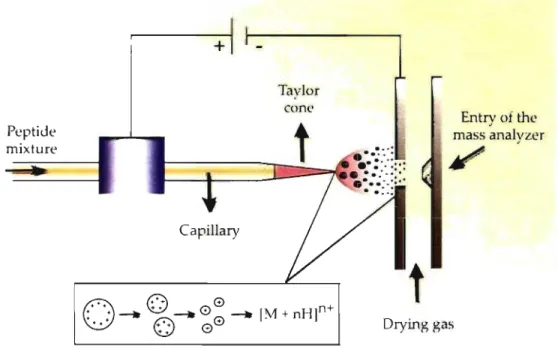
The atmospheric pressure interface of an ESI source is composed of a capillary where the sample solution is exposed to very high voltage (3-5 kY) applied to the tip. A stream of nebulising gas (nitrogen) can also be present around the capillary to facilitate the nebulisation of the analytes [71]. As the charged liquid first exits the emitter's tip, a cone shape, known as a Taylor cone, briefly fonns (Fig. 1.9). With the assistance of the drying gas (nitrogen), droplets decrease in size by solvent evaporation which forces the charges in the molecules closer together [71]. A Coulombic explosion occurs when there are too many charges being confined within a too small are a and start repelling each other (Rayleigh limit), causing the droplets to divide into two smaller droplets. This process repeats itself until the solvent is completely evaporated and the droplets have split up to become one single charged molecule (Fig. 1.9) [67]. This ionization process is very fast and occurs within milliseconds.
Peptide mixture
+
Capillary
Figure 1.9: Electrospray ionization chamber. Taylor cone
t
Entry of the mass analyzer/
Drying gas23
A new method named nano-eJectrospray ionization uses flow rate of sampJe injection of 20-40 nL/min and minimizes Joss of anaJyte [71]. The decrease in drop Jet size and inner diameter of the emitter enabled an ionization efficiency of two orders of magnitude greater than the conventional ESI [64]. Hence important parameters that influence the electrospray stability are the voltage applied, the flow rate and the emitter size. Overall, ESI provides a continuous stream of multiply charged ions that can be introduced in a multitude of mass analyzers.
1.4.2 L TQ-Orbitrap mass spectrometer
As technology progresses, more important advancements take place in the mass spectrometry field. For instance, several years ago, detection of about few hundreds phosphopeptides in a complex cell extract was considered very high for mass spectrometry analysis [52]. Recently, a new and very powerful mass spectrometer, the LTQ-ürbitrap, enabled the identification of almost 1000 phosphopeptides for large-scale phosphoproteomic analysis [66]. This observation shows that the extent to which biological information can be gained is completely dependent upon the analytical performance of the MS. The Linear Trap Quadrupole (LTQ)-Orbitrap recently invented by Makarov is one of the most efficient hybrid mass analyzer.
The orbitrap mass analyzer is an electrostatic trap containing an inner and outer electrode. Ions are injected tangentially in the trap and move in complex spiral patterns aIl around the inner electrode [72]. A Fourier transform performs the mass analysis based on image current detection of oscillation frequencies [73]. For this reason, the FTICR and orbitrap have similar level of mass accuracy and resolution [5]. The orbitrap has a mass accuracy below 2 ppm, a high dynamic range (~5000) and a maximum resolving power of 100,000 that can be reached in 1.6 sec of analysis and 400
m/z
[73, 74]. Even though the orbitrap is able to perform MS/MS analysis, a lower sensitivity is observed due to the loss of ions in the C-trap. Hence, it is more practical to link the orbitrap to another mass analyzer, the linear ion trap, which will perform MSIMS analysis with much higher sensitivity. This hybrid ofmass analyzers is referred to as Linear Trap Quadrupole (LTQ)-Orbitrap (Fig LlO).Autosamplcr
&
HPLC
LTQ Orbitrap
Figure 1.10: L TQ-Orbitrap hybrid mass spectrometer (Thermo Electron) cou pied to a nano-flow Le (Eksigent) with a Spark-Holland autosampler (Thermo Electron).
The L TQ-Orbitrap is composed of three main elements; the linear quadrupole ion trap, the C-trap and the orbitrap (Fig. 1.11). lnitially, the ions are accumulated into the linear quadrupole ion trap and then transferred into the C-shaped storage trap (C-trap). The C-trap is used to store and collisionally cool ions, which are subsequently pu1sed into the orbitrap [74].
ESI source Linear ion trap
l
L C-trap--
--===~ - = = ; - - -.!-.. - = Orbitrap
25
The MS and MS/MS analysis can be recorded independently or in tandem with both mass analyzers (linear quadrupole ion trap and orbitrap) according to the experimental requirements. The most popular approach used for peptide identification is to combine the fast MSn capabilities of the linear ion trap with the high resolution and mass accuracy of the orbitrap [73]. For the tandem MS analysis, the ion trap stores and stabilizes ions by applying a RF voltage. Maximum resolution and sensitivity is achieved by cooling down the ions with a damping gas. By increasing the RF voltage, ions are destabilized and ejected from the trap. As a result, only one peptide is kept where bond cleavage occurs upon vibrational excitation [67]. In a total time of 1 sec, the duty cycle of the LTQ-Orbitrap consists of 3-5 MS/MS analysis for one survey scan [73].
In summary, this higher number of acquired MSIMS spectrum considerably enhances the overall number of peptide identification. Aiso the faise positive rate of high-throughput peptide analysis is significantly smaller due to the instrument's high mass accuracy.
1.4.3 Tandem MS
Tandem MS is mainly applicable for small drug molecules and peptides. MS/MS spectrum offers structural information which can be pieced together to generate the complete peptide sequence. The process consists of isolating a particular ion, which subsequently coUides with neutral gas atoms (nitrogen, argon or helium) in the collision cell. Ions will absorb the kinetic energy which induces breakage of peptide bonds to form product ions [67]. Ali these fragmented ions are separated according to their m/z by the mass analyzer and finaUy provide a fingerprint pattern of the precursor ion under investigation [75]. Throughout one survey scan of a chromatographie run, the most abundant ion at a given time is kept in the trap and is further fragmented to obtain the MS/MS scan [69]. The success of de novo sequence interpretation largely depends upon the mass accuracy and resolution of the instrument.
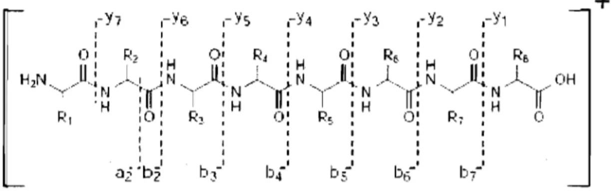
Three different peptide bonds (NH-CH, CH-CO and CO-NH bond) can be fragmented depending on the MS conditions. Upon bond breakage, two fragments are created where one is charged and the other neutral depending on the chemistry and proton affinity of the two species [67]. In ail, there are six different possible fragment ions; a, b and c ions when the charge is retained on the N-terminus and x, y and z ions for C-terminal charged molecules. Since it requîres less energy, the most common fragmentation pattern is the b and y type ions (Fig. 1.12) [75].
b--b 1
+
Figure 1.12: Peptide fragmentation in low collisional energy illustrating b and y ions
[69).
During tandem mass spectrometry, the phosphate moiety of phosphopeptides is predominantly lost and usually serves as a signature for phosphopeptides. Since the P~
bond is much weaker than a peptide bond for Serrrhr phosphorylated precursor, a satellite ion, [M+H-98t, is generated du ring fragmentation [76]. The unsaturated form of the serine (69.03 Da) and threonine (83.05 Da) will indicate the presence of a phosphate moiety in a MS/MS spectrum [71]. On the other hand, the phosphate group on tyrosine phosphorylated peptides usually remains attached to the side chain during MSIMS analysis due to tyrosine's stable ring structure [77]. Hence, the phosphorylated tyrosine can be identified with a mass of243.18 Da (163.18 + 80 Da) on the MSIMS spectrum.
The MSIMS spectrum can be interpreted manually or through different specialized algorithms (Mascot, Sequest and ProteinProspector) that try to match the fragments ions to theoretical values expected from a large protein database [69].
1.4.4 Data processing
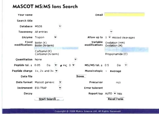
1.4.4.1 MS/MS ion search using Mascot
In
proteomics studies, Mascot from Matrix Science (http://www.matrixscience.comQ is a very popular database engine used to identify proteins from MSIMS spectrum and protein databases. Mascot is a useful tool for the interpretation of Jess informative MS/MS spectrum or complex protein mixtures [69]. Prior to the Mascot search, necessary information about the MS experiment needs to be added in the Mascot search engine page in order to minimize the [aise positive rate especially for the large scale proteomic analysis (Fig. 1.13).MASCOT MS/MS Ions Search
Your n~me
Search title
D~t~b~se MSDB T axonomy Ali entries
Enzyme TrYPsin Fixed Biotin (Ki
modific~tions Biotln (N-term)
Carbamyi (K)
Carbamyl (N-term)
Qu~ntitèltion None
Peptide toI. ± 0.05 Da .... # "e 0
.-Peptide charge 1 +, 2+ and 3+ ....
D~t~ file
O~t~ torm~t Mascot Q8n8nc ....
-Instrument ESI-TRAP T Decoy Em~il -....Allo,"" up to 1 .. mlssed cleavaQes
V~ri~ble OXldatlon (HW) modific~tion5 Oxidation (M) Proplonamlde (C) MS/MS toI. ± 0.5 Da .... Monoisotopic ... Averl!lQe Precursor mil Error toler~nt
Report top AUTO .... hits Reset Funn
-COPVPlght ç:, 2'J08 M,;tm~ ';':I4tflCa .. '.:d A.II P.II3"'~ R.~C"lIad
Figure l.13: Mascot search engine page showing aIl parameters for a MS/MS ion
search.
The general approach consists of first downloading the file containing ail MS/MS spectra (fragment ions with their intensity, m/z and RT) to Mascot. Then, the most suitable
database (MS DB, IPI, SwissProt and NCBInr) and species are determined according to the proteins characteristics. The sample preparation is taken into consideration by choosing the enzyme responsible for the digestion, the number of allowed missed cleavage sites and the
![Figure 1 . 11: A schematic representation of the LTQ-Orbitrap mass spectrometer [73]](https://thumb-eu.123doks.com/thumbv2/123doknet/2155813.9448/40.915.203.829.777.1034/figure-schematic-representation-ltq-orbitrap-mass-spectrometer.webp)