HAL Id: dumas-00824923
https://dumas.ccsd.cnrs.fr/dumas-00824923
Submitted on 22 May 2013HAL is a multi-disciplinary open access
archive for the deposit and dissemination of sci-entific research documents, whether they are pub-lished or not. The documents may come from
L’archive ouverte pluridisciplinaire HAL, est destinée au dépôt et à la diffusion de documents scientifiques de niveau recherche, publiés ou non, émanant des établissements d’enseignement et de
Amélioration de la survie globale des patients porteurs
de cancer du rein métastatique grâce aux thérapies
ciblées : revue monocentrique depuis 2000
Mélanie Tadj
To cite this version:
Mélanie Tadj. Amélioration de la survie globale des patients porteurs de cancer du rein métastatique grâce aux thérapies ciblées : revue monocentrique depuis 2000. Médecine humaine et pathologie. 2013. �dumas-00824923�
AVERTISSEMENT
Ce document est le fruit d'un long travail approuvé par le
jury de soutenance et mis à disposition de l'ensemble de la
communauté universitaire élargie.
Il n’a pas été réévalué depuis la date de soutenance.
Il est soumis à la propriété intellectuelle de l'auteur. Ceci
implique une obligation de citation et de référencement
lors de l’utilisation de ce document.
D’autre part, toute contrefaçon, plagiat, reproduction illicite
encourt une poursuite pénale.
Contact au SICD1 de Grenoble :
thesebum@ujf-grenoble.frLIENS
LIENS
Code de la Propriété Intellectuelle. articles L 122. 4
Code de la Propriété Intellectuelle. articles L 335.2- L 335.10
http://www.cfcopies.com/V2/leg/leg_droi.phpUNIVERSITÉ JOSEPH FOURIER FACULTÉ DE MÉDECINE DE GRENOBLE Année: 2013 N° AMÉLIORATION DE LA SURVIE GLOBALE DES PATIENTS PORTEURS DE CANCER DU REIN METASTATIQUE GRACE AUX THÉRAPIES CIBLÉES: REVUE MONOCENTRIQUE DEPUIS 2000. THÈSE PRÉSENTÉE POUR L’OBTENTION DU DOCTORAT EN MÉDECINE DIPLOME D’ÉTAT Mélanie TADJ Née le 23 Juillet 1981 à Suresnes (92) THÈSE SOUTENUE PUBLIQUEMENT À LA FACULTÉ DE MÉDECINE DE GRENOBLE* Le 25 Avril 2013 DEVANT LE JURY COMPOSÉ DE Président du jury: Madame le Professeur Mireille MOUSSEAU Membres: Monsieur le Docteur Mathieu LARAMAS, directeur de thèse Monsieur le Professeur Stéphane CULINE Monsieur le Professeur Jean‐Luc DESCOTES * La Faculté de Médecine de Grenoble n’entend donner aucune approbation ni improbation aux opinions emises dans les thèses; ces
REMERCIEMENTS
Aux membres du jury Madame le Professeur Mireille MOUSSEAU, vous m’avez fait l’honneur d’accepter la présidence de cette thèse. Je vous remercie de la confiance que vous m’avez accordée ce qui m’a permis d’acquérir une rapide autonomie. Je vous adresse ici ma reconnaissance et ma gratitude. Monsieur le Docteur Mathieu LARAMAS, je te remercie d’avoir accepté de diriger cette thèse. J’espère qu’à mon tour je saurai transmettre ce que tu m’a appris. Tout au long de mon internat, tu as été l’enseignant exigeant indispensable à ma formation sans oublier d’être l’ami bienveillant m’aidant à trouver le fragile équilibre entre vie professionnelle et familliale. A une époque où nous étions si peu nombreux, tu as été et restera mon “grand frère” de cancérologie!Monsieur le Professeur Stéphane CULINE, votre participation à ce jury m’honore particulièrement. Vous m’avez donné le goût de la cancérologie lors de mon passage dans votre service à Montpellier en tant qu’externe de 3eme année. Veuillez trouver ici le témoignage de toute ma considération.
Monsieur le Professeur Jean‐Luc DESCOTES, votre engagement en onco‐urologie et plus généralement en cancérologie n’est plus à démontrer. Votre participation à l’élaboration et à la critique de ce travail en est encore un exemple. Veuillez trouver ici l’expression de ma respectueuse et profonde considération.
A tous ceux avec qui j’ai eu le plaisir de travailler, un grand merci! A Valentin pour la réalisation de cette base de données sans quoi ce travail aurait été bien plus difficile. A Christine, Cristina et Oxana pour tous les bons moments partagés en service et ailleurs. A Cécile, Isabelle C et Armelle pour avoir fait de cette année en votre compagnie la meilleure de mon internat ainsi qu’à Guillaume et Jocelyne avec qui je suis impatiente de poursuivre mon apprentissage. Un merci particulier à Isabelle M qui sans le savoir, a influencé le medecin que je suis devenue.
A Prune, Sandrine, Claire, Laura, Jérome, Cécile pour votre enseignement et votre energie.
A Lysiane, Stephane et Remy pour m’avoir fait découvrir et apprécier l’hématologie.
A Violaine, Caroline, Julie, Isabelle pour m’avoir initié à la radiothérapie.
A Dominique, Lamia, Sylvie, Marie‐Héléne, Blandine, Barbara, Giacomo, Nicole, Géraldine pour avoir tenté de combler avec patience mes nombreuses lacunes anatomopathologiques.
A Gaétan et Claire pour avoir su cultiver ma fibre gériatrique devenue oncogériatrique!
A mes co‐internes d’oncologie qui depuis quelques années viennent grossir les rangs; Delphine, Medhi, Laure.
Aux internes croisés au grès de mon cursus avec qui nous avons partagés un plus que de la médecine: Philippe, Martin, Clara, Mathieu, Sylvain, Camille, Leïnag, Ségolène,
A Jean‐Cyril et Audrey pour avoir éveillé la curiosité et tissé des liens entre des spécialités parfois éloignées par l’intermédiaire de l’AJOI.
A Véronique, Anne‐Marie, Solange, Emmanuelle, Gaëlle, Angélique, Beatrice, Corinne pour votre investissement indispensable dans la recupération des données. Aux équipes paramédicales pour votre engagement auprès des malades. Merci à mes amis Caroline, Gaël, Stephanie et Germain qui me suivent depuis le lycée, quel chemin parcouru à vos cotés. Emilie, Marie, Marine que j’ai rencontrées à Montpellier sur les bancs de la faculté avec qui c’est toujours un plaisir de se revoir. Anne‐Claire, Mickaël, Janine, Franck, Christelle, Nicolas, Julie qui m’accompagnent depuis mon arrivée à Grenoble, votre soutien et votre amitié me sont précieuses. A ma famille Philip pour sa relecture attentive.
Mes parents pour leur soutien toujours indéfectible et leur écoute attentive, j’ai beaucoup de chance! Mon papa Valentin et Inès, vous grandissez trop vite … mais je suis fière des adultes que vous devenez ! A toi Emeric pour ton amour et l’équilibre que tu assures dans ma vie. A nos deux trésors Arthur et Zoé.
TABLE DES MATIÈRES
Résumé . . . p. 6 Abréviations . . . p. 9 Article . . . p. 11 ABSTRACT . . . p. 13 INTRODUCTION . . . p. 14 PATIENT AND METHOD . . . p. 16 Renal tumor database . . . p. 16 Inclusion criteria . . . p. 16 Objective study . . . p. 17 Statistical analysis . . . p. 17 RESULTS . . . p. 18 Cohort characteristic . . . p. 18 Overall survival . . . p. 18 Outcome in targeted therapy group . . . p. 19 Outcome in non‐targeted therapy group . . . p. 19 DISCUSSION . . . . . . p. 21 REFERENCES . . . p. 25 TABLE . . . p. 28 FIGURE . . . p. 29 Annexes . . . p. 33 Conclusion . . . p. 43RÉSUMÉ
Contexte: les traitements anti‐angiogéniques ont radicalement modifiés la prise en
charge thérapeutique des patients atteints de cancer du rein métastatique. Le but de ce travail est de déterminer dans la pratique clinique quotidienne l’impact de ces nouvelles thérapies sur la survie globale.Methodes: Etude rétrospective, mono‐centrique, non interventionnelle incluant les
patients porteurs d’un cancer du rein métastatique à cellules claires diagnostiqués depuis 2000 au Centre Hospitalier Universitaire de Grenoble. Les 2 cohortes ont été déterminées en fonction de la première ligne thérapeutique reçue (thérapies ciblées ou autres traitements).Résultats: Quatre‐vingt dix‐huit patients ont été inclus entre le 1er Janvier 2000 et le 31
Decembre 2010. Les caractéristiques démographiques et pathologiques des 2 cohortes étaient comparables, en particulier la distribution des profils pronostiques. En première ligne, 58 patients ont reçus une thérapie ciblée dont 71% ont été traités par sunitinib, 21% par bevacizumab, et 8% par temsirolimus. Dans l’autre cohorte (n=40), 37,5% des patients ont reçus des cytokines, 15% une chimiothérapie cytotoxique ou une hormonothérapie. La médiane de survie globale des patients traités par thérapie ciblée est significativement augmentée (30 mois contre 13 mois; p<.003, log‐rank test). Le Hazard Ratio (HR) de décès à 3 ans entre les deux groupes de traitement est de 0.53 (intervalle de confiance 95%, 0.33‐0.85; p=.008, log‐rank test). Le HR de décès à 3 ans ajusté sur le profil pronostique est de 0.43 (IC95%,0.27‐0.71).Conclusion: Cette étude rétrospective objective l’amélioraton de survie globale des
patients atteints de cancer du rein métastatique, quelque soit le groupe pronostique,des indications thérapeutiques ont radicalement modifié la stratégie thérapeutique dans le cancer du rein métastatique, pour le bénéfice des patients.
Abréviations
ABRÉVIATIONS
95%CI: 95% Confidence Interval FGI: French Group of Immunotherapy HR: Hazard Ratio mRCC: metastatic Renal Cell Carcinoma mTOR: mammalian Target Of Rapamycin OS: Overall survival PDGF: Platelet‐Derivated Growth Factor PFS: Progression Free Survival VEGF: Vascular Endothelial Growth FactorArticle
Targeted therapy improves overall survival in metastatic renal clear
cell carcinoma: Monocentric review since 2000.
M. Tadj. CHU de Grenoble, Oncologie Médicale, Pôle Cancérologie Medecine Aigüe Communautaire, France
V. Arnoux , MD. CHU de Grenoble, Département Urologie Transplantation, Pôle Digidune, France.
M.Mousseau, MD. PhD. CHU de Grenoble, Oncologie Médicale, Pôle Cancérologie Medecine Aigüe Communautaire, France
J.L. Descotes, MD. PhD. CHU de Grenoble, Département Urologie Transplantation, Pôle Digidune, France.
J.L. Quesada . CHU de Grenoble, Unité d’épidémiologie clinique, Centre d’Investigation Clinique,France.
L. Bensaadi, MD. HU de Grenoble, Departement d’Anatomie et Cytologie Pathologiques, France.
M.Laramas, MD. CHU de Grenoble, Oncologie Médicale, Pôle Cancérologie Medecine Aigüe Communautaire, France
Correspondence:
Mathieu LARAMAS, MD. Medical Oncology, University Hospital, CS 10217, 38043 Grenoble cedex 9, France.
Phone: 33 4 76 76 54 51 Fax: 33 4 76 76 56 61
E‐mail: mlaramas@chu‐grenoble.fr
ABSTRACT:
Introduction: Anti‐angiogenic treatment had radically modified therapeutic strategy in
metastatic renal cell carcinoma (mRCC). This study is aimed to determine the overall survival (OS) improvement in clinical practice.Patients and methods: Retrospective, monocentric and non‐interventional study in
mRCC diagnosed since 2000 with 2 cohorts of patients determined according to the first line treatment (targeted therapy or others treatment).Results: Between 1 January 2000 and 31 December 2010, 98 patients were included.
The 2 cohorts were balanced with regard to baseline disease and demographic characteristics in particular for prognosis profiles distribution. As first line, 58 patients received targeted therapy whose 71% were treated by sunitinib, 21% by bevacizumab and 8% by temsirolimus. In non‐targeted therapy cohort (n=40), 37.5% were treated by cytokines, 15% by cytotoxic chemotherapy or hormonal therapy. Patients treated with targeted therapy had a significantly longer median OS (30 months vs 13 months; p<.003, log‐rank test). The Hazard Ratio (HR) of death at 3 years was 0.53 (95% Confidence Interval, 0.33‐0.85; p=.008, log‐rank test). When adjusted to the prognosis profile, the HR of death was 0.43 (95%CI, 0.27‐0.71).Conclusions: This retrospective study demonstrated the improvement of OS due to
targeted treatments, for all prognostic risk groups. This result supported the complete change of care of mRCC patients with extension of therapeutic indications and efficient therapeutic lines.
INTRODUCTION
In the 1990s, immunotherapy was approved for treatment of metastatic renal cell carcinoma (mRCC) by using mainly α‐interferon or interleukin‐2 alone or a combination of both. Cytokines were effective on few patients and complete response was obtained in less than 15% of cases after 25 weeks of combined therapy. The impact on overall survival (OS) was small. Median survival time was 12 months for single therapies rising to 17 months with combined therapy.1 Toxic side effects like hypotension, fever,
performance‐status impairment and nausea or vomiting were frequently the limiting factor of these strategies in particular for interleukin‐2. High resistance to conventional chemotherapy, significant toxicity of cytokines and lack of alternative treatment when resistance to immunotherapy was achieved limited survival of mRCC.
Oncogenesis of mRCC is supported by inactivation of the Von Hippel‐Lindau (VHL) pathway that increased production of vascular endothelial growth factor (VEGF) and platelet‐derived growth factor (PDGF). A better comprehension of the neo‐angiogenesis mechanism has led to the emergence of new therapeutic agents: tyrosine kinase inhibitors (sunitinib, sorafenib), monoclonal antibody anti‐VEGF (bevacizumab) or mammalian Target Of Rapamycin (mTOR) inhibitors (temsirolimus, everolimus). Since 2006, therapeutic strategies include anti‐angiogenic treatment and therefore reduce use of immunotherapy. The first approved drugs were sunitinib and sorafenib. As first line, sunitinib increases Progression Free Survival (PFS) from 5 months with α‐interferon to 11 months.2 A phase III study demonstrated a statistically significant benefit in PFS for
sorafenib (5,5 months) versus placebo (2,8 months) in second line.3 With acceptable
The effect of anti‐angiogenic treatment on OS is partially established. Only temsirolimus has demonstrated a statistically significant lowering risk of death of 27% for poor prognosis patients with mRCC versus interferon alone.4 Pivotal trials of first VEGF‐
targeted agents (sunitinib, bevacizumab) were initially designed to prove such benefit. But when pre‐planned interim analysis was conducted, the important benefit in PFS led to amendment of the protocol. Subsequent lines of targeted therapy available during on‐ going trials and the possibility of crossover had strongly confounded the OS results. However, sunitinib showed a trend toward improved survival versus interferon (HR for death = 0,65; p=0,02).2 Survival data are missing to justify this change in clinical practice.
We reported the results of a retrospective, monocentric and non‐interventional study using data from adult kidney cancer database. The aim of this study was to determine in clinical practice whether anti‐angiogenic agents improve overall survival of mCCR compared with former therapeutic strategies.
PATIENTS AND METHODS
Renal tumour database
All patients aged 18 years or older with renal tumours recorded on the Grenoble University Hospital database. This database was retrospectively populated since 1 January 2000. Completeness of database was assured by crosschecking of three information sources: the Medical Information Department (request using Disease International classification v10), patient records (paper and digital with CristalNet Software v04) and the anatomopathologic database. Since 01 June 2009, the database was prospectively completed. In accordance with French data protection legislation, the database was declared to the “Commission Nationale de l’Informatique et des Libertés” and identified under number: 1500484 v 0. The database was designed according to the ethical principles of the Declaration of Helsinki and received approval from an independent ethics committee (Institutional Review Board 5891).
Inclusion criteria
Only patients aged 18 years and older with metastatic renal clear cell carcinoma were identified. Others histologic types were excluded. The 2 cohorts of patients were determined according to the first line treatment: one anti‐angiogenic targeted group (including bevacizumab, sunitinib or temsirolimus) and another group treated otherwise (including cytokines, cytotoxic chemotherapy and others treatment). Patients were classified into poor, intermediate and favorable prognosis according to the French Group of Immunotherapy (FGI) classification (considering performance status index, number of metastasis, liver metastasis and time to recurrence).
Objective study
The primary end‐point was overall survival defined as the time between the date for diagnosis of mRCC and death due to any cause. Patients without an event were censored at the last follow‐up assessment (31 October 2012). Subgroup analyses according to prognostic factor were planned. The secondary end‐point was to assess the impact of anti‐angiogenic treatment sequence on OS. Data on the use in clinical practice of targeted therapies was also collected: number of cycles of treatment, reason for stopping (progression or toxicity), dosage adjustment.
Statistical analysis
Data were summarized by size and frequency for categorical data and by mean scores ± standard deviation or by median and 25th‐75th percentiles when normality was rejected
for quantitative data. Comparison of categorical parameters was performed using Chi2 test, or Fisher exact test if needed. Comparisons of continuous parameters were performed using an unpaired Student t‐test or Mann–Whitney test if necessary. Normality was tested by the Shapiro–Wilk, and variance homogeneity by Levene tests. Survival analysis was performed using Kaplan–Meier curves, log‐rank univariate test and the Cox model for prognosis adjustment. The statistical analysis was performed using Stata® 12.1 (StataCorp LP, College Station, TX) PC‐Software. All data was analyzed independently in the Grenoble Clinical Research Center.
RESULTS
Cohort characteristics
Between 1 January 2000 and 31 December 2010, 766 patients with kidney cancer were identified of whom 124 were metastatic. Nineteen tumours were not clear cell carcinoma and 7 had an unknown histology. Ninety‐eight patients with mRCC were included: 58 received an anti‐VEGF or m‐TOR targeted therapy as first line and 40 received other treatments. At the time of analysis, 71 patients (72%) were dead. The median follow‐ups for targeted therapy treated patients and for non‐targeted therapy treated patients were 27,5 months (range, 1‐127) and 10,5 months (range, 1‐124) respectively. The 2 cohorts were balanced with regard to baseline disease and demographic characteristics in particular for prognosis profiles distribution (table 1). A greater percentage of a poor prognosis patient in the non‐targeted therapy group was observed (43% vs 30%) without difference statistically significant (p= .21).
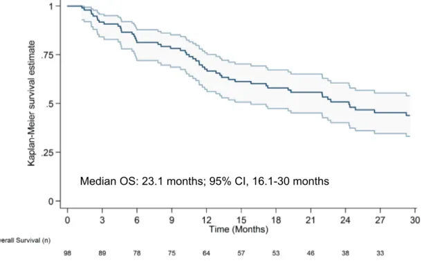
Overall survival
The median survival of all patients was 23.1 months (range, 16.1‐ 30) (fig 1). When stratified by FGI risk categories (fig 2), patients with poor risk had a significantly shorter overall survival (11 months; range, 5.8‐19.6) than patients with intermediate risk (29.7 months; range, 19.4‐30) or good prognostic patients (median overall survival not reached). Patient treated with targeted therapy as first line had a significantly longer median OS (30 months vs 13 months; p<.003, log‐rank test) (fig 3). The Hazard Ratio (HR) of death at 3 years was 0.53 (95% Confidence Interval, 0.33‐0.85; p=.008, Cox Proportional Hazards Model). When adjusted to the prognosis profile, the HR of death for
patient treated with targeted therapy as first line versus without was 0.43 (95%CI, 0.27‐ 0.71, log‐rank test).
Outcome in targeted therapy group
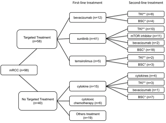
In the cohort treated by anti‐angiogenic therapy, patients received a median of 2 therapeutic lines (range 1 to 5). As first line, 71% of patients had received sunitinib (n=41), 21% bevacizumab (n=12), and 8% temsirolimus (n=5). The average duration of treatment as first line was 9, 10.5 and 4 months respectively for bevacizumab, sunitinib and temsirolimus. Dosage had been modified for 65% of patients treated by sunitinib: 21 patients with 25% of reduction and 6 with at least 50% of reduction. In 18% of cases (n=9), first line therapy had to be interrupted due to toxic effects. Only 3 long‐term stabilities (defined as non‐progressive disease during more than 2 years) and 2 complete responses had been observed. As second line, 12% of patients (n=4) received a monoclonal antibody, 59% received TKI (n=13 for sorafenib, n=7 for sunitinib) and 29% mTOR inhibitor (n=2 for temsirolimus, n=8 for everolimus). When stratified by anti‐ angiogenic treatment sequence, 23% (n=8) of patients received a monoclonal antibody‐ TKI sequence, 29% (n=10) a TKI‐mTOR inhibitor sequence and 35% (n=12) a TKI‐TKI sequence. Four patients were treated by TKI‐monoclonal antibody or mTOR inhibitor‐TKI sequence (fig 4). The median overall survival was not statistically different between these targeted therapy sequences.
Outcome in non‐targeted therapy group
time of clinical data cut‐off (93 months survival). Forty two percent of patient (n=17) did not receive active treatment: 8 patients had a poor prognosis profile and 6 where under observation during more than a year (survival ranging from 12 to 27 months). In this cohort, 7 patients received a targeted therapy during evolution: 4 as second line and 3 as third line or more.
DISCUSSION
This monocentric review of mRCC over the 11 past years demonstrates the significant improvement in OS by using targeted first line treatment (bevacizumab, sunitinib or temsirolimus) in routine clinical practice. This study among an unselected population of mRCC supports the profound modification of therapeutic strategy with the adoption of anti‐angiogenic treatment since 2006. This improvement was not demonstrated in the pivotal study of sunitinib or bevacizumab, but temsirolimus achieved a significant HR for death of 0.73 (95%CI, 0.58‐0.92; p=.008) versus placebo in poor prognostic population.4
Some others publications support the hypothesis of a benefit in OS. A population‐based study in British Columbia, Canada reported a significant improvement of median survival of 8.6 months for patients treated by sunitinib compared with cytokines (17.3 vs 8.7 months; p=.004).5 Less than 10% of 200 patients included received a second line therapy
and unsuitable patients for treatment were excluded. This study demonstrated the superiority in OS of sunitinib as exclusive first line treatment without highlighting profound modification of clinical practice: extending therapeutic indication and increasing number of therapeutic lines. A Swedish population‐based study sought to demonstrate the improvement in OS using a large dataset of 3 243 patients.6 The
RENCOMP analyses reported a HR for death of 0.76 (95%CI; 0.69‐0.83, p<.001) in mRCC patient diagnosed in the period 2006‐2008 versus 2000‐2005 with respectively median survival of 17.7 months (95%; 14.9‐20.5) and 10.2 months (95%CI; 9.1‐11.2). Nevertheless, important prognostic factors were not analysed such as risk group classification or histology data. Despite 40% of treatments having been omitted from the
In our study, because of retrospective data, patients could not been classified according to the MSKCC or Heng’s classification risk. The FGI classification is an effective prognostic model in predicting OS in mRCC patients treated in the era of targeted therapy.7 OS
observed in each FGI’s prognostic group are consistent with OS formerly reported in the corresponding prognostic group among Heng’s classification: 44 months for favorable‐ risk, 27 months for intermediate‐risk and 8.8 months for poor‐risk.8
The OS improvement is constant whatever the prognostic profile according to FGI classification with adjusted HR of death statistically significant (0.43; 95% CI, 0.27‐0.71). Particularly in poor prognosis patient group, targeted therapy led to an increase OS of 7 months. In our study, poor prognosis patients seemed to be overrepresented in the non‐ targeted therapy cohort but it was not statistically significant. The OS observed for poor prognosis treated by targeted therapy (13,5 months, data not shown) was concordant with median OS reported in same population treated by temsirolimus.4 Immunotherapy
or chemotherapy abstention for unsuitable patients was formerly standard practice and represented by 1 case per 5 patients. Even in the era of targeted therapy, there is still a small percentage of unfit patients who are not candidates for an active and potentially toxic treatment (less than 10% in our study).
This retrospective study could not grade adverse events. Clearly, toxicity limits had been approached since dosage modifications were frequently reported in patient records. Sixty five percent of sunitinib treated patients had a dosage reduction. Dosage modifications seem to be more frequent in usual clinical practice (32% of dose reduction was reported in the pivotal trial of sunitinib).2 Comorbidity could explain part of this reduction. The
Common Toxicity Criteria scale used in randomised studies was not designed for the assessment of toxicities in continuous therapy. Symptomatic toxicity (asthenia, mucosistis and diarrhoea) could be under‐estimated in an over‐selected population.9 Even if
toxicities could imply efficacy, the management of chronic adverse events are a challenge in an unselected population of mRCC.10
The number of patients collected does not allow a conclusion on the better therapeutic sequence. VEGF combinations do not improve OS but exacerbate toxicities.11,12 Recent
guidelines recommend sequential treatment with several options for second or third‐line drug.13 New drugs are available: pazopanib as first line for good or intermediate
prognosis, axitinib for second line.14,15 New options (e.g. tivozanib) are expected to be
approved in near future.16 Nevertheless, the immune pathway is not definitively
abandoned. Anti‐PD‐1 and anti‐PD‐L‐1 antibodies recently demonstrated in phase 1 studies up to 27% of cumulative response in renal cell cancer population.17,18
Beyond the effect of one single agent, the improvement of OS may be increased by introduction of new efficient therapeutic agents which multiple the number of available therapeutic lines. As in other metastatic disease (e.g. breast or colon cancer), defining the best therapeutic sequence is challenging. In the absence of the necessary strategic trials, clinicians must select treatment based on efficacy and toxicity of previous lines.
In conclusion, this retrospective study demonstrated the improvement of OS due to targeted treatments, for all prognostic risk groups. These results support the complete change of care in patients with the adoption of mRCC since 2006. In the era of targeted
number of therapeutic lines with efficient therapeutic agents for other patients is increasing. Each step of progress contributes to improve overall survival.
REFERENCES
1. Negrier S, Escudier B, Lasset C, et al: Recombinant human interleukin‐2, recombinant human interferon alfa‐2a, or both in metastatic renal‐cell carcinoma. Groupe Francais d'Immunotherapie. N Engl J Med 1998;338:1272‐8
2. Motzer RJ, Hutson TE, Tomczak P, et al: Sunitinib versus Interferon Alfa in Metastatic Renal‐Cell Carcinoma. N Engl J Med 2007;356:115‐24 3. Escudier B, Eisen T, Stadler WM, et al: Sorafenib in advanced clear‐cell renal‐cell carcinoma. N Engl J Med 2007;356:125‐34 4. Hudes G, Carducci M, Tomczak P, et al: Temsirolimus, Interferon Alfa, or Both for Advanced Renal‐Cell Carcinoma. N Engl J Med 2007;356:2271‐81 5. Heng DY, Chi KN, Murray N, et al: A population‐based study evaluating the impact of sunitinib on overall survival in the treatment of patients with metastatic renal cell cancer. Cancer 2009;115:776‐83 6. Harmenberg U, Wahlgren T, Kowalski J, et al: Treatment and overall survival (OS) in metastatic renal cell carcinoma (mRCC): A Swedish population‐based study (2000‐2008). J Clin Oncol 2012;30:abstr 389
7. Crepel M, Escudier BJ, Machiels JH, et al: Comparison of two major prognostic models for patients with metastatic renal cell carcinoma treated in the contemporary era of targeted therapies. ASCO Meeting Abstracts 2011;29:abstr 4660
8. Heng D, Xie W, Regan M, et al: Prognostic Factors for Overall Survival in Patients With Metastatic Renal Cell Carcinoma Treated With Vascular Endothelial Growth Factor–Targeted Agents: Results From a Large, Multicenter Study. J Clin Oncol 2009;27:5794‐99
9. Powles T, Sarwar N, Jones R, et al: An indirect comparison of the toxicity of sunitinib and pazopanib in metastatic clear cell renal cancer. Eur J Cancer 2012;48:3171‐6 10. Di Fiore F, Rigal O, Menager C, et al: Severe clinical toxicities are correlated with survival in patients with advanced renal cell carcinoma treated with sunitinib and sorafenib. Br J Cancer 2011;105:1811‐3 11. Molina AM, Feldman DR, Voss MH, et al: Phase 1 trial of everolimus plus sunitinib in patients with metastatic renal cell carcinoma. Cancer 2012;118:1868‐76 12. Negrier S, Gravis G, Perol D, et al: Temsirolimus and bevacizumab, or sunitinib, or interferon alfa and bevacizumab for patients with advanced renal cell carcinoma (TORAVA): a randomised phase 2 trial. Lancet Oncol 2011;12:673‐80
13. Escudier B, Eisen T, Porta C, et al: Renal cell carcinoma: ESMO Clinical Practice Guidelines for diagnosis, treatment and follow‐up. Ann Oncol 2012;23(Suppl 7): v65‐71
14. Sternberg CN, Davis ID, Mardiak J, et al: Pazopanib in locally advanced or metastatic renal cell carcinoma: results of a randomized phase III trial. J Clin Oncol 2010;28:1061‐85
15. Rini BI, Escudier B, Tomczak P, et al: Comparative effectiveness of axitinib versus sorafenib in advanced renal cell carcinoma (AXIS): a randomised phase 3 trial. Lancet 2011;378:1931‐9 16. Nosov DA, Esteves B, Lipatov ON, et al: Antitumor activity and safety of tivozanib (AV‐951) in a phase II randomized discontinuation trial in patients with renal cell carcinoma. J Clin Oncol 2012;30:1678‐85 17. Brahmer JR, Tykodi SS, Chow LQ, et al: Safety and activity of anti‐PD‐L1 antibody in patients with advanced cancer. N Engl J Med 2012;366:2455‐65
18. Topalian SL, Hodi FS, Brahmer JR, et al: Safety, activity, and immune correlates of anti‐PD‐1 antibody in cancer. N Engl J Med 2012;366: 2443‐54
Table 1‐ Baseline demographic and clinical characteristics
Targeted therapy Non-targeted therapy
n=58(%) n=40 (%) p values sex
male 38 (66) 33 (82) .06
female 20 (34) 7 (17)
age years (range) 61 (39-83) 63 (41-84) .25 performance status i 0 20 (37) 8 (26) .28 1 25 (46) 13 (42) .69 2 and more 9 (17) 10 (32) .09 prior nephrectomy 46 (80) 28 (70) .28 metachronous metastasis 26 (45) 10 (25) .04 Median time of delayed metastasis months
(25-75 percentil) 46 (12-82.5) 43.5 (4-79) .67 number of disease sites
1 24 (41) 17 (42) .91 2 and more 34 (59) 23 (58) Sites of metastasis bones 24 (41) 16 (40) .89 lung 40 (69) 22 (55) .16 liver 17 (29) 8 (20) .29 nodes 20 (34) 12 (30) .64
French Classification risk score ii
favorable 10 (18) 7 (19) .90
intermediate 29 (52) 14 (38) .18
poor 17 (30) 16 (43) .21
i: n=54 in targeted therapy group and n=31 in non-targeted therapy group; ii: n=56 in targeted therapy group and n=37 in non-targeted therapy group
Fig. 1 ‐ Kaplan‐Meier estimates of OS at 3 years follow‐up !"#$%&%'%()*+),'-."./%.012)3.0%45%67%)3%8%9.)/0%54++4:';*%%%%%%
Fig. 2 ‐ Kaplan‐Meier estimates of OS at 3 years follow‐up according to FGI risk categories !"#$%&%'%()*+),'-."./%.012)3.0%45%67%)3%8%9.)/0%54++4:';*%)<<4/=",#%34%!>?%/"0@%<)3.#4/".0$%% 5)A4/)B+.% ",3./2.=")3.% *44/%
Fig. 3 ‐ Kaplan‐Meier estimates of OS at 3 years follow‐up !"#$%&%'%()*+),'-."./%.012)3.0%45%67%)3%&%8.)/0%54++49':*$%%
Targeted therapy
OS: 30 months; 95% CI, 23.1-30
Others treatments
OS: 13 months, 95% CI, 6.1-22.8
Fig. 4 – Flow Chart !"#$%&'()*+,-./0' TKI** (n=8) BSC* (n=4) bevacizumab (n=12) TKI** (n=10) mTOR inhibitor (n=11) bevacizumab (n=2) BSC* (n=18) sunitinib (n=41) TKI** (n=2) BSC* (n=3) temsirolimus (n=5) Targeted Treatment (n=58) BSC* (n=7) bevacizumab (n=1) TKI** (n=3) cytokines (n=4) cytokine (n=15) cytotoxic chemotherapy (n=6) Others treatment (n=19) No Targeted Treatment (n=40) mRCC (n=98) !"/10+2"34'0/4.05430' 64,)37+2"34'0/4.05430' 8'9410'6:;;)/<=4'>./4' 88'?@/)1"34'A"3.14'B3-"C"0)/'
ANNEXE 1: masque de saisie des données !!"# #
$%
&''()(*#
!""#$#%&%!%'()*+#%,#%)(-)-#%,#)%,.""/#)%
#ANNEXE 2: Déclaration Commission Nationale de l’Informatique et des Libertés!""#$#%&%!%'()*+,+-./"%0123$
%
ANNEXE 3: Accord Comité d’Ethique