Full Terms & Conditions of access and use can be found at
http://www.tandfonline.com/action/journalInformation?journalCode=iern20
ISSN: 1473-7175 (Print) 1744-8360 (Online) Journal homepage: http://www.tandfonline.com/loi/iern20
Efficacy and mode of action of external trigeminal
neurostimulation in migraine
Jean Schoenen & Gianluca Coppola
To cite this article:
Jean Schoenen & Gianluca Coppola (2018) Efficacy and mode of action
of external trigeminal neurostimulation in migraine, Expert Review of Neurotherapeutics, 18:7,
545-555, DOI: 10.1080/14737175.2018.1488588
To link to this article: https://doi.org/10.1080/14737175.2018.1488588
Accepted author version posted online: 13
Jun 2018.
Published online: 26 Jun 2018.
Submit your article to this journal
Article views: 23
REVIEW
Efficacy and mode of action of external trigeminal neurostimulation in migraine
Jean Schoenen
aand Gianluca Coppola
ba
Headache Research Unit, University Department of Neurology CHR Citadelle Hospital, Liège, Belgium;
bResearch Unit of Neurophysiology of Vision
and Neuro-Ophthalmology, G. B. Bietti Foundation IRCCS, Rome, Italy
ABSTRACT
Introduction: Available preventive drug treatments for migraine lack complete efficacy and often have
unpleasant adverse effects. Hence, their clinical utility and therapeutic adherence are limited. Noninvasive
neurostimulation methods applied over various peripheral sites (forehead, mastoid, upper arm, cervical vagus
nerve) have raised great interest because of their excellent efficacy/tolerance profile. Among them external
trigeminal nerve stimulation (eTNS) was first to obtain FDA approval for migraine therapy.
Areas covered: All clinical trials of eTNS as preventive or acute migraine treatment published in
extenso or presented at congresses are reviewed. The paper analyzes neuroimaging and
neurophysio-logical studies on mechanisms of action of eTNS. As many of these studies point toward the anterior
cingulate cortex (ACC) as a likely eTNS target, the paper scrutinizes the available literature on the ACC
implication in migraine pathophysiology.
Expert commentary: eTNS is a viable alternative to standard pharmacological antimigraine strategies
both for prevention and abortive therapy. eTNS could chiefly exert its action by modulating the
perigenual ACC, which might also be of interest for treating other disorders like fibromyalgia or
depression. It remains to be determined if this might be a common mechanism to other peripheral
noninvasive neurostimulation methods.
ARTICLE HISTORY Received 2 March 2018 Accepted 11 June 2018 KEYWORDS Noninvasive neurostimulation; external trigeminal nerve stimulation; antimigraine treatment; perigenual anterior cingulate cortex
1. Introduction
1.1. Pharmacotherapy of migraine
Although cognitive-behavioral therapies can be useful as
add-on therapies in some patients, currently, migraine is mostly
managed with pharmacological treatments. Acute migraine
drugs like analgesics, nonsteroidal anti-inflammatory drugs
(NSAIDs) and triptans [
1
] are used to interrupt a migraine
attack and allow patients to recover normal functioning as
soon as possible [
2
]. Triptans, the agonists of 5-HT1B/D
sero-tonin receptors, are up to now the most effective drugs for
severe attacks. They can, however, induce class-specific side
effects like chest, face, and limb pain or discomfort that are
difficult to tolerate by many patients. Moreover, they are
vasoconstrictors and contraindicated in case of comorbid
car-dio- or cerebrovascular pathology. Migraine patients with
fre-quent attacks or attacks not responding adequately to acute
treatments are in need of a preventive antimigraine treatment
able to modify the disease course by decreasing attack
fre-quency and intensity [
3
] so that excessive consumption of
acute antimigraine drugs does not lead to headache
chronifi-cation, i.e. so-called medication overuse headache, which
wor-sens the patients
’ condition [
4
].
Effective preventive drugs include beta-blockers without
intrinsic sympathetic activity, certain calcium channel blockers
like flunarizine or verapamil, sartans, the antiepileptics
topir-amate and valproate, and possibly amitriptyline [
5
], as well as
nutraceuticals like riboflavin and coenzyme Q10 [
6
]. Preventive
migraine drugs have only partial efficacy with an overall
50
–60% success rate and, except for the nutraceuticals, they
tend to cause moderate to severe side effects, frequently
leading to dissatisfaction and discontinuation by the patients
[
7
–
9
]. Consequently, 80% of patients are willing to change
their current medication for a treatment with similar efficacy
but fewer side effects [
10
]. The therapeutic efficacy of
migraine preventives decreases sharply with migraine
chron-ification, which explains why up to 50% of chronic migraine
patients discontinue their prophylactic drug treatment after
2 months [
11
].
1.2. Neurostimulation as an alternative antimigraine
treatment
The shortcomings of pharmacological migraine management
and technological advances have allowed neurostimulation
methods to emerge as credible alternative antimigraine
treatments.
Since the 1990s, stimulation of pericranial nerves through
percutaneously implanted electrodes was found effective for
the treatment of various headaches in various studies [
12
,
13
].
Occipital nerve stimulation (ONS) was reported beneficial for
chronic migraine in some, but not all, sham-controlled trials,
but globally the effect size was modest [
14
–
16
]. Combining
percutaneous ONS with supraorbital nerve stimulation (SNS)
was claimed to have a better effect, but randomized
con-trolled trials are lacking [
17
]. These neurostimulation methods
CONTACTJean Schoenen jschoenen@ulg.ac.be University Department of Neurology, Headache Research Unit, CHR Citadelle Hospital, Boulevard du 12eme de Ligne 1, 4000 Liege, Belgium.
2018, VOL. 18, NO. 7, 545–555
https://doi.org/10.1080/14737175.2018.1488588
using implanted electrodes may cause various adverse effects
chiefly due to local infections, lead displacement or frequent
battery replacement, but their major drawback is that they are
invasive and indicated only for the most disabled,
drug-refrac-tory patients with very frequent and severe migraine
attacks [
18
].
With the advent of noninvasive transcutaneous stimulators
neurostimulation therapy became applicable to all migraine
patients irrespective of their level of disability or
drug-refrac-toriness [
19
]. In recent years various transcutaneous
stimula-tion methods were reported to be effective in migraine
treatment despite striking differences in targets: bilateral
mas-toid [
20
], cervical vagus nerve [
21
], or skin at the wrist [
22
].
Several studies of noninvasive neurostimulation in the
trigem-inal territory showing beneficial effects in various headache
types were published as early as 1985 [
23
–
26
]; among them,
the single-blinded, placebo-controlled trial by Solomon and
Guglielmo [
23
] appears to be the most convincing. Two
dec-ades later, technological advances permitted to produce a
portable, affordable and user-friendly external trigeminal
neu-rostimulator (eTNS) called Cefaly
®
(Cefaly Technology sprl,
Seraing, Belgium). This device delivers electrical pulses to the
forehead branches of the first division of the trigeminal nerve,
the ophthalmic nerve, though it can also be applied over the
greater occipital nerves via a suboccipital band. The pulses are
rectangular biphasic impulses with an electrical mean equal to
zero, impulse width of 250 µs, frequency of 60
–120 Hz,
max-imum intensity of 16 mA with an increment from 1 to 16 mA
over 14 min. The device has an inbuilt software to record time
of use. During the last 4 years several clinical trials have
explored the therapeutic effect of eTNS both for the
preven-tive and the acute treatment of migraine. We will review these
trials as well as studies aimed at determining the mode of
action of eTNS in migraine.
2. Clinical eTNS trials in migraine
For the sake of comprehensiveness, all clinical data of eTNS
with this device in migraine that are in the public domain are
displayed in
Table 1
, including manuscripts submitted for
publication and abstracts.
2.1. Migraine prevention
Eight studies have addressed the preventive effect eTNS,
ran-ging from an early small pilot trial to a randomized
sham-controlled trial [
27
–
34
]. In all of them there was a decrease in
migraine attack frequency after daily stimulation for time
periods between 1 and 4 months, both in episodic and chronic
migraine (two open label trials). We will focus briefly on the
PREMICE trial [
28
] where eTNS (
n = 34, 250 µs, 60 Hz, 16 mA)
was compared to sham stimulation (
n = 33, 30 µs, 1 Hz, 1 mA)
for a 3-month treatment period. After using the device with
the active stimulation protocol 38.1% of patients had
a ≥ 50%
reduction in monthly migraine days compared to
pretreat-ment baseline, while only 12.1% of patients did so with the
sham stimulation. There were no adverse events except for the
frontal paresthesias that are known to occur with electrical
stimuli. Up to now no trial comparing eTNS with a preventive
drug treatment is available. Topiramate, presently one of the
most effective drugs, is more efficacious than eTNS in the
pooled analysis of placebo-controlled trials reaching a 45.3%
of 50% responder rate [
29
], but it is hampered by unpleasant
side effects and low tolerability. For comparison, 1 out of 4
patients taking topiramate abandoned the drug because of
intolerable side effects [
29
], while in a survey of 2313 patients
using eTNS 4.3% reported one or more adverse effects, chiefly
intolerance to the forehead paresthesias (1.25%) and 2%
stopped treatment because of an adverse effect [
30
]. As a
result of the PREMICE trial, the device was the first medical
device to get approval from the FDA in 2014 for the
preven-tive treatment of migraine.
2.2. Acute migraine treatment
Since it was marketed, this eTNS device contains a stimulation
protocol with a 100 Hz frequency recommended for attack
treatment based on a pilot study [
27
]. In clinical practice many
migraineurs have been using this protocol to treat their
attacks effectively before evidence-based data became
avail-able. In an internet survey, 88.6% of 413 regular users applied
the device in 71.8% of attacks, which allowed reducing acute
migraine drug use in 42.6% of them [
31
]. Chou et al. [
32
,
33
]
were first to scientifically assess efficacy of eTNS for acute
migraine treatment in patients stimulated for 1 h in the
hos-pital for an untreated attack that had started
≥3 h before. In
the initial open study 77% of patients had
≥50% pain relief at
1 h; in the subsequent multicenter, sham-controlled ACME trial
[
32
,
33
], 63% of patients reached this end point compared to
31% in the sham arm and 29% were pain-free at 1 h versus 6%
with the sham stimulation. This beneficial effect was
con-firmed in a recent open label study of 48 patients who treated
a moderate to severe attack at home for 2 h [
34
]: 35.4% were
pain-free at the end of the stimulation. The 2 h pain-free rates
with eTNS are comparable, or even superior, to those reported
in recent drug trials of sumatriptan nasal powder (34.3%) [
35
],
the 5-HT1F agonist lasmiditan (28%) [
36
] or the CGRP receptor
antagonist ubrogepant (25.5%) [
37
]. They are also superior to
those found in open studies of noninvasive cervical vagus
nerve stimulation (21% and 22.9%) [
38
,
39
].
3. Mode of action
Some recent studies have shed some light on possible
mechanisms by which eTNS may reduce migraine attack
fre-quency and headache during an attack. eTNS can have an
effect on the peripheral and the central nervous system.
Transcutaneous electrical nerve stimulation (TENS), in use
since many years to relieve pain [
40
], is thought to act in part
by blocking nociceptive traffic at the segmental level by
acti-vating large A
β afferents according to the gate control theory
[
41
,
42
]. By analogy, pericranial nerve stimulation could have
the same effect by activating somatic afferents from the
tri-geminal territory or C2 dermatoma that are known to
con-verge with visceral trigeminovascular afferents on spinal
trigeminal nucleus second-order nociceptors. This might
apply to low intensity
–high frequency TENS.
Acupuncture-like high intensity
–low frequency TENS and high intensity–
high frequency TENS, resembling the Cefaly
®
stimulation
pro-tocols, more likely engage extrasegmental mechanisms such
as activation of subcortical pain control centers [
43
].
We will review the evidence for peripheral, segmental and
suprasegmental mechanisms possibly involved in the
antimi-graine eTNS effects.
3.1. Peripheral nervous system mechanisms
As opposed to the visceral afferents of the
trigeminovascu-lar system, somatic nociceptive afferents of the first division
of the trigeminal nerve are not supposed to play a major
role in migraine headache pathophysiology. Nonetheless,
branches of meningeal nociceptive fibers were recently
found to emerge at the level of cranial emissary canals
and fissures and to innervate the cranial periosteum and
muscles mainly in the temporal, parietal, and occipital areas
[
44
]. Because of its anatomical position and its reduced
surface, it seems thus unlikely that the device activates
such extracranial meningeal afferents.
High intensity
–low frequency TENS causes intense, but not
uncomfortable, muscle contraction able to activate muscle
affer-ents and to produce analgesia [
40
]. Given the small size of
fore-head muscles under the stimulator, this probably is not relevant
for the device
’s antimigraine effects. Interestingly, in chronic
migraine patients eTNS caused an increase of median frequency
and amplitude of the electromyographic signal in anterior
tem-poralis, auricularis posterior and middle trapezius muscles [
45
]. It
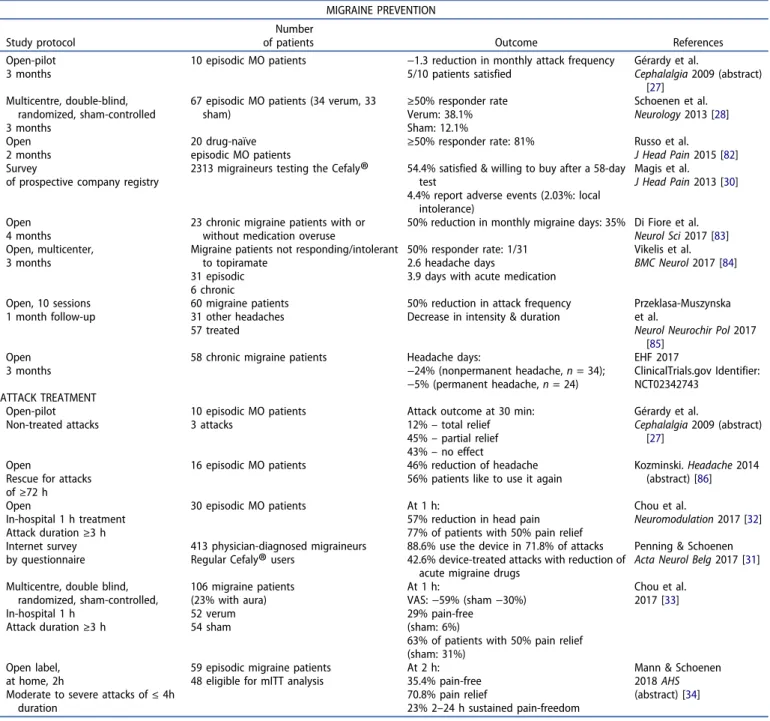
Table 1.eTNS with the Cefaly
®
: efficacy data for migraine prevention and acute treatment. MIGRAINE PREVENTION Study protocolNumber
of patients Outcome References Open-pilot
3 months
10 episodic MO patients −1.3 reduction in monthly attack frequency 5/10 patients satisfied Gérardy et al. Cephalalgia 2009 (abstract) [27] Multicentre, double-blind, randomized, sham-controlled 3 months
67 episodic MO patients (34 verum, 33 sham) ≥50% responder rate Verum: 38.1% Sham: 12.1% Schoenen et al. Neurology 2013 [28] Open 2 months 20 drug-naïve
episodic MO patients ≥50% responder rate: 81%
Russo et al. J Head Pain 2015 [82] Survey
of prospective company registry
2313 migraineurs testing the Cefaly
®
54.4% satisfied & willing to buy after a 58-day test4.4% report adverse events (2.03%: local intolerance)
Magis et al. J Head Pain 2013 [30]
Open 4 months
23 chronic migraine patients with or without medication overuse
50% reduction in monthly migraine days: 35% Di Fiore et al. Neurol Sci 2017 [83] Open, multicenter,
3 months
Migraine patients not responding/intolerant to topiramate
31 episodic 6 chronic
50% responder rate: 1/31 2.6 headache days
3.9 days with acute medication
Vikelis et al. BMC Neurol 2017 [84] Open, 10 sessions 1 month follow-up 60 migraine patients 31 other headaches 57 treated
50% reduction in attack frequency Decrease in intensity & duration
Przeklasa-Muszynska et al.
Neurol Neurochir Pol 2017 [85]
Open 3 months
58 chronic migraine patients Headache days:
−24% (nonpermanent headache, n = 34); −5% (permanent headache, n = 24) EHF 2017 ClinicalTrials.gov Identifier: NCT02342743 ATTACK TREATMENT Open-pilot Non-treated attacks 10 episodic MO patients 3 attacks
Attack outcome at 30 min: 12%– total relief 45%– partial relief 43%– no effect Gérardy et al. Cephalalgia 2009 (abstract) [27] Open
Rescue for attacks of≥72 h
16 episodic MO patients 46% reduction of headache 56% patients like to use it again
Kozminski. Headache 2014 (abstract) [86] Open In-hospital 1 h treatment Attack duration≥3 h 30 episodic MO patients At 1 h:
57% reduction in head pain 77% of patients with 50% pain relief
Chou et al.
Neuromodulation 2017 [32] Internet survey
by questionnaire
413 physician-diagnosed migraineurs Regular Cefaly
®
users88.6% use the device in 71.8% of attacks 42.6% device-treated attacks with reduction of
acute migraine drugs
Penning & Schoenen Acta Neurol Belg 2017 [31] Multicentre, double blind,
randomized, sham-controlled, In-hospital 1 h Attack duration≥3 h 106 migraine patients (23% with aura) 52 verum 54 sham At 1 h: VAS:−59% (sham −30%) 29% pain-free (sham: 6%)
63% of patients with 50% pain relief (sham: 31%)
Chou et al. 2017 [33]
Open label, at home, 2h
Moderate to severe attacks of≤ 4h duration
59 episodic migraine patients 48 eligible for mITT analysis
At 2 h: 35.4% pain-free 70.8% pain relief
23% 2–24 h sustained pain-freedom
Mann & Schoenen 2018 AHS (abstract) [34] MO: migraine without aura; VAS; visual analogue scale; mITT: modified intention-to-treat.
is doubtful that this is relevant for the mode of action of the
device, the more so that there was no increase in frontalis muscle
activity and that pericranial muscles have no role in the
patho-physiology of chronic migraine [
46
].
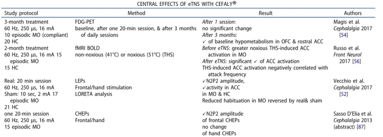
3.2. Central effects
The changes induced by single or multiple sessions of eTNS on
central nervous system activities are summarized in
Table 2
.
Several experimental studies have not confirmed the
hypothesis that pericranial nerve stimulation might decrease
trigeminal
nociception
by
a
segmental
gate
control.
Stimulation of the greater occipital nerve increased, rather
than decreased, activation by dural afferents of
trigemino-cervical nociceptive neurons in rats [
47
] and suboccipital
3 Hz nociceptive stimulation had no effect in humans on the
nociceptive blink reflex (nBR), a surrogate marker of spinal
trigeminal nucleus excitability [
48
].
By contrast, nBR amplitude and homotopic pain ratings were
found lastingly diminished by supraorbital 1 Hz noxious
stimu-lation in normal subjects and this was attributed to long-term
depression of second order nociceptors in the spinal trigeminal
nucleus [
49
]. An accompanying editorial [
50
] suggested that
low frequency
–high intensity acupuncture-like electrical
stimu-lation might be able to attenuate the long-term potentiation of
dorsal horn nociceptive synapses that contribute to
hyperalge-sia and allodynia and thus very promising for pain therapy.
Contrary to the previous study [
49
], eTNS uses high
fre-quency stimuli. Nonetheless, when the effect of one 20-min
session of stimulation (60 Hz, 16 mA) was tested on the nBR
in 10 migraine patients recruited for the abovementioned pilot
study [
27
], there was a mild, but significant decrease of nBR
amplitude and a more pronounced decrease of habituation.
One session of eTNS in migraine patients between attacks
also significantly decreased amplitude of contact heat-evoked
potentials (CHEPs) when they were obtained by
thermonoci-ceptive stimuli to the frontal skin, but not after stimulating the
wrist [
51
]. The fact that eTNS reduces CHEPs homotopically
suggests that it acts via trigemino-specific segmental or
supra-segmental pathways. Since the eTNS effect is greater on CHEPs
than on nBR, a suprasegmental mechanism could be more
likely.
Along the same line, amplitude of laser heat evoked
cortical responses was also significantly reduced after a
20min-session of real eTNS (250
μsec, 60 Hz, 16 mA) but
not after sham stimulation (2 mA) both in migraineurs
interictally and in healthy controls [
52
]. In this study, eTNS
was associated with reduced evoked EEG activity in anterior
cingulate cortex (ACC).
The confirmation that eTNS with this device might have
central effects came from a double-blinded, cross-over,
sham-controlled trial in 30 healthy volunteers measuring
vigilance and attention with psychomotor tests at the end
of a 20-min eTNS session at 2.5 or 120 Hz and of a session
with sham stimulation (1 mA) [
53
] (see
Table 2
). In this
study, there was a significant decrease in vigilance and
attention during high frequency eTNS. Whether such an
effect contributes to the therapeutic benefit of the eTNS
device is uncertain, as for migraine therapy the stimulation
frequencies used for acute and preventive treatment are
lower, respectively 100 and 60 Hz. The commercialized
device contains, however, a high frequency stimulation
pro-gram (120 Hz) that certain patients use for relaxation.
Using fluoro-deoxyglucose (FDG)-PET we have analyzed
changes in brain metabolism in 14 migraine without aura
patients before, after one 20-min session and after 3 months
of daily 20-min sessions of eTNS with the device (60 Hz,
16 mA) [
54
]. In patients compared to healthy controls,
base-line glucose uptake was significantly decreased bilaterally in
orbitofrontal (OFC), rostral anterior cingulate cortex (rACC),
and temporal lobe. This hypometabolism was not modified
after one eTNS session. However, in 10 patients who treated
themselves for 3 months and were compliant to eTNS as
revealed by the inbuilt software, monthly migraine days and
attack frequency significantly decreased, and metabolism
normalized
in
pretreatment
hypometabolic
areas
Table 2.Studies on central effect of eTNS with the Cefaly
®
.CENTRAL EFFECTS OF eTNS WITH CEFALY
®
Study protocol Method Result Authors 3-month treatment
60 Hz, 250μs, 16 mA 10 episodic MO (compliant) 20 HC
FDG-PET
baseline, after one 20-min session, & after 3 months of daily sessions
After 1 session: no significant change After 3 months:
↙ of baseline hypometabolism in OFC & rostral ACC
Magis et al. Cephalalgia 2017 [54] 2-month treatment 60 Hz, 250μs, 16 mA 15 episodic MO 15 HC fMRI BOLD non-noxious (41°C) or noxious (51°C) (THS)
Before eTNS: greater noxious THS-induced ACC activation in MO
After eTNS: significant↙ of ACC activation
THS-induced ACC activation negatively correlated with attack frequency
Russo et al. Front Neurol
2017 [56]
Real: 20 min session 60 Hz, 250μs, 16 mA Sham: 10 sec, 2 mA 17 episodic MO 21 HC LEPs Frontal/hand stimulation LORETA analysis ↙N2P2 amplitude, ↙activity in ACC in MO & HC
Reduced habituation in MO reversed by real& sham
Vecchio et al. Cephalalgia 2017
[52]
one 20-min session 60 Hz, 250μs, 16 mA 15 episodic MO CHEPs Frontal/hand ↙N2P2 amplitude of frontal CHEPs no change of hand CHEPs
Sasso D’Elia et al. Cephalalgia 2013 (abstract) [87] FDG-PET: fluorodeoxyglucose positron emission tomography; OFC: orbitofrontal cortex; ACC: anterior cingulate cortex; fMRI BOLD: functional magnetic resonance
(
Figure 1
). The change in rACC metabolism combined to the
reduction of migraine attack frequency overtime during
eTNS may be the consequence of a slow central
neuromo-dulatory action, similar to that of other peripheral nerve
stimulations (see [
50
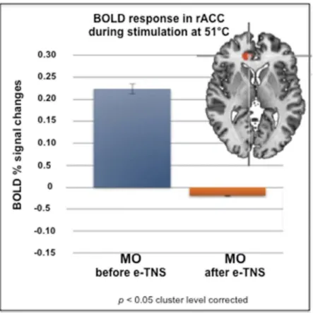
] for a review). Interestingly, Russo
et al. [
55
] found that blood oxygen-level dependent
(BOLD) activation in perigenual ACC (pgACC) by a noxious
heat stimulus on the cheek was significantly greater in
migraine without aura patients than in healthy volunteers.
In a therapeutic study of the eTNS device, the same authors
[
56
] found that 2 months of eTNS in 20 migraineurs
signifi-cantly reduced the noxious heat-induced BOLD activation in
pgACC as well as migraine attack frequency proportionally
to the functional magnetic resonance imaging (fMRI)
changes (
Figure 2
).
Between attacks migraine patients have smaller early bursts of
high-frequency oscillations (HFO1) embedded in somato-sensory
evoked potentials than healthy controls [
57
]. HFO1 are thought to
be generated by action potentials in thalamocortical afferents and
hence to reflect interictal thalamocortical dysrhythmia in migraine.
Late HFO (HFO2) are due to cortical (inhibitory) interneuron
dis-charges and of normal amplitude in migraine. Immediately after
one 20-min session of eTNS, SSEP HFO1 are significantly increased
for less than 5 min, while there is no effect on HFO2 (
Figure 3
) [
58
].
Whether the interictal low thalamocortical activity may be related
to the low metabolism in medial frontal cortices, and its
normal-ization by eTNS to the metabolic enhancement found after eTNS,
remains to be determined. It is worth mentioning, however, that
the electrophysiological changes were recorded after a single
eTNS session, whereas the metabolic changes were found after
chronic treatment, i.e. 3 months of daily eTNS.
4. ACC: the common denominator?
Taken together, the studies described above and summarized
in
Table 2
suggest that eTNS with the device acts
predomi-nantly at suprasegmental levels by modulating activity in
medial frontal cortical areas comprising the anterior cingulate
and OFC cortices. These areas exert multiple functions related
to decision-making, mood and the affective dimensions of
pain controlling in particular individual levels of central pain
modulation in healthy subjects [
59
]. They were found to
dys-function in chronic migraine [
60
], medication overuse
head-ache [
61
], and chronic cluster headache [
62
].
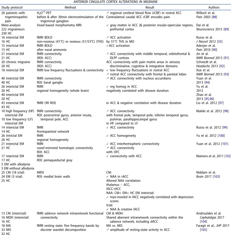
Schwedt et al. [
63
] have reviewed the data obtained with
functional MRI in migraine. An update of the literature review
on the topic depicted in
Table 3
shows that the ACC, a major
target of eTNS as described above, was found abnormal in a
number of studies assessing brain morphology, connectivity, or
function. The pgACC is of particular interest, as its activity was
found modified by eTNS in three studies with convergent results
but with different methods, FDG-PET [
54
], fMRI [
56
], and
pain-related evoked potentials [
52
]. The pgACC has been implicated in
various functions and diseases. For instance, it plays a role in
Figure 1.Histogram of changes in monthly migraine attack frequency before and after 1, 2, and 3 months of Cefaly
®
eTNS treatment in 10 compliant migraine without aura (MO) patients. Brain areas with significantly different glucose uptake on FDG-PET overlaid over an MRI anatomical map. Anterior cingulate cortex, orbitofrontal cortex and temporal lobe were hypometabolic in patients before treatment compared to healthy controls; all these areas normalized after 3 months eTNS.several psychiatric disorders in which its dysfunction is
asso-ciated with a variant in the CACNA1C gene [
64
], but also in the
self and resting state activity in the default mode network where
it is modulated by glutamate [
65
,
66
]. With regard to migraine,
however, the most relevant pgACC function is the one related to
pain control where it mainly seems to be involved in the affective
and emotional dimension. The pgACC was overall less activated
on fMRI in chronic pain patients than in healthy controls in a
paradigm where subjects had to estimate perceived pain
inten-sity on pictures depicting human limbs in painful and
non-pain-ful situations [
67
]. It is thought to mediate in part the analgesic
effects of motor cortex stimulation [
68
] and to play a pivotal role
in the central opioïdergic pain control system. The pgACC is rich
in opioid receptors and selectively activated during opioid
[
69
,
70
], but also placebo analgesia [
71
], providing evidence that
both analgesic modalities are mediated by activation of
descend-ing antinociceptive pathways.
Admittedly, abnormalities of the pgACC are not specific to
migraine. In cluster headache, in particular, the pgACC was found
hypometabolic outside of a bout but its metabolism increased
during a bout [
72
]. The same authors using PET with the opioid
receptor ligand [11C]diprenorphine found an inverse relationship
between duration of the cluster headache disorder and opioid
receptor availability in pgACC and ipsilateral hypothalamus [
73
].
In drug-resistant chronic cluster headache patients, we have
shown in a FDG-PET study that metabolism in the pgACC is
increased in responders to chronic ONS compared to
nonrespon-ders [
61
]. In migraine and cluster headache, both neurovascular
disorders, it remains controversial whether the dysfunction of
descending pain control systems is a primary causative
phenom-enon or secondary to repeated painful attacks. Interestingly, both
cluster headache [
74
] and migraine [
75
] are accompanied by
hypothalamic activation as a possible generator of an attack. The
pgACC has strong connectivity with the hypothalamus [
76
], which
might thus explain why it is involved in both disorders.
Finally, the facts that involvement of the pgACC seems not
specific for migraine, nor limited to pain control, and that eTNS
can change its activity and thalamocortical circuits, may explain
why pericranial neurostimulation, including eTNS, was reported
to have also therapeutic effects in tension-type headache [
77
],
fibromyalgia [
78
], depression [
79
], and epilepsy [
80
]. Given that
transcranial stimulation methods are able to change activity in
underlying cortical and subcortical structures including the
pgACC, it remains to be determined if they have therapeutic
potentials in migraine. Based on this rationale and the
above-mentioned study showing pgACC activation in refractory chronic
cluster headache patients ameliorated by ONS [
62
], we have
recently found in a proof-of-concept trial that transcranial direct
current stimulation (tDCS) with a montage targeting the pgACC
also has beneficial therapeutic effects in those patients [
81
].
5. Expert commentary
We have reviewed published evidence showing that eTNS
with this device is effective for the preventive and the
acute treatment of migraine. Effect size for prevention is
Figure 2.BOLD responses induced in right ACC (Talairach coordinates (x, y, z: 12, 35, 7) by a thermo-nociceptive stimulus are increased in migraine without aura (MO) patients (n = 20) before eTNS. They are significantly reduced after 2 months of daily eTNS. Insert: T-map of statistically significant differences between MO and healthy controls overlaid onto a Talairach transformed Colin-27 T1 high-resolution anatomical template. Bar graphs of percent BOLD signal changes during noxious trigeminal heat stimulation at 51°C in MO patients before and after eTNS (modified after Russo et al. 2017 [56]).
somewhat lower compared to the available most effective
preventive drugs, but tolerability is far better. Published
studies on the mechanisms of action of eTNS suggest that
modulation of central brain areas involved in pain control,
in particular of the ACC, either directly or via changes in
thalamocortical activity, could be pivotal for the beneficial
clinical effects. A segmental action on the trigeminal
sys-tem in the brain ssys-tem may contribute to the beneficial
acute eTNS effect. We have analyzed the imaging literature
in migraine and were struck by the fact that the ACC was
found abnormal in most studies, be they focused on its
morphological, connectivity or functional properties. The
pgACC may thus be the crucial mediator of the device
’s
therapeutic effects, which may explain why eTNS can
miti-gate other painful and non-painful conditions in which this
brain area has a pathophysiological role. Given that
non-invasive neurostimulation at very different sites such as
mastoids [
20
], upper limb [
22
] or vagus nerve in the neck
[
21
], appears to have comparable beneficial effects in
migraine, it seems worthwhile to explore modulation of
ACC as a common mechanism of action for noninvasive
peripheral neurostimulation.
6. Five-year view
Several clinical trials with this eTNS device are ongoing or
planned, exploring, for example, its utility in treating headache
in the emergency ward, in preventing the full development of a
migraine attack when applied at the very beginning of an attack,
or in mitigating withdrawal headache during the weaning period
in medication overuse headache. There are also plans to conduct
a randomized controlled trial in tension-type headache, but also
in other chronic pain syndromes like fibromyalgia and in
epi-lepsy. Evidence of efficacy should be sought for the suboccipital
eTNS that has shown promising results in open trials of chronic
migraine. The possible advantage of stimulating simultaneously
or alternatively the frontal and suboccipital regions has to be
investigated. It is also of great interest to develop a device
combining eTNS with transcranial direct current stimulation,
since the latter allows direct modulation of the cortical
respon-sivity known to be abnormal in migraine while the former seems
to act more generally on areas of the limbic and pain systems.
Despite the recent avenue of novel pharmacotherapies
for migraine with an excellent efficacy/tolerability profile
like the monoclonal antibodies against CGRP or its
recep-tor,
it
is
likely
that
a
niche
for
noninvasive
Figure 3.Upper: illustrative recording in a migraine patient of high frequency oscillations (HFO, 450–750Hz) in somato-sensory evoked potentials before (pretreatment) and immediately (Post 1) and 5 min after (Post 2) one 20-min session of eTNS with the Cefaly
®
. HFO1: early burst reflecting thalamocortical activity; HFO2: late burst reflecting discharges of inhibitory cortical neurons.neurostimulation methods as alternative or add-on
thera-pies in primary headaches will persist. The future will
determine which of the various methods is most valuable
and for each headache type.
Key issues
● External trigeminal neurostimulation (eTNS) with the
Cefaly
®
device has evidence for a preventive therapeutic
effect in episodic migraine as well as for an abortive effect
during migraine attacks. There is circumstantial evidence
for its beneficial effect in chronic migraine and
tension-type headache.
● Its efficacy/tolerability profile is excellent, exceeding that of
most drug treatments for headaches.
● Available data cannot exclude that eTNS device may have an
analgesic effect via a gating effect in the trigeminal pain
pathway.
● Recent imaging studies, however, suggest that its
predo-minant acute and chronic effect is modulation of activity
Table 3.Functional imaging studies showing anterior cingulate cortex alterations in migraine.
ANTERIOR CINGULATE CORTEX ALTERATIONS IN MIGRAINE
Study protocol Method Result Authors 20 patients with
trigeminopathic pain
HéO15-PET
before & after 30min electrostimulation of the trigeminal ganglion
↗ regional cerebral blood flow (rCBF) in rostral ACC Contralateral caudal ACC rCBF encodes pain
Willoch et al. Pain 2003 [88] Meta-analysis
222 migraineurs 230 HC
Voxel-based morphometry MRI ↙ gray matter in ACC (& posterior insular-opercular regions, prefrontal cortex Dai et al. Neuroscience 2015 [89] 16 interictal EM 16 HC fMRI BOLD non-noxious (41°C) or noxious (51/53°C) (THS) ↗ ACC activation by 51°C THS in MO Russo et al. J Neurol 2012 [55] 15 interictal EM 15 HC fMRI BOLD after nasal ammonia
↙ACC activation Aderjan et al. Pain 2010 [90] 21 interictal EM
21 HC
fMRI connectivity (ROI: ACC)
↗ ACC connectivity with middle temporal, orbitofrontal & DLPF cortices Jin et al. NMR Biomed 2013 [91] 20 chronic migraine 20 HC fMRI connectivity (ROI: ACC)
ACC connectivity with pain matrix areas in sensory discriminative, cognitive & integrative domains
Schwedt et al. Headache 2013 [92] 18 interictal EM fMRI low frequency fluctuations & connectivity ↙ low frequency fluctuations in rostral ACC
↗ rostral ACC connectivity with frontal & parietal lobes Xue et al.NMR Biomed 2013 [93] 40 interictal EM
40 HC
fMRI connectivity ROI: basal ganglia
↗ ACC connectivity with nucleus accumbens Yuan et al. 2013 [94] 26 interictal EM 26 HC 40 interictal EM 20 HC fMRI
regional homogeneity (whole brain)
↙ reg homog in ACC
negatively correlated with disease duration
Yu et al. 2012 Zhao et al. 2013 [95,96] 43 interictal EM 43 HC
fMRI (90 ROI) in ACC & negative correlation with disease duration Liu et al. 2012 [97] 10 high frequency (HF)
interictal EM 10 low frequency (LF)
interictal EM
fMRI connectivity
ROI: postcentral gyrus, anterior insula, temporal pole, ACC
↗ ACC connectivity
with frontal pole, temporal pole, inferior temporal gyrus, pulvinar, parahippocampal gyrus
in HF compared to LF Maleki et al. 2012 [98] 14 interictal EM 14 HC fMRI frontoparietal network
↙ ACC connectivity Russo et al. 2012 [99] 26 intrictal EM
26 HC
fMRI
regional homogeneity
↙ ACC homogeneity Yu et al. 2012 [100] 21 interictal EM
21 HC
fMRI
voxel-mirrored homotopic connectivity ROI: ACC
↙ ACC interhemispheric connectivity ↗ ACC connectivity with OFC Yuan et al. 2012 [101] 17 interictal EM 17 HC 5 EM with allodynia 5 EM without allodynia fMRI
ROI: periaqueductal gray ↙ connectivity with ACC
Mainero et al. 2011 [102]
25 CM (18 ictal) 24 EM (3 ictal) 25 HC
MRSI
ROI: medial brain walls
CM:
↙ NAA in rACC Altered NAA correlation thalamus– ACC, lACC-rACC
NAA: CM< EM< HC EM interictal:
↙ myo-inositol in lACC negatively correlated with depression scores
Ictal:
↙ NAA & creatine lACC
Niddam et al. Brain 2017 [103]
13 CM (interictal) 16 MOH (interictal) 16 HC
fMRI salience network intranetwork functional connectivity
CM & MOH:
Shared aberrant intranetwork connectivity within the salience network, including dACC
Androulakis et al. Cephalalgia 2017 [104] 18 MA 33 MO 32 HC
fMRI resting state: five frequency bands by discrete wavelet decomposition
MA vs. MO:
↗ amplitude of resting-state activity in ACC Faragò et al., JHP 2017[105] rCBF: regional cerebral blood flow; EM: episodic migraine; ROI: region of interest; DLPFC: dorsolateral prefrontal cortex; CM: chronic migraine; MRSI: magnetic
in the anterior cingulate cortex (ACC), a crucial limbic
area belonging to the salience/pain network.
● A comprehensive literature review indicates that the ACC is
precisely an area found abnormal in most morphological or
functional imaging studies of migraine.
Funding
The contribution of the G.B. Bietti Foundation in this paper was supported by the Italian Ministry of Health and Fondazione Roma.
Declaration of interest
J Schoenen is a consultant for Cefaly
®
Technology. The authors have no other relevant affiliations or financial involvement with any organization or entity with a financial interest in or financial conflict with the subject matter or materials discussed in the manuscript apart from those disclosed.Reviewer disclosures
Peer reviewers on this manuscript have no relevant financial or other relationships to disclose.
References
Papers of special note have been highlighted as either of interest (•) or of considerable interest (••) to readers.
1. Evers S, Áfra J, Frese A, et al. EFNS guideline on the drug treatment of migraine–revised report of an EFNS task force. Eur J Neurol.
2009;16:968–981.
2. Marmura MJ, Silberstein SD, Schwedt TJ. The acute treatment of migraine in adults: the American headache society evidence assess-ment of migraine pharmacotherapies. Headache.2015;55:3–20. 3. Silberstein S, Latsko M, Schoenen JSJ. Preventive Antimigraine
Drugs. In: Fernandez-de-las-Penas C, Chaitow L, eds. Multidisciplinary management of migraine : pharmacological, man-ual, and other therapies. Burlington USA: Jones & Bartlett Learning;
2013. p. 91–102.
4. Lipton RB, Buse DC, Serrano D, et al. Examination of unmet treat-ment needs among persons with episodic migraine: results of the American Migraine Prevalence and Prevention (AMPP) Study. Headache.2013;53:1300–1311.
5. Paemeleire K, Magis D, Migraine SJ. Medical Treatment of migraine. In: Reference Module in Neuroscience and Biobehavioral Psychology. Elsevier; 1–5.
6. Schoenen J, Magis D. Migraines and Other Headache Disorders. Lipton R, Bigal M, eds. New York: Informa Healthcare;2006. p. 363–374. 7. Gallagher RM, Kunkel R. Migraine medication attributes important for
patient compliance: concerns about side effects may delay treatment. Headache.2003;43:36–43.
8. Berger A, Bloudek LM, Varon SF, et al. Adherence with Migraine Prophylaxis in Clinical Practice. Pain Practice.2012;12:541–549. 9. Blumenfeld AM, Bloudek LM, Becker WJ, et al. Patterns of use and
reasons for discontinuation of prophylactic medications for episo-dic migraine and chronic migraine: results from the second inter-national burden of migraine study (IBMS-II). Headache.
2013;53:644–655.
10. Turner DP, Golding AN, Houle TT. Using a graphical risk tool to examine willingness to take migraine prophylactic medications. Pain.2016;157:2226–2234.
• This important study shows that patients’ willingness to take topiramate or valproate for migraine prevention is low because of the unfavorable efficacy/adverse effect profile. 11. Hepp Z, Dodick DW, Varon SF, et al.. Persistence and switching
patterns of oral migraine prophylactic medications among patients
with chronic migraine: A retrospective claims analysis. Cephalalgia.
2017;37:470–485.
• This study shows that 50% of migraine patients abandon their prophylactic drug treatment after 2 months.
12. Ahmed HE, White PF, Craig WF, et al. Use of percutaneous electrical nerve stimulation (PENS) in the short- term management of head-ache. Headhead-ache.2000;40:311–315.
13. Ghoname EA, Craig WF, White PF. Use of percutaneous electrical nerve stimulation (PENS) for treating ECT-induced headaches. Headache. 39:502–505.
14. Saper JR, Dodick DW, Silberstein SD, et al. Occipital nerve stimula-tion for the treatment of intractable chronic migraine headache: ONSTIM feasibility study. Cephalalgia.2011;31:271–285.
15. Silberstein SD, Dodick DW, Saper J, et al. Safety and efficacy of peripheral nerve stimulation of the occipital nerves for the manage-ment of chronic migraine: results from a randomized, multicenter, double-blinded, controlled study. Cephalalgia.2012;32:1165–1179. 16. Dodick DW, Silberstein SD, Reed KL, et al. Safety and efficacy of
peripheral nerve stimulation of the occipital nerves for the man-agement of chronic migraine: long-term results from a randomized, multicenter, double-blinded, controlled study. Cephalalgia.
2015;35:344–358.
17. Reed KL, Black SB, Banta CJ, et al. Combined occipital and supraor-bital neurostimulation for the treatment of chronic migraine head-aches: initial experience. Cephalalgia.2010;30:260–271.
18. Schwedt TJ. Neurostimulation for primary headache disorders. Curr Neurol Neurosci Rep.2009;9:101–107.
19. Schoenen J, Roberta B, Magis D, et al. Noninvasive neurostimula-tion methods for migraine therapy: the available evidence. Cephalalgia.2016;36:1170–1180.
20. Juan Y, Shu O, Jinhe L, et al. Migraine prevention with percuta-neous mastoid electrical stimulator: A randomized double-blind controlled trial. Cephalalgia.2017;37:1248–1256.
• First study indicating that bilateral mastoid neurostimulation can have a preventive effect in migraine, presumably by acting on the fastigial nuclei in the cerebellum.
21. Silberstein SD, Calhoun AH, Lipton RB, et al.. Chronic migraine headache prevention with noninvasive vagus nerve stimulation. Neurology.2016;87:529–538.
22. Yarnitsky D, Volokh L, Ironi A, et al. Nonpainful remote electrical stimulation alleviates episodic migraine pain. Neurology.
2017;88:1250–1255.
• First evidence for antimigraine effect of electrical stimulation at the wrist.
23. Solomon S, Guglielmo KM. Treatment of headache by transcuta-neous electrical stimulation. Headache.1985;25:12–15.
•• This pioneer study was first suggesting that noninvasive peri-cranial neurostimulation might have beneficial effects in migraine patients.
24. Lapeer GL. High-intensity transcutaneous nerve stimulation at the Hoku acupuncture point for relief of muscular headache pain. Literature Review Clinical Trial Cranio.1986;4:164–171.
25. Heydenreich A. [Localized transcutaneous electric nerve stimula-tion with high voltage impulses in funcstimula-tional chronic headache and migraine]. Z Arztl Fortbild (Jena).1991;85:37–39.
26. Taubert K. [Transcutaneous electric nerve stimulation (TENS) in headache and facial pain]. Z Arztl Fortbild (Jena).1991;85:31–36. 27. Russo A, Tessitore A, Conte F, et al. Transcutaneous supraorbital
neurostimulation in‘de novo’ patients with migraine without aura: the first Italian experience. J Headache Pain.2015;16:69.
28. Di Fiore P, Bussone G, Galli A, et al. Transcutaneous supraorbital neurostimulation for the prevention of chronic migraine: a pro-spective, open-label preliminary trial. Neurol Sci.2017;38:201–206. 29. Vikelis M, Dermitzakis EV, Spingos KC, et al.. Clinical experience with transcutaneous supraorbital nerve stimulation in patients with refrac-tory migraine or with migraine and intolerance to topiramate: a prospective exploratory clinical study. BMC Neurol.2017;17:97. • Open trial suggesting that eTNS with the Cefaly° also has a
30. Przeklasa-Muszyńska A, Skrzypiec K, Kocot-Kępska M, et al. Non-invasive transcutaneous Supraorbital Neurostimulation (tSNS) using Cefaly
®
device in prevention of primary headaches. Neurol Neurochir Pol.2017;51:127–134.31. Kozminski M. Transcutaneous supraorbital nerve stimulation as a rescue therapy. Headache.2014;54:12.
32. Gérardy P, Fabry D, Fumal A, et al. A pilot study on supra-orbital surface electrotherapy in migraine. Cephalalgia.2009;29:134. 33. Schoenen J, Vandersmissen B, Jeangette S, et al. Migraine
preven-tion with a supraorbital transcutaneous stimulator: a randomized controlled trial. Neurology.2013;80:697–704.
•• This is the pivotal randomized sham-controlled trial that proved a preventive effect of treatment with the Cefaly° in episodic migraine and led to FDA approval of the device for migraine prevention.
34. Bussone G, Diener HC, Pfeil J, et al.. Topiramate 100 mg/day in migraine prevention: a pooled analysis of double-blind randomised controlled trials. Int J Clin Pract.2005;59:961–968.
35. Magis D, Sava S, d’Elia TS, et al. Safety and patients’ satisfaction of transcutaneous supraorbital neurostimulation (tSNS) with the Cefaly
®
device in headache treatment: a survey of 2,313 headache sufferers in the general population. J Headache Pain.2013;14: 95. 36. Penning S, Schoenen J. A survey on migraine attack treatment withthe CEFALY
®
device in regular users. Acta Neurol Belg.2017;117:547–549.
37. Mann J, Schoenen J. Abortive treatment of migraine with e-TNS: pilot trial. Headache.2018. (in press).
38. Chou DE, Gross GJ, Casadei CH, et al. External Trigeminal Nerve Stimulation for the Acute Treatment of Migraine: open-Label Trial on Safety and Efficacy. Neuromodulation.2017;20:678–683. 39. Chou DE, Yugrakh MS, Gross G, et al. Acute treatment of migraine
with e-TNS: a multi-center, double-blind, randomized, sham-con-trolled trial. Cephalalgia.2017;37:323.
• This is the first large trial showing that eTNS also has an effect during migraine attacks.
40. Cady RK, McAllister PJ, Spierings ELH, et al.. A randomized, double-blind, placebo-controlled study of breath powered nasal delivery of sumatriptan powder (AVP-825) in the treatment of acute migraine (The TARGET Study). Headache.2015;55:88–100.
41. Färkkilä M, Diener H-C, Géraud G, et al. Efficacy and tolerability of lasmiditan, an oral 5-HT(1F) receptor agonist, for the acute treatment of migraine: a phase 2 randomised, placebo-con-trolled, parallel-group, dose-ranging study. Lancet Neurol.
2012;11:405–413.
• First evidence for the effectiveness of lasmiditan, a 5-HT1F receptor agonist, in acute migraine treatment.
42. Voss T, Lipton RB, Dodick DW, et al. A phase IIb randomized, double-blind, placebo-controlled trial of ubrogepant for the acute treatment of migraine. Cephalalgia.2016;36:887–898.
• First evidence for the effectiveness of ubrogepant, a CGRP receptor antagonist without hepatotoxicity, in acute migraine treatment.
43. Goadsby PJ, Grosberg BM, Mauskop A, et al. Effect of noninvasive vagus nerve stimulation on acute migraine: an open-label pilot study. Cephalalgia.2014;34:986–993.
• First evidence for the effectiveness of transcutaneous cervical vagus nerve stimulation in acute migraine treatment. 44. Barbanti P, Grazzi L, Egeo G, et al.. Non-invasive vagus nerve
stimulation for acute treatment of high-frequency and chronic migraine: an open-label study. J Headache Pain.2015;16:542. 45. Ersek RA. Transcutaneous electrical neurostimulation: a new
ther-apeutic modality for controlling pain. Clin Orthop Relat Res.
1977;128:314–324.
46. Melzack R, Wall PD. Pain mechanisms: a new theory. Science.
1965;150:971–979.
47. Melzack R, Wall PD. Acupuncture and transcutaneous electrical nerve stimulation. Postgrad Med J.1984;60:893–896.
•• Milestone publication for the understanding of pain processing and the mechanisms of action of various analgesic treatments.
48. Jones I, Johnson MI. Transcutaneous electrical nerve stimulation. Contin Educ Anaesth Crit Care Pain.2009;9:130–135.
49. Schueler M, Neuhuber WL, De Col R, et al. Innervation of rat and human dura mater and pericranial tissues in the parieto-temporal region by meningeal afferents. Headache.2014;54:996–1009. 50. Didier HA, Di Fiore P, Marchetti C, et al. Electromyography data in
chronic migraine patients by using neurostimulation with the Cefaly
®
device. Neurol Sci.2015;36(Suppl S1):115–119.51. Coppola G, Schoenen J. Cortical excitability in chronic migraine. Curr Pain Headache Rep.2012;16:93–100.
52. Sasso D’Elia T, Fataki M, Sava S, et al. Effect of Anodal Transcranial Direct Current Stimulation Over the Visual Cortex on Thermosensitivity Assessed by Quantitative Sensory Testing and Contact Heat Evoked Potentials. Cephalalgia.2013;33(232):P341. 53. Said G, Lacroix C, Planté-Bordeneuve V, et al. Nerve granulomas
and vasculitis in sarcoid peripheral neuropathy: a clinicopathologi-cal study of 11 patients. Brain.2002;125:264–275.
54. Jürgens TP, Busch V, Opatz O, et al. Low-frequency short-time nociceptive stimulation of the greater occipital nerve does not modulate the trigeminal system. Cephalalgia.2008;28:842–846. 55. Aymanns M, Yekta SS, Ellrich J. Homotopic long-term depression of
trigeminal pain and blink reflex within one side of the human face. Clin Neurophysiol.2009;120:2093–2099.
56. Cruccu G, Truini A. Neurostimulation therapy (acupuncture-like) and long-term depression: a challenge for the clinical neurophy-siologist. Clin Neurophysiol.2009;120:2004–2005.
• Thought-provoking review on neurostimulation therapy for pain and its mode of action.
57. Schoenen J Scientific report. TRADONI Convention.2014. 58. Vecchio E, Gentile E, Franco G, et al. Effects of external trigeminal
nerve stimulation (eTNS) on laser evoked cortical potentials (LEP): A pilot study in migraine patients and controls. Cephalalgia.
2017;33310241772874.
59. Piquet M, Balestra C, Sava SL, et al. Supraorbital transcutaneous neurostimulation has sedative effects in healthy subjects. BMC Neurol.2011;11:135.
60. Magis D, D’Ostilio K, Thibaut A, et al. Cerebral metabolism before and after external trigeminal nerve stimulation in episodic migraine. Cephalalgia.2017;37:881–891.
•• Pivotal study showing pretreatment hypometabolism of ante-rior cingulate and orbitofrontal cortices in migraine and its normalization after 3 months of eTNS with Cefaly°.
61. Russo A, Tessitore A, Esposito F, et al. Pain processing in patients with migraine: an event-related fMRI study during trigeminal noci-ceptive stimulation. J Neurol.2012;259:1903–1912.
62. Russo A, Tessitore A, Esposito F, et al.. Functional Changes of the Perigenual Part of the Anterior Cingulate Cortex after External Trigeminal Neurostimulation in Migraine Patients. Front Neurol.
2017;8:282.
• Confirmatory study showing that eTNS with Cefaly° modulates heat pain-induced activation of anterior cingulate cortex in migraine patients.
63. Coppola G, Vandenheede M, Di Climente L, et al. Somatosensory evoked high-frequency oscillations reflecting thalamo-cortical activity are decreased in migraine patients between attacks. Brain.2004;128:98–103.
• First study favoring an abnormal interictal thalamo-cortical drive in migraine.
64. Di Lenola D, Coppola G, Serrao M, et al.. O024. Transcutaneous supraorbital nerve stimulation enhances somatosensory thalamic activity in migraine between attacks: a central mechanism of clin-ical efficacy?. J Headache Pain. 2015;16. Epub ahead of print. doi:10.1186/1129-2377-16-S1-A160.
65. Bogdanov VB, Viganò A, Noirhomme Q, et al.. Cerebral responses and role of the prefrontal cortex in conditioned pain modulation: an fMRI study in healthy subjects. Brain.
2015;281:187–198.
66. Matharu MS, Bartsch T, Ward N, et al. Central neuromodulation in chronic migraine patients with suboccipital stimulators: a PET study. Brain.2004;127:220–230.
67. Fumal A, Laureys S, Di Clemente L, et al. Orbitofrontal cortex involvement in chronic analgesic-overuse headache evolving from episodic migraine. Brain.2006;129:543–550.
68. Magis D, Bruno MA, Fumal A, et al. Central modulation in cluster headache patients treated with occipital nerve stimulation: an FDG-PET study. BMC Neurol.2011;11: 25.
• This study suggests the mode of action of percutaneous occi-pital nerve stimulation in cluster headache is activation of the perigenual anterior cingulate cortex.
69. Schwedt TJ, Chiang CC, Chong CD, et al. Functional MRI of migraine. Lancet Neurology.2015;14:81–91.
•• Extremely comprehensive review of functional MRI studies performed in migraine.
70. Willoch F, Gamringer U, Medele R, et al.. Analgesia by electrosti-mulation of the trigeminal ganglion in patients with trigemino-pathic pain: a PET activation study. Pain.2003;103:119–130. 71. Dai Z, Zhong J, Xiao P, et al.. Gray matter correlates of migraine and
gender effect: A meta-analysis of voxel-based morphometry stu-dies. Neuroscience.2015;299:88–96.
72. Aderjan D, Stankewitz A, May A. Neuronal mechanisms during repetitive trigemino-nociceptive stimulation in migraine patients. Pain.2010;151:97–103.
73. Jin C, Yuan K, Zhao L, et al. Structural and functional abnormalities in migraine patients without aura. NMR Biomed.2013;26:58–64. 74. Schwedt TJ, Schlaggar BL, Mar S, et al. Atypical resting-state
func-tional connectivity of affective pain regions in chronic migraine. Headache.2013;53:737–751.
75. Xue T, Yuan K, Cheng P, et al. Alterations of regional spontaneous neuronal activity and corresponding brain circuit changes during rest-ing state in migraine without aura. NMR Biomed.2013;26:1051–1058. 76. Parkerson HA, Noel M, Pagé MG, et al. Factorial validity of the English-language version of the Pain Catastrophizing Scale–child version.. J Pain.2013;14:1383–1389.
77. Yu D, Yuan K, Zhao L, et al. Regional homogeneity abnormalities affected by depressive symptoms in migraine patients without aura: a resting state study. PLoS One.2013;8:10.
78. Zhao L, Liu J, Dong X, et al. Alterations in regional homogeneity assessed by fMRI in patients with migraine without aura stratified by disease duration. J Headache Pain.2013;14:85.
79. Liu J, Zhao L, Li G, et al.. Hierarchical alteration of brain structural and functional networks in female migraine sufferers. PLoS One.2012;7:10. 80. Maleki N, Becerra L, Brawn J, et al.. Concurrent functional and structural
cortical alterations in migraine. Cephalalgia.2012;32:607–620. 81. Russo A, Tessitore A, Giordano A, et al.. Executive resting-state
network connectivity in migraine without aura. Cephalalgia.
2012;32:1041–1048.
• This is one of the first studies showing abnormal connectivity of anterior cingulate cortex with various brain areas including the salience/pain matrix in migraine.
82. Yu D, Yuan K, Zhao L, et al. Regional homogeneity abnormalities in patients with interictal migraine without aura: a resting-state study. NMR Biomed.2012;25:806–812.
83. Yuan K, Qin W, Liu P, et al. Reduced fractional anisotropy of corpus callosum modulates inter-hemispheric resting state functional con-nectivity in migraine patients without aura. PLoS One.2012;7:10. 84. Mainero C, Boshyan J, Hadjikhani N. Altered functional magnetic
resonance imaging resting-state connectivity in periaqueductal gray networks in migraine. Ann Neurol.2011;70:838–845. 85. Niddam DM, Lai K-L, Tsai S-Y, et al.. Neurochemical changes in the
medial wall of the brain in chronic migraine. Brain. 2017. Epub ahead of print 11 December. doi:10.1093/brain/awx331
86. Androulakis XM, Rorden C, Peterlin BL, et al. Modulation of salience network intranetwork resting state functional connectivity in women with chronic migraine. Cephalalgia.2017;33310241774857. 87. Faragó P, Tuka B, Tóth E, et al. Interictal brain activity differs in
migraine with and without aura: resting state fMRI study. J Headache Pain.2017;18:8.
88. Erk S, Meyer-Lindenberg A, Schmierer P, et al. Hippocampal and Frontolimbic Function as Intermediate Phenotype for Psychosis: evidence from Healthy Relatives and a Common Risk Variant in CACNA1C. Biological Psychiatry.2014;76:466–475.
89. Enzi B, Duncan NW, Kaufmann J, et al. Glutamate modulates resting state activity in the perigenual anterior cingulate cortex - a com-bined fMRI-MRS study. Neuroscience.2012;227:102–109.
90. Horn DI, Yu C, Steiner J, et al.. Glutamatergic and resting-state functional connectivity correlates of severity in major depression -the role of pregenual anterior cingulate cortex and anterior insula. Front Syst Neurosci. Epub ahead of print. 2010. doi:10.3389/ fnsys.2010.00033.
91. Noll-Hussong M, Otti A, Wohlschlaeger AM, et al. Neural correlates of deficits in pain-related affective meaning construction in patients with chronic pain disorder. Psychosom Med.
2013;75:124–136.
92. Garcia-Larrea L, Peyron R. Motor cortex stimulation for neuropathic pain: from phenomenology to mechanisms. Neuroimage.2007;37 (Suppl 1):S71–9.
93. Casey KL, Svensson P, Morrow TJ, et al. Selective opiate modulation of nociceptive processing in the human brain. J Neurophysiol.
2000;84:525–533.
94. Wagner KJ, Sprenger T, Kochs EF, et al. Imaging human cerebral pain modulation by dose-dependent opioid analgesia: a positron emission tomography activation study using remifentanil. Anesthesiology.
2007;106:548–556.
95. Petrovic P, et al. Placebo and opioid analgesia– imaging a shared neuronal network. Science.2002;295:1737–1740.
96. Sprenger T, Ruether K, Boecker H, et al. Altered Metabolism in Frontal Brain Circuits in Cluster Headache. Cephalalgia.2007;27:1033–1042. 97. Sprenger T, Willoch F, Miederer M, et al. Opioidergic changes in the
pineal gland and hypothalamus in cluster headache: a ligand PET study. Neurology.2006;66:1108–1110.
98. May A, Bahra A, Büchel C, et al. Hypothalamic activation in cluster headache attacks. Lancet.1998;352:275–278.
• First study demonstrating hypothalamic increased blood flow during cluster headache attacks.
99. Schulte LH, May A. The migraine generator revisited: continuous scanning of the migraine cycle over 30 days and three sponta-neous attacks. Brain.2016;139:1987–1993.
100. Boehringer A, Tost H, Haddad L, et al. Neural Correlates of the Cortisol Awakening Response in Humans. Neuropsychopharmacology.
2015;40:2278–2285.
101. Mousavi SA, Mirbod SM, Khorvash F. Comparison between effi-cacy of imipramine and transcutaneous electrical nerve stimu-lation in the prophylaxis of chronic tension-type headache: a randomized controlled clinical trial. J Res Med Sci.2011;16:923– 927.
102. Plazier M, Dekelver I, Vanneste S, et al.. Occipital nerve stimula-tion in fibromyalgia: a double-blind placebo-controlled pilot study with a six-month follow-up. Neuromodulation.
2014;17:256–63–4.
103. Cook IA, Abrams M, Leuchter AF. Trigeminal Nerve Stimulation for Comorbid Posttraumatic Stress Disorder and Major Depressive Disorder. Neuromodulation.2016;19:299–305.
• Study showing that external trigeminal neurostimulation is beneficial in post-traumatic stress disorder and depression. 104. DeGiorgio CM, Soss J, Cook IA, et al. Randomized controlled trial of
trigeminal nerve stimulation for drug-resistant epilepsy. Neurology.
2013;80:786–791.
105. Sava S, Cosseddu A, D’Ostilio K, et al. Anodal transcranial direct stimulation (tDCS) targeting the anterior cingulate gyrus for the preventive treatment of chronic cluster headache: a proof-of-con-cept trial. Cephalalgia.2015;35: 91.
• Proof-of-concept trial showing that frontal anodal transcranial neurostimulation may have a preventive effect in difficult-to-treat chronic cluster headache patients.