Université du Québec
Institut National de la Recherche Scientifique Centre Institut Armand-Frappier
L'IMPLICATION DE L'AUTOPHAGIE DANS L'INFECTION PAR
LE VIRUS DE L'HÉPATITE C
Par
Ahmed Mohamed Fahmy
Thèse présentée pour l’obtention du grade de Philosophiae doctor (Ph.D.)
en Virologie et Immunologie
Jury d’évaluation
Président du jury et Pr. Terence Ndonyi Bukong Examinateur interne INRS Institut Armand-Frappier
Examinateur externe Pr.Guy Lemay
Université de Montréal
Examinateur interne Pr. Hugo Soudeyns
Université de Montréal Directeur de recherche Pr. Patrick Labonté
INRS Institut Armand-Frappier
<<Look deep into nature, and then you will understand everything better>>
Albert_Einstein
iii
ACKNOWLEDGEMENTS
Foremost, I would like to express my deepest gratitude to my PhD supervisor Professor Patrick Labonté for his caring guidance, patience, extraordinary support, and for excellently conveying the spirit of adventure and excitement regarding research. Without his guidance and continuous support this dissertation would not have been possible.
I would like to thank all my present and former lab members Dr. Matthieu Blanchet, Dr. Carl Guevin, Tuan Le Quoc, Marwa Khabir for helping and supporting me during my research.
I would like to thank my committee members, Professor Terence Ndonyi Bukong, Professor Guy Lemay, and Professor Hugo Soudeyns for participating in the evaluation of my research.
A special thanks to my lovely wife for her unbelievable patience and encouragement throughout the whole journey of my research. She has been my inspiration and motivation for continuing to improve my knowledge and move my career forward. She is my only love and my rock, and I dedicate this dissertation to her. I also thank my sweetheart daughter Salma and our beautiful new addition Talya for always making me smile and for understanding on those weekend mornings when I was busy working on my research project instead of playing with them. I would like to thank my parents, my brilliant father and my amazing mother, for allowing me to follow my passion and ambition throughout my childhood.
I would like also to thank my brothers and sisters, my parents-in-law for their love and encouragement with their best wishes.
I would like to thank all my friends and colleagues at the INRS specially Slimane Dridi, Asma Lamine, Guillermo Arango Duque, Hossam Draz, Mohamed Haddad, Ahmed Soliman, Ghislain Paka, Edward Kwarting, Mustapha Iddir, Akil Hammami, Ahlem Saghmi, Hajer Habouria and Emre Yurdusev for their support and providing a stimulating and fun environment during my studies.
v
RÉSUMÉ EN FRANÇAIS
L'infection par le virus de l'hépatite C (VHC) est un problème majeur de santé publique avec plus de deux cent millions d’individus chroniquement infectés dans le monde. Malgré le développement d'antiviraux très puissants à action directe (AAD) pour le traitement de l'infection chronique par le VHC, il reste encore de nombreux cas de personnes infectées. Ce nombre devrait augmenter dans les prochaines années en raison du coût élevé de ces médicaments antiviraux qui en limite l’accès et le grand nombre d'infections occultes. Bien que de nombreuses avancées majeures aient été réalisées dans la compréhension de la pathogénèse et de la réplication virale, les liens étroits entre le VHC et la cellule hôte sont encore mal compris.
L'autophagie, un mécanisme cellulaire de la cellule hôte, s'est révélée être modulée par le VHC. L'autophagie est un mécanisme de dégradation cellulaire qui vise à recycler les protéines ainsi que les organites qui ne sont plus nécessaires mais également à protéger la cellule d'éventuels agents pathogènes. Nous avons montré que le VHC exploite l'autophagie pour se répliquer. Le mécanisme par lequel le VHC détourne à son avantage l'autophagie et/ou les composantes autophagiques est encore débattu. Dans une étude récente au sein de notre laboratoire, nous avons montré que l'ARN-polymérase dépendante de l'ARN du VHC (NS5B) interagit avec la protéine autophagique ATG5. De plus, dans des cellules arborant un réplicon du VHC, la protéine ATG5 colocalise avec un marqueur des usines de réplication membranaire du VHC, la protéine NS4B. Finalement, le « knock-down » d’ATG5 a permis de diminuer l'ARN du virus ainsi que les niveaux d'expressions des protéines virales.
Puisque ATG5 est une composante du complexe d'élongation de l'autophagie (12/16L1), l'objectif principal de ce projet de recherche est d'approfondir le rôle de ATG5-12/16L1 dans le cycle de réplication du VHC.
Dans le premier article, nous avons évalué la localisation du complexe ATG5-12/16L1 dans une infection chronique in vitro. En effet, nous avons montré que les composants du complexe ATG5-12/16L1 colocalisent avec les composantes de la réplicase du VHC (NS3, NS4B, NS5A et NS5B).
vi
De plus, en utilisant la méthode de ligation de proximité « Proximity Ligation Assay» (PLA), nous avons montré, in situ, que ATG5-12 interagit avec les composantes de la réplicase du VHC. Bien que ATG16L1 colocalise avec la réplicase de VHC, aucune interaction entre ATG16L1 et la réplicase du HCV n'a été observée.
Ces résultats suggèrent que l'interaction de ATG5-12/16L1 avec la réplicase virale se produit via ATG5-12 et non pas avec ATG16L1.
Contrairement à nos attentes, la protéine LC3, un marqueur autophagique, n'a pas été recrutée avec le complexe d'élongation au site de réplication du VHC et aucune colocalisation des protéines virales avec sa forme active (LC3II) n’a été observée.
En utilisant l'expression de dominants négatifs des protéines ATG, nous avons démontré que le conjugué ATG5-12 est nécessaire pour la réplication virale du VHC. Par ailleurs, LC3II ne semble pas impliquée dans la réplication virale.
Dans le deuxième article, nous avons étendu nos travaux afin de déterminer à quelle étape du cycle de réplication du VHC, le complexe ATG5-12/16L1 est nécessaire.
En utilisant la méthode de «knock-down», nous avons montré que ATG5-12/16L1 est nécessaire pour la réplication de l'ARN viral du VHC. Cependant, ATG5-12/16L1 n'est pas nécessaire pour l'entrée du virus ou pour la formation de particules virales infectieuses. En contraste, LC3 semble requise pour la traduction du génome viral au début de l’infection.
En purifiant les usines de réplication du VHC, nous avons détecté les composants du complexe d'élongation par immuno-buvardage de type western. Par contre, tel qu’anticipé, la protéine LC3 n’a pas été détecté dans ces échantillons.
Nous avons également montré que la diminution de l'expression d’ATG12 ou ATG7 est associée à l'agrégation subcellulaire des protéines de la réplicase du VHC.
L'analyse en microscopie électronique a révélé une diminution drastique du nombre et de la taille des vésicules à double membranes qui sont des composantes majeures des usines de réplication membranaire du VHC. Finalement, nous avons démontré une disparition complète des vésicules multi-membranaires dans ces mêmes usines de réplication en contexte de « knock-down » d’ATG12.
vii
En conclusion, l'ensemble de ces résultats ont mis en évidence un nouveau rôle pour le complexe ATG5-12/16L1 dans la réplication du génome et dans la formation des usines de réplication membranaire du VHC.
i
ENGLISH SUMMARY
Hepatitis C virus (HCV) infection is a major health problem that accounts for around 200 million chronic infections worldwide. Albeit the development and approval of highly potent direct acting antivirals (DAAs) for the treatment of HCV infection, the burden of HCV infection remains and is expected to increase in the coming years due to the high cost of these drugs, the limited accessibility to the treatment, and the large number of occult infections. Even though, major breakthroughs have been achieved in the understanding of viral pathogenesis and replication, the tight link between HCV and host cell is still poorly understood.
One host mechanism that has been shown to be modulated by HCV is autophagy. Autophagy is a cellular degradation mechanism that functions in the recycling of unwanted cellular proteins and organelles and to protect the cell from invading pathogens. Uniquely, HCV was shown to exploit autophagy for the purpose of viral replication. The mechanism by which HCV usurps autophagy and/or autophagic components is widely argued. In a previous report, our lab revealed that HCV RNA-dependent RNA polymerase (NS5B) interacts with the autophagy protein ATG5. Furthermore, ATG5 colocalized with the HCV NS4B, a viral nonstructural protein that is associated with the HCV-induced membranous web (MW), in HCV replicon cells. Silencing of ATG5 has been shown to attenuate HCV RNA and proteins level. Since ATG5 is one component of what is called the autophagy elongation complex (ATG5-12/16L1), the main objective of this research project was to further investigate the role of ATG5-12/16L1 in HCV replication cycle. In the first article, we assessed the localization of ATG5-12/16L1 complex in chronically infected cells. Clearly, ATG5-12/16L1 components colocalized with HCV viral replicase (NS3, NS4B, NS5A, and NS5B). In addition, we showed that ATG5-12 interacts in situ with HCV replicase components by using proximity ligation assay (PLA). Although ATG16L1 colocalized with HCV replicase, no interaction between ATG16L1 and the HCV replicase was observed suggesting that the interaction of ATG5-12/16L1 with viral replicase occurs via ATG5-12 but not ATG16L1. Surprisingly, LC3 was not recruited along with the elongation complex to the site of HCV replication and no colocalization of LC3-II with HCV proteins was observed. By using dominant negative forms of ATG proteins, we
ii
demonstrated that ATG5-12 conjugate, but not LC3-II formation, was vital for HCV replication.
In the second article, we extended our research to investigate at which stage of HCV replication cycle the ATG5-12/16L1 was required. By using siRNA approach, we showed that ATG5-12/16L1 was required for HCV RNA replication but not HCV entry or infectious virus particle formation. In contrast, LC3 was required only for the onset of HCV genome translation. By isolating HCV-induced membranous web, we were able to detect the elongation complex component by using western blotting whereas LC3 was undetectable in the isolated membranes. Interestingly, we revealed that silencing of ATG12 or ATG7, but not LC3, has led to subcellular aggregation of the HCV replicase proteins. Electron microscopy analysis has revealed a dramatic decrease in number and size of double membrane vesicles, a major component of HCV MW, and a complete disappearance of multimembrane vesicles from the MW composition upon ATG12 silencing.
In conclusion, the results represented in this thesis highlights a novel role of the ATG5-12/16L1 in HCV genome replication and the formation of HCV MW.
iii
TABLE OF CONTENTS
ACKNOWLEDGEMENTS ... III RÉSUMÉ EN FRANÇAIS ... V ENGLISH SUMMARY ... I TABLE OF CONTENTS ... III LIST OF FIGURES ... V LIST OF ABBREVIATIONS ... VI
CHAPTER 1: INTRODUCTION ... 1
1 HEPATITIS C VIRUS ... 2
1.1 HEPATITIS C HISTORY AND DISCOVERY ... 2
1.2 HCV PREVALENCE AND GENOTYPES DISTRIBUTION ... 4
1.3 HCV GENOME ORGANIZATION ... 5
1.3.1 IRES-mediated translation initiation ... 7
1.4 STRUCTURAL PROTEINS ... 10 1.4.1 Core Protein ... 10 1.4.2 E1 and E2 glycoproteins ... 11 1.5 NONSTRUCTURAL PROTEINS ... 13 1.5.1 P7 ... 13 1.5.2 NS2 ... 13 1.5.3 NS3-4A complex ... 14 1.5.4 NS4B ... 15 1.5.5 NS5A ... 16 1.5.6 NS5B ... 17 1.6 HCV VIRION STRUCTURE ... 18 1.7 HCV ATTACHEMENT ... 21
1.7.1 Scavenger receptor class B type I ... 22
1.7.2 CD81 ... 23
1.7.3 Claudin 1 and Occludin ... 23
1.7.4 Receptor tyrosine kinases ... 24
1.7.5 Niemann-Pick C1-like 1 cholesterol absorption receptor ... 25
1.7.6 Transferrin receptor 1 ... 25
iv
1.8 HCV REPLICATION AND MEMBRANOUS WEB FORMATION ... 27
1.9 HCV ASSEMBLY AND RELEASE ... 29
2 AUTOPHAGY ... 31
2.1 THE AUTOPHAGY MACHINERY ... 31
2.2 THE NON-CANONICAL AUTOPHAGY ... 33
2.3 SELECTIVE AUTOPHAGY ... 35
3 AUTOPHAGY AND VIRUSES ... 37
3.1 AUTOPHAGY AS ANTI-VIRAL RESPONSE ... 38
3.2 AUTOPHAGY IN VIRAL IMMUNE RESPONSE ... 39
3.3 VIRUS SUBVERSION OF AUTOPHAGY ... 40
3.4 AUTOPHAGY AS A PROVIRAL MECHANISM... 41
3.5 AUTOPHAGY AND HCV ... 43
3.5.1 ER-stress in HCV-induced autophagy ... 48
3.5.2 Autophagy modulation by HCV proteins ... 50
3.5.3 Autophagy in HCV replication ... 51
HYPOTHESIS AND OBJECTIVES ... 54
CHAPTER 2: PUBLICATIONS ... 55
THE AUTOPHAGY ELONGATION COMPLEX (ATG5-12/16L1) IS RECRUITED AT HCV REPLICATION SITE AND IS REQUIRED FOR VIRAL REPLICATION ... 56
THE AUTOPHAGY ELONGATION COMPLEX (ATG5-12/16L1) POSITIVELY REGULATES HCV REPLICATION AND IS REQUIRED FOR WILD-TYPE MEMBRANOUS WEB FORMATION ... 100
CHAPTER 3: DISCUSSION ... 137
4.1 ATG5-12/16L1 COMPLEX FORMATION IN HCV-INFECTED CELLS ... 139
4.2 ATG5-12/16L1 COLOCALIZES WITH HCV REPLICATION COMPLEX ... 140
4.3 ATG5-12/16L1 COMPLEX IS RECRUITED TO HCVMW ... 141
4.4 ATG12 CONJUGATION TO ATG5, BUT NOT LC3 LIPIDATION, IS REQUIRED FOR HCV REPLICATION ... 143
4.5 THE ATG5-12 CONJUGATE IS IMPLICATED IN THE HCV LIFECYCLE AT A POST-TRANSLATIONAL STEP ... 143
4.6 ATG5-12 CONJUGATE IS CRUCIAL FOR THE PROPER MORPHOLOGY OF HCV-INDUCED MW ... 145
4.7AUTOPHAGY AS A POTENTIAL TARGET FOR HCV TREATMENT ... 146
CONCLUSION AND PERSPECTIVES ... 148
v
APPENDIX ... 177
LIST OF FIGURES
FIGURE 1. SCHEMATIC DIAGRAM FOR THE FIRST SUCCESSFUL IDENTIFICATION OF HCV. 3. ... 3FIGURE 2. WORLD HCV PREVALENCE AND GENOTYPES DISTRIBUTION ... 5
FIGURE 3.HCV GENOME ORGANIZATION AND POLYPROTEIN PROCESSING ... 6
FIGURE 4. HCV IRES STRUCTURE AND MODEL FOR 80S COMPLEX FORMATION ... 8
FIGURE 5.HCVE1 AND E2 GLYCOPROTEINS..12 ... 13
FIGURE 6.THE COMPONENTS OF EACH HCV PARTICLE TYPE ... 20
FIGURE 7.MODEL OF HCV ENTRY ... 26
FIGURE 8.HCV INDUCED MEMBRANOUS WEB FORMATION ... 29
FIGURE 9. SCHEME OF AUTOPHAGY MECHANISM ... 33
FIGURE 10.NON-CANONICAL AUTOPHAGY. ... 35
FIGURE 11. DIFFERENT TYPES OF MAMMALIAN SELECTIVE AUTOPHAGY AND THEIR RESPECTIVE RECEPTORS. ... 37
FIGURE 12. THE ANTIVIRAL ROLE OF AUTOPHAGY. ... 39
FIGURE 13. THE PROVIRAL ACTIVITY OF AUTOPHAGY. ... 43
FIGURE 14. THE MECHANISM OF UNFOLDED PROTEIN RESPONSE ... 49
LIST OF TABLES
TABLE 1.HOST FACTORS INVOLVED IN HCVRNA ... 9vi
LIST OF ABBREVIATIONS
aa: Amino acid AH1: Alpha helix 1 AH2: Alpha helix 2 apo: Apolipoprotein
ATF6: Activating transcription factor 6 ATG: Autophagy-related gene
ATG12-DN2: ATG12 dominant negative ATG4BDN2: ATG4B dominant negative ATG5-DN2: ATG5 dominant negative CDK: Cyclin-dependent kinase
CEs: Cholesteryl esters CKII: casein kinase II CLDN1: Claudin-1
cLDs: Cytoplasmic lipids droplets CMA: Chaperon-mediated autophagy coV: Coronaviruses
CypA: cyclophilin A
D1: Core N-terminal hydrophilic domain D2: Core C-terminal hydrophobic domain DAAs: Direct Acting Antivirals
DENV: Dengue virus
vii DGAT1: Diacylglycerol acyltransferase-1
EGFR: Epidermal growth factor receptor EIA: Enzyme immunoassay
EM: Electron microscopy EphA2: Ephrin receptor A2 ER: Endoplasmic reticulum
ERAD: ER-associated degradation gHV68: Murine gamma-herpesvirus 68 GSK3: Glycogen synthase kinase 3 HAV: Hepatitis A virus
HBV: Hepatitis B virus
HCC: Hepatocellular carcinoma HCMV: Human cytomegalovirus HCV: Hepatitis C virus
HCVcc: Cell culture grown HCV HCVpp: HCV pseudoparticles HDLs: High-density lipoproteins
hPLIC1: Human homolog 1 of protein linking integrin-associated protein and cytoskeleton HSPG: Heparan sulfate proteoglycan
HSV1: Herpes Simplex Virus 1 HVRs: Hyper variable regions IAV: Influenza A virus
igVR: Intergenotypic variable region IRE1: Inositol-requiring enzyme 1
viii IRES: Internal ribosomal entry site
IRGM: Immunity-associated GTPase family M KSHV: Kaposi's sarcoma-associated herpesvirus LCS: Low complexity sequences
LDLR: LDL receptor
LDLs: Low-density lipoproteins LDs: Lipid droplets
LVP: Lipo-viro-particle
MAVS: Mitochondrial antiviral signaling protein
MDA-5: Melanoma differentiation-associated protein 5 MEFs: Mouse embryonic fibroblasts
MHV: Mouse hepatitis virus
MKNK1: MAPK interacting serine/threonine kinase 1 MNV: Murine norovirus
mTORC1: Mechanistic/mammalian target of rapamycin MW: Membranous web
NANBH: Non-A, Non-B hepatitis NBM: Nucleotide-binding motif
NPC1L1: Niemann-Pick C1-like 1 cholesterol absorption receptor NS: Nonstructural protein
nt: Nucleotide
NTR: Untranslated region OCLN: Occludin
ix PAS: Phagophore assembly site
PERK: PKR-like ER kinase PLA: Proximity ligation assay
PLEKHM1: Pleckstrin homology domain containing protein family member 1 pRb: Retinoblastoma tumor suppressor protein
RdRp: RNA-dependent RNA polymerase RLRs: RIG-I-like receptors
RTK: Receptor tyrosine kinases
SARS: Severe Acute Respiratory Syndrome SLI: Stem-loop I
SMVs: Single-membrane vesicles
SNARE: Soluble N-ethylmaleimide-sensitive factor attachment protein receptor SPP: Signal peptide peptidase
SR-BI: Scavenger receptor class B type I STING: STimulator of INterferon Genes TC-PTP: T-cell protein tyrosine phosphatase TFA: Tubule forming agent
TfR1: Transferrin receptor 1 TM: Transmembrane
TMD: Transmembrane domain
TRIF: Toll/IL-1 receptor homology domain-containing adaptor inducing IFN-β
Ub: Ubiquitin
UBDs: Ub-binding domains
x
UVRAG: UV radiation resistance-associated gene product VCP: Valosin-containing protein
1
2
1
HEPATITIS C VIRUS
1.1 Hepatitis C history and discovery
Hepatitis C virus (HCV) is a pathogen with globally high prevalence and is a leading cause of liver cirrhosis, hepatocellular carcinoma (HCC), and death. The initiation of the field of viral hepatitis in the 1940s and 1960s was by the recognition of infectious serum hepatitis (Krugman et al., 1962). Later on, it was identified as hepatitis A virus (HAV) infection by Dr. Feinstone and colleagues (Feinstone et al., 1973) and hepatitis B virus (HBV) infection by Dr. Blumberg and Dr. Prince (Bayer et al., 1968). Following the development of serological tests to detect HAV and HBV infections in the 1970s, the majority of parentally transmitted hepatitis were surprisingly not due to either virus (Feinstone et al., 1975). Thus, it was termed Non-A, Non-B hepatitis (NANBH). Initial studies on viral hepatitis used chimpanzees as a reliable model for passaging infection from human materials (Alter et al., 1978, Hollinger et al., 1978). The use of chimpanzees as a model provided evidences about the existence of NANBH agents. These agents have been shown to cause the formation of tubule-like structures within the cytoplasm of the chimpanzee hepatocytes (Shimizu et al., 1979). The tubule forming agent (TFA) has appeared to be a lipid-enveloped agent as it could be inactivated upon organic solvents treatment and it could be filtered through a 80 nM pore-size filter (Bradley, 1985).
In the 1980s, it was suggested that it could be a novel enveloped virus that is related to either the flaviviridae, togaviridae or hepatitis delta (Bradley, 1985). Simultaneously, it has been reported that with gradual progression of NANBH disease about 20% of infected patients slowly develop liver cirrhosis over many years (Dienstag et al., 1986). During this period, several trials to propagate the NANBH agents in culture were performed. However, no successful cell culturing of NANBH was observed during this period. In 1989, Dr. Michael Houghton and colleagues in Chiron company were able for the first time to isolate and identify the NANBH agent, named as HCV, by immunoscreening bacterial expression cDNA libraries from NANBH patient sera (Figure 1) (Choo et al., 1989). Later on, the same group developed the first enzyme immunoassays (EIA) tests
3
specific for NANBH-specific antibodies, which showed that HCV was the main cause of parenterally-transmitted NANBH around the world (Kuo et al., 1989).
Figure 1. Schematic diagram for the first successful identification of HCV. A lambda gt11 cDNA library
from both RNA and DNA present in the ultra-centrifuged pellet NANBH chimpanzee plasma was generated using random primers and screened with serum obtained from a NANBH patient with high serum ALT levels. One small clone of 150 bp, named clone 5-1-1, was found to be derived from HCV genome using the criteria in the figure (Houghton, 2009) .
4
1.2 HCV prevalence and genotypes distribution
The most recent estimation of HCV global burden showed an increased seroprevalence over the last 15 years to more than 185 million people worldwide. The global distribution of HCV infection is markedly variable (Figure 2). The highest prevalence is reported in Africa and Middle East with lower prevalence in Europe, Australia and North and South America (Hajarizadeh et al., 2013). Based on phylogenetic and sequence analysis, HCV strains are classified into seven genotypes (1-7). Different genotypes vary in 30-35% of nucleotide content. Genotypes are further classified into 67 confirmed subtypes, 20 provisional and 21 unassigned ones. Strains that belong to the same subtype differ in less than 15% of nucleotide content (Smith et al., 2014).
Globally, genotype 1 accounts for the highest number of HCV cases (46% of cases) compared to other genotypes with over one third of its cases located in East Asia. Genotype 3 is the second most abundant and accounts for 30.1% of cases, nearly three-quarters of which occur in South Asia. The majority of the remaining cases worldwide are due to genotype 2,4, and 6 (9.1%, 8.3%and 5.4% of the cases respectively). Genotype 2 and 6 are mainly located in East Asia while genotype 4 is located mainly in North Africa and the Middle East. Genotype 5 was estimated to account for the fewest HCV cases in the world, less than 1% of all HCV cases, most of which occur in Southern and Eastern sub-Saharan Africa (Messina et al., 2015). Until now, only three genotype 7 subjects have been reported. These subjects were all Central African immigrants located in Canada (Murphy et al., 2015).
5
Figure 2. World HCV prevalence and genotypes distribution (Hajarizadeh et al., 2013)
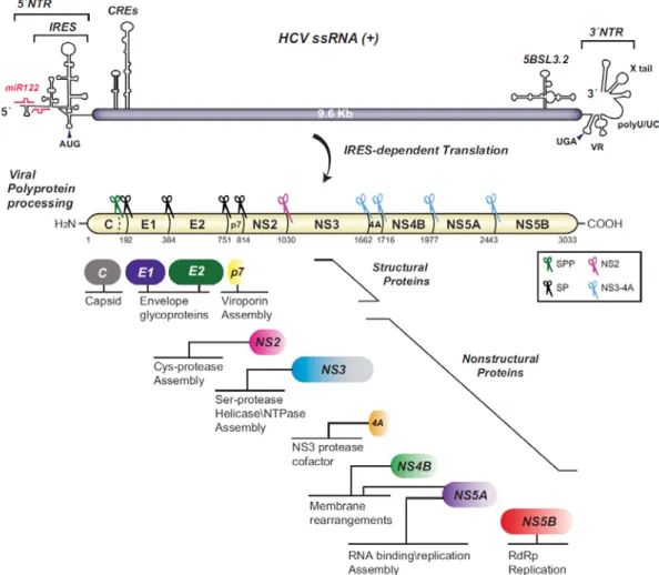
1.3 HCV genome organization
HCV in an enveloped positive-sense single-stranded RNA virus and is grouped with the genus Hepacivirus within the Flaviviridae family (Lindenbach et al., 2013).HCV genomic RNA is 9,6 kb in length that is composed of a single open reading frame (ORF) of nearly 3040 codons flanked by two untranslated regions (NTR) the 5’ and 3’ NTRs (Figure 3). The viral genome is directly translated by a mechanism mediated by the internal ribosomal entry site (IRES) to a single polyprotein that is localized to the endoplasmic reticulum (ER). This polyprotein is cleaved co- and post-translationally by the action of viral and cellular proteases into 10 mature structural and nonstructural proteins (NS). The one third amino-terminal region of the polyprotein encodes the structural proteins E1, E2 and Core, which are incorporated, into viral particles. The remaining two thirds of the carboxy-terminal region encodes the NS proteins which includes P7, NS2, NS3, NS4A, NS4B, NS5A and NS5B (Figure 3). These NS proteins are not incorporated in viral particles. Instead, they function to orchestrate different activities required for HCV RNA replication, forming what is called the replication complex (Moradpour et al.,
6
2013). The expression of the NS protein has been shown to induce a massive membrane remodeling within the cytoplasm of the infected cell forming the replication factories, termed as the membranous web (MW) (Egger et al., 2002).With the help of host factors and viral structural proteins, the NS proteins have also been shown to participate in virion maturation (Lindenbach, 2013). In addition, several NS proteins modulate host immune defense and play a role in establishing chronicity (Lindenbach, 2013, Morikawa et al., 2011).
Figure 3. HCV genome organization and polyprotein processing. The HCV RNA single strand
(SS) with positive polarity is shown on the top. The RNA Secondary structures include cis-acting RNA elements (CREs) in the untranslated regions (NTRs) are also shown on the top. miR-122 binding sites in the 5′ NTR containing the internal ribosome entry site (IRES) are shown. The proteins resulting from the single polyprotein cleavage and their functions are indicated. Scissors represent proteases that cleaves the virus polyprotein. SP, signal peptidase; SPP, signal peptide peptidase. (Paul et al., 2014).
7
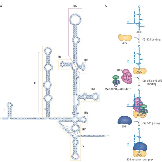
1.3.1 IRES-mediated translation initiation
HCV protein translation initiation is directed by a group of regulatory structural RNA elements at the 5’ NTR that define IRES. The HCV IRES spans a region of ~341 nucleotides (nt) and is composed of three distinctly structured domains II, III, and IV. The boundaries of the IRES have been carefully mapped to be between 25 and 46 nt of the 5′NTR, stretching up to about 24-40 nt within the coding region. The first domain of the IRES, stem-loop I (SLI), is not required for translation, but is important for replication (Figure 4a). SLII is comprised of a stem having several internal loops (Figure 4a, boxed in yellow, left side). The larger domain SLIII consists of branching hairpin stem–loops (IIIabcdef) organized in 3- and 4-way junctions and a pseudoknot near the base of the stem–loop. SLIV is a small structure that resembles a hairpin and contains the start codon AUG at position 342. After its binding with the 40S ribosomal subunit, the central IRES core domain with the double pseudoknot positions the start codon on the 40S into the mRNA binding cleft (Berry et al., 2011). To form preinitiation 48S ribosomal complexes, the HCV IRES requires just three initiation factors, eukaryotic initiation factor (eIF) 2, eIF3, and eIF5. The eIF3 binds to the apical region of SLIII (Sizova et al., 1998) (Figure 4a, boxed in magenta) and associates with the ribosomal 40S subunit (Jackson et al., 2010). The eIF2 associates with the initiator tRNA and GTP to form the ternary complex eIF2-GTP-Met-tRNAiMet which brings the Met-tRNAiMet to the
40S subunit (Pestova et al., 1998). Consequently, eIF5 promotes start codon recognition by the ternary complex eIF2-GTP-Met-tRNAiMet and acts as a
GTPase-activator protein for eIF2 (Pestova et al., 1998). This is sufficient to allow the bound 40S subunit to lock onto the HCV initiation codon and associate with the large ribosomal 60S subunit to form translation-competent 80S (Pestova et al., 1998, Terenin et al., 2008) (Figure 4b).
In addition to these canonical translation initiation factors, several host factors have been identified to interact with HCV IRES and/or 3’NTR and modulate viral RNA translation (Table 1). Since the IRES-mediated translation is a critical step in HCV life cycle, the IRES-mediated translation became an important target for developing therapeutic strategies to eradicate HCV.
8
Figure 4. HCV IRES structure and model for 80s complex formation. (a) The secondary
structure of the HCV 5’NTR consisting of 4 different domains (I, II, III, IV). The domains for 40S binding are boxed in yellow while the domain for eIF3 binding is boxed in magenta. The translation start codon is in red color (b) a model for HCV IRES mediated initiation of translation. [modified from (Fraser et al., 2007)]
9
Table 1. Host factors involved in HCV RNA translation [adopted from (Hoffman et al., 2011)] Cellular factors Effect on
HCV translation
Proposed mechanisms Cellular functions References
Gemin5 - Binds to IRES and forms complex with eIF3a, b, c. Addition of purified Gemin5 resulted in down-regulation of HCV IRES activity, while shRNA knockdown of Germin5 resulted in increase of HCV IRES activity
snRNA binding protein of SMN complex, which is involved in biogenesis of snRNPs, a component of mRNA slicing machinery. Also, involved in down-regulation of cellular translation in which a complex containing eIF4E is formed
(Pacheco et al., 2009)
HMGB1 + Facilitates viral replication by interaction with the HCV RNA
A nuclear protein involved in a group of diseases, including infectious diseases, metabolic and immune disorders, and cancer.
(R. Yu et al., 2015)
hnRNP D (AUF1) + Interacts with SLII of HCV IRES. Knockdown inhibits translation but increases replication suggesting a role in balancing viral translation and replication
Involved in mRNA decay, telomere maintenance and translation initiation
(Paek et al., 2008)
hnRNP L + Binds to 30 -end of HCV IRES. hnRNP L-specific RNA aptamers inhibited IRES function in dose dependent manner
Role in mRNA processing including alternative splicing, mRNA export and mRNA stability
(Hahm et al., 1998, B. Hwang et al., 2009)
Hu antigen R
(HuR) +
Binds to polyU/UC region of 30 -UTR. Overexpression enhances HCV IRES activity while knockdown down-regulates HCV IRES activity
Selectively binds and stabilizes AU-rich element containing mRNAs
(Rivas-Aravena et al., 2009, Spangberg et
al., 2000)
IGF2BP1 (IMP-2) + Binds to 50 - and 30 -UTRs. siRNA knockdown down-regulates HCV IRES activity. May stimulate translation by promoting RNA circularization and/or recruitment of eIF3
Binds to and regulates the translation of certain mRNA such as insulin-like growth factor 2 and b-actin
(Weinlich et al., 2009)
La autoantigen + Binds near the initiation AUG codon of IRES. Thought to alter the conformation which facilitates formation of the initiation complex and stimulate internal initiation of translation
Multifunctional protein with roles in RNA biogenesis. Binds to the 30 termini of many newly synthesized RNAs, particularly those made by RNA pol III, protecting the 30 ends from exonucleases. Autoantigen in systemic autoimmune diseases
(Ali et al., 1997, Pudi et al., 2004)
LSm1-7 + LSm1-7 ring binds to 50 -UTR dependent on SLIII. Also binds to poly(U/UC) tract of 30 -UTR. Silencing of LSm1 selectively down-regulates HCV translation
Component of P-bodies. Involved in mRNA turnover. Binds to short oligo (A) tracts at 30 -end of de-adenylated mRNA and inhibits 30 degradation while promoting decapping and 50 –30 degradation
(Scheller et al., 2009)
miR-122 + Binding to S1 and S2 sites in 50 -UTR upstream of the IRES enhances translation. Thought to enhance the association of 40S ribosomal subunit and HCV RNA. May protect 5’-end of viral genome from degradation
Well-conserved, highly abundant, liver specific microRNA. Binding to 30 -UTR of cellular mRNA encoding cationic amino acid transporter CAT-1 results in down-regulation of CAT-1 protein levels
(Henke et al., 2008, Jangra et al., 2010, Jopling et al., 2005)
miR-199a ?(-) Target sequence in domain II of the HCV IRES. Over-expression results in down-regulation of viral replication
Implicated in the post-transcriptional regulation of gene expression for various genes such as Ceruloplasmin (CP)
(Murakami et al., 2009)
miR-196 - Potential target sequence present in HCV NS5A coding region. Over-expression results in down-regulation of HCV protein expression and replication
Implicated in the post-transcriptional regulation of gene expression for various genes such as Bach1
(Hou et al., 2010)
Nucleolin ?(+) No data using HCV reporter. Identified by mass spectrometry as a component of HCV IRES bound 40S ribosomal subunit. Bound by yeast inhibitor RNA (IRNA). Stimulates poliovirus IRES
Implicated in a variety of processes including rRNA maturation, and ribosome assembly
(Izumi et al., 2001)
NSAP1 (SYNCRIP, hnRNP Q)
+ Binds to adenosine rich region downstream of AUG start codon in core coding sequence. Over-expression enhances IRES activity while knockdown down-regulates IRES activity
A member of hnRNP family of proteins implicated in mRNA processing mechanisms. Component of the spliceosome
(Ali et al., 1995)
PTB ? Interacts with both 50 - and 30 -UTRs of HCV RNA. Role in HCV protein translation is unclear with contradictory reports suggesting stimulation, inhibition, or no effects on HCV protein translation
Implicated in the regulation of pre-mRNA splicing
(Ali et al., 1995, Tischendorf et al.,
10
PSMA7 + Knockdown by siRNA or ribozymes results in significant inhibition of HCV translation
Human 20S proteasome a-subunit type 7 is a component of 20S core structure of proteasome
(Kruger et al., 2001)
Staufen 1 + Knockdown by siRNA decreased virus secretion a dsRNA-binding protein involved in the regulation of translation, trafficking, and degradation of cellular RNAs
(Blackham et al., 2013)
1.4 Structural proteins
1.4.1 Core Protein
The first protein to be translated from the ORF is the core protein. The presence of an internal signal sequence between core and E1 directs the nascent polyprotein to the ER where cleavage of the signal sequence takes place by the action of signal peptidase (SP) (Figure 3). This cleavage yields immature core protein with 191 amino acid (aa). Further processing of the C-terminus by signal peptide peptidase (SPP), an intramembrane cleaving protease, yielding the mature 21-kDa core protein, with ~177 amino acid (aa) (Figure 3) (Oehler et al., 2012, Santolini et al., 1994). Mature core is a homodimeric membrane protein stabilized by the formation of disulfide bond at Cys 128 (Kushima et al., 2010). HCV core has two main domains, the N-terminal hydrophilic domain (D1) that is rich in basic aa residues with highly flexible intrinsic disordered structure that allows an expansion of interaction with viral and host molecules (Uversky, 2011). It also harbors critical residues required for Core function. Indeed, core D1 can bind HCV RNA promoting RNA encapsidation. As other nucleocapsid proteins, D1 has RNA chaperone activity required for structural remodeling and packaging of the RNA during virion formation (Cristofari et al., 2004). In addition, core D1 can regulate translation of viral proteins (Boni et al., 2005, Lourenco et al., 2008). Moreover, the C-terminus of D1 harbors motifs for BH3 interaction capable of binding cellular proteins to regulate apoptosis (Mohd-Ismail et al., 2009). The other core protein domain is the C-terminal hydrophobic domain (D2), which mediates the binding with lipid droplets (LDs). This association with LDs impedes core nuclear localization exerted by a nuclear localization signal found in the N-terminal RNA binding domain (Suzuki et al., 1995). Core D2 contains a central hydrophobic
11
loop that connects two amphipathic α-helices and interacts with phospholipids interface on LDs (Boulant et al., 2007).
It has been shown that the association of core with LD and its interaction with NS5A plays a crucial role in virion assembly process (Miyanari et al., 2007, Shavinskaya et al., 2007).
1.4.2 E1 and E2 glycoproteins
The envelope glycoproteins E1 and E2 are type 1 transmembrane with an ectodomain at the N terminus (~160 aa and ~360 aa for E1 and E2 respectively) facing ER lumen and a short transmembrane domain (TMD) of ~30 aa at C terminus (Moradpour et al., 2013) (Figure 5a). During their biogenesis, the ectodomains of E1 and E2 translocate into the ER lumen and inserts their TMDs (shown in black, Figure 5a) into the ER membrane. The TMDs are strongly implicated in the functions of E1 and E2, including membrane anchoring and ER retention. In addition, they participate in the formation of E1–E2 noncovalent heterodimer, which is supposed to represent the building units for HCV envelope. The E1 and E2 embedded in the virus envelope were shown to form large covalent complexes stabilized by disulfide bonds (Vieyres et al., 2010). The folding of these two proteins, that occurs in the ER, is a complex process that requires ER chaperones, disulfide bridging, and glycosylation. Indeed, E1 and E2 contain up to 6 and 11 glycosylation sites (trees in Figure 5a), respectively. Despite the high genetic variability, this extensive glycosylation is quite conserved across all genotypes (Brown et al., 2010, Goffard et al., 2005).
The genes that encode E1 and E2 are uniquely variable with several hyper variable regions (HVRs) that have been identified within E2 (shown in red, Figure 5a). These HVRs differs by up to 80% across the different genotypes. The HVR1 consists of the first 27 aa of E2 and is responsible for eliciting type-specific neutralizing antibodies (Figure 5a) (Penin et al., 2001). HVR2, from aa 91 to 97 of E2, has up to 100% sequence diversity across genotypes (Figure 5a). The intergenotypic variable region (igVR), aa 187 to 197, is widely variable between different genotypes while it contains a single conserved N-linked sugar moiety
12
(Figure 5a). HVR2 and igVR are essential for the structural integrity and hence for the function of HCV glycoproteins (McCaffrey et al., 2011). However, in contrast to HVR1, they are not targets for humoral immune response.
Recently, two independent crystal structures have been provided for the core ectodomain of E2 protein (Khan et al., 2014, Kong et al., 2013) (Figure 5b and c, respectively) and amino-terminal domain of E1 (El Omari et al., 2014) (Figure 5d). The structures of E1 and E2 lack the hallmarks of viral membrane fusion proteins suggesting a possible new entry mechanism for HCV.
a
b c d
Figure 5. HCV E1 and E2 glycoproteins. (a) Schematic of the HCV E1 and E2 that shows the
conserved glycosylation sites (trees). (b) and (C) Two structures of the core ectodomain of E2. (D) Represents the structure of the amino terminal domain of E2.
13
1.5 Nonstructural proteins
1.5.1 P7
P7 is a small, 63 aa, integral membrane protein located at the junction between structural and nonstructural proteins (Lin et al., 1994).It is classified as viroporin, as influenza virus M2 protein, based on its ability to alter membrane permeability (Nieva et al., 2012). It comprises two transmembrane α-helices with an N and C termini oriented towards ER lumen connected by a cytosolic loop (Carrere-Kremer et al., 2002, Vieyres et al., 2013). P7 forms hexameric or heptameric structures with cation channel activity to facilitate the formation of infectious viral particles (Chandler et al., 2012). It was shown that trafficking of viral and cellular glycoproteins is delayed by the action of P7. Furthermore, P7 could act to prevent acidification in intracellular compartment to promote infectious viral particles production (Wozniak et al., 2010). Recently, it was shown that, in addition to its function as viroporin, P7 could act as lipid raft adhesion factor for HCV budding process (G. Y. Lee et al., 2016). Although P7 is required for virus assembly but not replication, the exact function of P7 has not been elucidated yet (Scull et al., 2015).
1.5.2 NS2
NS2 is a 217 aa integral membrane protein. It binds to intracellular membranes via its hydrophobic N-terminal domain that contains 3 transmembrane segments (Jirasko et al., 2008). NS2 gene encodes for a cysteine protease responsible for its autocleavage from the HCV polyprotein precursor at NS2/NS3 junction. The activity of this cysteine protease has been shown to be greatly improved by one third N-terminal of the NS3 protein (Schregel et al., 2009). The catalytic activity resides at the C-terminal domain. The crystal structure of this domain revealed a dimer with two active sites (Lorenz et al., 2006). Despite the fact that NS2 is dispensable for RNA replication, NS2 plays a crucial organizing role in the assembly of infectious viral particles. This action was shown to be independent of its protease activity and may involve interaction network with other structural and
14
nonstructural proteins (Boson et al., 2011, Stapleford et al., 2011). Recently, it was proposed that NS2 protease activation in modulated by a conserved NS3 surface patch (Isken et al., 2015).
1.5.3 NS3-4A complex
NS3 and its co-factor, NS4A, form a non-covalent complex (J. L. Kim et al., 1996). NS3 is a 70 kDa multifunctional protein with a serine protease located in the N-terminal (aa 1-180) and an NTPase/RNA helicase in the C-N-terminal (aa 181-631). The activity of both enzymes is well characterized and their crystal structures have been resolved (Morikawa et al., 2011, Raney et al., 2010, Yao et al., 1999). NS3-4A protease activity is required for cleavage at four junctions in the HCV polyprotein, NS3/NS4A, NS4A/NS4B, NS4B/NS5A, and NS5A/NS5B (Bartenschlager et al., 1993, Grakoui et al., 1993). In addition, NS3-4A protease was shown to target, so far, three cellular factors for cleavage. These include T-cell protein tyrosine phosphatase (TC-PTP), mitochondrial antiviral signaling protein (MAVS), and toll/IL-1 receptor homology domain-containing adaptor inducing IFN-β (TRIF), which may be implicated in the development of chronic infection and HCC (K. Li et al., 2005, Meylan et al., 2005). Accordingly, NS3-4A protease plays essential roles not only in the replication but also in the persistence and pathogenesis of HCV (Morikawa et al., 2011). This made the NS3-4A protease a primary target for DAAs. The binding of NS4A to NS3 was shown to stabilize the NS3 structure and hence enhances its catalytic activity and directs it to cellular membrane localization via the hydrophobicity of the N-terminal transmembrane α-helix of NS4A (Abian et al., 2010). Furthermore, the C-terminal acidic portion of NS4A is involved in the regulation of HCV RNA replication and virus assembly by interacting with other viral nonstructural proteins forming what is called the replication complex (Morikawa et al., 2011). NS4A was also shown to regulate HCV replication through its role in NS5A hyperphosphorylation (Lindenbach et al., 2007). The NS3 NTPase/RNA helicase belongs to the superfamily 2 DExH/Dbox helicases (Raney et al., 2010). It combines ATP hydrolysis activity to its capability
15
to unwind either double-stranded RNA or single-stranded RNA regions with extensive secondary structure. Although the NS3 helicase is essential for HCV RNA replication and also plays a role in viral particle assembly, its precise role (s) in HCV life cycle remain(s) still not determined.
1.5.4 NS4B
NS4B is a hydrophobic 27 kDa protein that is poorly characterized (Hugle et al., 2001). It is an integral protein consists of a N-terminal part (aa 1-69), a central portion with at least four predicted transmembrane (TM) domains (aa 70-190), and a C-terminal part (aa 191-261) (Gouttenoire et al., 2009b, Hugle et al., 2001, Lundin et al., 2003). The N-terminal part comprises two amphipathic α-helices, AH1 (aa 3-35) and AH2 (aa 42-66). The AH2 is conserved across different genotypes and is crucial for HCV replication (Elazar et al., 2004, Gouttenoire et al., 2014). This segment plays an important role in the assembly of a functional replication complex. The C-terminus comprises a highly conserved amphipathic α-helix, H1, (aa 201–213) and a membrane-associated amphipathic α-helix, H2, (aa 229–253), and two other palmitoylation sites (Gouttenoire et al., 2009b, G. Y. Yu et al., 2006). Upon NS4B dimerization/ multimerization, the N-terminus can translocate its AH1 part into the ER lumen promoting recruitment of the replication complex via its role in the formation of membranous web (MW), a specific membrane alteration consisting of confined membranous vesicles that serves as a scaffold for the HCV replication complex, thus allowing HCV RNA replication (Egger et al., 2002, Gosert et al., 2003, Gouttenoire et al., 2009a, Romero-Brey et al., 2012). In addition, NS4B interacts with other viral nonstructural proteins and can bind viral RNA (Einav et al., 2008). The central portion of NS4B harbors a nucleotide-binding motif (NBM) Walker A located between TM2 and TM3 domains with NTPase activity. This NBM walker A exists in all HCV genotypes and shows importance in HCV life cycle (Einav et al., 2004, Jones et al., 2009). Altogether, NS4B is considered as a master organizer of HCV replication complex formation.
16
Therefore, NS4B is also considered as a potential target for antivirals (Cannalire et al., 2016, Rai et al., 2011).
1.5.5 NS5A
NS5A is a 447-aa membrane-associated phosphoprotein that plays an important role in modulating HCV RNA replication and particle formation. It can be found in basally phosphorylated (56 kDa) and hyperphosphorylated (58 kDa) forms. NS5A has N-terminal membrane anchoring domain in addition to three other domains, D1, D2 and D3 separated by two low complexity sequences (LCS). D1 (aa 36-213) and D2 (aa 250-342) are mainly involved in HCV RNA replication while D3 (aa 356-447) is implicated in viral assembly (Appel et al., 2008, S. Kim et al., 2011, Tellinghuisen et al., 2008, Tellinghuisen et al., 2004). Moreover, D1 was shown to be involved in lipid droplets (LDs) binding (Miyanari et al., 2007) whereas D2 is involved in the interaction with core protein (Masaki et al., 2008). The basal phosphorylation of NS5A protein is a conserved feature within flaviviruses (Reed et al., 1998). In the case of HCV NS5A, it occurs at the C-terminal and the central portion of the protein while hyperphosphorylation of the protein requires the serine residues 225, 229, and 232 in the first LCS. Indeed, cell culture adaptive changes often affect these centrally located serine residues suggesting that the phosphorylation state of NS5A modulates the efficiency of HCV RNA replication (Appel et al., 2005, Evans et al., 2004, Neddermann et al., 2004). Cellular protein kinases in the CMGC kinase family, named by the initials of some members of the family, mediate NS5A phosphorylation. These include cyclin-dependent kinase (CDK), mitogen activated protein kinase (MAPK), glycogen synthase kinase 3 (GSK3) and casein kinase II (CKII) (Asabe et al., 1997, Koch et al., 1999, Lindenbach et al., 2007, Macdonald et al., 2004, Tellinghuisen et al., 2008). The subcellular distribution of both forms of NS5A seems to be similar. Thus, the phosphorylation level of NS5A does not alter its localization to the ER membrane (Tanji et al., 1995). However, the phosphorylation of NS5A is shown to enhance its degradation and decreases the protein half-life (Y. Huang et al., 2007b). NS5A
17
is also involved in the production of infectious virions through its interaction with core protein, which needs a basal phosphorylation of NS5A (Masaki et al., 2008). The role of NS5A in HCV RNA replication and virion formation made it an attractive target for DAA (Adler et al., 2014)
1.5.6 NS5B
NS5B is a conserved 68 kDa protein with RNA-dependent RNA polymerase (RdRp) activity. It is the key enzyme responsible for the initiation of complementary negative-strand RNA synthesis and subsequent synthesis of genomic positive-strand RNA using this negative-positive-strand RNA as template. Due to the lack of proofreading of RdRp, HCV replication is error-prone. This 591 aa viral enzyme has been widely studied and characterized (Behrens et al., 1996, Lesburg et al., 1999, Lohmann et al., 1997, Simister et al., 2009). The NS5B catalytic domain is located at the N-terminal 530 aa and contains motifs that are common in all RdRps. These include the hallmark GDD sequence within motif C, and the classical fingers, palm, and thumb subdomain organization of a right hand. The catalytic domain and the C-terminal membrane anchor are separated by a 40-aa linker between (aa 570-591) that occludes the active site. Within the palm and thumb domains, there are four allosteric sites which serve as potential targets for DAA development (Beaulieu, 2009, Court et al., 2016, Pierra Rouviere et al., 2016). NS5B can interact with other viral proteins such as NS3, NS4A and NS5A (Ishido et al., 1998). In addition, it can interact with cellular proteins like hVAP-33, which facilitates the formation of the viral RNA replication complex (Tu et al., 1999). Recently, it was shown to interact with Valosin-containing protein (VCP), which is a AAA+ATPase that can modulate viral replication (Yi et al., 2016). It can also form a complex with the retinoblastoma tumor suppressor protein (pRb) and promote pRb degradation in a ubiquitin-dependent manner, thus contributing to HCC development (Munakata et al., 2007). Moreover, NS5B was found to interact with ubiquitin-like protein hPLIC1 (human homolog 1 of protein linking integrin-associated protein and cytoskeleton) and causes NS5B ubiquitination. The
18
ubiquitination modification of NS5B through hPLIC1 binding promotes ubiquitin-dependent proteasome degradation, resulting in reduction of NS5B level (Gao et al., 2003). As NS5B primarily functions in RNA replication, decrease in NS5B through proteasome degradation leads to decrease in HCV RNA replication. Thus up-regulation of NS5B ubiquitination could be a potential target for anti-viral development.
1.6 HCV virion structure
Despite the significant progress in cell culture systems which enables the production of viral particles as well as several biochemical and morphological studies, the structure of the HCV particle remains elusive.
Viral particles purified from patients’ sera or cell culture media by filtration and visualized by electron microscopy (EM) revealed that, HCV has a spherical morphology of different sizes (range 40-80 nm diameter) (Catanese et al., 2013, Kaito et al., 1994, X. Li et al., 1995). The HCV genome interacts with the core protein to form the nucleocapsid that is surrounded by a lipid membrane, called the viral envelope, in which are anchored two envelope glycoproteins, E1 and E2. These two glycoproteins assemble as a E1E2 heterodimer that is stabilized by disulfide bonds on the viral particle where they represent the major viral determinants of HCV entry (Figure 6).
One unique feature of HCV virion is its association with apolipoproteins (apo) such as apoE, apoB, apoA1, apoC1, apoC2 and apoC3 (Andre et al., 2002, Catanese et al., 2013, Chang et al., 2007, Meunier et al., 2008, Nielsen et al., 2006). Furthermore, analysis of cell culture-produced viral particles by mass spectrometry indicates that their lipid composition resembles the one of the very low-density lipoproteins (VLDLs) and low-density lipoproteins (LDLs) with cholesteryl esters (CEs) accounting for almost half of the total HCV lipids (Merz et al., 2011). Purified infectious virions showed a pleomorphic nature of HCV virions and show viral particles with a rather smooth and even surface (Merz et al., 2011).
19
The type of the interactions occurring between HCV virion components and the lipoprotein remains elusive. One proposed model suggested that HCV particles form a lipo-viro-particle (LVP), a combination of a virion moiety and a lipoprotein moiety (see figure 6A) (Andre et al., 2002, Bartenschlager et al., 2011). However, alternative models have also been suggested, with lipoproteins peripherally associated with canonical viral particles via interaction between apolipoproteins and HCV envelope lipids or proteins (Lindenbach, 2013). In both models, the interaction of virus particles with lipoprotein may contribute to the protection of virus particles from circulating neutralizing antibodies (Andre et al., 2002, Catanese et al., 2013, Dao Thi et al., 2012). Importantly, apolipoprotein(s) associated with HCV particles play a critical role in HCV entry. The difficulty to investigate natural HCV, or LVP, and its different lipoprotein or immunoglobulin-associated forms is due to low infectivity of serum-derived HCV either in primary hepatocytes or hepatoma cell lines (Fournier et al., 1998, Rumin et al., 1999). In addition, the mechanism(s) by which LVPs mediate cell entry, leading to release of HCV genetic material and RNA replication, have not been fully characterized.
20
Figure 6. The components of each HCV particle type. The apolipoprotein composition of each
type of viral particle is indicated in the figure. [modified from (Douam et al., 2015)]
The development of several surrogate in vitro assays to overcome these limitations has been successfully achieved. Among them, two main assays consist of cell culture grown HCV (HCVcc) derived from a fulminant hepatitis C 1 and JFH-1-derived recombinant genomes (Lindenbach et al., 2005, Wakita et al., 2005) and of HCV pseudoparticles (HCVpp) harboring authentic E1E2 glycoproteins which is particularly beneficial to study viral entry (see figure 6C) (Bartosch et al., 2003a, Drummer et al., 2003). HCVcc exists as a mixture of infectious and noninfectious particles and displays a broad density profile similar to HCV derived from patients’ sera. Higher specific infectivity was shown to be associated to low-density fractions (Gastaminza et al., 2006, Lindenbach et al., 2006, Podevin et al., 2010). The lipid composition of HCVcc particles was determined and found similar to that of VLDL and LDL (Merz et al., 2011). Highly purified HCVcc particles were found to contain
21
several apoE molecules on their surface as well as apoC-I and, to some extent, apoB (see figure 6B). This comes in line with the finding that HCV formation and secretion rely on VLDL assembly and secretion pathway (Chang et al., 2007, Gastaminza et al., 2008, H. Huang et al., 2007a, Jiang et al., 2009, Meunier et al., 2008). While HCVcc, which are produced in human hepatoma cells typically Huh7 and Huh-7.5 cells, further permit investigation of the late steps of infection, HCVpp provide a flexible platform to study the structure/ function relationship of HCV glycoproteins in vitro. It is noteworthy that, since HCVpp are produced from 293T kidney cells, they are not associated to lipoproteins (Flint et al., 2004), which, in turn, allows to investigate the cell entry events that are specifically linked to the functions of the E1E2 glycoproteins (Bartosch et al., 2005). In addition, this provides a suitable tool to reconstitute some of the interactions of HCV with lipoproteins or apolipoproteins (Dreux et al., 2007, Dreux et al., 2006, Meunier et al., 2005). Altogether, utilizing HCVcc and HCVpp allows the dissection of the cellular and viral factors involved in HCV infection.
1.7 HCV attachement
Hepatocytes are the main target cells for HCV infection. However, infection of dendritic cells, B cells, and other cell types have also been reported. Viral entry into the host cell involves a complex multistep series of interactions including attachment, entry and fusion. The initial capture of HCV virion by its cell surface receptor/co-receptors may involve HVR1 in HCV E2 (Flint et al., 2000). Depending on virion density, this step may be mediated by heparan sulfate proteoglycan (HSPG) syndecan-1 and/or syndecan-4 (Lefevre et al., 2014, Shi et al., 2013) or by the scavenger receptor class B type I (SR-BI) (Figure 7) (Dao Thi et al., 2012). Although, the initial binding of virions to HSPG or SR-BI was thought to be through HCV glycoproteins (Barth et al., 2003) (Scarselli et al., 2002), a more recent data showed that apoE, rather than HCV glycoproteins, could be implicated in this binding (Dao Thi et al., 2012, Jiang et al., 2013). As HCV particle interacts with
22
lipoproteins, the LDL receptor (LDLR) has also been suggested to be involved in the early phase of HCV entry (Agnello et al., 1999). However, this interaction may result in non-productive entry pathway and can potentially lead to viral particle degradation (Albecka et al., 2012). After initial attachment step, and in addition to HSPG, SR-BI, and LDLR, several entry factors for HCV have been identified, including CD81, tight junction proteins claudin-1 (CLDN1) and occludin (OCLN), the receptor tyrosine kinases (RTK) epidermal growth factor receptor (EGFR), ephrin receptor A2 (EphA2), Niemann-Pick C1-like 1 cholesterol absorption receptor (NPC1L1) (Evans et al., 2007, Liu et al., 2009, Lupberger et al., 2011, Pileri et al., 1998, Ploss et al., 2009, Sainz et al., 2012), and the two recently identified entry factors transferrin receptor 1 and E-cadherin (Q. Li et al., 2016, Martin et al., 2013).
1.7.1 Scavenger receptor class B type I
The SR-BI is a 509 aa cell surface glycoprotein receptor that is present on many cell types with higher expression within the liver (Meredith et al., 2012). It has been identified as HCV E2 binding partner along with CD81(Pileri et al., 1998, Scarselli et al., 2002). SR-BI is involved in the lipid metabolism and is known to be a major receptor for high-density lipoproteins (HDLs) promoting its selective uptake into hepatocytes (Acton et al., 1996). It can also bind other lipoprotein classes such as the VLDL and LDL (Van Eck et al., 2008). A role of SRB1 in HCV entry was first suggested through its binding of HCV E2 HVR1 (Bartosch et al., 2003b, Catanese et al., 2007, Scarselli et al., 2002). The lipid transfer capability of SRB1 may be required for HCV cell entry as HCV entry is enhanced by HDL and could be inhibited by oxidized LDL (Dreux et al., 2009a, Meuleman et al., 2012). It was shown that SR-BI mediates primary attachment of HCV particles that have intermediate density to cell surface and that these interactions require apolipoproteins, such as apoE (Dao Thi et al., 2012). Recently it was shown that, introducing three specific mutations (Y507L, V514A, and V515A) located within E2 neutralizing epitope modulated HCV dependence on the viral receptor SRB1
23
(Lavie et al., 2014). SRB1 antagonist was shown to inhibit HCV replication with additive to synergistic potency when used in combination with other antiviral therapeutics (Zhu et al., 2012).
1.7.2 CD81
Human CD81 is a tetraspanin adaptor cell surface molecule that is broadly expressed in many cell types (van Spriel et al., 2010). It is involved in many cellular functions including adhesion, morphology, proliferation and differentiation. It harbors four transmembrane domains, two short intracellular domains and two extracellular loops, named SEL and LEL (Meredith et al., 2012). The involvement of CD81 in HCV entry is likely to be after the very early attachment phase (i.e. after SRB1), promoting a conformational change in the HCV E1/E2 glycoproteins. This conformational change facilitates low pH-dependent fusion and viral endocytosis (Sharma et al., 2011). HCV, through its envelope glycoprotein E2, binds CD81 large extracellular loop to facilitate HCV entry (Petracca et al., 2000, Pileri et al., 1998). The CD81 large extracellular loop sequence is conserved between humans and chimpanzees. However, CD81 from other species, including species that are not permissive to HCV infection, can support HCV entry in vitro suggesting that CD81 sequence variability is not sufficient to define HCV susceptibility (Flint et al., 2006).
1.7.3 Claudin 1 and Occludin
Claudin 1 and Occludin are two tight junction proteins that were identified after screening of a cDNA library of potential cellular factors implicated in HCV entry utilizing HCVpp (Evans et al., 2007, Ploss et al., 2009). Neither CLDN1 nor OCLN directly interact with HCV envelope. Although, CLDN1 may interact with CD81 as a part of the HCV receptor complex (Harris et al., 2010, Harris et al., 2008). It was suggested that CLDN1 and OCLN are involved in a later phase of HCV entry, after SRB1 and CD81, although their exact roles are still elusive. It was also shown that
24
HCV envelope glycoproteins promote co-endocytosis of CD81 and CLDN1 and fusion with early endosome (Farquhar et al., 2012). It was shown that anti-CLDN1 monoclonal antibodies can inhibit HCV infection in primary hepatocytes in vitro as well as in cell lines by neutralizing the interactions between HCV E2 and CLDN1 (Fofana et al., 2010, Krieger et al., 2010). Synchronized infection assays showed that HCV utilizes CD81, Claudin-1, and Occludin in this order during the attachment and entry. In addition, downregulation of Occludin reduces both HCV entry and glycoprotein-mediated cell fusion (Sourisseau et al., 2013).
1.7.4 Receptor tyrosine kinases
The two RTKs including the EGFR and EphA2 were identified as HCV entry co- factors using a functional RNAi kinase screen (Lupberger et al., 2011). EGFR is known to regulate several key processes including cell proliferation, survival, and differentiation during development, tissue homeostasis and tumorigenesis (Schneider et al., 2009). While EphA2 is involved in cell positioning, cell morphology, polarity and motility (Lackmann et al., 2008). These RTKs appear to participate in HCV entry after the initial engagement step, by regulating CD81 and CLDN1 co-receptor interactions and viral E2-mediated membrane fusion by EGFR-dependent signaling pathways (Lupberger et al., 2011). Indeed, the small GTPase HRas (Harvey Rat Sarcoma oncogene homologue) was identified as a host signal transducer that represents the physical link between the HCV–CD81– CLDN1 complex and the EGFR/Shc1/Grb2/HRas signaling pathway (Zona et al., 2013). Stimulation of EGFR activates the Ras/MEK/ERK pathway which promotes MAPK interacting serine/threonine kinase 1 (MKNK1) activation. Later on, MKNK1 was identified as a host factor in HCV entry, which possibly act to facilitate the steps that are downstream of the EGFR (S. Kim et al., 2013a).
25
1.7.5 Niemann-Pick C1-like 1 cholesterol absorption receptor
Niemann-Pick C1-like 1 cholesterol absorption receptor (NPC1L1) is a 13 transmembrane-domain cell surface cholesterol-sensing receptor expressed on the apical surface of intestinal enterocytes and human hepatocytes. NPC1L1 is responsible for cellular cholesterol absorption and whole-body cholesterol homeostasis (Castro-Torres et al., 2014, L. Yu, 2008). HCV entry was shown to be inhibited by the available NPC1L1 antagonist ezetimibe which is FDA-approved to treat hypercholesterolemia (Sainz et al., 2012). Based on an experiment comparing the susceptibility of HCVpp and HCVcc entry to ezetimibe, it was demonstrated that the cholesterol content of viral particles correlates with NPC1L1-mediated infection (Sainz et al., 2012).
1.7.6 Transferrin receptor 1
Transferrin receptor 1 (TfR1) is the main receptor for cellular iron uptake into cells and is ubiquitously expressed in all tissues. After TfR1 binds to its extracellular ligand, iron-bound transferrin, the TfR1-trafficking protein (TTP) facilitates its internalization via clathrin-mediated endocytosis. Once iron is released in the cytoplasm, TfR1 will be recycled back to the cell surface (Tosoni et al., 2005). It has been shown that HCV infection can alter the expression of TfR1. In addition, blocking of the cell surface TfR1 inhibits HCVcc and HCVpp infection, demonstrating that TfR1 acts at the level of HCV glycoprotein-dependent entry. The role of TfR1 in HCV entry was suggested to be beyond CD81 and might be specifically involved in HCV particle internalization (Martin et al., 2013).
1.7.7 E-cadherin
E-cadherin, encoded by the CDH1 gene, is a major classical adherent junction protein. It is a transmembrane glycoprotein that plays an important role in maintaining cell-cell adhesion. In addition, it performs a vital role in establishing
26
epithelial architecture and maintaining cell polarity and differentiation (Stemmler, 2008, van Roy et al., 2008). Recently, E-cadherin was identified as key modulator of HCV entry (Q. Li et al., 2016). Depletion of E-cadherin has been shown to drastically diminishes the cell-surface distribution of the CLDN1 and OCLN in various hepatic cell lines. Thus, suggesting that E-cadherin plays an important regulatory role in CLDN1/OCLN localization on the cell surface (Q. Li et al., 2016).
Figure 7. Model of HCV entry. The main HCV entry receptors and cofactors are indicated. The
entry is composed of 7 main steps as indicated. (Douam et al., 2015)
HCV entry to the cell occurs by clathrin-mediated endocytosis, a dynamin-dependent pinocytosis endocytic mechanism (Figure 7) (E. Blanchard et al., 2006b). During this process, particle attachment induces the inward budding of the plasma membrane and the formation of a clathrin pit that internalizes the particle– receptor complex. Down regulation of the clathrin heavy chain was shown to inhibit
27
HCVpp and HCVcc infection (A. A. Blanchard et al., 2006a). Moreover, internalization of CD81–Claudin-1 complexes through clathrin-dependent endocytosis has been observed during HCV infection. This was shown to be consistent with the fact that CD81–Claudin-1 association is critical for virus internalization and that virus internalization occurs prior to the fusion step (Farquhar et al., 2012, Harris et al., 2010, Harris et al., 2008, Krieger et al., 2010). Following endocytosis, HCV-receptor complexes migrate to the RAB5A-positive endosomal compartments. Fusion is thought to occur at these compartments to allow the release of HCV RNA (Coller et al., 2012, Meertens et al., 2006). The release of viral RNA is believed to be through membrane fusion which is mediated by endosomal acidification. This is based on the observation that the use of bafilomycin A1 which prevents the endosomal re-acidification, has been shown to block HCV infection (Meertens et al., 2006, Sharma et al., 2011). By using transdominant-negative mutants in an HCVpp experiment, it has been shown that HCV membrane fusion occurs in an early endosomal compartment that was not positive for RAB7 (Meertens et al., 2006). Few evidences suggested that the interaction of HCV with CD81 may trigger a conformational change permitting low pH-induced membrane fusion (Sharma et al., 2011). Altogether, the mechanisms behind the activation of HCV for low pH-induced fusion, the fusion step, and the identity of the fusion peptide(s) are still unknown.
1.8 HCV replication and membranous web formation
A hallmark of all positive-strand RNA viruses is the induction of extensive cytoplasmic membrane proliferation and remodeling (Romero-Brey et al., 2014). Depending on the virus type, the induced membrane structures derived from either ER, Golgi apparatus, mitochondria or even lysosomes provide suitable microenvironments for viral replication. The exact role of membranes in viral RNA synthesis is not understood. Evidences suggest that they may serve to increase local concentration of host and viral factors required for efficient RNA replication,
28
to facilitate spatial coordination of different steps of the viral replication cycle, and to protect viral RNA and proteins from degradation and immune defense (Paul et al., 2013, Paul et al., 2014, Romero-Brey et al., 2015). The induced membrane rearrangement by positive-strand RNA viruses can be confined to two main morphological subclasses: the invaginated vesicle/spherule type within the ER and the double-membrane vesicle (DMV) type (Chatel-Chaix et al., 2014, Romero-Brey et al., 2014). Unlike other related flaviviruses, HCV is known uniquely to induce a matrix of DMVs, designated membranous web (MW) (Figure 8). EM analysis of HCV polyprotein expressing cells in recent studies revealed that the diameter of these DMVs is with an average of 150 nm. and contain active replicase complexes, supporting the model that they form to support viral RNA replication (Paul et al., 2013, Romero-Brey et al., 2012). The outer membranes of ∼50% of DMVs connect to the ER membrane via a neck-like structure. Biochemical analyses of purified vesicles revealed the presence of ER calnexin and calreticulin proteins, confirming the ER as a major source of membranes for MW formation (Ferraris et al., 2010, Miyanari et al., 2003)
For a period of time, it was believed that viral NS4B protein was responsible for the induction of MW formation during HCV infection since its sole expression can induce massive membrane rearrangements (Egger et al., 2002). Moreover, mutations in NS4B inhibiting self-interaction affect DMVs morphology and block HCV RNA replication (Paul et al., 2011). However, by using a replication-independent polyprotein expression approach, it has been found that NS5A was the only protein capable of inducing DMVs, while only single-membrane vesicles (SMVs) were observed in NS3/4A-, NS4B-, and NS5B-expressing cells (Reiss et al., 2011, Romero-Brey et al., 2012). The contribution of SMVs to HCV replication is unknown, but one model postulates that local, HCV-induced exvaginations form SMVs and, while the vesicles remain attached to the ER, a secondary invagination produces DMVs (Romero-Brey et al., 2012). Since none of the HCV proteins expressed solely was capable of inducing a MW, this suggests that MW formation most likely requires a concerted action of the replicase proteins.


![Table 1. Host factors involved in HCV RNA translation [adopted from (Hoffman et al., 2011)]](https://thumb-eu.123doks.com/thumbv2/123doknet/5006239.124996/27.918.37.885.154.1042/table-host-factors-involved-hcv-translation-adopted-hoffman.webp)


