Récepteur Wnt Non-Canonique Frizzled-6 Régule Hématopoïèse
Induite Par Les Stress
Non-canonical Wnt Receptor Frizzled-6 Regulates
Stress-Induced Hematopoiesis
by
Belma Melda Abidin
Thèse présentée pour l’obtention du grade de Philosophiae doctor en immunologie et virologie
Jury d’évaluation
© Belma Melda Abidin, 2017 Président du jury et examinateur interne Examinateur externe Examinateur externe Directeur de recherché
Claude Daniel
INRS-Institut Armand Frappier Christian Beauséjour
Université de Montréal
Département de pharmacologie et physiologie Tatiana Scorza
L'Université du Québec à Montréal (UQAM)
Département des sciences biologiques Krista Heinonen
Robert Heinlein said, “Love is that condition in which the happiness of another person is essential to your own.” With this ideal in mind, I dedicate my thesis to my rock, my wonderful mother, for her endless love, measureless support, encouragement and understanding, and to the memory of my father for his constant love and support from the Heaven. Without them, this journey would not have been possible.
L’homéostasie du sang est maintenue par l’équilibre entre l’auto-renouvellement et la différenciation des cellules souches/progénitrices hématopoïétiques (HSPC). Bien que les cellules souches hématopoïétiques (HSC) soient principalement maintenues dans un état de dormance, elles peuvent entrer rapidement dans le cycle cellulaire et se différencier en précurseurs lymphoïdes et myéloïdes pour reconstituer les cellules immunitaires suite à une myelosuppression ou à des infections systémiques. La signalisation de Wnt a été suggérée pour maintenir l’équilibre dynamique du pool de HSPC en régulant leur division et les signaux provenant du microenvironnement de la cellule souche. Récemment, notre groupe a montré que la protéine Wnt4 augmente l’expansion des HSPC par une voie de la polarité planaire cellulaire (PCP). Cependant, l’impact physiologique de la voie PCP sur l’hématopoïèse reste à être identifié. Pour aborder cette question, nous avons examiné le rôle d’un récepteur central de la PCP, Frizzled-6 (Fzd6), dans la spécification de la lignée et la fonction des HSPC pendant l’homéostasie et la réponse au stress. À l’état de base, nous n’avons observé aucune différence dans le nombre de HSC phénotypiquement enrichis dans la moelle osseuse (MO) Fzd6-/-. Cependant, nos expériences de transplantations compétitives ont démontré que les HSPC des souris Fzd6-/- n’ont pas réussi à reconstituer une hématopoïèse à court et long terme chez les souris receveuses en raison d’une forte activation de la caspase-3.
Pour tester si les HSC Fzd6-/- répondent à une hématopoïèse d’urgence induite par une inflammation, nous avons injecté les souris Fzd6-/- avec une dose sous-létale de lipopolysaccharides bactériens (LPS) et de parasites Leishmania donovani qui établissent une infection chronique dans la MO et la rate. Les HSPC Fzd6-/- se sont peu multipliées et ont produit moins de progéniteurs myéloïdes dans les deux modèles. Cette diminution est accompagnée d’une production réduite de monocytes inflammatoires Ly6Chi dans la MO et d’une diminution de l’accumulation de cellules myéloïdes dans la rate. Par conséquent, les souris Fzd6-/- sont plus sensibles à l’inflammation aiguë induite par l’endotoxine, mais présentent un faible taux de parasites dû à un nombre moins élevé de cellules monocytes/macrophages. Nos résultats établissent un lien mécanique entre la signalisation de Fzd6 et la régulation de la réponse des HSC pendant le stress. Nous pensons que la signalisation de Fzd6 est une
cible thérapeutique prometteuse pour moduler l’activation des HSC pour de nouveaux concepts de traitement afin de surmonter les limites de la transplantation de MO. En outre, nos observations reliant Fzd6 à la myélopoïèse peuvent également avoir des implications pour les infections aiguës et chroniques.
Mots clés: cellules souches hématopoïétiques, signalisation Wnt/Frizzled, auto-renouvellement, myélopoïèse, inflammation, infection chronique
iii
Blood homeostasis is maintained by the fine balance between self-renewal and differentiation of hematopoietic stem/progenitor cells (HSPCs). Although hematopoietic stem cells (HSCs) are maintained in a predominantly quiescent state, they can rapidly enter the cell cycle and differentiate into committed lymphoid and myeloid precursors to replenish immune cells in response to hematopoietic injury or systemic infections. Wnt signaling has been suggested to maintain the dynamic balance of HSPC pool by regulating HSPC divisions and the signals derived from the stem cell microenvironment. Recently our group has shown that non-canonical Wnt4 signaling increases HSPC expansion through a planar cell polarity (PCP)-like pathway. However, the wider physiological impact of PCP signaling on hematopoiesis is yet to be identified.
To address this question, we examined the role of a core PCP receptor, Frizzled-6 (FzdFrizzled-6) in the lineage specification and the function of HSPCs during homeostatic maintenance and the stress response. At steady-state, we did not observe any differences in the numbers of phenotypically enriched HSCs in Fzd6-/- bone marrow (BM). However, our competitive transplantation experiments demonstrated that HSPCs from Fzd6-/- mice failed to reconstitute short and long term hematopoiesis in recipient mice due to a strong activation of caspase-3 and defective engraftment.
To test whether Fzd6-/- HSCs respond to inflammation-induced emergency hematopoiesis, we challenged Fzd6-/- mice with a sub-lethal dose of bacterial endotoxin lipopolysaccharide (LPS) and Leishmania donovani parasites that establish a chronic infection in the BM and spleen. Fzd6-/- HSPCs expanded poorly and produced significantly fewer myeloid progenitors in both models. This decrease was accompanied by a reduced production of Ly6Chi inflammatory monocytes in BM and decreased accumulation of myeloid cells in spleen. L. donovani specifically induced the production of BM-derived factors that promote myeloid differentiation in both Fzd6-/- and wild-type BM. However, more pronounced elevations in inflammatory cytokines in Fzd6-/- BM resulted in decreased HSPC expansion and myeloid differentiation both in culture and in vivo. As a result, Fzd6-/- mice were more susceptible to endotoxin-induced acute inflammation but presented with lower parasite burden due to a corresponding decrease in monocyte/macrophage lineage cells.
Our results establish a mechanistic link between Fzd6 signaling and demand adapted regulation of HSC response during stress. We anticipate that Fzd6 signaling is a promising therapeutic target to modulate HSC activation for new treatment concepts to overcome the limitations of BM transplantation. Furthermore, our observations linking Fzd6 to myelopoiesis may also have implications for acute and chronic infections.
Key words: Hematopoietic stem cell, Wnt/Frizzled signaling, self-renewal, myelopoiesis, inflammation, chronic infection
L’hématopoïèse chez l’adulte consiste en une production de cellules sanguines suite à un processus d’engagement et de différenciation des cellules souches et progénitrices. Une cellule souche hématopoïétique (hematopoietic stem cell; HSC) est une cellule unique qui peut, à elle seule, reconstituer le système hématopoïétique d’un hôte suite à une irradiation létale (Osawa et al., 1996). Pour que la reconstitution se produise, la cellule souche doit se doter de trois caractéristiques importantes. Premièrement, elle doit pouvoir s’auto-renouveler afin de produire une copie identique à la cellule d’origine et de maintenir son état indifférencié. Deuxièmement, elle doit posséder un potentiel de prolifération extensive pour repeupler le système sanguin. Finalement, elle doit aussi avoir la capacité de différenciation pour donner naissance à tous les éléments sanguins, autant lymphoïdes, myéloïdes qu’érythroïdes (Seita et al., 2010).
Au sommet de la hiérarchie du système hématopoïétique, les HSC à long terme (long-term HSC; LT-HSC) possèdent le plus haut degré d’auto-renouvellement et la capacité de repeupler in vivo. Elles fournissent une reconstitution hématopoïétique à vie. Les LT-HSC ne se divisent qu’environ cinq fois au cours de la vie d’une souris adulte en l’absence de facteurs de stress externes (Wilson et al., 2008). Cette quiescence profonde protège les HSC de l’épuisement fonctionnel pour soutenir la production sanguine et prévient le développement de tumeurs malignes (Nakamura-Ishizu et al., 2014). En aval des LT-HSC, les HSC à court terme (short-term HSC; ST-HSC) maintiennent l’équilibre hématopoïétique en produisant des progéniteurs multipotents (MPP). Ces trois populations cellulaires sont dans la fraction Lin- Sca-1+ c-KIT+ (LSK) de la moelle osseuse (MO). Contrairement aux LT-HSC, leur progéniture engagée prolifère activement pour préserver l'intégrité de l’ensemble du tissu hématopoïétique (Wilson et al., 2008). Les cellules lymphoïdes et myéloïdes sont produites par des progéniteurs engagés qui comprennent les progéniteurs lymphoïdes communs (CLP), les progéniteurs myéloïdes communs (CMP), les progéniteurs lymphoïdes multipotents (LMPP), les progéniteurs de granulocytes et macrophages (GMP) et les progéniteurs de mégacaryocytes et érythrocytes (MEP). Ces progéniteurs sont générés par des MPP et ont une capacité de différenciation limitée (Adolfsson et al., 2001, Morrison, 2002, Muller-Sieburg et al., 2012).
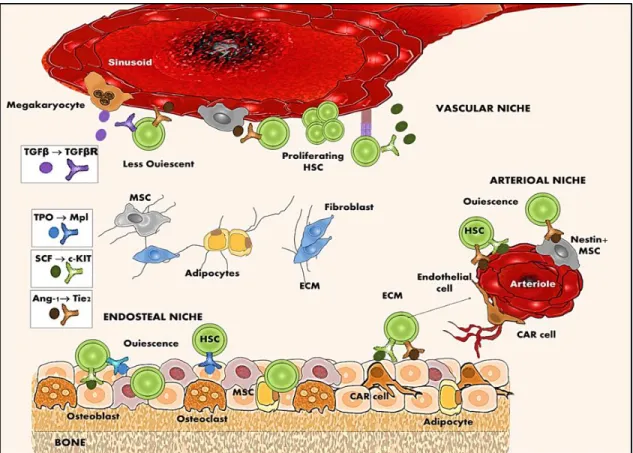
Les populations de cellules souches/progénitrices hématopoïétiques (HSPC) résident dans un microenvironnement spécialisé (niche) entourées de cellules stromales dans la MO (Boulais et al., 2015). Normalement, l’équilibre entre la division et la différenciation des HSC est contrôlé par différentes voies de signalisation intrinsèques à la cellule et des signaux de la niche provenant des cellules voisines dans la MO (Cheung et al., 2013, Ema et al., 2012, Nakamura-Ishizu et al., 2014). Cependant, un certain nombre de conditions de stress telles les radiations et les infections peuvent renverser l’état de dormance des LT-HSC et les faire entrer en cycle cellulaire pour remplacer les cellules perdues (Allakhverdi et al., 2009, MacNamara et al., 2011a, Matatall et al., 2016, Wright et al., 2002). La manière dont les HSPC coordonnent des mécanismes opposés comme l’auto-renouvellement et la différenciation en conditions de stress reste à être clarifiée.
Il est bien établi que la signalisation de Wnt regroupe un large éventail de processus physiologiques et développementaux, comprenant la croissance, la division et la différenciation cellulaire (Cadigan et al., 1997, Eisenmann, 2005). Différentes études ont rapporté que le dérèglement de cette cascade de signalisation joue un rôle central dans le développement des maladies hématologiques malignes et des leucémies (Johnson et al., 2006, Lento et al., 2013). Les protéines Wnt sont sécrétées par divers types de cellules stromales et agissent sur de courtes distances comme molécules de signalisation. Elles agissent également sur de longues distances dans la MO en suivant un gradient. Les protéines Wnt transmettent le signal via des récepteurs transmembranaires Frizzled (Fzd) pour activer au moins deux types de voies de signalisation intracellulaire. La voie canonique de Wnt contrôle la transcription des gènes cibles et est dépendante de la β-caténine pour induire une réponse cellulaire. Les voies de signalisation non canoniques sont β-caténine indépendantes et incluent les voies Wnt-Ca2+ et polarité planaire cellulaire (PCP). L’activation de la voie Wnt/Ca2+ conduit à la mobilisation de calcium (Ca) intracellulaire et à l’activation de la protéine kinase II dépendante à la calmoduline. La voie de la polarité cellulaire planaire (PCP) passe par la Jun N-terminal kinase (JNK) pour organiser le cytosquelette de manière asymétrique et polariser les cellules dans le tissu (Kikuchi et al., 2007, Komiya et al., 2008).
La majorité des études suggère que les voies de signalisation non canonique de Wnt jouent un rôle essentiel pour le maintien à long terme d’un pool de cellules HSC dans la M(Nemeth et al., 2007, Povinelli et al., 2014, Sugimura et al., 2012).
Récemment, notre laboratoire a démontré que la signalisation de Wnt4 augmente l’expansion des HSPC via une voie de type PCP (Heinonen et al., 2011b, Louis et al., 2008). Le récepteur de la voie PCP Fzd6 est au moins partiellement requis pour l’expansion de HSPC médiée par Wnt4. Cependant, le rôle fonctionnel de la signalisation Fzd6 dans les cellules engagées dans la lignée hématopoïétique est très peu connu. Dans le système hématopoïétique, il a été démontré que les HSPC et les cellules formant le sang mature chez l’humain et la souris expriment Fzd6 avec un niveau d’expression plus élevé chez les cellules les plus immatures (Wagner et al., 2004, Yokota et al., 2008). Par conséquent, nous avons proposé que la signalisation de Fzd6 puisse réguler l’hématopoïèse chez l’adulte à l’état d’équilibre et sous des conditions de stress.
Pour étudier le rôle de Fzd6 dans l’hématopoïèse, nous avons croisé les souris Fzd6+/- avec des souris C57Bl/6 pendant 10 générations, ce qui a permis de minimiser la variabilité génétique dans nos expériences. Nous avons effectué une analyse détaillée de l’hématopoïèse in vivo chez les souris Fzd6-/- pour caractériser le phénotype
hématopoïétique des souris Fzd6-/- pendant l’hématopoïèse chez l’adulte.
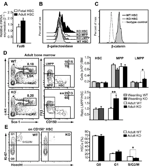
À l’état de base, la proportion de LSK CD150+ qui contiennent des LT- et
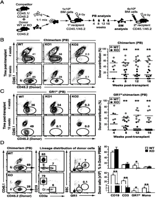
ST-HSC est normal dans la MO Fzd6-/-. Cependant, nous avons observé une augmentation de MPP amorcé vers la lignée lymphoïde chez les souris Fzd6-/-. À l’inverse, nous n’avons détecté aucune différence dans le développement lymphoïde par rapport aux souris de type sauvage (wild-type; WT). Pour examiner la repopulation et le potentiel de différenciation des HSC Fzd6-/- in vivo, nous avons effectué des tests de repeuplement compétitifs et suivi la reconstitution dans le sang périphérique et la MO en comparant la greffe de cellules de MO Fzd6-/- avec des cellules témoins WT. Nous avons montré que le manque de Fzd6 entraîne une perte progressive des HSPC du donneur en raison d’une forte activation de la caspase-3 dès 7 à 8 jours après la transplantation. Cela indique que la greffe, le repeuplement et l’auto-renouvellement des HSPC Fzd6
sont inefficaces.
Afin de déterminer si les HSC Fzd6-/- répondent à d’autres types de stress dans leur microenvironnement, nous avons ensuite soumis les souris Fzd6-/- à une dose sous-létale de lipopolysaccharide (LPS), une endotoxine bactérienne. Des études antérieures ont montré que l’hématopoïèse d’urgence induite par LPS « réveille » les HSC en dormance et les fait entrer en cycle cellulaire avec un changement dans la différenciation favorisant la production de cellules myéloïdes (Boettcher et al., 2012,
Scumpia et al., 2010). Nous avons montré qu’un traitement systémique de LPS entraîne une diminution rapide de la cellularité de la MO qui s’accompagne d’une augmentation de 10 à 15 fois du nombre de HSC. Malgré la grande proportion de HSC (~65-70%) qui résident dans la phase G0 du cycle cellulaire pendant l’homéostasie, plus de la moitié de ces HSC en dormance ont été retrouvés dans les phases G1 ou S-G2-M du cycle cellulaire suite à l’injection de LPS. Nous n’avons observé aucune différence dans le comportement cyclique entre les HSC Fzd6-/- et WT lorsqu’analysé avec la coloration Ki67/Hoechst. Cependant, les souris Fzd6-/- ne parviennent pas à obtenir l’expansion prévue de la population HSPC. Ces résultats suggèrent que Fzd6 est nécessaire pour une expansion efficace des HSPC de la MO et leur auto-renouvellement, non seulement après la transplantation, mais aussi suite à un stress prolifératif comme une inflammatoire aiguë.
L’inflammation aiguë se caractérise par une génération rapide et une mobilisation de cellules effectrices myéloïdes de la MO conduisant à une leucocytose inflammatoire. La capacité des HSPC à répondre aux infections en augmentant le nombre de progéniteurs et en produisant des cellules myéloïdes capables de détruire les agents pathogènes microbiens tout en préservant un pool de cellules souches intactes est une caractéristique critique de la défense de l’hôte (Scumpia et al., 2010, Ueda et al., 2005). Lors d’une administration sous-létale de LPS, la moelle des souris injectées démontre une réduction du nombre absolu de cellules lymphoïdes et de granulocytes. Nous avons aussi observé une augmentation relative de l’accumulation de HSC et de cellules myéloïdes effectrices (granulocytes et monocytes) dans la rate. Nous avons observé une augmentation significative des GMP Sca-1+, également nommés GMP d’urgence (emergency GMP; eGMP) car ils n’apparaissent que pendant la myélopoïèse d’urgence. Même si des nombres similaires de HSC ont été observés dans les rates des souris Fzd6-/- et WT, la rate Fzd6-/- contient beaucoup moins d’eGMP. Cette diminution est accompagnée d’une production réduite de monocytes inflammatoires Ly6Chi dans la MO et une diminution de l’accumulation de cellules myéloïdes effectrices dans la rate. Des essais réciproques de greffe de MO ont révélé que des facteurs dépendants du stroma et intrinsèques aux cellules jouent un rôle dans la différenciation myéloïde inefficace des HSPC Fzd6-/-. Collectivement, nos données suggèrent que Fzd6 joue un rôle essentiel dans la détermination du sort des HSPC vers les lignées myéloïdes lors de l’inflammation aiguë induite par des endotoxines. Les mécanismes sous-jacents la défectuosité de la réponse myéloïde des HSPC doivent
encore être identifiés. Pour répondre à cette question, nous avons poursuivi en étudiant l’importance de Fzd6 dans l’activation des HSPC dans une infection chronique en utilisant un modèle de souris de leishmaniose viscérale.
La leishmaniose viscérale est une maladie transmissible par un vecteur et causée par le protozoaire intracellulaire Leishmania donovani. Des études antérieures ont montré que le parasite L. donovani n’infecte pas directement les HSC ou leurs progéniteurs en aval. Il établit plutôt une infection persistante dans les macrophages de la MO, ce qui corrèle avec une amélioration de la production des cellules myéloïdes par la MO et la rate (Cotterell et al., 2000a, Cotterell et al., 2000b). Cependant, la contribution de l’amélioration de la myélopoïèse au cours de l’infection n’est pas bien comprise. Étant donné que la déficience en Fzd6 réduit l’activation des HSC en réponse à une inflammation induite par l’endotoxine, nous avons analysé le compartiment hématopoïétique de souris Fzd6-/- et WT au cours de l’infection. De 14 à 28 jours après l’infection (post-infection; pi), l’expansion du parasite dans la MO et la rate a entraîné une augmentation graduelle de la fréquence et du nombre de LT- et ST-HSC dans la MO, suivi de l’accumulation de HSPC dans la rate. 21 jours après l’infection (21 pi), la majorité des HSC se sont divisés (>75-80%). La progression des HSC dans le cycle cellulaire et leur expansion sont accompagnées d’une augmentation de l’activation de la β-caténine au cours de l’infection, ce qui suggère que la signalisation Wnt/Frizzled (Fzd) joue un rôle dans l’activation des HSPC dans la leishmaniose.
Parallèlement à la charge parasitaire, la proportion et le nombre de eGMP Sca-1+ ont également augmenté, atteignant un plateau entre les jours 21 et 28, selon la force de l’infection. La progéniture myéloïde de ces eGMP consiste principalement en des monocytes Ly6Chi avec un phénotype régulateur de type suppresseur de cellules. Étonnamment, nous avons observé que les parasites L. donovani n’infectent pas seulement les macrophages de la MO, mais également les précurseurs de monocytes et les monocytes matures, confirmés par une coloration Giemsa de la MO provenant des souris infectées au jour 28 pi. Pour savoir si les cytokines de l’environnement dans la MO influencent la différenciation des HSPC et augmentent la charge parasitaire dans la MO, nous avons analysé le surnageant de la totalité des cellules de la MO en comparant des souris non infectées à des souris infectées par L. donovani à différents jours après l’infection. Nous avons détecté une augmentation significative dans la MO du niveau d’expression de différentes cytokines et chimiokines qui pourraient contribuer à l’expansion et à la différenciation myéloïde des HSPC dans la phase chronique de
l’infection. Pour tester si les monocytes générés dans la MO infectée sont fonctionnellement différents des monocytes normaux différenciés in vitro, nous avons exposés les cultures aux amastigotes fluorescents de L. donovani. Les surnageants provenant de la MO infectée ont augmenté non seulement la proportion de monocytes infectés par des parasites, mais aussi la réplication du parasite dans ces cellules avec le temps. Ces résultats suggèrent que l’expansion du parasite L. donovani dans la MO active efficacement les HSC et les pousse à produire un grand nombre de cellules myéloïdes, qui sont parmi les cibles préférées du parasite, et deviennent encore plus sensibles à l’infection. À l’inverse, les HSPC Fzd6-/- ont généré moins de progéniteurs
myéloïdes et, par conséquent, moins de monocytes Ly6Chi et une réduction de l’accumulation de cellules myéloïdes dans la rate Fzd6-/- par rapport aux témoins WT.
Cette diminution dans la différenciation myéloïde corrèle avec la diminution de l’expansion du parasite, la charge parasitaire ayant été mesurée dans la MO et la rate. Cela établissant un lien entre la signalisation Fzd6 et la réponse aux infections des HSPC. Par conséquent, nos résultats démontrent un rôle important pour l’activation des HSPC dépendante de Wnt/Fzd dans la régulation de l’infection chronique par les parasites.
Compte tenu du potentiel thérapeutique des HSC, il est nécessaire de mieux comprendre les mécanismes impliqués dans le contrôle des HSPC. Nous prévoyons que la signalisation non canonique de Fzd6 est une cible thérapeutique prometteuse pour de nouveaux traitements pour moduler l’activation des HSC et le développement des cellules myéloïdes et surmonter les limites de la transplantation de moelle osseuse et le traitement des maladies inflammatoires aiguës et chroniques.
xi
First, I would like to express my heartfelt thanks to my enthusiastic supervisor, Krista Heinonen, for believing in my potential, for her inspirational guidance and enthusiastic encouragement. Her passion in science, her perfectionism, sharp critics and brilliant suggestions combined with her creativity have an influential impact on my academic life and beyond, to become an independent and productive scientist.
I would like to thank Simona Stäger for being a dedicated mentor and providing a positive and inspiring working atmosphere. Her positive outlook, her confidence in science and her heart-warming support have given me great confidence as a researcher. I would like to express my deep appreciation to my generous research advisory committee members, Pascale Duplay and Christian Beausejour for their dedication, sharing their knowledge and invaluable advice along this project. I also wish to thank my internal examiner, Claude Daniel and my external examiner, Tatiana Scorza for their time and intellectual contributions to my work. My sincerest thanks are extended to Albert Descoteaux, Alain Lamarre and Maritza Jaramillo for their helpful advice in science. Special mention goes to Jessy Tremblay for long-lasting scientific discussions, his friendly attitude and for the smell of happiness coming from his coffee.
Special thanks go to Akil, Mondher and Mouna for being great friends and bringing some gangsta into my PhD life. I am indebted to Roxann for not only her loyal friendship but also for her precious time in French translation. I extend my thanks to Daniel for his positive attitude and his tasty yoghurt; Mirtha for always making me smile; Slimane and Aymeric for always being calm, kind and generous, I had never seen them upset; Soumia for her warm company in the cold FACS room; Ahmed for always being there to listen, his generosity and encouragement; Constance and Mitra for their heart-warming love, generosity, and strong friendship; Tania, Christine and Guillermo for gladly helping out in time of need; Sasha, Melina and Linh for adding joyful moments to my days; Renaud for giving me the worst cold virus all the time and for his mad-scientist attitude. I am also so much indebted to Quattrociocche family for never letting me doubt myself, for their tremendous amount of encouragement and endless love.
Finally, but by no means least, wholehearted thanks go to my brother for his constant support and my mother, the most important person in my world, for her almost unbelievable encouragement, extreme patience and love during my PhD and my entire life. This journey would not have been possible without their moral support, encouragement and inspiration.
TABLE OF CONTENTS
RÉSUMÉ ... i
ABSTRACT ... iii
SYNOPSIS ... v
ACKNOWLEDGEMENT ... xi
TABLE OF CONTENTS ... xii
LIST OF FIGURES ... xv
LIST OF TABLES ... xvii
LIST OF ABREVIATIONS ... xviii
CHAPTER 1 INTRODUCTION ... 1
1. Matter of life: Blood ... 2
2. Ontogeny of hematopoietic stem cells ... 4
3. Transition towards an adult phenotype ... 6
4. Steady-state hematopoiesis ... 9
4.1. Maintenance of hematopoietic stem cell quiescence ...14
4.1.1. Intrinsic regulation of the cell-cycle ...14
4.1.2. Regulation of quiescence by hematopoietic stem cell niches ...16
4.2. Hematopoietic progenitor cells ...22
4.3. The role of classical cytokines in the hematopoietic diversity ...26
5. Stress response of hematopoietic stem cells ...30
5.1. Homing and cell kinetics of transplanted hematopoietic stem cells ...31
5.2. Inflammation- and pathogen-induced emergency hematopoiesis ...35
5.2.1. Direct recognition of pathogens by hematopoietic stem cells ...41
5.2.2. Effect of pro-inflammatory signals on emergency hematopoiesis ...44
6.1. Wnt proteins acts as morphogens ...50
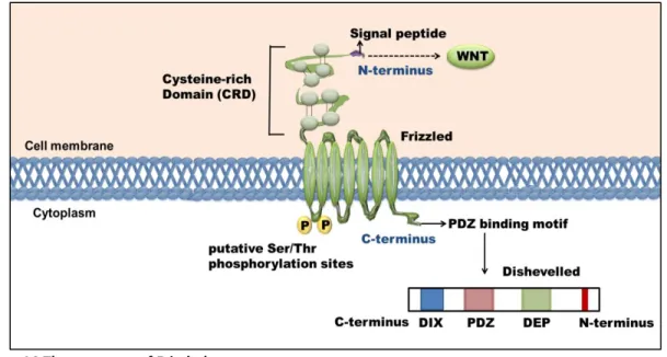
6.2. Wnt recognition by Frizzled receptors ...51
6.3. Wnt signaling pathways ...53
6.4. Multi-faced role of Wnt ligands in hematopoiesis ...57
6.5. Frizzled receptors in hematopoiesis ...64
6.6. The role of Wnt signaling in inflammation ...68
7. OBJECTIVES ...70
CHAPTER 2 PUBLICATION NO.1 ...72
FRIZZLED-6 REGULATES HEMATOPOIETIC STEM/PROGENITOR CELL SURVIVAL AND SELF-RENEWAL ...73
1. SUMMARY ...75
2. INTRODUCTION ...76
3. RESULTS ...78
3.1. Fzd6 deficiency does not affect intracellular β-catenin levels in HSPCs ...78
3.2. Fzd6 has an age-dependent effect on hematopoietic progenitor cell maintenance ...80
3.3. Fzd6 negatively regulates Cdc42/JNK signaling in HSPCs ...82
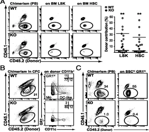
3.4. Fzd6 is essential for competitive repopulating capacity of HSCs and long-term granulocytic reconstitution...84
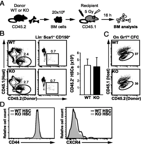
3.5. Long-term reconstitution defects of Fzd6 deficient HSPCs are not due to altered homing but rather to defective survival and expansion in the recipient bone marrow...87
3.6. Fzd6 deficient HSPCs expand poorly in response to emergency signals ...90
4. DISCUSSION ...92
5. MATERIAL AND METHODS ...95
CHAPTER 3 PUBLICATION NO.2 ...99
INFECTION-ADAPTED EMERGENCY HEMATOPOIESIS PROMOTES VISCERAL LEISHMANISIS ... 100
2. AUTHOR SUMMARY ... 103
3. INTRODUCTION ... 104
4. RESULTS ... 106
4.1. L. donovani induces the expansion of HSC-like cells in the bone marrow and spleen ... 106
4.2. Induction of myelopoiesis during L. donovani infection results in the generation of altered progeny with a regulatory phenotype ... 109
4.3. Fzd6 promotes bone marrow response to L. donovani ... 115
4.4. Enhanced myelopoiesis correlates with increased parasite burden ... 121
4.5. Decreased parasite expansion in Fzd6-/- mice is not due to enhanced T lymphocyte activity ... 129
4.6. Bone marrow cytokine environment promotes the generation of permissive monocytes ... 132
5. DISCUSSION ... 137
6. EXPERIMENTAL PROCEDURES ... 140
CHAPTER 4 GENERAL DISCUSSION ... 145
1. Fzd6 is indispensable for the self-renewal and repopulation of hematopoietic stem cells ... 146
2. Fzd6 regulates endotoxin-induced expansion and myeloid differentiation of hematopoietic stem/progenitor cells... 151
3. Fzd6 signaling accelerates the progression of Leishmania donovani parasite infection in mice ... 156
4. Conclusion: a new role for Fzd6 signaling in homeostatic and stress-induced hematopoiesis ... 159
CHAPTER 5 REFERENCES ... 161
ANNEX-I The role of Fzd6 in endotoxin-Induced inflammation ... 195
LIST OF FIGURES
CHAPTER 1: INTRODUCTION
Figure 1.Establishment of primitive and definitive hematopoiesis in mice ... 5
Figure 2.HSC transition from fetal and adult properties ... 8
Figure 3. Self-renewal and differentiation of hematopoietic stem cells ...10
Figure 4.Cycling activity of hematopoietic stem and progenitor cells ...13
Figure 5.Cell-intrinsic regulation of hematopoietic stem cell quiescence ...15
Figure 6. Maintenance of quiescent hematopoietic stem cells in the bone marrow ...18
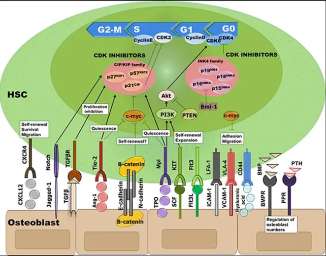
Figure 7. Crosstalk signaling between hematopoietic stem cells and osteoblasts in the niche ...20
Figure 8. The current scheme of hematopoietic hierarchy ...24
Figure 9. Cytokine regulation of hematopoietic differentiation in semisolid medium ...27
Figure 10. Hematopoietic stem cell homing following bone marrow transplantation ...33
Figure 11. Initial stages of inflammation-induced emergency hematopoiesis ...36
Figure 12. Direct and indirect recognition of danger signals by hematopoietic stem cells ...43
Figure 13. G-CSF-mediated mobilization of hematopoietic stem/progenitor cells ...46
Figure 14. The structure of Frizzled receptors ...51
Figure 15. Canonical Wnt/β catenin signaling pathway ...54
Figure 16. Non-canonical Wnt signaling pathways ...56
Figure 17. Frizzled receptor expression on hematopoietic/progenitor cells ...64
CHAPTER 2: PUBLICATION NO.1
Figure 1. Non-canonical Fzd6 is expressed on CD150+ HSCs and influences the ratio of Flt3+ vs CD150+ progenitors ...79Figure 3. Fzd6-/- HSPCs display defective long-term engraftment and self-renewal in vivo. ...85 Figure 4. Fzd6-/- HSPCs display defective long-term engraftment and self-renewal in vivo ...86 Figure 5. Defective long-term reconstitution of Fzd6-/- HSPCs is not due to altered homing ...88 Figure 6. Fzd6-/- HSPCs cannot expand and die by apoptosis in the first week after transplant. ...89 Figure 7. Fzd6-/- HSPCs exhibit poor emergency hematopoiesis. ...91
CHAPTER 3: PUBLICATION NO.2
Figure 1. Parasite expansion coincides with proliferation and accumulation of bone marrow hematopoietic stem/progenitor cells ... 107 Figure 2. Bone marrow HSCs switch their differentiation towards non-classical
myeloid progenitors ... 110 Figure 3. Leishmania parasite expansion promotes myeloid output in the bone
marrow ... 113 Figure 4. Frizzled-6 is required for parasite-induced expansion and myeloid
differentiation of HSPCs ... 117 Figure 5. Diminished myeloid output in Fzd6-/- mice correlates with a reduced
parasite burden during the chronic phase of infection. ... 122 Figure 6. Decreased accumulation of myeloid cells is accompanied with reduced
parasite burden in Fzd6-/- spleen ... 125 Figure 7. Fzd6-/- T lymphocytes are functionally indistinguishable from their Fzd6+/+
counterparts ... 130 Figure 8. Fzd6-/- bone marrow microenvironment is enriched in pro-inflammatory
cytokines and chemokines ... 133 Figure 9. Infected bone marrow microenvironment directly promotes HSPC
xvii
Table 1.Antigenic proteins and glycoproteins used to purify hematopoietic stem cells ...11
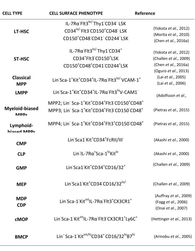
Table 2. Cell surface phenotype of identified hematopoietic stem /progenitor cells ...25
Table 3. Cytokines and hormones regulating hematopoietic cells ...29
Table 4. Pathogen specific response of hematopoietic stem/progenitor cells...40
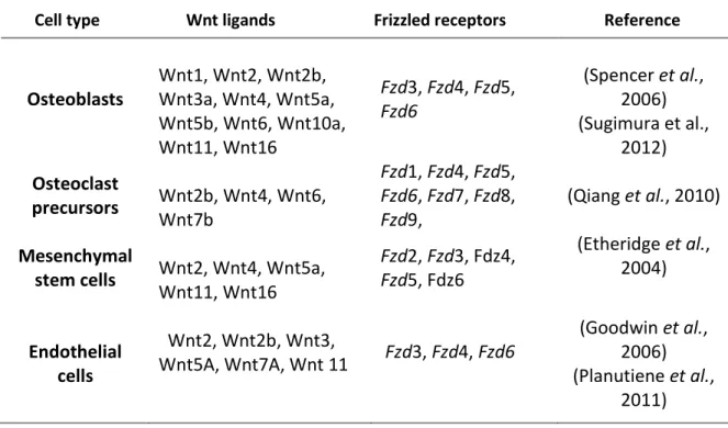
Table 5. Expression of Wnt ligands and Frizzled receptors in the bone marrow ...57
Table 6. Wnt ligands and their function in hematopoietic stem cell regulation ...63
Table 7. Known function of Frizzled receptors in hematopoiesis ...67
LIST OF ABREVIATIONS
5-FU: 5- FluorouracilABC: ATP-binding cassette ACD: Asymmetric cell division
AGM: Aorta-gonad-mesonephros region Ang-1: Angiopoietin-1
APC: Adenomatous polyposis coli Axin: The axis inhibition protein-1 BaP: Basophil progenitor
BM: Bone marrow
BMDM: Bone marrow-derived macrophages BMCP: Basophil-mast cell progenitor
BMP:Bone morphogenic protein Ca2+: Calcium
CAR: CXCL12-abundant perivascular cell CDK: Cyclin dependent kinase
CDP: Common-dendritic cell progenitor CFU: The colony-forming unit
CFU-E: Colony-forming-unit-erythroid CFU-G: Colony-forming unit-granulocytes
CFU-GEMM: Colony-forming unit-granulocyte-erythrocyte-monocyte- megakaryocyte CFU-GM: Colony-forming unit-granulocyte-monocyte
CFU-M: Colony-forming unit-monocyte CK1α: Casein kinase 1α
CLP: Common lymphoid progenitor cMOP: Common monocyte precursors
CMP: Common myeloid progenitor CRD: Cysteine-rich domain
DAMP: Danger-associated molecular pattern Dkk1: The Dickkopf protein -1
Dsh: Dishevelled (in Drosophila) Dvl: Dishevelled (in vertebrates) ECM: Extracellular matrix EGF: Epidermal growth factor ELP: Early-lymphoid progenitor EOP: Eosinophil progenitor EP:Erythroid progenitor EPO: erythropoietin
ETP: Early thymic progenitor
FACS: Fluorescence activated cell sorting FGF: Fibroblast-growth factor
FL: Fetal liver
Fz: Frizzled in Drosophila
Fzd: Frizzled ortholog in mammals
G-CSF: Granulocyte colony stimulating factor
GM-CSF: Granulocyte-monocyte colony stimulating factor
GM-CSFR: Granulocyte-macrophage colony-stimulating factor receptor GMP: Granulocyte-monocyte progenitor
GPCR: G-protein-coupled receptor GSK-3β: Glycogen synthase kinase-3β HGF: Hepatocyte growth factor
HSPC: Hematopoietic stem/progenitor cell ICAM-1: Intercellular adhesion molecule-1 IFN: Interferon
IFNAR: Interferon-α/β receptor IGF-1: insulin-like growth factor-1 IL: Interleukin
IRF: Interferon regulatory transcription factor Jnk: c-Jun N-terminal kinase
LCMV: Lymphocytic choriomeningitis virus LEF: Lymphoid enhancer binding factor LFA-1: Lymphocyte-function antigen-1 LIF: Leukemia Inhibitory Factor
Lin: Lineage
LMPP: Lymphoid-primed multipotent progenitor LRP: Low density lipoprotein receptor-related protein LPS: Lipopolysaccharide
LT-HSC: Long-term hematopoietic stem cell
Madcam1: Mucosal vascular addressin cell adhesion molecule-1 MCP: Mast cell progenitor
M-CSF: Macrophage colony-stimulating factor
M-CSFR: Macrophage colony-stimulating factor receptor MDP: Macrophage-dendritic cell progenitor
MEP: Megakaryocyte-erythrocyte progenitor Mk: Megakaryocyte
MkP: Megakaryocyte progenitor
MMP: Matrix metallopeptidase MPP: Multipotent progenitor MSC: Mesenchymal stem cell
MyRP: Myeloid progenitor with repopulating ability NK: Natural killer
NP: Neutrophil-progenitor OB: Osteoblast
PAMP: Pathogen-associated molecular pattern PCP: Planar cell polarity
PDE: Phosphodiesterase
PDGF-BB: Megakaryocyte-secreted platelet-derived growth factor PKC: Protein kinase C
PLC: Phospholipase C
PRR: Pattern recognition receptor
PSGL-1: P-selectin glycoprotein-ligand-1 PTH: Parathyroid hormone
Rb: Retinoblastoma
ROS: Reactive oxygen species SCD: Symmetric cell division SCF: Stem cell factor
SDD: Symmetric differentiation division SFRP: Secreted Frizzled-related protein
SLAM: Signaling lymphocyte activation molecule ST-HSCs: Short-term hematopoietic stem cell TCF: T-cell factor
Th: T-helper
TLR: Toll-like receptor TNF: Tumor necrosis factor
TNFR: Tumor necrosis factor receptor TPO: Thrombopoietin
VCAM-1: Vascular-adhesion protein-1
VEGF: Vascular endothelial growth factor
VLA-4: Very-late antigen-4/ Integrin α4β1 Wg: Wingless
WIF: Wnt Inhibitory Factor
1
2
1. Matter of life: Blood
Over centuries, blood has been considered as a magical symbol, which is often linked to courage, power and immortality in many faiths and religions. Due to its mysterious healing properties, blood was described as ‘the centre of life itself’ (Learoyd, 2006, Meletis et al., 2010). Hematopoiesis—the Greek haima, meaning “blood” and poiesis, meaning “formation”— refers to the process of mature and functional blood cell generation (Damjanov, 2013).
Our understanding of hematopoiesis is the combination of ongoing research and extensive effort that have been carried out for several decades. The concept of hematopoiesis was introduced in 19th century by the establishment of bone marrow (BM) as the source of blood cells (Cooper, 2011). The existence of a “mother cell” for all blood elements was first proposed in Maximov’s theory in 1909, while its potential participation in radiation damage was postulated by Sabin in 1932 (Friedenstein, 1989, Sabin et al., 1932). The threat of continued atomic warfare in the period of mid-1940s and 1960s, marked a new era in the hematopoietic research, which is mainly based on studying the effects of irradiation and BM transplantation. Jacobson and his colleagues found that mice could survive an otherwise lethal irradiation exposure if a hematopoietic organ is protected by lead foil (Jacobson et al., 1949). Soon afterwards, it became apparent that adult BM cells transplanted into syngeneic recipients were capable of rescuing lethally irradiated mice from hematopoietic failure (Lorenz et al., 1952). But, it was not until early 1960s that first in vivo evidence provided by Till and McCulloch that radioprotection was due to the transplanted multipotent stem cells (Till et al., 1961). The ability of BM cells to reconstitute the entire hematopoietic system in recipients provided an in vivo assay for the identification of hematopoietic stem cells (HSCs), thus setting the basis towards further evaluation (Becker et al., 1963, Siminovitch, 1964).
While it was initially thought that HSCs are a homogenous population, the development of in vitro clonal assays progressively revealed the heterogeneity of hematopoietic stem/progenitor cells (HSPCs) in various aspects (Bradley et al., 1966, Pluznik et al., 1965). Consequently, the description of methods to characterize HSCs and their progeny did not occur another decade or more (Metcalf, 1980, Spangrude, 1991, Spangrude et al., 1988). Today, we can enrich adult HSCs using the combination of monoclonal antibodies and high-speed fluorescence activated cell sorting (FACS)
technique based on the expression of different cell surface markers (Adolfsson et al., 2001, Ikuta et al., 1992, Oguro et al., 2013). However, there is not any unique marker to identify true HSCs, thus more fastidious approaches are required for the functional characterization. Since the discovery of transplantable multipotent HSCs in adult BM to the present day, BM transplantation assays are considered “the gold standard” to demonstrate HSC function. The term ‘HSC’ refers to a single cell which is sufficient to rescue a host from otherwise lethal irradiation exposure and completely establish its entire hematopoietic system (Osawa et al., 1996). This reconstitution ability requires: (i) self-renewal; to make an identical copy of itself while preserving the undifferentiated state, (ii) extensive proliferation potential; to repopulate, and (iii) potency; to differentiate and give rise to all blood elements.
2. Ontogeny of hematopoietic stem cells
During embryogenesis and fetal development, hematopoiesis occurs in different hematopoietic sites (niches) including the extraembryonic yolk sac (YS), the intraembryonic aorta-gonad-mesonephros region (AGM), fetal liver (FL), spleen and BM (Figure 1). Primitive hematopoiesis is initiated in the YS of the developing mouse embryo at around embryonic day 7.5 (E7.5). During the primitive streak stage, development of a putative YS, also known as the hemangioblast, marks the onset of hematopoiesis (Baron, 2003, Kennedy et al., 2007). First hematopoietic cells produced in the YS are primitive red blood cells, termed erythroblasts, which contain embryonic hemoglobin. These large nucleated erythroblasts are surrounded by a layer of endothelial cells, which form blood islands. Macrophages and megakaryocytes are also generated at this stage (Kennedy et al., 2007, Sasine et al., 2016). At a slightly later stage, lymphoid precursors are also found in YS (Godin et al., 1995). However, the first adult-type HSC, which can reconstitute the hematopoietic system are generated in AGM at ~E10.5 (Sasine et al., 2016). Emergence of HSC from AGM marks the establishment of definitive hematopoiesis. Definitive HSCs circulate via umbilical vessels and migrate to YS and placenta. These sites provide potent hematopoietic niches for the maturation of immature “pre-HSCs” into functional HSCs (Gekas et al., 2010, Swiers et al., 2010). At E11.5, HSCs migrate to the FL in which they will be directed towards differentiation and give rise to definitive hematopoietic progenitors (Gekas et al., 2010, Sasine et al., 2016). Thereafter, hematopoietic activity stops in AGM, but still detected in YS and placenta (de Bruijn et al., 2000, Sasine et al., 2016). HSCs daily double their numbers in FL to seed the newly forming hematopoietic niches in the spleen and BM. Before the birth, the primitive HSCs migrate and colonize spaces filled out with embryonic bone and cartilage, establishing a close connection with BM microenvironment in early life (Taichman et al., 1998). During transition from FL to BM, the frequency of HSCs dramatically declines. By the time of birth, the BM remains the primary hematopoietic site in which all HSC activity is retained during the adult life (Calvi et al., 2003, Kunisaki et al., 2013).
Figure 1.Establishment of primitive and definitive hematopoiesis in mice
Primitive hematopoiesis occurs between embryonic day E7.5 and E10. Main purpose of the primitive hematopoiesis is to provide the oxygenation of the developing embryo. Both endothelial cells and hematopoietic cells are generated from a common progenitor, the hemangioblast. The extraembryonic yolk sac serves as the source of primitive erythrocytes. The extraembryonic yolk sac blood islands, a structure formed by endothelial cells surrounds primitive erythrocytes. Primitive macrophages and megakaryocytes are also found in the yolk sac during this stage. Emergence of HSCs from the intraembryonic AGM around day E10.5, marks the beginning of definitive hematopoiesis which comprises: (i) expansion of ‘pre-HSC’ and maturation to functional HSC in fetal liver, yolk sac and placenta and (ii) differentiation of HSC into multipotent hematopoietic progenitors and generation of definitive hematopoietic cells in the fetal liver which is the major hematopoietic site during mid-gestation. Before the time of birth, fetal HSCs migrate to hematopoietic niches in BM in which all
hematopoietic activity is retained in late-gestation and during adult life. AGM:
aorta-gonad-mesonephros region, HSC: hematopoietic stem cell. Adapted from “Baron et al., 2012; Boisset et al., 2012; Ginhoux et al., 2013; Medvinsky et al., 2011”.
3. Transition towards an adult phenotype
During the development and throughout aging, HSCs undergo phenotypic and functional changes. Mouse fetal HSCs actively cycle in order to expand the stem cell pool and seed newly forming hematopoietic organs in the developing embryo. The post-natal switch in HSC turnover occurs around 3 to 4 weeks of age at which HSCs become predominantly quiescent with only 5 to 10% remain cycling (Makio Ogawa, 1993, Pietras et al., 2011). While both adult BM and FL stem cells show an extensive capacity for self-renewal, FL has a greater hematopoietic reconstitution potential in transplant settings when compared to adult (Christensen et al., 2004, Morrison et al., 1995). Throughout the ontogeny, HSCs acquire different differentiation potentials. FL HSCs have a strong bias towards myeloid differentiation when compared to adult HSCs, which give a balanced lymphoid-myeloid lineage output (Bowie et al., 2007). HSPC populations generate distinct types of hematopoietic cells at different stages of the development. Lymphoid and myeloid cell subtypes differ in phenotype and function (Ghosn et al., 2012, Mold et al., 2010), suggesting that HSCs generate distinct hematopoietic cell types at different stages of the development.
These developmental changes are linked to the differences in the gene expression profiles, transcriptional programs and cell cycle regulation of fetal and adult HSCs. Some of the cell-autonomous mechanisms, which selectively regulate fetal and adult HSCs, are depicted in Figure 2. The generation of HSCs from hemogenic endothelium requires Notch signaling (Kumano et al., 2003). Notch signaling occurs through the interaction of transmembrane receptors (Notch1-4 in mice) and Notch ligands (Jagged and Delta) on the signal receiving cells, which leads to the transcriptional regulation of Notch target genes (Kim et al., 2014). Notch1 and Jagged-1 display specific expression patterns within the arterial endothelium in the developing mouse embryo (Robert-Moreno et al., 2005). While Notch1 signaling plays an essential role in the generation of HSCs within the AGM region (Kumano et al., 2003), it is dispensable for primitive or definitive hematopoiesis in the YS (Hadland et al., 2004, Robert‐Moreno et al., 2008). Jagged-1 coupling with Notch1 initiates direct transcriptional activation of GATA2 in the hemogenic endothelium (Robert-Moreno et al., 2005). A transcriptional network, which include GATA2, Ets and Scl contributes to the early hematopoietic expression of Runx1 (also known as Aml1), which is essential for the generation of definitive HSCs in mice (Nottingham et al., 2007). Thus, mice that are
deficient in either GATA2 or Aml/Runx1 are embryonically lethal due to the lack of definitive hematopoietic cells (Okuda et al., 1996, Tsai et al., 1994). It is demonstrated that Aml/Runx1 and Notch1 are not required for the maintenance of adult HSCs, while GATA2 plays a key role also in adulthood (Ichikawa et al., 2004, Mancini et al., 2005, Rodrigues et al., 2005).
After HSCs are formed, fetal specific genes such as the polycomb-group gene Ezh2, which regulates Lin28/let7 pathway plays an essential role in the acquisition of a fetal gene signature (Oshima et al., 2016). The Lin28/let7 pathway is critically required for the higher self-renewal activity of fetal HSCs and production of fetal-type lymphocytes (Copley et al., 2013). Conversely, the transcription factor, Sox17 acts in the downstream of Aml-1/Runx1 and Gata-2 and maintains fetal HSCs prior to the acquisition of an adult phenotype (Kim et al., 2007). Germline deletion of Sox17 reduces the expression of Dickoff-1 (DKK-1) which negatively regulates canonical Wnt pathway (Kim et al., 2007). Canonical Wnt signaling is transmitted by binding of Wnt secreted glycoproteins to the Frizzled (Fz) family of cell surface receptors, which leads to the formation of a larger surface complex with the co-receptors, the low-density lipoprotein-related receptor (LRP) 5 and 6 (Malhotra et al., 2009). Activation of canonical Wnt pathway regulates the amount of a multifunctional adaptor protein β-catenin and directs transcriptional activation of intracellular signaling cascades (Komiya et al., 2008, Schubert et al., 2013). In the developing embryo, Wnt/β-catenin signaling regulates HSC emergence in a dose-dependent paracrine fashion. At around E10.5, Wnt/β activity is restricted to endothelial non-hematopoietic cells localized at the base of hematopoietic clusters in the AGM. Inactivation of Wnt/β-catenin signaling at this stage not only decreases the numbers of newly generated HSCs but also alters the function of these cells as shown in transplantation settings (Ruiz-Herguido et al., 2012). Interestingly, already formed HSCs contained in the E11.5 AGM appear to be β-catenin independent, suggesting a transient activation of Wnt/β-catenin signaling in HSC emergence (Ruiz-Herguido et al., 2012). Although Wnt/β-catenin activity is dispensable for the stem cell maintenance after E11.5 in fetal life, it is involved in the regulation of adult HSPC pool in a dose-dependent manner (Luis et al., 2011, Ruiz-Herguido et al., 2012). Likewise, transcriptional regulators including the proto-oncogene Bmi-1 and the transcriptional repressor Gfi-1 play an essential role in for the generation and maintenance of adult HSCs, while they are not required for the emergence and maintenance of fetal HSCs (Hock et al., 2004, Park et al., 2003). Interestingly, the changes in the cycling status of
HSCs, which occur by 3 weeks of age in mice also appears to be a cell-intrinsic developmental event. Evidence shows that the transcription factor C/EBPα which negatively regulates fetal HSCs self-renewal is at least partially required for the acquisition and maintenance of adult HSCs and for in vivo switch in HSC cycling activity in mice (Ye et al., 2013). These observations suggest that intrinsically determined molecular signals mediated by different developmental regulators collectively control properties and appropriate numbers of HSCs during fetal and adult life.
Figure 2. HSC transition from fetal and adult properties
A molecular switch during the transition from fetal to adult phenotype, results in decreased HSC expansion, cycling, self-renewal ability and myeloid differentiation potential. Aml/Runx-1, Notch1 and GATA2 are essential for the HSCs in the developing embryo, but dispensable for adult HSC maintenance. When HSCs are formed, transcription factor Sox17 regulates the acquisition of an adult phenotype via promoting Wnt/β-Catenin signaling. Ezh2 and its downstream Lin28b are crucial for the maintenance of fetal HSCs while Bmi-1 is required for HSC maintenance/function only in adult. PTEN and the transcription factors Gfi-1 and c-myb are dispensable for the primitive stages of hematopoiesis but essential for the definitive hematopoiesis while Wnt/β-Catenin activity regulates HSC maintenance/function transiently in a dose-dependent manner throughout developmental stages and adult life. Compiled from “Copley et al., 2013; Dejana et al., 2017; Ichikawa et al., 2004; Jang et al., 2007; Luis et al., 2011; Magee et al., 2012; Mancini et al., 2005; Oshima et al., 2016; Rodrigues et al., 2005; Ruiz-Herguido et al., 2012”.
4. Steady-state hematopoiesis
Adult hematopoietic system consists of stem and progenitor cells, which continuously feed blood cell production through the commitment and differentiation processes. HSC self-renewal occurs through symmetric and asymmetric cell divisions. Symmetric divisions can yield either two identical stem cell daughters for stem cell expansion (SCD), or two committed daughter cells, termed symmetric differentiation division (SDD). Asymmetric divisions (ACD) generate one daughter that remains in a quiescent state as a reservoir for the stem cell pool and a committed daughter, which proliferates and differentiates to sustain hematopoiesis (Ito et al., 2012, Ting et al., 2012, Yamamoto et al., 2013). The progenitor cells are a subset between HSCs and mature blood cells. The restricted-differentiation potential and lack of self-renewal capacity distinguish committed-progenitors from HSCs (Seita et al., 2010).
Evidence from a growing number of single-cell transplantation and lineage tracing studies indicates to a significant heterogeneity in HSC subtypes with differential repopulation kinetics (Dykstra et al., 2007, Sieburg et al., 2006), self-renewal ability (Yamamoto et al., 2013), cell cycle status (Mallaney et al., 2014, Wilson et al., 2008) and multi-lineage differentiation output (Dykstra et al., 2007, Ema et al., 2014). Accordingly, short-lived HSCs (ST-HSCs) provide radioprotection and the initial regeneration of hematopoiesis for up to 3-4 months post-transplant while their ancestors, long-term repopulating HSCs (LT-HSC), retain their repopulation and multi-lineage reconstitution activity for the life time of a recipient mouse (Harrison et al., 1993, Yamamoto et al., 2013).
During the commitment, self-renewal capacity and multi-lineage differentiation potential gradually lost (Figure 3), whereby a developmental switch occurs in genetic, metabolic and transcriptional programs associated with cell fate. Multipotent progenitors (MPPs) generate all mature blood lineages but possess transient reconstitution ability due to the lack of self-renewal capacity (Adolfsson et al., 2001, Ikuta et al., 1992, Morrison et al., 1997, Osawa et al., 1996, Weissman et al., 2001). Notably, steady-state hematopoiesis is largely sustained by ST-HSCs and progenitor cells rather than LT-HSCs. Thus, the frequency of LT-HSCs is considerably lower than any other progenitor population within BM (Busch et al., 2015).
Figure 3. Self-renewal and differentiation of hematopoietic stem cells
Hematopoietic stem cells (HSCs) are a rare slow-dividing fraction of the bone marrow (~0.01% of all marrow). HSCs are identified with their unique self-renewal ability to reserve their pool and
multi-lineage differentiation capacity to give rise to the committed progenitors, which supply mature blood
lineages. HSC self-renewal occurs through symmetric and asymmetric cell divisions. At the top of hierarchy, LT-HSCs have the highest self-renewal and in vivo repopulation ability and provide a life-time hematopoietic reconstitution. When LT-HSCs divide, the self-renewal ability and function are gradually lost. When transplanted, ST-HSCs provide radioprotection and initial reconstitution of recipient’s hematopoietic system for a limited time. MPPs lack self-renewal ability but have multi-lineage potential. LT-HSC: long-term hematopoietic stem cell, ST-HSC: short-term hematopoietic stem cell, MPP:
multipotent progenitor cell.Figure adapted from “Passegué et al., 2003; Suda et al., 2011; Wilson et al., 2006”.
FACS is commonly used for the identification of HSCs. Isolation of HSCs with this technique includes a negative selection using lineage (Lin) markers and a positive selection (Morrison, 2002). Lin selection cocktail, typically includes; CD3, CD4 and CD8 to exclude T lymphocytes, CD19 and B220 for B lymphocytes, CD11b and Gr-1 to remove myeloid cells and glycophorin-A or Ter119 to eliminate erythroid cells. After the removal of Lin- cells, HSCs are further characterized based on the presence of stem-cell specific antigens on their surface. Some of the cell surface molecules expressed on mouse HSPCs are summarized in Table 1.
Table 1.Antigenic proteins and glycoproteins used to purify mouse hematopoietic stem cells
Antigen Known function Reference
CD34
a type I transmembrane phosphoglycoprotein which regulates cell cycle activity of HSCs and mediates HSCadhesion within hematopoietic niche
(Krause et al.,
1996)
CD117
(c-Kit)
the receptor tyrosine kinase which supports the maintenance and survival of HSCs via binding its ligand stem cell factor
(Broudy, 1997)
Sca-1
stem cell antigen-1; facilitates HSC maintenance andlineage specification
(Bradfute et al.,
2005)
Thy-1
(CD90)
a heavily N-glycosylated glycophosphatidylinositol-linked surface protein which is postulated to participate in lymphocyte-cell interactions and adhesion
(Barda-Saad et
al., 1999)
CD150
a member of the signaling lymphocytic activation molecule (SLAM) family receptors
expressed on megakaryocytes and HSPCs
(Oguro et al.,
2013)
CD48
a member of SLAM family receptors required for the initiation of T-cell activation and proliferation; absent on HSCs; upregulated on MPPs during inflammation
(González-Cabrero et al.,
1999)
(Boles et al.,
2011)
CD244
a ligand for CD48regulates the activation and cytotoxicity of T and NK cell functions during infections; not expressed on HSCs
(Waggoner et
al., 2012)
IL-7Rα
(CD127)
a receptor for interleukin 7 (IL-7) which plays a critical role in lymphocyte development; not expressed on HSCs
(Barata, 2013)
Flt3
(Flk2 or
CD135)
a fms-related tyrosine kinase which contributes to the proliferation and differentiation of BM hematopoietic cells
(Gilliland et al.,
2002)
CD41
mainly expressed on megakaryocytes and platelets but transiently expressed on HSPCs at different stages of development and in response to stress
(Gekas et al.,
2013)
(Pietras et al.,
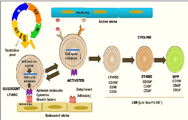
The most primitive HSC/HPC population remains within Lin−Sca-1+c-KIT+ (LSK) fraction of the mouse BM (Ikuta et al., 1992, Minetaro Ogawa et al., 1991, Gerald J Spangrude et al., 1988). LSK fraction consists of LT-HSCs, ST-HSCs and MPPs (Figure-4).These populations are distinguished based on the expression of Thy1.1, CD34 and a HSC-specific fms like tyrosine kinase, CD135/Flt3. LT-HSCs are identified as IL-7Rα-Flt3lo/-Thy1-CD34- LSK fraction, which give rise to ST-HSCs (IL-7Rα-Flt3
lo/-Thy1-CD34+ LSKs) whose further downstream is MPPs (IL-7Rα-Flt3lo/hiThy1-CD34+ LSKs) (Adolfsson et al., 2001, Ikuta et al., 1992, Morrison et al., 1997, Osawa et al., 1996, Weissman et al., 2001). Differential expression of SLAM (the signaling lymphocytic activation molecule) markers, CD150, CD48 and CD244 enable further separation of distinct HSC and MPP subpopulations. While ST-HSCs express CD48 and its ligand CD244 on their surface, LT-HSCs lack these antigens. Lack of CD150 expression distinguishes MPPs from LT and ST-HSCs (Morita et al., 2010, Oguro et al., 2013).
Similar to mouse HSPCs, human HSPC populations can be isolated using a combination of monoclonal antibodies and FACS cell-sorting technology. Lin surface markers, which are used for the negative selection of human HSPCs include CD2 and CD3 for T lineage, CD19 for B cells, CD16 for NK cell lineage, Glycophorin A for red blood cells and CD14 and CD15 for myeloid cells. Following negative selection, HSCs are further enriched into Thy-1+, Thy-1+CD38-lo, or CD133+ fractions within CD34+ cells (Reitsma et al., 2002). In contrast to mouse HSPCs, the SLAM receptor CD150 does not appear to be differentially expressed on human HSCs compared to more differentiated progenitors. Conversely, CD48 expression is found not only on committed human hematopoietic progenitor cells but also non-hematopoietic cell populations including endothelial cells. Interestingly, CD244, a ligand for CD48 is specifically expressed on human HSCs (Sintes et al., 2008, Zaiss et al., 2003).These observations suggest differential expression pattern of surface cell markers that differs on mouse and human HSCs.
Figure 4. Cycling activity of hematopoietic stem and progenitor cells in mice
The most primate HSCs are found in the LSK (Lin-Sca-1+c-Kit+) fraction within the bone marrow. LSK cell subset is composed of HSC, ST-HSC and MPPs. Under steady-state conditions, the majority of LT-HSCs are found in G0 of the cell cycle phase while only >2% remain cycling (S-G2-M). Quiescence is maintained by cell-intrinsic signals and cell-niche interactions. Conversely, only 39% of ST-HSCs are found in G0 whereas MPPs are actively proliferating (only 16% in G0) (Wilson et al., 2008). HSC:
hematopoietic stem cell, LT-HSC: long-term hematopoietic stem cells, MPP: multi-potent progenitor cell, ST-HSC: short-term hematopoietic stem cell. Figure adapted from “Arai et al., 2008; Nakamura-Ishizu et al., 2014; Raaijmakers, 2010”.
The cell cycle is divided into four phases: G1, S, G2 and M phases (Figure-4). Two key checkpoints control the cell cycle and interrupt its progression when DNA damage occurs or the cells have failed to satisfy the requirement due to growth factor /nutrition deprivation. As a consequence, cells permanently exit the cell cycle and enter into an inactive state (G0) before apoptosis. The G0 phase is associated with the loss of cycling potential, senescence and death (Pietras et al., 2011). In contrast to short-lived somatic cells, the majority of HSCs remain resting in the G0 phase. As they divide and differentiate into MPPs, which possess restricted lineage potential, the majority of the committed progeny become active in the S, G2, and M phases to maintain blood cell production (Steinman, 2002).
4.1. Maintenance of hematopoietic stem cell quiescence
Quiescence is crucial to protect HSCs from oxidative stress, DNA damage and the accumulation of replication-associated mutations. Stress resistance in quiescent HSCs is regulated by distinct programs. For instance, HSCs have specific ATP-binding cassette (ABC) transporter gene expression, which provides high drug efflux ability. The ABC transporters protect stem cells against toxic compound accumulation by pumping them across their cell membranes (Tang et al., 2010). Dormant HSCs also display distinct apoptotic programs (Kosan et al., 2015), which probably facilitates their resistance to cytotoxic agents including ultraviolet light, ionizing radiation, and chemicals (Blanpain et al., 2011). The proper balance between HSC quiescence and expansion is critical to maintain homeostatic hematopoiesis and prevent hematopoietic failure or excessive expansion of stem pool that can lead to exhaustion or cancer formation. During steady-state hematopoiesis, HSC quiescence is tightly controlled by cell-intrinsic mechanisms, which are modulated by the niche signals in the BM.
4.1.1. Intrinsic regulation of the cell-cycle
The cell cycle regulation plays an important role in longevity and functional potential of HSCs. Yet, increased cell cycle activation in HSCs can lead to premature depletion and eventually exhaustion of the stem cell pool (Thompson et al., 2008). The cell cycle activity of HSCs is dynamically controlled by cyclins and cyclin dependent kinases (CDKs) as depicted in Figure 5. During quiescence, the retinoblastoma (Rb) proteins prevent cell cycle progression by a complex mechanism, which includes the inactivation of transcription factors such as the E2F family. Progression through G1 and S phase requires the phosphorylation and suppression of the Rb protein by D cyclin/Cdk4/Cdk6 complex. Inactivation of the Rb protein activates the E2F transcription factors, which in turn induces HSC entry into the S phase (Steinman, 2002, Walkley et al., 2007).