Use of clinical biology techniques in clinical practice:
injections of platelet-rich plasma to heal tendon
Use of clinical biology techniques in clinical practice:
injections of platelet-rich plasma to heal tendon
KAUX JF1, Le Goff C², Drion P³, Pascon F4, Libertiaux V4, Gothot A², Cescotto S4, Defraigne JO5, Rickert M6, Crielaard JM1
1. Physical Medicine Service, Department of Motility Sciences, University Hospital of Liège, University of Liège, Belgium. 2. Department of Clinical Biology, University Hospital of Liège, University of Liège, Belgium.
3. Animal Facility of University Hospital of Liège, ULg-GIGA-R, University of Liège, Belgium. 4. Department Argenco, University of Liège, Belgium.
5. CREDEC, Laboratory of Experimental Surgery, University of Liège, Belgium. 6. Department of Orthopaedic Surgery, University of Heidelberg, Germany.
1. Introduction: A tendon is a tissue which does not heal easily. Tendinopathy is a condition which often becomes chronic in the case of bad or overdue management. Several studies, essentially in vitro and, more recently, a few in clinical practice, have demonstrated the positive effects of platelets on the healing process of tendons. A local injection of platelet–rich plasma (PRP), which releases many growth factors, seems to have the potentiality to enhance the tendon healing process. The aim of our experiment was to ascertain whether the use of PRP could accelerate the healing process of an Achilles tendon after a surgically induced lesion.
A. PRP preparation: PRP was obtained from the blood of 12 Sprague Dawley rats by cardiac puncture under general anaesthesia until the heart stopped beating. Quantities of 1mL of anticoagulant, adenosine-citrate-dextrose-acid (ACD-A), were added immediately to each 4,5mL of blood. The blood was then centrifuged at 180g for 10 minutes. To improve platelet concentration of the PRP, the supernatant
B. Experiment: A 5mm defect was surgically induced in the Achilles tendon of 60 rats (Fig. 1-2). Rats were divided into 2 groups of 30: A: control group (without injection) and B: PRP treatment. The rats of group B received a PRP injection 1 hour after the surgery inside the site of the lesion of the Achilles tendon. Fifty micro-litres of PRP were injected in each rat of the PRP group (Fig. 3). Platelets were activated by the local presence of 2. Methods (*):
platelet concentration of the PRP, the supernatant was centrifuged for a second time at 1000g for 10 minutes. The platelets were then collected using a gauge pipette. Cell and platelet counts were made by an auto-analyser. Platelet concentration was around 2.2 to 2.9 x10⁶/mm³.
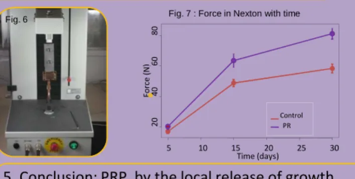
3. Results: We demonstrated that the force necessary to induce tendon rupture during biomechanical tensile testing was greater over time for tendons which had been submitted to an injection of PRP (Fig. 7). These results were observed and significant (p<0.05) from day 5 onwards.
4. Discussion: This experimentation showed that PRP injections could accelerate the tendon healing process and increase the force needed to break tendons in their healing process. This “accelerating” process can be observed and is significant (p<0.05) as early as day 5.
5. Conclusion: PRP, by the local release of growth factors, would be a new therapeutic tool to accelerate tendon healing.
6. Acknowledgement: This experimentation was partially financed by “Standard de Liège” and “Lejeune-Lechien” grants.
(*) All experimental procedures and protocols used in this investigation were reviewed and approved by the Institutional Animal Care and Use Committee of the University of Liège.
(Fig. 3). Platelets were activated by the local presence of collagen in the wound. Afterwards, the rats of both groups were placed in their cages without immobilization. After 5, 15 and 30 days, 10 rats of each group were euthanized. The traumatized Achilles tendon of each rat was dissected and removed (Fig. 4). Immediately after sampling, tendons were submitted to a biomechanical tensile test up to rupture, using a tensile machine with a “Cryo-jaw” (Fig. 5-6).
Time (days) F o rc e ( N ) 5 10 15 20 25 30 2 0 4 0 6 0 8 0 Control PR Fig. 1 Fig. 2 Fig. 3 Fig. 4 Fig. 5