J. Clin. Endocrinol. Metab. 1998 83: 3454-3458, doi: 10.1210/jc.83.10.3454
P. Petrossians, P. Delvenne, P. Flandroy, P. Jopart, A. Stevenaert and A. Beckers
An Unusual Pituitary Pathology
Society please go to: http://jcem.endojournals.org//subscriptions/
or any of the other journals published by The Endocrine
Journal of Clinical Endocrinology & Metabolism
To subscribe to
P. PETROSSIANS, P. DELVENNE, P. FLANDROY, P. JOPART, A. STEVENAERT, AND A. BECKERS
Departments of Endocrinology (P.P., B.A.), Neurosurgery (S.A.), Pathology (D.P.), and Radiology (F.P.), University of Lie`ge; Department of Endocrinology (J.P.), Jolimont, Haine-St-Paul, Belgium
N
ONADENOMATOUS lesions of the pituitaryrepre-sent a small part of the intrasellar processes. How-ever, when encountered, they may present a diagnostic dif-ficulty (1).
Most of them are discovered because of the tumoral syn-drome, due to the compression of the pituitary and the sur-rounding structures, and may lead to hypopituitarism, dia-betes insipidus, secondary hyperprolactinemia, visual field disturbances, and oculomotor abnormalities. None of these signs are fairly specific of these lesions, and, with the ex-ception of diabetes insipidus which is more rare, they could very often be seen in pituitary adenomas. In some cases however, these tumors may be incidentalomas, provoking no pituitary or ocular dysfunction.
Making a more precise diagnosis on the basis of biological, hormonal, and radiological examinations is of primary im-portance, as it may lead to a less aggressive, although very efficient, medical therapy in some cases.
The case history we are presenting here shows the diffi-culties encountered in the diagnosis of these unusual pitu-itary processes and highlights the potential interest of re-cently developed techniques in the endocrinological exploration of these patients.
Case Report
A 54-yr-old menopausal woman of Caucasian origin was admitted to the hospital complaining of extreme weakness, headache, and vomiting. The patient had no previous med-ical history and was taking no medication. At clinmed-ical exam-ination, she presented with signs of hypopituitarism and a mild diabetes insipidus. The basic laboratory tests showed an inflammatory syndrome, with a rise of sedimentation rate to 81 mm/h, fibrinogen to 8 g/L, but normal white blood cell count. Endocrine tests at admission showed a complete an-terior pituitary deficiency, with undetectable gonadotropin and TSH values for a low estradiol and free T3-T4, low insulin-like growth factor I, and no stimulation of GH and cortisol by insulin tolerance test.
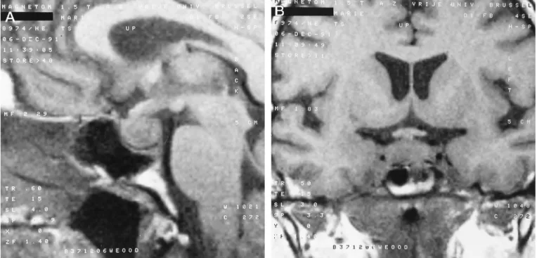
Pituitary computed tomography scan and magnetic res-onance imaging (MRI) examinations were performed (Fig. 1),
showing an endosellar mass and a thickening of the pituitary stalk. There were no intrasellar calcifications. The clinical history and the radiological imaging were not in favor of a pituitary adenoma. Diabetes insipidus is rare even in non-functionning adenomas. Moreover, a pituitary stalk involve-ment by an intrasellar adenoma is very rare.
These examinations suggested that this lesion belongs probably to other types of sellar lesions, which are listed in Table 1.
Because of the nonavailability of gadolinium at that time, contrast images were not performed. However, the T1 and T2 weighted images were not in favor of a cystic lesion nor of a craniopharyngioma. Other diagnoses like intrasellar he-matoma, melanoma, lipoma, vascular (2) or bony tumor were unlikely because of the lack of the MRI signal modifications usually found in these lesions. Moreover, the very high sed-imentation rate could not be explained by these lesions. An inflammatory lesion or a secondary tumor were therefore considered, and a general screening was started to better determine the diagnosis.
Pituitary abscesses (3) usually appear in a context of sep-ticemia, which was not found in our patient. These could be associated with a purulent central nervous system fluid and, in some cases, a rise in white blood cells, none of which were present.
Tuberculosis skin test was negative, and body fluid cultures for bacteria and mycobacteria were sterile. More-over, radiological examinations did not show any primary tuberculous lesion. Syphilitic serology (VDRL and TPHA) was negative. All autoimmunity markers, including an-tineotrophil cytoplasmic antibodies, were normal. Screen-ing for a primary neoplasia, includScreen-ing chest X-ray and computed tomography, gastroscopy, abdominal echoto-mography, gynecological examination, and mammogra-phy, did not unveil any tumorous lesion. For all these reasons, an abscess, or a tuberculous (4, 5), syphylitic, or metastatic (6) lesion seemed to be unlikely.
A pituitary localization of a lymphoma or a plasmocytoma was very unlikely, as this sole localization has rarely been described (7).
The clinical history and the age of the patient were not in favor of a lymphocytic hypophysitis (8, 9), which usually occurs after a pregnancy and presents a less dramatic occurrence.
Received June 11, 1998. Accepted June 22, 1998.
Adress all correspondence and requests for reprints to Professor Al-bert Beckers, Service d’Endocrinologie, CHU de Lie`ge, Dom. Univ. Du Sart Tilman, Lie`ge Belgium.
Langerhans-cell histiocytosis usually shows the involve-ment of other organs and does not cause a rise in sedimen-tation rate (10). However a unique pituitary localization is possible, and the definite diagnosis may only be given by a biopsy of the lesion (11). Granular cell tumors are usually asymptomatic (12). Neurosarcoidosis is a rare diagnosis (5% of all sarcoidosis cases), but a possible diagnosis for our case (13, 14). Laboratory tests showed, however, normal angio-tensin-converting enzyme values that, without excluding this disease, gave another argument against it. Wegener’s granulomatosis (15, 16) was possible too, despite the lack of involvement of other organs and the presence of normal ANCAs. Because of the lack of definite arguments, an in-flammatory pituitary granuloma was considered the most likely diagnosis.
The patient was treated by hormonal replacement therapy (0.1 mg tyroxine and 25 mg hydrocortisone daily). She was then discharged from the hospital and referred to our
insti-tution to discuss the indications for a pituitary biopsy. She was retested for infectious and autoimmunity markers. The results were similar to those previously obtained. The screen-ing for a primary tumor was continued by an examination of the upper respiratory tract. A granulomatous lesion of the cavum was found and biopsied.
First histological examination
Histological examination of the cavum biopsy showed an ill-defined granulomatous inflammation with the presence of epithelioid cells, giant plurinucleated cells, and small areas of nonspecific necrosis. On the basis of histology alone, a diagnosis of either Wegener’s granulomatosis or tuberculosis was first proposed.
Several special stains were then performed. The orcein staining demonstrated some vascular modifications in medium-sized arteries that may accompany Wegener’s gran-ulomatosis. No organisms were seen on Zielh-Neelsen stains
FIG. 1. A and B: Pituitary magnetic resonance imaging at time of admission, showing an intrasellar mass and a thickening of the pituitary stalk.
TABLE 1. Some of the nonadenomatous pituitary tumors already described in the literature (1)
Noninflammatory tumors Inflammatory tumors Cysts Others
Granulous cell Abscess Rathke Hamartomas Craniopharyngioma Sarcoidosis Arachnoid Gangliocytomas
Chordoma Lymphocytic hypophysitis Epidermoid Accessory salivary glands Metastatic Giant cell granuloma Dermoid
Meningioma Histiocytosis Sarcoma Wegener Melanoma Tuberculosis Lymphoma Plasmocytoma Glioma Schwanoma Germinal cell Vascular Bony Lipoma
of the tissue. On the basis of these results, the diagnosis of Wegener’s granulomatosis was preferred.
Clinical course
The patient was therefore treated with corticosteroids (prednisolone). She was then lost for follow-up. Two months later she again presented to the hospital. She was still under corticosteroids and complained of the reappearance of head-ache and vomiting. Clinical examination showed the pres-ence of cavernous and chiasmatic syndromes. At MRI ex-amination, the lesion showed an increase in size with a suprasellar extension, a chiasmatic compression, and a
lat-eral extension in the cavernous sinuses (Fig. 2). Contrast images showed a peripheral shell with a necrotic center. There was no meningial involvement.
The patient underwent a pituitary surgery by a transsphe-noidal approach. The neurosurgeon (A.S.) proceeded to a large debulking of the tumor, relieving the chiasmatic com-pression. Tissue samples were sent to the pathology department.
Second histological examination
The histological aspect was quite similar to that described for the cavum. At low magnification, the normal pituitary
FIG. 2. A, B, C: Pituitary magnetic resonance imaging, showing A, an increase in size of the intrasellar mass; B, a suprasellar extension; and
tissue was replaced by an extensive inflammation. At higher magnification, granulomatous features were observed with the presence of epithelioid cells and giant plurinucleated cells (Fig. 3). Focal areas of necrosis were also present but did not exhibit the typical aspect of caseation. As in the previous sample, no acid-fast bacilli were detected by the Zhiel stain-ing. A diagnosis of granulomatous hypophysitis was pro-posed and probably related, because of the history of the patient, to a Wegener’s granulomatosis.
Clinical course
On the basis of clinical suspicion, the worsening of the symptoms under corticoids, and the fact that pathological examination could not exclude an infectious etiology, the patient was empirically treated by antituberculosis trith-erapy. Three months after the beginning of this treatment, there were a tremendous improvement in the clinical con-ditions and a complete disappearance of the inflammatory syndrome. To definitely determine wheter the patient had a mycobacterial infection, a detection of Mycobacterium tu-berculosis DNA by the polymerase chain reaction (PCR) technique was performed in the pathology departement. Amplification of Mycobacterium Tuberculosis DNA by PCR
Crude DNA was first extracted from the paraffin-embed-ded biopsy as previously described (17). A nested PCR was then performed on 10mL of extracted DNA. We used a pair of primers derived from the sequence of the insertion ele-ment IS6110, which is specific for M. Tuberculosis, and situ-ated outside of the primers proposed by Savic et al. (18). These primers were used in a first ampliflication step, giving an amplified DNA fragment of 559 bp (19). A second am-plification was then performed on an aliquot of the first amplification product by using primers described by Savic et
al. (18), delimiting a DNA fragment of 263 bp within the
originally amplified DNA sequence. The first amplification was carried out using PCR Master (Boehringer Mannheim, Mannheim, Germany) in a final volume of 50mL containing 20 pmol of each sense IS1A and antisense IS2A oligonucle-otide primer. After initial denaturation of the DNA sample for 3 min at 94 C, the reaction was run for 40 cycles consisting of 30 sec at 94 C for denaturation, 2 min at 60 C for annealing, and 2 min at 72 C for extension. The second amplification was performed on 1mL of the originally amplified DNA using the primers and PCR conditions (initial denaturation of the DNA for 13 min at 98 C followed by 40 cycles consisting of 1 min at 92 C for denaturation, 1 min at 72 C for annealing/exten-sion) described by Savic et al. (18). The amplified product was
FIG. 3 Hematoxylin-eosin stained
sec-tion demonstrating a granulomatous inflammation with epitheloid cells and a central multinucleated giant cell (magnification,3200).
FIG. 4 Agarose gel analysis of polymerase chain reaction (PCR) prod-ucts visualized by ultraviolet light after ethidium-bromide staining. Amplified M tuberculosis DNA was detected by PCR at the expected size (263 bp) in the DNA solution obtained from sections of the biopsy specimens (lanes e and f). Primers amplifying a 370 bp fragment in a human genomic sequence were also used to rule out inability to amplify DNA (lane d). H2O and DNA from an empty paraffin block
were used as negative controls (lanes b and c). DNA from a M
tuber-culosis strain was used as positive control (lane a). Lane MW
con-tained DNA molecular weight standards.
Clinical course
Five-year follow-up shows no signs of reappearance of the inflammatory syndrome. The patient presents no complaints under hormonal replacement therapy. Pituitary MRI shows an empty sella with no sign of tumor reappearance.
Discussion
Before the discovery of antituberculosis drug therapy, tu-berculomas represented as much as 30% of the intracranial tumors in adults and 50% in children (20). The frequency is now between 0.25% and 4% in western countries (21). A unique localization in the pituitary is very rare, as meninges are also usually involved. These cases present mainly as a secondary complication of a known tuberculosis or at least as the first manifestation of an easily diagnosed tuberculosis, with a more-or-less obvious entry point.
Moreover, the imaging findings of pituitary tuberculomas are nonspecific and may resemble other pituitary granulo-mas caused by a specific lesion such as syphilis, sarcoidosis, Langerhans cell histiocytosis, Wegener’s granulomatosis, and even macroadenoma or lymphocytic hypophysitis.
The diagnostic puzzle presented by our patient was as-sociated with the unique pituitary localization, with no ob-vious primary infection site, until the finding of a granulo-matous inflammation in the nasal cavities. The normal skin test and sterile body fluid cultures were other factors that made the diagnosis of tuberculosis less likely. The patho-logical examination, while not excluding a mycobacterial infection, was more in favor of a noninfectious granuloma-tous lesion. This diagnosis was reinforced by the clinical presentation and the complementary examinations, and led finally to corticoid therapy.
Fortunately, this treatment was not associated with any dramatic complication except for the growth of the pituitary tumor.The early and transient improvement in the com-plaints presented by the patient was probably related to the antiinflammatory effect of the corticoids.
The pathological examination of the pituitary tissue ob-tained by transsphenoidal surgery did not help in refining the diagnosis, but confirmed the inflammatory nature of this tumor. The antituberculosis therapy was mainly motivated by clinical suspicion and the ineffectiveness of the corticoids. Moreover, a mycobacterial infection was never entirely ex-cluded in any part of the history of the patient.
PCR examination was not performed earlier in the history of the patient because it was, at that time, a new technique whose usefulness was just being evaluated. The patient un-derwent a 9-month, 3-drug therapy, and the treatment was
or from a hematogenous contamination from the cavum. A secondary extension of purulent bacterial infections has al-ready been described from preexisting peritonsillar abcesses (22).
In conclusion, this case report illustrates the diagnosis difficulty of nonadenomatous pituitary lesions. An accurate diagnosis is important, however, because it allows the ad-ministration of an efficient medical treatment in some cases.
References
1. Rohmer V, Chanson P, Dupas B, Beckers A. 1997 Intrasellar non-adenoma-tous expansive process. Ann Endocrinol. 58:11–19.
2. Asa SL, Kovacs K. 1983 Histological classification of pituitary disease. Clin Endocrinol Metab. 12(3):567–596.
3. Bjerre P, Riishede J, Lindholm J. 1983 Pituitary abscess. Acta Neurochir (Wein). 68:187–193.
4. Eposito V, Fraioli B, Ferrante L, Palma L. 1987 Intrasellar tuberculoma: case report. Neurosurgery. 21:721–723.
5. Delsedime M, Aguggia M, Cantello R, et al. 1988 Isolated hypophyseal tuberculoma: case report. Clin Neuropathol. 7:311–313.
6. Teears RJ, Silverman EM. 1975 Clinicopathologic review of 88 cases of car-cinoma metastatic to the putuitary gland. Cancer. 36:216 –220.
7. Bitterman P, Ariza A, Black RA, Allen WE, Lee SH. 1986 Multiple myeloma mimicking pituitary adenoma. Comput Radiol. 10:201–205.
8. Hashimoto K, Takao T, Makino S. 1997 Lymphocytic adenohypophysitis and lymphocytic infundibuloneurohypophysitis. Endocr J. 44:1–10.
9. Pressman EK, Zeidman SM, Reddy UM, Epstein JI, Brem H. 1995 Differen-tiating lymphocytic adenohypophysitis from pituitary adenoma in the peri-partum patient. J Reprod Med. 40:251–259.
10. Tien RD, Newton TH, McDermott MW, Dillon WP, Kucharczyk J. 1990 Thickened pituitary stalk on mr images in patients with diabetes insipidus and Langerhans cell histiocytosis. Am J Neuroradiol. 11:703–708.
11. Nishio S, Mizuno J, Barrow DL, Takei Y, Tindall GT. 1987 Isolated histio-cytosis3 of the pituitary gland: case report. Neurosurgery. 21:718–721. 12. Cone L, Srinivasan M, Romanul FC. 1990 Granular cell tumor (choristoma)
of the neurohypophysis: two cases and a review of the literature. Am J Neu-roradiol. 11:403– 406.
13. Bell NH. 1991 Endocrine complications of sarcoidosis. Endocrinol Metab Clin North Am. 20:645– 654.
14. Timsit J, Valeyre D, Chanson P, et al. 1993 Isolated pseudotumoural sar-coidosis of the hypothalamic-pituitary area. Eur J Med. 2:505–506. 15. Lohr KM, Ryan LM, Toohill RJ, Anderson T. 1988 Anterior pituitary
in-volvement in Wegener’s granulomatosis. J Rheumatol. 15:855– 857. 16. Czarnecki EJ, Spickler EM. 1995 MR demonstration of Wegener
granuloma-tosis of the infundibulum, a cause of diabetes insipidus. Am J Neuroradiol. 16:968 –970.
17. Delvenne P, Fontaine MA, Delvenne C, Nikkels A, Boniver J. 1994 Detection of human papillomaviruses in paraffin-embedded biopsies of cervical intra-epithelial lesions. analysis by immunohistochemistry, in situ hybridization, and the polymerase chain reaction. Mod Pathol. 7:113–119.
18. Savic B, Sjo¨bring U, Alugupalli S, Larsson L, Mio¨rner H. 1992 Evaluation of polymerase chain reaction, tuberculostearic acid analysis, and direct micros-copy for the detection of Mycobacterium tuberculosis in sputum. J Infect Dis. 166:1177–1180.
19. Fauville-Dufaux M, Waelbroeck A, De Mol P, et al. 1996 Contribution of the polymerase chain reaction to the diagnosis of tuberculous infections in chil-dren. Eur J Pediatr. 155:106 –111.
20. Garlan HG, Armitage G. 1933 Intracranial tuberculoma. J Pathol Bacteriol. 37:461– 471.
21. Kummerr R, Storch B, Ranch M, Krause KM. 1981 A case of multiple intra-cranial tuberculomas followes by serial computerized tomography. Nerve-narzt. 52:344 –347.
22. Berger SA, Edberg SC, G David. 1986 Infectious disease of the sella turcica. Rev Infect Dis. 8:747–755.