Université de Montréal
P
REDICTIVE VALUES OF NEUROLOGICAL EXAMINATION,
OTOSCOPIC EXAMINATION AND BRAINSTEM AUDITORY EVOKEDRESPONSE
(BAER)
IN CALVES WITH OTITIS MEDIA-
INTERNApar
Andrea Finnen
Département de sciences cliniques
Faculté de médecine vétérinaire
Mémoire présenté à la Faculté de médecine vétérinaire
en vue de l’obtention du grade de
maître ès sciences (M. Sc.)
en sciences vétérinaires
option sciences cliniques
Avril 2011
Université de Montréal
Faculté de médecine vétérinaire
Ce mémoire intitulé
P
REDICTIVE VALUES OF NEUROLOGICAL EXAMINATION,
OTOSCOPIC EXAMINATION AND BRAINSTEM AUDITORY EVOKEDRESPONSE
(BAER)
IN CALVES WITH OTITIS MEDIA-
INTERNAprésenté par
Andrea Finnen
a été évalué par un jury composé des personnes suivantes
Dr Sébastien Buczinski, président-rapporteur
Dre Joane Parent, directrice de recherche
Summary in English
Currently, the antemortem diagnosis of otitis media-interna is based upon the presence of appropriate clinical signs and adjunctive diagnostic imaging including radiography and computed tomography. The purpose of this prospective study was to evaluate predictive values of neurological examination, otoscopic examination and BAER in calves for the diagnosis of otitis media-interna using computed tomography as the gold standard. The second objective was to define BAER reference values in normal calves and to describe BAER abnormalities in calves affected with otitis media-interna. Seventeen Holstein calves between 5 and 7 weeks of age were included. All calves had a neurological examination, otoscopic examination and BAER. Calves were sedated with intravenous xylazine (0.05-0.15 mg/kg [0.02-0.07 mg/lb]) for computed tomography of the tympanic bullae to evaluate for the presence of otitis media-interna. Based upon computed tomographic results, 11 of 17 calves were affected with otitis media, 4 unilaterally and 7 bilaterally. Five waveforms were consistently identified on BAER traces from 6 normal calves. The positive predictive value of BAER, neurological examination and otoscopic examination were 94.7%, 91.7% and 66.7% respectively. Clinically, the most reliable non-invasive diagnostic test to diagnose otitis media-interna in the calf is the BAER. Abnormalities were observed on BAER before the development of neurological deficits in approximately 40% of calves allowing earlier diagnosis.
Résumé en français
Présentement, le diagnostic d’otite moyenne-interne chez le veau est basé sur la présence de signes cliniques appropriés ainsi que les tests diagnostiques tels que la radiographie et la tomodensitométrie. L’objectif de cette étude prospective était d’évaluer les valeurs prédictives de l’examen neurologique, l’examen otoscopique et le test des potentiels auditifs évoqués (PAE) dans le diagnostic d’otite moyenne-interne chez le veau, en utilisant la tomodensitométrie comme test standard. Le deuxième objectif était de définir les valeurs de référence pour le PAE chez le veau normal et d’en décrire les anomalies chez des veaux atteints d’otite moyenne-interne. Dix-sept veaux de race Holstein entre 5-7 semaines d’âge ont été inclus. Tous les veaux ont eu un examen neurologique, un examen otoscopique et une évaluation des PAEs. Les veaux ont été tranquillisés avec de la xylazine intraveineuse (0,05-0,15mg/kg) pour la tomodensitométrie des bulles tympaniques afin d’évaluer pour la présence d’otite moyenne-interne. Selon les résultats de la tomodensitométrie, 11 des 17 veaux étaient atteints avec otite moyenne, 4 de façon unilatérale et 7 bilatéralement. Cinq ondes ont été identifiées de façon constante sur les tracés des PAEs des 6 veaux normaux. Les valeurs positives prédictives pour le PAE, l’examen neurologique et l’examen otoscopique étaient 94,7%, 91,7% et 66,7% respectivement. D’un point de vue clinique, le test le plus fiable dans le diagnostic d’otite moyenne-interne chez le veau est le PAE. Les anomalies ont été observées au PAE avant le développement des signes neurologiques chez certains veaux.
Mots clés : Veau, Potentiels auditifs évoqués, tomodensitométrie, otite
TABLE OF CONTENTS
Summary………...iv Résumé en français………....v Table of Contents………..vi List of Tables………....ix List of Figures………x List of Abbreviations………xi Introduction………11. Anatomy of the Ear………2
1.1 External ear………..3
1.2 Middle ear………3
1.3 Inner ear………...4
1.4 Otoscopic examination.………...………....5
2. Neurological structures of the ear………..6
2.1 Neurological structures of the middle ear………6
2.1.1 The facial nerve….………...6
2.1.2 Sympathetic innervation………..6
2.2 Neurological structures of the inner ear………...7
2.2.1 Vestibular receptors……….7
2.2.2 Auditory receptors………...8
3. BAER.………..………11
3.1 Nomenclature……….11
3.2 Neuroanatomic generators……….12
4. Performing the BAER……….14
4.1 Acoustic stimulation………..14
4.2 Masking noise………15
4.3 Electrode placement………...15
4.4. Signal averaging system………16
5. BAER analysis……….18
5.1 Identification of waves………...18
5.2 Latency………...21
5.3 Amplitude and ratios………..22
6. Non-pathologic factors that influence the BAER………24
6.1 Equipment factors………..24
6.1.1 Polarity of stimulus………24
6.1.2 Number of clicks per second………..25
6.1.3 Intensity………..26 6.2 Patient factors……….27 6.2.1 Age……….27 6.2.2 Species………...27 6.2.3 Size……….28 6.2.4 Sex……….29 6.2.5 Temperature………...29
6.2.6 Pharmacologic agents………30
7. Pathologic factors that influence the BAER………31
7.1 Sensorineural hearing loss……….31
7.2 Conductive hearing loss……….31
7.3 Brainstem disease………...32
Summary of literature review………..33
Article………...34
Abstract………35
Introduction………..36
Materials and methods……….38
Results………..41 Discussion………44 Conclusion………...48 Footnotes………..50 References.………...51 Figures………..55 Tables…...………58 General Discussion………..60 Conclusion………...68 Bibliography……….69
LIST OF TABLES
Table I: BAER latencies of 5 different species………..………..28
ARTICLE
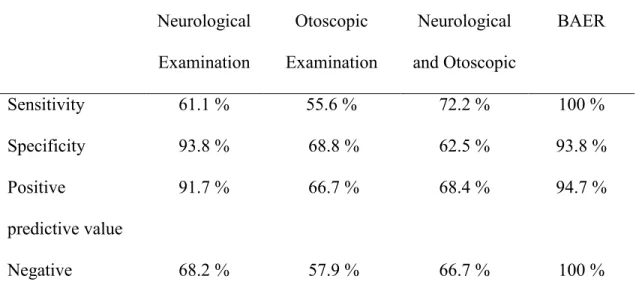
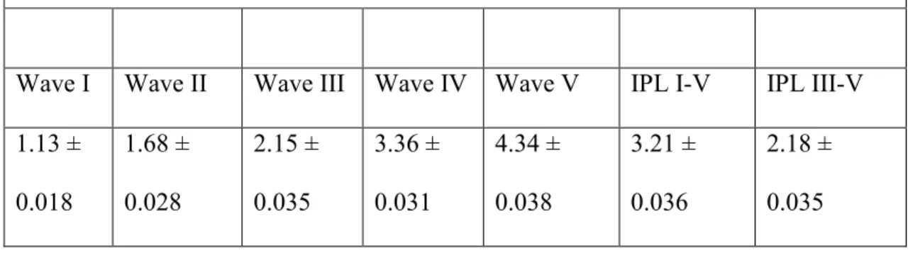
Table I: Sensitivity, specificity and predictive values...………..58 Table II: BAER latencies in 6 normal calves.………..59
LIST OF FIGURES
Figure 1: Anatomy of the Human Ear……….……...…………...2
Figure 2: Normal Human BAER……….………12
Figure 3: Normal Equine BAER……….…….………19
Figure 4: Normal Bovine BAER……….…….…………....20
Figure 5: Normal Canine BAER……….…….………20
Figure 6: BAER wave latencies and interpeak latency (IPL) in a calf…..………..22
ARTICLE
Figure 1: Normal BAER in a calf...…….………55Figure 2: BAER in calf with otitis media………56
LIST OF ABBREVIATIONS
BAER: Brainstem Auditory Evoked Response dB: DecibelCNS: Central Nervous System
CN VII: Cranial nerve VII, Facial nerve
CN VIII: Cranial nerve VIII, Vestibulocochlear nerve IPL: Interpeak latency
ARTICLE
BAER: Brainstem Auditory Evoked Response CT: Computed Tomography
1
INTRODUCTION
The brainstem auditory evoked response (BAER) test is a non-invasive diagnostic tool evaluating the auditory function and the auditory pathway from the internal ear through the brainstem. The BAER is useful in the evaluation of peripheral hearing loss or brainstem lesions in humans and other mammals.[1] Otitis media is a frequently encountered disease of young cattle that can cause significant morbidity and mortality. As otitis media is frequently associated with otitis interna, they are often referred together as one entity (otitis media-interna).[2-4] Young calves under 3 months of age are most commonly affected with bacterial agents that are thought to develop from ascending infection via the Eustachian tube. Clinical signs include ear droop, facial weakness, head tilt, pharyngeal weakness, epiphora, inappetance and fever.[2, 5-8] Early identification and treatment of affected animals can reduce economic losses from this disease. Numerous diagnostic tests exist to evaluate for the presence of otitis media-interna including brainstem auditory evoked response (BAER), neurological examination and otoscopic examination. BAER can be abnormal in cases of otitis due to sound transmission failure, consistent with conductive hearing loss.[9] Neurological deficits such as facial and/or vestibulocochlear nerve dysfunction can also be observed in cases of otitis media-interna.[10] A thorough understanding of the anatomy of the ear including neurological function is essential to interpret the results from the BAER, otoscopic and neurological examinations.
1. Anatomy of the ear
The ear is also referred to as the ‘vestibulocochlear’ organ as it not only allows transmission of sound and the ability to hear, but is also important for maintaining equilibrium.[11] Sound waves are collected and concentrated in the external ear canal, transmitted through the middle ear and finally are transformed into nerve impulses in the inner ear. The nerve impulses generated in the inner ear are propagated along the brainstem eventually reaching the auditory cortex for the perception of sound. In all species, the ear is divided into three anatomical and functional regions. From the most exterior to the most interior the regions include: the external ear, the middle ear and the inner ear.
1.1.
The external ear
The external ear is composed of two parts, the pinna or auricle, and the external acoustic meatus. The pinna is the externally visible portion of the ear and is shaped like a funnel in order to collect and direct sound waves internally into the external acoustic meatus. Unlike in humans, cattle and most animals are able to move the pinna towards the source of sound.[11, 13] As well, the left and right pinnae can move independently of one another, allowing the animal to focus on sounds coming from different directions without moving the head. The external acoustic meatus is a curved canal that concentrates the sound waves collected by the pinna and directs them internally towards the tympanic membrane. Sebaceous and tubular ceruminous glands are found in the skin lining the external acoustic meatus and are responsible for secreting cerumen to prevent dust and other particles from reaching and damaging the delicate tympanic membrane. [11] The external ear ends at the tympanic membrane, an extremely thin membrane that covers the opening of the temporal bone. The membrane functions to transmit sound waves from the external acoustic meatus to the auditory ossicles within the middle ear. This membrane is under tension and is well vascularized and innervated.[13]
1.2.
The middle ear
The middle ear is an air-filled space within the temporal bone that is also known as the tympanic cavity. This cavity can be further divided into three portions: dorsal, middle and ventral. The dorsal part is found above the level of the tympanic membrane and contains the chain of auditory ossicles (malleus, incus, staples). The auditory ossicles
mediate the transmission of sound across the tympanic membrane, through the middle ear and to the inner ear for the perception of sound.[11] The middle part of the tympanic cavity includes the medial aspect of the tympanic membrane and opening of the auditory tube, which connects to the nasopharynx. The auditory or Eustachian tube connects the tympanic cavity to the nasopharynx and is important in equalizing pressure over the two faces of the tympanic membrane.[11, 13] The tube has a narrow lumen and is usually collapsed. When swallowing, the tube temporarily opens permitting drainage of the tympanic cavity secretions. Two other openings are found in the medial wall of the tympanic cavity: the vestibular window and the cochlear (round) window. The vestibular window is occupied by the staples and acts to mechanically transmit stimuli from sound waves to the inner ear whereas the cochlear window leads to the cochlear cavity and is covered by a thin secondary tympanic membrane. The ventral part of the tympanic cavity is an enlarged extension of the temporal bone also known as the tympanic bulla. The anatomy of the bulla varies among species and is further subdivided into cells in some species, including cattle.[13, 14]
1.3.
The inner ear
The internal ear is completely enclosed within the petrous temporal bone, the densest bone in the body.[15] The inner ear serves two main functions: the perception of sound (hearing) and the maintenance of equilibrium (vestibular system). Numerous interconnected chambers and ducts containing endolymph are collectively referred to as the membranous labyrinth. The labyrinth is subdivided into the vestibular labyrinth and the cochlear labyrinth and is surrounded by the osseous labyrinth.[13] The space in
between the osseous and membranous labyrinths is filled with perilymph, fluid that is similar in composition to cerebrospinal fluid, and is connected to the subarachnoid space surrounding the brain via the endolymphatic duct.[13] Movement of the endolymph, fluid contained within the membranous labyrinth, stimulates sensory cells within the membranous walls to send impulses along the vestibulocochlear nerve to the brainstem.[3, 11]
1.4.
Otoscopic examination
The external acoustic meatus can be visually examined via a hand-held or video otoscope. The meatus is relatively straight in the dog and cat, which allows for easy visualization of the canal and tympanic membrane.[16] However in the horse and cow, the canal is long and curved, which renders examination difficult. The tympanic membrane can be visualized in adult large animals yet it can be physically difficult and may not be possible without chemical restraint or general anesthesia.[14] Otoscopic examination is performed routinely to evaluate for the presence of otitis externa and can also occasionally indicate the presence of otitis media. In small animals, thickening, proliferation, ulceration and malodorous secretions are often observed in cases of otitis externa whereas rupture or other abnormalities of the tympanic membrane such as opacification, hemorrhage or bulging are found in cases of otitis media.[16] Due to the anatomic location of the inner ear, otoscopic examination cannot evaluate for otitis interna.
2. Neurological structures of the ear
The main function of the ear is sound transmission and the perception of sound yet numerous other important neurological structures are associated with the ear.
2.1.
Neurological structures of the middle ear
2.1.1.
The facial nerve
The facial nerve (cranial nerve VII) arises from the facial nucleus in the rostral medulla and travels in close association with the vestibulocochlear nerve when exiting the skull via the internal acoustic meatus within the petrous temporal bone.[17] The nerve then travels though the facial canal and courses adjacent to the dorsal portion of the tympanic cavity where it is unprotected by a bony canal.[10] The nerve emerges through the stylomastoid foramen and branches to innervate muscles of the external ear, muscles of the eyelid, nostril, cheek, lips, and digastricus.[10, 11] Dysfunction of the facial nerve at any point along its course results in paresis or paralysis of the innervated muscles and subsequent facial asymmetry.
2.1.2.
Sympathetic innervation
Sympathetic preganglionic fibres originate in the lateral column of the thoracolumbar portion of the spinal cord and pass via spinal nerves into the sympathetic trunks, coursing rostrally towards the head. Fibres synapse in the cranial cervical ganglion, found near the base of the skull, ventral and medial to the tympanic bulla.[11, 18] The
route of the postganglionic fibres from this ganglion to the ocular structures is not well defined and differs from dogs and cats to horses and other ruminants.[10, 19] In the cat, the sympathetic fibres of the deep petrosal nerve join the greater petrosal nerve (CN VII), pass through the tympanic bulla and continue rostrally to innervate ocular structures such as the smooth muscle of the periorbita, third eyelid and dilator muscles of the pupil.[11, 18] In the bovine, the sympathetic fibres course deeper, following the internal carotid artery to eventually reach the orbit.[18, 19] Infections or neoplasia of the middle ear can affect these fibres and lead to sympathetic dysfunction such as Horner’s syndrome in dogs and cats. Due to the different anatomy in the bovine, Horner’s syndrome is not observed in this species associated with otitis media.[3]
2.2.
Neurological structures of the inner ear
2.2.1.
Vestibular receptors
The vestibular receptors are associated with the vestibular labyrinth, composed of the utriculus, sacculus, and three semi-circular ducts, within the petrous temporal bone. At the base of each semi-circular duct is a dilation called the ampulla. The dendritic zones of the vestibular neurons are found at the base of hair cells found within the crista, a specialized area within each ampulla. Movement of the endolymph in the semi-circular ducts causes movement of the hair cells and stimulation of the vestibular neurons. As the semi-circular canals are arranged at 90 angles to one another, movement of the head in any direction will stimulate the vestibular receptors. A similar receptor called the macula is found within the utriculus, oriented in a horizontal plane. Movement of the
hair cells associated with the macula stimulates an impulse in an associated vestibular neuron. The macula is influenced by gravitational forces and is responsible for sensation of head position at rest and in response to linear acceleration or deceleration.[3] Neurons from each ampulla and macula converge together to form the vestibular portion of the vestibulocochlear nerve and travel through the internal acoustic meatus and facial canal towards the rostral medulla.[17] The majority of neurons terminate in the vestibular nuclei of which there are four. The remaining neurons continue to the flocculonodular lobes of the cerebellum. The vestibular system maintains the position of the eyes, trunk and limbs in relation to the position and movement of the head and is important for maintaining balance.[3]
2.2.2.
Auditory receptors
The second function of the inner ear is the perception of sound. The cochlea, the structure responsible for perception of sound, is found within the petrous temporal bone and is a snail-like structure with 2.5 to 4 turns depending on the species.[13] The cochlea winds around a central core of hollow bone called the modiolus, which contains the nerve fibres forming the cochlear nerve.[13] A bony shelf projects from the modiolus into the cochlea called the spiral lamina, which divides the cochlea into two portions: scala vestibuli and scala tympani, both of which contain perilymph. The cochlear duct is part of the membranous labyrinth and as such, contains endolymph. It spirals through the cochlea and is bordered dorsally by the scala vestibuli and ventrally by the scala tympani. These two structures are continuous at the end of the cochlea, called the helicotrema, as the cochlear duct does not reach the apex of the cochlea.[3]
The auditory receptors are similar to the vestibular receptors; hair cells found in the spiral organ (organ of Corti) within the cochlear duct are in synaptic relationship with the dendritic zones of the neurons. Sound waves transmitted from the external ear cause movement of the ossicular chain of the middle ear, which in turn causes movement of the vestibular window. The perilymph of the scala vestibuli is set into motion by the movement of the vestibular window and subsequently causes movement of the endolymph of the cochlear duct. The endolymph can also be stimulated via the scala tympani as the scala vestibuli and scala tympani are continuous. Movement of the hair cells of the spiral organ generates impulses in the associated neuron. These neurons course through the modiolus to the spiral ganglion and continue with the vestibular portion of the nerve to the rostral medulla. These neurons synapse in the cochlear nucleus and impulses continue rostrally through the brainstem and eventually to the auditory cortex.[3, 11]
2.3.
Neurological examination
Neurological examination of the ear involves evaluation of the facial (CN VII) and vestibulocochlear (CN VIII) nerves. Evaluation of these two nerves is part of the complete cranial nerve examination.[4] The facial nerve is evaluated when testing the menace response, the palpebral reflex and assessing facial symmetry however; the most reliable test is considered the palpebral reflex.[10] Neurological deficits related to facial nerve dysfunction include absent or decreased menace response with normal vision, absent or weak palpebral closure, and ear and/or lip droop. Deficits may range from mild weakness to complete paralysis.
Evaluating physiological nystagmus assesses the vestibular portion of CN VIII. The presence of a head tilt or vestibular ataxia are also deficits that indicate vestibular dysfunction. The cochlear portion of CN VIII is evaluated with the BAER to assess the integrity of the auditory receptors in the inner ear. Behavioural tests to assess the ability to perceive sound can be attempted yet are not specific and false negatives can occur. The BAER remains the preferred test for evaluating auditory function. [1]
When evaluating the neurological structures of the ear, a complete neurological examination should be completed. The presence of other cranial nerve deficits, presence of somnolence or proprioceptive ataxia, in addition to CN VII and CN VIII deficits may indicate involvement of central components such as the brainstem and help to further localize the lesion.
3. BAER
The BAER is a non-invasive, sensitive, and consistent far-field recording of electrical activity in the auditory pathway from the inner ear through the brainstem. BAER waveforms represent an average of brainstem activity recorded within the first 10 ms after an auditory stimulus to the ear. In humans, up to 7 waves can be identified that correlate to specific neural generators in the brainstem and subcortical regions.[1] This test provides an objective measure of auditory system function that is relatively unaffected by the level of arousal, sedatives or general anesthesia. The BAER has been reported in numerous other mammals such as the dog, cat, horse, cow, and is similar in morphology to humans.[20-22] BAER testing is useful for evaluating peripheral deafness and various brainstem diseases. In the bovine species, BAER has been reported in cases of neomycin ototoxicity, otitis media, and bovine spongiform encephalopathy.[23-25]
3.1.
Nomenclature
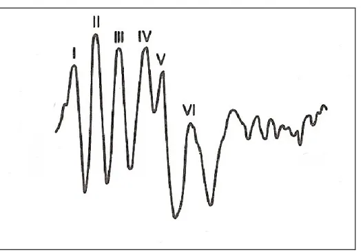
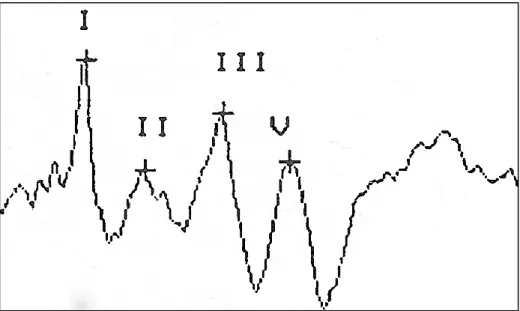
Each wave of the BAER is labeled with a roman numeral from I to VII to facilitate analysis of amplitude, absolute latencies and interpeak latencies. The two most important waves to identify are waves I and V. Wave I is easily identified as the first wave produced after the stimulus and Wave V is identified by the large negative trough following the wave. Wave III is identified as a peak equally spaced in between waves I and V. Waves II, IV, VI and VII are not consistently identified or of clinical importance and are labeled accordingly if present.[1] In horses and cows, not all waves are present
or identifiable with only 4 or 5 waves recognized consistently. In horses and cows, wave IV and V are often fused, as in humans and observed occasionally in dogs. [20, 21, 26-28]
Figure 2 – Normal human BAER tracing demonstrating the presence of 6 peaks. Note the presence of wave IV as distinct from wave V. [1]
3.2.
Neuroanatomic generators
Controversy exists as to the neural generators of each BAER waveform. Waves may be generated from synapses in brainstem nuclei, by electrical conduction of the action potential in the white matter brainstem tracts, or by a combination of synapses and conduction.[1] Specific neural generators have been identified that correspond to BAER waveforms however it is possible that more than one neural structure or the summation of neural activity at more than one location may combine to produce one wave. It must
be noted that electrical activity generated from one cochlear nerve will ascend the brainstem bilaterally as the pathways cross at least twice in the brainstem leading to bilateral activation of the auditory cortex with unilateral stimulation.[29, 30] Wave I originates from action potential volley in the cochlear portion of the vestibulocochlear nerve (CN VIII). Wave II is generated by the intracranial/extramedullary portion of the cochlear nerve or synapse in the cochlear nucleus in the rostral medulla. Wave III originates from the nucleus of the trapezoid body. Waves IV and V are generated in the upper pons with wave IV from the lateral lemniscus and wave V from the caudal colliculus. Waves VI is generated by the medial geniculate nucleus and wave VII from the auditory radiations however these waves are not commonly observed.[1]
4. Performing the BAER
BAER is a far-field recording (electrical activity generated from deeper structures of the brain) of brainstem electrical activity in response to acoustic stimulation.[30] To perform the BAER, specialized equipment must be utilized that provides acoustic stimulation, records electrical activity over the scalp, and registers the tracings for interpretation.
4.1.
Acoustic stimulation
The most common source of auditory stimulation is a repetitive square click of short duration (100-200 msec). The click is directed into the ear by earphone inserts or headphones and plastic tubing attached to the stimulator source. Generally, 1000 clicks are delivered at a rate of 10 clicks per second. Each ear is stimulated separately (monoaurally) and compared to the opposite side.[1] The intensity of the click is determined by the evaluator and should be appropriate to the species and age of the patient. Intensity of the click is measured in decibels (dB). Hearing threshold differs according to species and is assessed in humans as the midpoint value between intensities at which the clicks can and cannot be heard. The BAER is usually then evaluated at 65-70 dB greater than the threshold value to obtain diagnostic tracings.[1] Assessing threshold in domestic mammals is more difficult and is assessed as the value at which the BAER tracing is no longer visible. This value differs according to species and is considered to be approximately 5-20 dB in dogs, 50 dB in horses and 65-70 dB in cows.[20, 21, 26, 27, 31]
4.2.
Masking noise
When performing the BAER, the click stimulus of the test ear can stimulate the non-test ear by way of air or bone transmission of the sound. As the auditory pathways cross, both sides of the brainstem are activated with unilateral stimulation. This crossover effect is not significant if the auditory system is normal bilaterally. However, if significant differences exist between the two sides with respect to peripheral hearing loss, inadvertent stimulation of the opposite normal ear can affect the results of the abnormal ear.[1] For example, stimulating an ear with peripheral hearing loss due to congenital deafness will produce a flat BAER with no waveforms. If the opposite normal ear is stimulated by bone vibration of the auditory stimulus of the deaf ear, due to the cross over effect in the brainstem, a wave or numerous waves may be recorded when evaluating the deaf ear, giving the impression of sound transmission.[32] To avoid stimulation of the non-test ear, the use of white masking noise is recommended. When stimulating one ear, white noise of all frequencies randomly is applied at 20-40 dB below the test ear intensity. No effects on wave latency or amplitude are observed when using masking noise.[22] This practice is recommended in human and veterinary medicine when performing the BAER to avoid confounding results.[1, 32, 33]
4.3.
Electrode placement
Needle electrodes placed over the skull detect the electrical activity of the propagating action potential in the brainstem. The waves are generated due to an electrical difference in voltage from one electrode compared to another. Three subcutaneous needle
electrodes are placed over the skull: the active, the reference and the ground electrodes. Surface electrodes are used in human medicine however are impractical in veterinary medicine due to hair coat and patient co-operation. No significant difference in latency or amplitudes is appreciated between the two recording electrode types.[1] In dogs, the active electrode is placed at the vertex, reference electrode at the base of the tympanic bulla of the test ear, and ground electrode at the base of the opposite ear.[33] Numerous variations to the electrode placement exist with minor effects observed to the BAER waveforms.[27, 34, 35] The most important consideration is placement of the negative electrode as this will affect the polarity of the waveforms.[1] The waves are representative of negative activity however convention states that the BAER waves are displayed as an upward deflection from baseline. An upward deflection is obtained when the positive electrode is placed at the vertex and negative electrode at the base of the ear. [1]
4.4.
Signal averaging system
The BAER wave voltage is extremely low and therefore a signal amplifier is required in order to extract the waveforms from background noise, such as muscle and heart activity.[1] Excessive muscle activity can cause significant artifact that can completely obscure the BAER waveforms.[28] The interface between the electrodes and patient is an important factor referred to as impedance. Any resistance to conduction through the electrodes can significantly affect the registration of the BAER activity and therefore the results. Impedance should be evaluated before each registration of the BAER and verified to be less than 5 k (kilo-ohms).[1] The signal averaging system will extract
the low voltage BAER activity from the higher voltage background noise. The BAER activity is recorded within 10 ms following the auditory stimulus and in order to extract this low voltage activity, the test is repeated numerous times, at least 500-1000 times.[1] As the BAER activity is time-locked to the stimulus, with repeated measurements this activity will be detected and amplified compared to the background noise, which is random and not associated with the stimulus.
5. BAER analysis
Before analyzing the BAER, the trace must be determined to be repeatable. Therefore, a minimum of 2 tests must be performed that are similar in appearance before the tracing can be analyzed. Waves can be expected to vary slightly, however only 0.1-0.2 ms difference between subsequent recordings is considered acceptable. After registering the first tracing, the second tracing is performed and superimposed upon the first tracing to evaluate for repeatability. If the waves on each trace do not superimpose, the test must be repeated until at least two similar if not identical tracings are obtained. This quality control step is to ensure that the recorded waves are from the BAER and not due to random background activity. Once the test has been determined to be repeatable, analysis can proceed.[1]
5.1.
Identification of waves
Analysis of the BAER tracing begins with identification of waveforms. Waves I and V are the most prominent and easy to identify waveforms and are therefore identified first. Wave I is important to identify as this wave is integral in measuring interpeak latencies. [1] Wave I is the first wave encountered and in the bovine, is found approximately 1.5-1.6 ms after the stimulus at 95 dB.[20, 26] Wave V is the wave followed by a large negative trough and is usually the last wave to be observed on a bovine BAER. Wave IV is not always visible as a distinct wave and interacts in different ways with wave V. Wave IV is reported to be absent in the majority of BAER tracings in the bovine, equine and other species as this wave is speculated to be combined with wave V.[20, 21, 26,
36] Waves II and III are then identified as waves in between wave I and V, spaced at approximately 1 ms intervals. Waves I, III and V are most important to identify due to the clinical significance of these waves in calculating interpeak latencies. Waves VI and VII are rarely observed in veterinary BAER tracings and are not of clinical significance.[1]
Figure 3 – Normal BAER from an adult horse demonstrating the presence of 5 waves. (90dB rarefaction clicks, 55 clicks/sec, 8000 responses).[21]
Figure 4 – Normal BAER in an adult cow demonstrating the presence of 4 waves. (105dB rarefaction clicks, 11.4 clicks/sec, 2000 responses).[20]
Figure 5 – Normal BAER in an adult dog demonstrating the presence of 7 waves. (90dB, alternating clicks, 20 clicks/sec, 1024 responses).[22]
5.2.
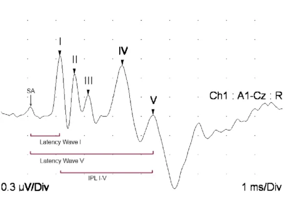
Latency
Latency is measured from the stimulus artifact to the peak of the wave and is measured in milliseconds (ms).[1] The stimulus artifact is the deflection associated with the stimulus at the beginning of the test, which is often removed in new software programs and therefore not visible.[1] The latency corresponds to the time taken by the action potential to travel through one neural structure to another. Absolute latencies are important to consider and references exist for numerous species. However, absolute latencies are not as clinically significant as interpeak latencies (IPL). IPLs from wave I to III and wave I to V represent the conduction time through the early brainstem components and entire brainstem respectively. The latter is referred to as the central conduction time.[1, 33] Once electrical activity is generated in the cochlear nerve, action potentials are propagated in a time locked manner. Therefore, in cases of peripheral hearing loss due to otitis or ruptured tympanic membrane, despite prolongation of wave I latency due to sound attenuation in the middle ear, the IPL will be normal as electrical activity will continue at the same speed through the brainstem once the activity has been initiated. As well, if wave I latency is normal and conduction through the brainstem is slowed due to disease, the IPL will be prolonged which localizes a problem to the brainstem.[1, 33] Latencies are influenced by numerous pathologic and non-pathologic factors and these factors should be taken into consideration before making any conclusions in regards to latency. In order to measure latency, waves must be present. In cases of peripheral deafness, latencies cannot be evaluated due to the absence of any waves. Any defects to the generator of wave I, the cochlear nerve, greatly inhibits the
evaluation of the BAER as the influx of auditory stimulation by this nerve is required to produce the BAER.
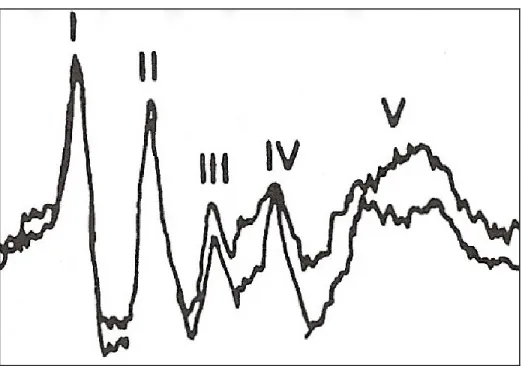
Figure 6: BAER wave latencies and interpeak latency (IPL) in a calf (95dB, alternating clicks, 11.3 clicks/sec, 500 responses). SA = stimulus artifact.
5.3.
Amplitude and ratios
Absolute amplitude of waves is measured in microvolts and is generally measured from the highest point of the upward deflection to the lowest point of the downward deflection.[1] Amplitude can vary significantly even in the same patient with the same parameters. It is also influenced by numerous factors such as electrode placement, stimulus intensity and stimulus rate.[1] As well, amplitudes become less reliable with peripheral hearing loss. The use of absolute amplitudes clinically in cases of otitis media-interna is therefore not useful. However, calculating an amplitude ratio can be
clinically useful to evaluate for the presence of brainstem disease.[1] Using the wave amplitudes from the same patient, the wave V/wave I ratio can be used for interpretation. A reduction in this amplitude results from a decrease in wave V amplitude in relation to wave I. This implies reduced conduction within the brainstem, as the generators of these waves differ with wave I generated in the peripheral nervous system while wave V is generated in the central nervous system. This value is only important in cases where brainstem disease is suspected and not of importance when evaluating the peripheral hearing apparatus.[1]
6. Non-pathologic factors that influence the BAER
Numerous factors can affect the BAER and knowledge of these confounding factors is necessary when performing and interpreting BAER results. Obviously, pathologic factors such as middle/inner ear disease or brainstem lesions will affect the BAER, however numerous non-pathologic factors can also significantly affect the results obtained.
6.1.
Equipment factors
Before performing the BAER, the operator should be aware of the various parameters of the machine that can affect the BAER tracing.
6.1.1.
Polarity of stimulus
When delivering the click stimulus, the tympanic membrane can be either pushed away from the sound or can be pulled towards the sound. The latencies and morphology of the waves differ depending on the polarity of the stimulus utilized. Condensation polarity is encountered when the tympanic membrane is pushed away from the sound source and causes a longer latency of wave I. Rarefaction polarity is encountered when the tympanic membrane is pulled towards the sound source and causes the shortest wave I latency and better separates waves IV and V in humans and waves III and IV in dogs.[1, 22] Alternating polarity uses both condensation and rarefaction and alternates between the two in subsequent clicks. Rarefaction polarity is recommended as the polarity of
choice when performing the BAER because wave I amplitude tends to be larger which in turns allows easier identification of this most important wave.[1] However, alternating polarity is recommended if a large stimulus artifact is encountered or wave I cannot be easily identified.[1]
6.1.2.
Number of clicks per second
Due to the necessity of signal averaging to produce the BAER waveforms, the click is repeated numerous times, at least 500 and up to 2000 times.[1] The number of clicks delivered per second influences the amount of time that it takes to perform the BAER and also influences the results of the BAER. As a general rule, 10 clicks per second is the recommended value in a clinical setting in adults.[1] As stimulus rates increase there is a progressive decrease in waveform resolution with concurrent increased latency and decreased amplitude of all BAER waveforms. Using high stimulus rates has clinical application in humans when high stimulus rates are used to screen children for hearing deficits.[1] Using high stimulus rates reduces the time to perform the BAER which may be an advantage in veterinary medicine when patient co-operation and movement complicate testing. When the number of clicks per second is increased to greater than 20/sec, BAER waveform latency increases and amplitude decreases therefore high stimulus rates are not recommended.[22]
6.1.3.
Intensity
The intensity of the click stimulus is measured in decibels (dB) and is determined by the evaluator. The unit of measure is based upon the base standard scale, dB sound pressure level (SPL). The dB SPL is the root mean square pressure of a sound relative to the lowest intensity that can be heard by the most sensitive ear.[1] Determining the intensity for the BAER test is dependent upon the click threshold for the species tested. Click threshold is determined by decreasing the intensity of the click by 5 dB intervals until the click is no longer heard. Click threshold is then defined as the midpoint between the two intensities that the click could no longer be heard. Once the click threshold is determined, the BAER is usually performed at 65-70 dB higher than the threshold to obtain diagnostic results.[1] The latency and amplitude of BAER waveforms are greatly influenced by the stimulus intensity. With a progressive increase in the stimulus intensity, more and more fibres are recruited with eventually all fibres, including the fastest conducting fibres, conducting the action potential. With the fastest conducting fibres conducting the electrical activity, the fastest latency is obtained. Therefore, at supra-maximal stimulation, the fastest conducting fibres are recruited and the most accurate representation of the conduction velocity is obtained.[1] As the intensity increases, the waveform latency decreases and amplitude increases. The exact opposite trend is observed as the stimulus intensity decreases with increasing latency and decreasing amplitude of all waveforms. The most resilient wave, which is the last to disappear with reducing stimulus intensity, is wave V.[1] This phenomenon is observed in conductive deafness, which acts to attenuate the stimulus intensity and mimics the effects seen with reducing stimulus intensity.[1, 9, 33]
6.2.
Patient factors
6.2.1.
Age
Maturation of the central nervous system occurs with age due to increased myelination of axons and increased efficiency of post-synaptic transmission.[1] The central nervous system is more mature at birth in precocious species such as chickens, horses, cattle and other ungulates compared to altricous species such as dogs, cats, monkeys and humans. As the nervous system is more mature in precocious species, less maturational changes are observed with age.[35] In the bovine species, the BAER and other evoked potentials (Visual Evoked Potential, Electroretinogram) are similar to adult values at birth and minimal changes are observed with age.[37] In comparison, significant maturational changes occur in humans with the BAER reaching adult values at 1 year of age.[1] Similarly, dogs demonstrate changes with age and obtain adult values at 6 - 8 weeks of age.[35] With increasing maturation, latencies decrease and amplitudes increase.[1, 35] At the other end of the spectrum, changes to the BAER are also observed with increasing age into adulthood. In humans, a small increase in latencies of all waves is noted into advanced age. Increases in BAER latencies have also been observed in dogs older than 10 years of age.[1, 28, 38]
6.2.2.
Species
BAER differs according to the species tested and therefore using reference values from one species cannot be extrapolated to another. Latencies, amplitudes and even waveform morphology can differ depending on the species evaluated due to differing anatomy,
neural generators and head size.[1, 34, 39] For example, latency of wave I in adult dogs is much faster at 1.21 ms compared to humans at 1.7 ms. Amplitudes are also greater in dogs compared to humans, horses and cows.
TABLE I – BAER Latencies (in ms) of 5 Different Species. [1, 20-22, 40]
Human (60dB) Cow (105dB) Horse (90dB) Dog (90dB) Cat (90dB)
Wave I 1.7 +/- 0.15 1.48 +/- 0.14 1.44 +/- 0.07 1.21 +/- 0.02 1.02 +/- 0.04 Wave II 2.8 +/- 0.17 2.48 +/- 0.17 2.70 +/- 0.23 2.00 +/- 0.02 1.84 +/- 0.04 Wave III 3.9 +/- 0.19 3.67 +/- 0.19 3.81 +/- 0.50 2.78 +/- 0.05 - Wave IV 5.1 +/- 0.24 - 4.58 +/- 0.25 - - Wave V 5.7 +/- 0.25 4.65 +/- 0.27 5. 87 +/- 0.59 3.65 +/- 0.06 3.53 +/- 0.04 Wave VI - - - - 4.31 +/- 0.12 6.2.3.
Size
In humans, a correlation has been demonstrated between BAER latencies and size of the head. This relationship is likely due to the size difference of the brainstem in humans with different head sizes. With larger heads, the brainstem is proportionally larger which infers a longer auditory pathway. Larger head sizes have longer BAER latencies due to the increased length of travel.[1] A similar correlation was found when evaluating BAER in the horse compared to the pony with longer latencies found in the horse.[21] This correlation has also been demonstrated in dogs when comparing different sized breeds.[34] Given this conclusion, BAER reference values should be determined for each breed in dogs given the extremes in size between different breeds. This factor is not
as important in the bovine species as the head size of most breeds of cattle is similar. However, this factor should be kept in mind when evaluating the BAER of smaller breeds or younger adult animals that have smaller heads. The breed of animal does not play as important a role compared to the size of the head.
6.2.4.
Sex
Again in humans, a correlation between sex and BAER has been demonstrated with women having shorter BAER latencies than men. However, this relationship may be related to the smaller head size of women compared to men and may not actually be related to the sex.[1] Controversy exists about this conclusion and similar results have not been found in other mammals.
6.2.5.
Temperature
BAER latencies are significantly affected with decreasing body temperature. This effect has been demonstrated in both human and veterinary medicine.[1] Latencies increase by approximately 0.2 msec with every 1 degree Celsius drop in temperature.[1] It is important when performing BAER under general anesthesia or heavy sedation that the body temperature of the patient is kept within reference limits to prevent falsifying latencies.
6.2.6.
Pharmacologic agents
The BAER is not significantly affected by the mental state of the patient or by most sedatives or anesthetics. No significant difference is observed when evaluating the BAER in the awake, asleep or comatose patient making this test highly valuable to evaluate brainstem function in debilitated patients that are non-responsive.[1] As well, most tranquillizers, sedatives, hypnotics and general anesthetic medications do not significantly alter BAER latencies or waveforms.[1] Even with induction of coma with barbiturates, the BAER is recordable. In dogs, BAER latencies were found to be prolonged with the use of thiopental yet in cats, anesthetic doses of barbiturates and ketamine did not alter the BAER.[38, 41] It must be noted that when using general anesthesia to perform the BAER that the patient’s body temperature often decreases and this factor may play a more important role in the prolonged latencies rather than the anesthetic medication.[1] No alteration in the BAER was found in horses sedated with acepromazine or in cattle sedated with xylazine.[21, 26]
Other medications can significantly alter the BAER and knowledge of these medications is important to consider before evaluating the BAER. Aminoglycoside antibiotics are ototoxic and have been demonstrated to significantly alter the BAER in humans and cattle.[1, 23] In addition, intoxicating doses of alcohol has significant effects on BAER in humans.[1]
7. Pathologic factors that influence the BAER
The BAER is a useful diagnostic test to evaluate the presence of hearing loss due to peripheral or central lesions. Central lesions of the brainstem can also be identified that do not necessarily cause hearing loss. Despite the ability to identify anatomically where a lesion exists, the BAER cannot give any etiologic information to the cause of the lesion.
7.1.
Sensorineural hearing loss
Sensorineural hearing loss is due to the dysfunction of any portion of the auditory pathway that decreases the threshold of audition. BAER is used frequently to diagnose sensorineural hearing loss due to congenital deafness in dogs, cats, and humans.[1, 32, 42] Cochlear agenesis and spiral organ degeneration manifest as a flat line BAER with no generation of wave I or subsequent waves.[32, 42]
7.2.
Conductive hearing loss
Conductive hearing loss is observed in otitis externa, otitis media, tympanic membrane rupture or damage to the ossicular chain.[9, 33, 43] Sound transmission to activate the auditory receptors in the inner ear is slowed down or attenuated which leads to prolonged latency of wave I and subsequent waveforms, and decreased amplitude of waves.[28] The BAER abnormalities of conductive hearing loss are identical to the changes observed from decreasing the auditory stimulus.[1] As the lesion lies
peripherally, the conduction time through the brainstem is normal and therefore the interpeak latencies are not affected.[1]
7.3.
Brainstem disease
The BAER evaluates both peripheral and central components of the auditory pathway and a lesion within the brainstem will cause BAER abnormalities. Abnormalities such as prolonged wave latency, reduced wave amplitude and even absence of waveforms can be observed depending on the severity of the lesion. In brainstem disease, waves I and II can be expected to be normal as the generators of these waves are located peripherally. Waves III to VII can be abnormal as well as the interpeak latencies. Even with crossing over of impulses within the brainstem at numerous levels, the BAER abnormalities are generally observed ipsilateral to the brainstem lesion.[1]
33
SUMMARY OF LITERATURE REVIEW
The anatomy of the ear is complex and has numerous associated neurological structures and functions. Complete neurological examination can identify neurological deficits that may localize a lesion to the middle or inner ear however similar deficits can be observed with central brainstem disease. Otoscopic examination of the ear can be helpful to evaluate for ear disease however is limited to visualization of the external ear and tympanic membrane. The BAER is an electrodiagnostic test that is non-invasive and sensitive to objectively evaluate the auditory pathway from the inner ear through the brainstem. Abnormalities of the BAER can help localize a lesion to the peripheral or central nervous system auditory components. Numerous pathologic conditions can affect the BAER and an abnormal result can aid in localizing an anatomic location for the lesion. However, an abnormal BAER result cannot give etiologic information as to the cause of the lesion. The BAER is useful to evaluate sensorineural or conductive hearing loss and brainstem disease. Numerous equipment and patient factors can influence the BAER and must be taken into consideration before analyzing the results.
34
ARTICLE
Predictive values of neurological examination, otoscopic examination and
brainstem auditory evoked response (BAER) in calves with otitis media-interna.
Andrea M. Finnen DVM, ACVIM (Neurology); David Francoz DMV, MSc, ACVIM (Large Animal); Frédéric Sauvé DMV, MSc, ACVD; Laurent Blond Dr Vét, MSc, ACVR; Malcolm J. Gains DVM, PhD, ACVP; and Joane M. Parent DMV, MVetSc, ACVIM (Neurology)
From the Département de sciences cliniques, Faculté de Médecine Vétérinaire, Centre Hospitalier Universitaire Vétérinaire, 1525 des Vétérinaires, Saint-Hyacinthe, Québec J2S 7C6
This manuscript represents a portion of a thesis submitted by Dr. Finnen to the
Université de Montreal Département de Sciences Cliniques as partial fulfillment of the requirements for a Master of Science degree.
Funded by Fonds du centenaire and Fonds de recherche Clinique Pfizer
Acknowledgements: The authors would like to thank Robert Barger for assistance with data collection and Guy Beauchamp for assistance with statistical analysis.
35
Abstract
Objective: The purpose of this study was to evaluate predictive values of neurological examination, otoscopic examination and BAER in calves for the diagnosis of otitis media-interna using computed tomography as the gold standard. The second objective was to define BAER reference values in normal calves and describe abnormalities in calves affected with otitis media-interna.
Design: Prospective study.
Animals: Seventeen Holstein calves between 5 and 7 weeks of age.
Procedures: All calves had a neurological examination, otoscopic examination and BAER. Calves were sedated with intravenous xylazine (0.05-0.15 mg/kg [0.02-0.07 mg/lb]) for computed tomography of the tympanic bullae to evaluate for the presence of otitis.
Results: Based upon computed tomographic results, 11 of 17 calves were affected with otitis media, 4 unilaterally and 7 bilaterally. Five waveforms were consistently identified on BAER traces from 6 normal calves. The positive predictive value of BAER,
neurological examination and otoscopic examination were 94.7%, 91.7% and 66.7% respectively.
Conclusions and Clinical Relevance: Clinically, the most reliable non-invasive
diagnostic test to diagnose otitis media-interna in the calf is BAER. Abnormalities were observed on BAER before the development of neurological deficits in approximately 40% of calves allowing earlier diagnosis.
36
Abbreviations: Computed tomography – CT
Brainstem auditory evoked response – BAER Interpeak latency - IPL
Introduction
Otitis is classified according to the anatomic portion of the ear that is affected. Otitis externa is inflammation of any structure from the pinna to the tympanic membrane, otitis media relates to inflammation of the tympanic cavity and/or auditory ossicles, and otitis interna to inflammation of the cochlear and/or vestibular labryrinths.1 Otitis media appears to be a common disease of young calves however the true prevalence has not been determined. Reported morbidity rates in calves range from less than 1% to 30%, depending on the geographical region and season.2-5 Otitis has been reported in calves as early as 4 days of age, however most calves are affected between 3 and 8 weeks of age.2,5-7 It has been suggested in previous reports that males appear to be more affected than females.5,8 However, a retrospective study at a University referral hospital indicated significantly more affected females.7 This over-representation may be due to the higher economic value of female calves and the subsequent referral for treatment.
Numerous etiologic agents have been implicated, mainly of bacterial origin such as
Mycoplasma bovis,4-9 Pasteurella haemolytica,2 Pasteurella multocida,2,3 Actinomyces pyogenes, Streptococcus pneumoniae,2 Corynebacterium pseudotuberculosis,3 Escherichia coli, Pseudomonas spp. and Acinetobacter spp.10 Infection of the middle ear can occur due to ascending infection via the Eustachian tube which is thought to be
37
the most common, secondary to bacteremia, or from direct extension of otitis externa.9,11-13 Respiratory infection appears to be a common predisposing and concomitant disease.5,7,8,11 Clinical signs of otitis media include facial nerve dysfunction such as incomplete eyelid closure, facial asymmetry and ‘dropped’ ear as the facial nerve travels in close association with the tympanic bulla.12 Other clinical signs may include discharge from the external ear, fever, epiphora, and decreased appetite.2,3,5,7,8,10 Some reports suggest that otitis media can be clinically undetected with the actual prevalence being higher than initially thought.2,6 Otitis media can progress to involve the inner ear and cause vestibular dysfunction such as head tilt and ataxia and infrequently, pharyngeal weakness and reduced esophageal motility.14 Calves can be affected unilaterally or bilaterally and reported incidence of bilateral disease ranges from 11-47%.2,7,9 Despite bilateral disease, affected calves may only have clinical signs unilaterally.11 As otitis media is frequently associated with otitis interna, they are often referred together as one entity (otitis media-interna). 11,15,16
The antemortem diagnosis of otitis is based upon the presence of appropriate clinical signs and adjunctive diagnostic imaging including radiography and computed tomography.7,13,14 In a recent study, CT was demonstrated to be superior to radiography for the diagnosis of otitis media in calves.17 Due to the associated costs and limited availability of CT, this imaging modality is not frequently utilized in the diagnosis of otitis in calves. However, given the almost perfect correlation with necropsy diagnosis in this study, this modality can be used as the gold standard antemortem diagnostic test.17
38
Brainstem auditory evoked response (BAER) is a non-invasive diagnostic test evaluating the auditory pathway from the internal ear through the brainstem.18 BAER tracings can be abnormal with otitis externa or otitis media due to sound transmission failure, or, due to deafness secondary to auditory receptor damage in otitis interna.18-21 BAER reference values have been described in the adult cow and it has been demonstrated that the BAER in the calf is similar to that of the adult.22-24 In addition, a case report exists of BAER in a calf with otitis and in a study evaluating ototoxicity of neomycin.25,26 However, the validity of this diagnostic test in the diagnosis of otitis media-interna has not been reported in the bovine species. In addition, the BAER abnormalities associated with otitis media-interna have not been described outside of individual case reports.
This prospective study evaluates predictive values of neurological examination, otoscopic examination and BAER testing in calves with otitis media-interna using CT as the gold standard for diagnosis. The BAER reference values in normal calves as well as BAER abnormalities in calves with otitis media-interna are also described.
Materials and Methods
Fifteen dairy calves randomly obtained from local producers purchased for an unrelated study were subsequently recruited for the study of otitis. The calves had no history or clinical signs of otitis at the time of purchase. Two client owned calves referred for suspicion of otitis were also included in this prospective study. There were 14 males and
39
3 females and all calves were between 5-7 weeks of age ranging in weight from 38.5-56.5kg. The calves were housed individually in pens at the Centre Hospitalier Universitaire Vétérinaire (CHUV) bovine clinic. A complete physical examination and otoscopic examination were performed followed by BAER testing. Neurological examination was performed as previously described and otitis was suspected based upon the presence of peripheral facial and/or vestibular nerve deficits.15 The otoscopic examination was performed by using a hand-held otoscope with a plastic 7-mm cone introduced into the external ear canal.a Each ear was evaluated for signs of otitis externa (erythema, purulent secretions, erosions or ulcers, and ceruminous gland hyperplasia). The integrity of the tympanic membrane was also evaluated for signs of perforation, deformation or increased opacity that were considered suggestive of otitis media. The study was approved by the Comité d’éthique de l’utilisation des animaux (CÉUA).
BAER tracings were recorded on a Cadwell Sierra Wedge II® electromyogram/evoked potential systemb. The majority of calves remained in sternal recumbency during the examination however a few preferred to stand. An alternating (rarefaction and condensation) click stimulation was delivered via internal ear plugsc with disposable ear tipsd placed in the external auditory meati. Tracings were obtained at intensity levels of 80 db, 90 db and 95 db nHL with a stimulation rate of 11.33 repetitions per second (Hz). White (masking) noise was applied to the contralateral ear at an intensity of 30 dB lower than the testing ear stimulus. Three sub-dermal stainless steel needle electrodese were placed as follows: the reference electrode at the vertex on midline, the active electrode just ventral to the tympanic bulla of the ear being tested, and the ground electrode over
40
the dorsal neck midway from the head to the withers. Amplifier sensitivity was set at 1 μV/division, sweep speed at 1ms/division with a bandpass filter of 100 Hz/3KHz and automatic artefact rejection. Each trace was the average of 500 responses recorded over the first 10 ms following acoustic stimulation. Both ears were tested at least twice at each intensity and were verified to be repeatable in all calves giving a total of 6 traces per calf (12 traces in total). Data was analyzed using a computer based programf. A 1 ms correction was applied to account for tubal insert delay. Waves I through V were identified using manually placed cursors. Absolute latencies for peaks I, II, III, IV and V as well as I-III and I-V interpeak latencies (IPL) were calculated for each trace. Increased latency was defined as a value greater than the mean plus 3 standard deviations.18
Calves were then sedated with xylazine (Bayer Inc® Toronto, Ontario) intravenously at 0.05-0.15mg/kg (0.02-0.07 mg/lb) for CT of the tympanic bullae. Non contrast-enhanced computed tomography for all calves was performed using an on-site, single-detector row, third generation scanner.g Contiguous 2-mm transverse slices were obtained at 1-mm intervals from the temporomandibular joints to the caudal margin of the petrous temporal bones. Computed tomography parameters were 130 kVp, 105 mA, and a 1 sec tube rotation time. Images were reconstructed with a bone algorithm, field of view (FOV) of 20 cm and matrix of 512 x 512. Images of each bulla were evaluated in DICOM format on a diagnostic work stationh by a board-certified radiologist. Images were evaluated for the presence of increased soft tissue opacity within the bulla, thickened bulla wall, enlarged bulla, and osteolysis of the bulla wall and trabeculations
41
indicating the presence of otitis, as previously described.17 Each bulla was then considered unaffected or affected with otitis media based upon the previously mentioned criteria.
The sensitivity, specificity and predictive values were calculated for each diagnostic test using CT as the gold standard. The BAER wave latencies were analyzed with a repeated measures linear model with side (left or right) and dB level as within-subject factors and their interaction. The pairs of means were compared using a priori contrasts with the sequential Bonferroni procedure. This procedure adjusted comparison-wise alpha levels to ensure a family-wise level of 5%. The level of statistical significance was set at p < 0.05.i
Results
Based upon CT images17, 11 of 17 calves were affected with otitis, 4 unilaterally and 7 bilaterally giving a total of 18 affected bulla. Of the calves affected unilaterally, all were affected on the left side. Prevalence of otitis in this group of calves was 64.7%.
Neurological Examination
The neurological examination identified 9 calves with neurological deficits, 6 had evidence of unilateral neurological deficits and 3 had evidence of bilateral neurological deficits. Seven affected bullae (38.9%) were not associated with neurological deficits (false negatives). One false positive was identified, i.e. unilateral mild facial weakness
42
was diagnosed in a calf without otitis media. Of the 8 remaining affected calves with neurological deficits, 5 had unilateral left facial nerve deficits and 3 had bilateral facial nerve deficits with unilateral vestibular deficits. Facial nerve deficits ranged from mild paresis characterized by ear droop and incomplete eyelid closure to facial paralysis. Vestibular deficits were characterized by head tilt to the side of the lesion and mild vestibular ataxia. No pathologic nystagmus was noted in any of the 3 calves with vestibular dysfunction. Based upon gold standard CT diagnosis, the neurological examination correctly identified 11/18 affected bullae giving a sensitivity of 61.1%, specificity of 93.8%, positive predictive value of 91.7% and negative predictive value of 62.3%. [Table 1].
Otoscopic Examination
Otoscopic examination was well tolerated in all calves. The tympanic membrane was not easily visualized due to the long and curved horizontal canal. Brown, ceruminous secretions in the external canal were infrequently encountered (27.8%) consistent with mild otitis externa. No ear mites were observed. In only 27.8% of cases, copious amounts of white to yellow mucoid discharge were observed in the external ear canal consistent with tympanic membrane rupture secondary to otitis media. Otitis media was suspected based upon the presence of tympanic membrane rupture, the presence of copious amounts of white to yellow mucoid discharge in the external canal, or deformation/mass effect of the external canal at the region of the tympanic membrane.27 Based upon gold standard CT diagnosis, the otoscopic examination correctly identified
43
10/18 affected bullae giving a sensitivity of 55.6% and specificity of 68.8%, positive predictive value of 66.7% and negative predictive value of 57.9% [Table 1].
When results of both the neurological and otoscopic examinations were combined, more true positives were identified giving a sensitivity of 72.2% and specificity of 62.5% [Table 1]. The combined predictive values were similar to those of the otoscopic examination with a positive predictive value of 68.4% and negative predictive value of 66.7%.
BAER
The BAER tracings of normal calves consistently identified five waves: I, II, III, IV and V [Figure 1]. As the sound intensity increased, absolute latencies decreased. The latencies for all waves were significantly longer at 80 dB compared to 95 dB (p<0.001). There was no statistical significant difference between values obtained at 90 dB compared to 95 dB (p>0.05). The interpeak latencies (IPL I-V and III-V) were not statistically different at different dB levels (IPL I-V: p=0.06, IPL III-V: p=0.90). The 95 dB waveforms were used to calculate reference latency values (Table 2). BAER abnormalities were noted in all (100%) calves affected with otitis and included increased wave latency of more than 1 wave (44.4%), absence of more than one waveform (11.1%), and absence of all waveforms (44.4%). [Figures 2 and 3] One trace was abnormal in an unaffected bullae (false positive). Based upon gold standard CT diagnosis, the BAER correctly identified 18/18 affected bullae giving a sensitivity of
![Figure 1 – Normal anatomy of the human ear. [12]](https://thumb-eu.123doks.com/thumbv2/123doknet/12166280.313163/12.918.171.740.549.973/figure-normal-anatomy-human-ear.webp)
![TABLE I – BAER Latencies (in ms) of 5 Different Species. [1, 20-22, 40]](https://thumb-eu.123doks.com/thumbv2/123doknet/12166280.313163/38.918.162.848.313.628/table-i-baer-latencies-ms-different-species.webp)