The present work was possible by a fellowship from CIFRE (2012/0572, jointly financed by the BIOMIN Holding GmbH, Association Nationale de la Recherche Technique (ANRT) and the laboratory of INRA-ToxAlim, UMR 1331.
I would like to thank all researchers who have accepted to be part of my jury:
Prof.Chiara Dall’Asta, Dr. Sven Dänicke and Prof. Alexis Valentin, who accepted to evaluate my work.
Dr. Wulf-Dieter Moll, for having accepted to be my co-director during this PhD, for his support and all his advice and for trusting me during these 4 years.
Dr. Isabelle Oswald :
Isabelle, merci à toi de m’avoir donné ma chance en stage d’ingénieur, puis d’avoir continué à me faire confiance pour une thèse. Merci de m’avoir permis de faire de nombreuses expérimentations, supplémentaires, non prévues… Je te remercie de m’avoir permis de participer à de nombreux congrès et réunions à l’étranger qui m’ont fait énormément progresser et évoluer! Enfin, merci de m’avoir suivie, conseillée, à toute heure, jour et nuit, 7 jours sur 7, pour rendre sans cesse mon travail meilleur. Ta capacité de travail et de gestion de nombreux projets simultanément est impressionnante. En un mot j’ai adoré ma thèse sous ta direction.
BIOMIN :
I would like to thank the company for funding my thesis, allowing me to make many varied experiments in my research during my PhD. I thank BIOMIN France and BIOMIN Austria, as well as Gerd Schatzmayr, Chirstian Tenier, Wulf-Dieter Moll to make possible this collaboration. They gave me the opportunity to visit them several times in Austria, go to several congresses where I met many researchers. All of this was very rewarding. Dieter, Gerd, it was a great pleasure to work with you during this period.
Merci à tous les collaborateurs qui ont participé directement ou indirectement à la réalisation de ces travaux. Merci à Ting Zhou et à Franz Berthiller pour nous avoir procuré les différentes molécules nécessaires à mes analyses.
Un grand merci au Professeur Ana-Paula Loureiro Bracarensede l’école vétérinaire de Londrina au Brésil, pour avoir réalisé toutes les coupes et analyses histologiques des expérimentations in vivo. J’ai été enchantée de te rencontrer et de bénéficier de tes conseils.
Je tiens aussi à remercier toutes les personnes qui m’ont accompagnée pendant ces années de travail au sein de ToxAlim, de près ou de loin, et que j’ai eu le plaisir de
rencontrer ; qui m’ont aidé, conseillé, soutenu, encouragé et avec qui j’ai eu le plaisir de papotter!
A ma chère équipe, tu vas me manquer! Ces 4 années et 4 mois tout compris, que j’ai passé au sein de cette équipe ont été super, riches en enseignement et en rencontre. Merci à tous les membres qui la composent, qui font fonctionner la machine et qui font en sorte que notre séjour, plus ou moins long, soit toujours agréable ! Ce sera difficile de trouver une équipe aussi chouette, avec des personnes aussi travailleuses, dynamiques, enjouées et compétentes que celles qui la compose.
Joëlle, telle une abeille butineuse tu t’affaires sans relâche pour que toute la ruche fonctionne correctement et ait toujours assez pour se nourrir et fonctionner! Merci Joëlle.
Philippe, tel le sage de la montagne, tu sais tout ! Toujours patient, tu écoutes et promulgue soit un bon conseil, une astuce ou encore des précautions! Je suis contente d’avoir pu travailler avec toi.
Anne, que dire…, peut-être que de passer la meilleure des journées c’est de la passer avec toi et les cochons! Anne je te souhaite tout le meilleur pour la suite et encore plus.
Je remercie aussi tous les autres membres de l’équipe pour leur disponibilité, conseils et bonne humeur : Olivier, Sophie, Soraya, Sylviane, Jean-Denis, Arlette ; cela a été un plaisir de travailler à vos côtés.
Une pensée à tous les stagiaires, thésards, post-docs que j’ai eu le plaisir de rencontrer et de côtoyer de plus ou moins près pendant ma thèse, et pour tous les bons moments passés en vôtre compagnie : Pascal, Sabria, Julien, Julie, Joanna, Rhoda, Patricia, Yann, Stella, Anwar, HK, Stéphanie, Florian, Inès, Pauline, Duncan, Joanne, Leticia, Viviane, Sophal, Joya… j’espère que je n’oublie personne. Aux post-docs, Delphine, Selma et Imourane courage pour la suite et les concours !
Pendant cette thèse j’ai eu l’occasion de rencontrer des étudiantes supers (eh oui que des filles), dynamiques, intelligentes et courageuses, pour certaines prêtes à faire leurs études loin de leur famille. Les filles tout le meilleur pour la suite avec une super thèse, un super post-doc et un super échantillon à la clé… ! ;)
Elisabeth, Marion, Yann et Sarah, merci pour tous les moments de rigolades partagés, les longues discussions…, les aprems détentes/jeux, tout le meilleur pour la suite ! Courage Marion et Sarah bientôt la fin pour vous aussi !;)
Une pensée spéciale aux filles sublimes et exotiques! ;), qui m’ont soutenu, aidé avec plein d’entrain et de bonnes humeur!
Joanna, Rhoda, Joanne, Joya et Christelle, merci de m’avoir fait découvrir la culture libanaise à travers vous. Christelle merci énormément pour tes corrections d’anglais de dernière minute !
Isaura, Amaranta, associées aux brésiliennes, merci de m’avoir fait découvrir un brin de votre culture et la joie de vivre des filles d’Amérique du Sud !;)
Bianca, j’ai été ravie de découvrir ton beau pays !
Je tiens aussi à remercier et à citer Beyoncé pour son soutient (même si elle n’est pas au courant) pendant toutes ces soirées de travail et de rédaction… : « I'm a survivor, I'm not gonna give up, I'm not gon stop, I'm gon work harder… ».
Une pensée pour tous mes cochons, décédés mais non moins aimés. A mon Marshmallow trop vite parti…, ainsi que Baloo et tous les autres.
Un grand merci à mon club de Kung Fu, mon équipe de volley et à mes compagnons de sorties roller et danse…, qui m’ont permis de bien me défouler et de me vider la tête au besoin.
Merci à tout mon entourage, de plus ou moins loin, qui m’ont soutenu, réconforté et aussi permis de me changer les idées, merci d’être là ! Je citerai en particulier Sarah (ma BF), Florent (mon BF), Christelle pour votre soutien actif !
Un mot à ma famille (plus ou moins éloignée), inébranlable, parfaite, qui m’a toujours soutenue, encouragée, portée, cette thèse est aussi pour vous. Je vous aime : Jacky, Jojo, Maman.
Original papers
Pierron A., Mimoun S., Murate L. S., Lippi Y., Loiseau N., Bracarense A-P., Liaubet L., Schatmayr G., Berthiller F., Moll W.D. and Oswald I. P. Intestinal toxicity of the masked mycotoxin deoxynivalenol-3-β-D-glucoside (D3G). 2016. Archive of Toxicology
Pierron A., Mimoun S., Murate L. S., Lippi Y., Loiseau N., Bracarense A-P., Zhou T., Schatmayr G., Moll D. and Oswald I. P. 2016. Microbial biotransformation of DON reduces its intestinal toxicity. Scientific Report (In press).
Pierron A., Cossalter A-M., Bracarense A-P., Schwartz-Zimmermann H., Pinton P., Laffitte J., Schatzmayr G., Moll D. and Oswald I. P. Evaluation of metabolisation and toxicity of purified deepoxy-deoxynivalenol (DOM-1) in piglets (Under writing).
Review
Pierron A., Alassane-Kpembi I., Payros D., Pinton P. and Oswald I. P. Masked mycotoxins : the risk on porcine production ? 2016. 48th days of Journée de la recherche Porcine (JRP).
Pierron A., Alassane-Kpembi I. and Oswald I. P. 2016. Impact of mycotoxins on immune response and consequences for pig health. Animal Nutrition.
Pierron A., Alassane-Kpembi I. and Oswald I. P. 2016. Feed mycotoxins: impact on pig intestinal health. PHM (Under review).
Communications (Oral and Poster)
A. Pierron, S. Mimoun, L. S. Murate, Y. Lippi, L. Liaubet, F. Berthiller, G. Schatmayr, W.D. Moll and I. P. Oswald. Intestinal toxicity of the masked mycotoxin deoxynivalenol-3-β-D-glucoside (D3G). Day of PhD student of SEVAB, Toulouse (France), March 18, 2016. (Oral).
A.Pierron and I. P. Oswald. Masked mycotoxins : the risk on porcine production ? Journée de la recherche Porcine, February, Paris (France), February3-4, 2016. (Oral)
A.Pierron, S. Mimoun, L. S. Murate, Y. Lippi, L. Liaubet, F. Berthiller, G. Schatmayr, W.D. Moll and I. P. Oswald. Microbial de-epoxidation of deoxynivalenol (DON) decreases its immunotoxicity for piglets. EVIW2015, Vienna (Austria), September 2-4, 2015. (Oral)
A.Pierron, S. Mimoun, L. S. Murate, Y. Lippi, L.Liaubet, F. Berthiller, G. Schatmayr, W.D. Moll and I. P. Oswald. Intestinal toxicity of the masked mycotoxin deoxynivalenol-3-β-D-glucoside (D3G). Journée des doctorants, Toxalim (Toulouse), June 26, 2015. (Oral)
Mimoun S., Pierron A., L. S. Murate, Y. Lippi, L.Liaubet, F. Berthiller, G. Schatmayr, W.D. Moll and I. P. Oswald. Intestinal toxicity of the masked mycotoxin deoxynivalenol-3-β-D-glucoside. 37th Mycotoxin Workshop, Bratislava (Slovakia), June 1-3, 2015.
A.Pierron, S. Mimoun, L. S. Murate, Y. Lippi, L.Liaubet, F. Berthiller, G. Schatmayr, W.D. Moll and I. P. Oswald. Intestinal toxicity of the masked mycotoxin deoxynivalenol-3-β-D-glucoside (D3G). EFS13, Martina Franca (Italy), May 10 - 14, 2015. (Oral).
Souto P.C.M.C., Jager A.V., Tonin F.G., Bordin K., Pierron A., Cossalter A.M., Laffitte J., Oswald I.P., Oliveira C.A.F. 2014. Immune response and urinary fumonisin B1 excretion in piglet fed low dietary levels of Fumonisin. The World Mycotoxin 8th Forum Conference, Vienna (Austria), November 10-12, 2014.
Pierron A., Mimoun S., Lippi Y., Gourbeyre P., Liaubet L., Zhou T., Berthiller F., Schatmayr G., Moll D. and Oswald I.P. Plant or microbial transformation of DON reduce its intestinal toxicity. The 6th World Nutrition Forum, Munich (Germany), October 15-18, 2014. (Poster)
Pierron A., Mimoun S., Lippi Y., Gourbeyre P., Liaubet L., Zhou T., Berthiller F., Schatmayr G., Moll D. and Oswald I.P. Masked DON and other DON metabolites do not exert intestinal toxicity : Transcriptomic analysis of jejuna explants. The World Mycotoxin 8th Forum Conference, Vienna (Austria), November 10-12, 2014. (Poster)
Pierron A., Mimoun S., Lippi Y., Gourbeyre P., Liaubet L., Zhou T., Berthiller F., Schatmayr G., Moll D. and Oswald I.P. Plant or microbial biotransformation of DON reduce its intestinal toxicity. 36th Mycotoxin Workshop, Göttingen (Germany), June 16 - 18, 2014. (Oral)
F. Accensi, P. Pinton, M. Sibila, A. Pierron, A.M. Cossalter, J. Segalés, I.P. Oswald. Effect of deoxynivalenol (DON) intoxication on a PCV2 infection in piglets. 23th International Pig Veterinary Society Congress, Cancun (Mexico), June 8-11, 2014.
Mimoun S., Pierron A., Lippi Y., Gourbeyre P., Liaubet L., Zhou T., Berthiller F., Schatmayr G., Moll D. and Oswald I.P. La décontamination biologique du DON réduit sa toxicité intestinale chez le porc. 5ème journée Mycotoxines, France (Montpellier), Juin 5-6 , 2014,
Awards and distinctions
Marasas Oral Prize Winner :
A.Pierron, S. Mimoun, L. S. Murate, Y. Lippi, L.Liaubet, F. Berthiller, G. Schatmayr, W.D. Moll and I. P. Oswald. Intestinal toxicity of the masked mycotoxin deoxynivalenol-3-β-D-glucoside (D3G). EFS13, Martina Franca (Italy), May 10 - 14, 2015. (Oral).
TABLE OF CONTENTS
List of Abbreviations
List of tables and figures
INTRODUCTION
I.Context general of study
II.Problem of DON contamination III.Literature review
A. Mycotoxins in Feed: impact on pig intestinal health (Review n°1)………... Introduction
1. Toxicity of the main mycotoxins in pig feed 2. Effects of mycotoxins on the pig intestine
3. Consequences of intestinal toxicity of mycotoxins for pig health Conclusion
References
B. Impact of mycotoxin on immune response and consequences for pig health (Review n°2)…. Introduction
1. Effect of major mycotoxins on the pig immune response
2. Consequences of mycotoxin induced immunomodulation for pig health 3. The problem of mycotoxins co-contamination
Conclusion References
C. Masked mycotoxins: a risk in pig production? (Review n°3)……….…….… Original Version (French)
English Version Introduction
1. The mycotoxins regulated in pig’s feed 2. ”Modified” mycotoxins in pig feed
3. Assessment of the exposure and characterization of the risk for the “modified” mycotoxins in pig
Conclusion References
AIMS OF THE THESIS
EXPERIMENTAL STUDIES
I. Toxicity assessment of derivatives of deoxynivalenol (DON)...
A. Intestinal toxicity of the masked mycotoxin deoxynivalenol-3 -glucoside (Article n°1).. Introduction
Materials and Method Results
Discussion References
B. Microbial biotransformation of DON: molecular basis for reduced toxicity (Article
n°2)... Introduction
Materials and Methods Results 17 25 28 38 39 59 69 115 129 13 23 105 111 21 113
Discussion References
II. In vivo toxicity of purified deepoxy-deoxynivalenol (DOM-1) in piglets (Article
n°3)... Introduction
Materials and Method Results
Discussion References
GENERAL DISCUSSION & PERSPECTIVES
I. Discussion……….…
A. Discussion on the analysis performed in the thesis……….…
1. In silico analysis of the interaction between DON, DON derivatives and the ribosome 2. Pan genomic analysis of the effect of DON and its derivatives
3. The in vivo experimental protocol
B. Toxicity assessment of biological strategies to reduce toxic effects of DON…………....……
1. Efficiency of detoxifying strategies a) Bacterial transformation b) Plant transformation II.Perspectives………..…
CONCLUSION
183 207 223 209 216 209 212
List of Abbreviations
15ADON 15-acetyl-deoxynivalenol
3ADON 3-acetyl-deoxynivalenol
ADONs acetylated forms of deoxynivalenol
AFB1 aflatoxin B1 AFB2 aflatoxin B2
AFs aflatoxines
ATP adenosine triphophaste
BC before Christ
BW Body Weight
Caco-2 human colonic adenocarcinoma cell line
DOM-1 deepoxy-deoxynivalenol DON deoxynivalenol DON15GlcA deoxynivalenol-15-β-D-glucuronide DON3G deoxynivalenol-3-β-D-glucoside DON3GlcA deoxynivalenol-3-β-D-glucuronide DON3S deoxynivalenol-3-α-sulfate DON-GlcA deoxynivalenol-glucuronide 3-epi-DON 3-epi-deoxynivalenol
EFSA European Food Safety Authority
ERK extracellular signal-regulated kinases
EtOH ethanol
F fumonisin
FB1 fumonisin B1
FB2 fumonisin B2
FHB Fusarium head blight
GSTs glutathione S-transferases
HT2-Glc HT-2 toxin
HT2-Glc HT-2 toxin-glucoside
IARC International Agency for Research on Cancer
IC50 half maximal inhibitory concentration
Ig immunoglobulin
IL interleukin
ILSI International Life Science Institute IPEC-1 porcine intestinal epithelial cell line
IPEC-J2 porcine intestinal epithelial cell line derived from the jejunum JECFA Joint FAO/WHO Expert Committee on Food Additives
LOAEL low observed adverse effect level
MAPK mitogen-activated protein kinases
mRNA messenger ribonucleic acid
NA not available
NIV nivalenol
NS non significant
OTA ochratoxine A
OVA ovalbumin A
PAT patulin
PCR polymerase chain reaction
PCV2 porcine circo virus type 2
PHA phytohemagglutinin
ppm part per million
PRRSV porcine reproductive and respiratory syndrome virus
PTC peptidyl transferase center
q-PCR quantitative-PCR
RIN RNA integrity number
RNA ribonucleic acid
rRNA ribosomal ribonucleic acid
RT reverse transcriptase
SEM standard error mean
T-2 T-2 toxin
T2-G T-2 toxin-glucoside
TEER trans epithelial electrical resistance TCT trichothecene
TDI tolerable daily intake
Th T helper cell
TNF-α tumour necrosis factor-α
ZEN zearalenone ZEN14G zearalenone-14-glucoside ZEN14S zearalenone-14-sulfate ZEN16G zearalenone-16-glucoside α-ZEL α-zearalenol β-ZEL β-zearalenol
List of figures and tables
List of figures and tables not included in paper or review: from Introduction, Chapter 2 and General Discussion & Perspectives parts.
Introduction
p21: Figure 1 – Major mycotoxins produced by fungi and naturally found in several food products
p22: Figure 2 – Structural diversity of major mycotoxins
p23: Figure 3 – “Saint Antoine tentation” painted between 1512 – 1516 by Grünewald (on the left). Ears of rye contaminated with ergot (on the right).
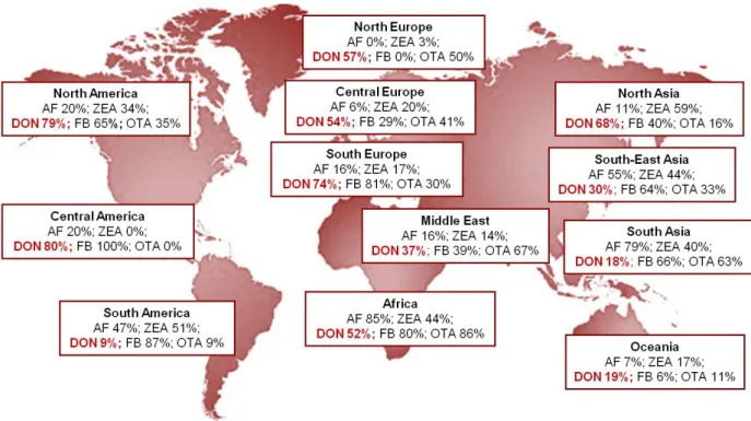
p24: Figure 4 - Global mycotoxin prevalence in surveyed regions.
Chapter 2
p175: Figure 1 - Experimental design of the in vivo experiment
p179: Figure 2 - Mean weight gain per piglets measured on animals submitted to the gavage to treatment
p180: Figure 3 - Mean weight gain per piglets measured on animals submitted by gavages to treatment
p181: Figure 4 - Histological intestinal samples from piglets submitted by gavage to treatment
p182: Figure 5 - The mRNA expression levels of markers of inflammation
p183: Figure 6 - Histological liver samples from piglets submitted by gavage to treatment p184: Figure 7 - Histological spleen from piglets submitted by gavage to treatment p184: Figure 8 - Total Immune response for each treatment
p185: Figure 9 - IgG-antiOVA during the experiment per treatment
p189: Annex 1- BELECLA: Complete feed for first age piglet p190: Annex 2 - STIMIOUTI : Complete feed for second age piglet
Discussion general & Perspectives
I.
Context of the study
Plants and cereals are subject to numerous fungal contaminations occurring either in fields or during storage processes. Many species of fungi exist and are able to grow on various types of cereals (maize, wheat, barley, soybean, rice, rye…) or on food commodities (seeds, peanuts, fruits, spices, forages…) (Figure 1) (AFSSA 2009).
Figure 1 – Major mycotoxins produced by fungi and naturally found in several food products
These fungi are able to produce several toxic molecules, called mycotoxins (from Greek μύκης (mykes, mukos) ‘fungus’ and Latin (toxicum) ‘poison’) (Aiko and Mehta 2015). Mycotoxins are secondary metabolites; and unlike primary metabolites, they are not essential to the development and survival of the fungus but could constitute an advantage during the colonization of ecological niche when in competition with other microorganism. These molecules also discourage predators from eating the fungus (Keller et al. 2005).
Mycotoxins can be divided into polyketoacids, terpenes, cyclopepetides and nitrogen metabolites, depending on their origins and their structures (AFSSA 2009). They can also be classified according to their toxic effects. Mycotoxins considered important in terms of food safety are aflatoxins (AF), ochratoxins (OT) (in particular ochratoxin A (OTA)), patulin (PAT), fumonisins (FB), zearalenone (ZEA) and trichothecenes (TCT), especially deoxynivalenol (DON) (Figure 2) (Bennet and Klich 2003). Several factors control fungal growth and mycotoxin production, such as weather conditions, agricultural practices or storage conditions (Hesseltine 1976).
Figure 2 – Structural diversity of major mycotoxins
The toxic effects of moulds and fungi were already known in ancient times. Historically, many illnesses linked to mycotoxicoses have been reported (AFSSA 2009). The most famous case, which occurred in the Middle Ages, is known under the name of ignis sacer (sacred fire) or St Anthony's fire. It was caused by toxins of Claviceps purpurea, the ergot alkaloids of rye (Figure 3). Ergotism reached epidemic proportions, mutilating and killing thousands of people in Europe. Victims of ergotism suffered from delirium, prostration, acute pain, abscess and gangrene of the extremities, leading to serious and incurable infirmity. Epidemics occurred from the 8th to the 15th century due to the bad quality of food and contamination with fungal sclerotia. Similarly, fusariotoxins (toxin T2 and ZEA),
seem to have been involved in the decline of the Etruscan civilization 5 centuries B.-C (Richard 2003). “Yellow rice disease” or shoshin-kakke disease in Japan was also a mycotoxicosis caused by unhygienic conditions and practices, which is induced by the Citreoviridin, a metabolite produced by Penicillium citreonigrum. This fungus used to grow readily on rice during its storage (after harvest), especially in the colder regions of Japan. New hygiene measures applied, more rigorous than before, made it disappear (Udagawa and Tatsuno 2004). In 1960, the turkey X disease has been an important episode of mycotoxicosis on animals. It killed thousands of turkey, ducklings and other domestic animals in England. This allowed the discovery of aflatoxins, main mycotoxin produced by Aspergillus flavus, and present in a high quantity in groundnut flour designed for poultry’s food (Blount 1961).
Figure 3 – “Saint Antoine tentation” painted between 1512 – 1516 by Grünewald (on the left). Ears of
rye contaminated with ergot (on the right).
Most mycotoxins have an acute toxicity, but nowadays it’s exceptional to be exposed to such high doses in Europe (Européenne C. 2003). In this part of the world chronic contamination is the most threatening, due to the persistence of these mycotoxins in food and the repeated ingestion by animals for example. In 2004, a worldwide survey showed that 72% of more than 19000 samples analyzed contained detectable amounts of AF, FB, DON, ZEA or OTA (Figure 4). Among them, 38% represented a co-contamination by 2 or more mycotoxins (Schatzmayr and Streit 2013). Several toxic effects can be induced, depending of the mycotoxin and the organ targeting. At high doses, mycotoxins exposure can leads to general cytotoxicity, biochemical lesions and impact on early cellular functions in the cascade of events (Bryden 2012; Maresca and Fantini 2010). At low doses, various functions of tissues
and organs can be impaired. And some mycotoxins are also genotoxic, carcinogenic and teratogenic (Maresca and Fantini 2010).
Figure 4 - Global mycotoxin prevalence in surveyed regions (adapted from Schatzmayr and Streit
2013). Aflatoxins (AF), zearalenone (ZEA), deoxynivalenol (DON), fumonisins (FB), ochratoxin A (OTA).
II.
Problem of DON contamination
A.
Occurrence
DON is produced by Fusarium fungi, one of the most common mycotoxin in the world. It and can be found in many cereals and raw materials, like wheat, barley, oat, rye, maize and sometimes on rice, sorghum and triticale. A worldwide survey to assess the contamination by mycotoxins in feed and feed raw materials, done on 19,000 samples, shows that DON was present in 56% of the tested samples (Schatzmayr and Streit 2013).
Fungal infection and DON production are difficult to predict and regulate; because largely dependent on the weather, high humidity and low temperature, and so vary greatly from year to year and between areas (Rotter et al. 1996). In developed countries, where storage conditions are well managed and controlled, DON contamination is especially a pre-harvest problem. While, in developing countries, DON can also be produced during the storage stage. So, DON can be commonly detected at low levels (< 1 ppm) and sporadically at higher levels (5 to 20 ppm) on cereals intended to be given to animals or humans (Abouzied et al. 1991). It
can also be present in end products, such as the cereal-based food for adults and infants or even at low levels in beer (Lombaert et al. 2003; Scott 1996).
Economic losses due to DON contamination are difficult to evaluate. Nevertheless a computer simulations evaluated that the annual costs for DON in the USA were $637 million in crop losses (mainly wheat and corn), $18 million in feed losses and $2 million in livestock losses (EFSA 2013).
B.
Toxicity
The toxicity of DON is well known and numerous studies bring information on its toxic effects at high and chronic doses (Maresca 2013; Pestka 2010; Wang et al. 2014). A high concentration of DON causes effects and symptoms that are similar to those observed during an exposure to ionizing radiation, such as abdominal distress, salivation, discomfort, diarrhea, vomiting, leukocytosis and gastrointestinal bleeding. It also has high emetic and anorexic effects, that are equal or even higher to those of observed with the most toxic trichothecene B (Pestka and Smolinski 2005). Actually, the first name of DON was “vomitoxin” due to its emetic effects seen in pigs (Vesonder et al. 1973).
A chronic exposure can impact growth (by anorexia and disregulation of nutrients efficacy), immunity (increased or decreased) and reproduction in animals. At acute doses it can induces emesis, abdominal distress, malaise, diarrhea and increases the salivation (Pestka 2010). At low dose it impairs the growth and the immune function in human and interferes with nutritional efficiency on pigs (Rotter et al. 1996). At higher doses it causes diarrhea, emesis, leukocytosis, hemorrage, endotexemia and ultimately shock-like death (Ueno 1983).
C.
Detoxification methods for DON
The effectiveness of detoxification methods of mycotoxins depends on several parameters, the nature of the food/feed, the environmental conditions such as moisture content, temperature, as well as the type of mycotoxin, its concentration and the extent of binding between mycotoxin and constituents (Grenier and Oswald 2011).
DON resist to most of the industrial processes; it is stable at high temperature, due to its high chemical stability and can be found in numerous final processed products (Hazel and Patel 2004). Actually DON is completely stable at 120°C, quite stable at 180°C and partially stable at 210°C (OMS 2001). At concentrations below 1mg/kg, DON is mainly found on the
seed surface but at higher concentrations, it can be found in the entire grain (Charmley and Prelusky 1994). To reduce the occurrence and the impact of mycotoxins and especially DON, several detoxifying strategies were established in the feed chain, including the prevention of fungal growth and the production of mycotoxin, strategies to reduce or eliminate mycotoxins from contaminated raw or finished materials or even in diverting contaminated product to low risk uses including animal feeds (Bryden 2012). However, the amount of information related to mycotoxins detoxifying methods is still limited. From the described detoxifying strategies there are three principal categories used: the physical, the chemical and the biological methods.
1.
Physical methods
Some processes used to detoxify mycotoxins (such as milling, irradiation, ethanol fermentation or extrusion) were initially developed for other purposes, and some were specifically developed for the detoxification itself (such as sorting, cleaning or washing). These practices, are linked to the FAO guidelines, namely fulfilled: cheap and simple, no production of toxic metabolites, and no change in the nutritional value or properties of raw materials. However, all these approaches present some inconvenient. The standard processes, like milling and baking, do not allow the elimination of DON with efficacy (Abbas et al. 1985; Hart and Braselton 1983). Dry milling, permit an elimination that is up to 40% of DON present in the flour; sieving or cleaning can reduce the concentration in DON by over 60% (Pestka and Smolinski 2005). The problem of the milling and grain separation process, commonly used for human’s food, is that it concentrates all the mycotoxins in bran and Germ, fractions will be used later for animal feed. However, in the sieving and cleaning procedures, an important loss of grains is reported. In their study, Trenholm et al. (1991) did observe a 73% reduction of DON, but they have also observed that up to 69% of the total weights of the corns was removed as well. And after flotation and washing, the cost of drying grains is high.
2.
Chemical methods
Several chemical processes, using molecules like ammonia, calcium hydroxide, chlorine, hydrochloric acid, ozone, sodium bisulphate and sodium hydroxide are able to degrade DON. In fact ammoniation has been proved to reduce the aflatoxin levels but this process is not accepted in all countries and is quite expensive (Norred et al. 1991; Park and
Price 2001). With alkalization, DON can be transformed into different products, with various toxicity (Bretz et al. 2005; Bretz et al. 2006; Young et al. 1986).
In majority, the chemical methods can reduce mycotoxins’ levels, but they can also severely damage the nutrient quality of the grains and can be health hazards on their own. Not only that, they can result in the formation of degraded products that might be constituted of new and unknown biologically active mycotoxins (Humpf and Voss 2004).
3.
Biological methods
Two strategies are possible to manage DON, once present in plants and cereals. The first strategy consist on preventing the production of DON in infected crop by controlling the plant pathogens (Fusarium spp.). Equipping crops with DON detoxification activities can reduce the concentration of mycotoxin in grain and also increase the resistance against infection (Karlovsky 2011). It was shown that DON plays a role in the infection; host plants inoculated with fungal strain not able to produce TCT can’t be able to infect the plant (Maier et al. 2006; Proctor et al. 1995). Another study shows that a major QTL responsible for the resistance of wheat to FHB co-segregated with the ability to detoxify DON by glycosylation (Lemmens et al. 2005); It has been proved that by selecting a plant naturally resistant to
Fusarium, its capacity to glycosylate DON into D3G can be increased by 2.7 times more
(Sasanya et al. 2008).
Some companies also tried to build transgenic plants, by transferring the 3-O-acetyltransferase gene issued from F. sporotrichioides to the plant in order to reduce the pathogenicity of Fusarium (Karlovsky 2011).
The second strategy consists in detoxifying DON that has been produced, by physical and chemical methods as we saw but more innovative by biological methods. The bio-detoxification of mycotoxins, by isolating microorganisms and/or enzymes that will degrade or metabolize the mycotoxins, is currently an innovative and promising strategy aiming to control mycotoxicoses in animals (Schatzmayr et al. 2006). (Cheng et al. 2010) obtained two Bacillus strains able to detoxify DON in wheat and maize. In another study, Bacillus sp. LS100, which transforms deoxynivalenol (DON) to a less toxic chemical de-epoxy DON (DOM-1) has been assessed. This intestinal bacteria, Genus novus species novus of family Coriobacteriaceae BBSH 797, isolated from digestive tracts, is able to de-epoxydize DON to DOM-1 (Fuchs et al. 2002). There is also the bacterial strain Devosia mutans 17-2-E-8, isolated from an alfalfa soil enriched with F. graminearum-infested corn that is able to highly
reduce DON level, in producing an epimer, the 3-epi-DON (He et al. 2015b; Zhou and He 2009, 2010).
Bringing the enzymatic kit to animals, by the use of bacteria, will allow them detoxify mycotoxins, and easily and effectively protect them against the toxic effects of mycotoxins. Definitely, since decontaminated or detoxified crops are cheaper (since they are considered as products of lower quality), they are mainly used for feed production and animal feeding (Grenier and Oswald 2011), in which explains why animals are very exposed. The need of feed additives preventing the absorption of mycotoxins and by that occurrence of their toxic effects in farm animals has increased significantly. Indeed, the adsorption is not a viable option regarding trichothecenes, zearalenone and ochratoxins, that’s why the mycotoxin inactivation by biotransformation is a very promising strategy to detoxify these mycotoxins. However, all the additives and bacterial products have to be tested before coming into the market to assure their efficiency and safety. In vitro and in vivo tests are mostly important to check and follow their scientific development and improvement. Sensitive parameters such as biochemistry, gross pathology, histopathology, immune parameters and animal performances have to be measured to evaluate their toxicity. This is why the aim of this thesis was to evaluate the toxicity of the products issued from biological detoxification and to assess the efficiency of this process in order to protect animals or humans from the toxic effects of DON.
References
Abbas HK, Mirocha CJ, Pawlosky RJ, Pusch DJ. 1985. Effect of cleaning, milling, and baking on deoxynivalenol in wheat. Appl Environ Microbiol 50:482-486.
Abouzied MM, Azcona JI, Braselton WE, Pestka JJ. 1991. Immunochemical assessment of mycotoxins in 1989 grain foods: Evidence for deoxynivalenol (vomitoxin) contamination. Appl Environ Microbiol 57:672-677.
AFSSA. 2009. Evaluation des risques liés à la présence de mycotoxines dans les chaînes alimentaires humaine et animale. Rapport final.
Aiko V, Mehta A. 2015. Occurrence, detection and detoxification of mycotoxins. J Biosci 40:943-954. Bennett JW, Klich M. 2003. Mycotoxins. Clin Microbiol Rev 16:497-516.
Blount WP. 1961. Turkey "X" Disease. Turkeys 9.
Bretz M, Beyer M, Cramer B, Humpf HU. 2005. Synthesis of stable isotope labeled 3-acetyldeoxynivalenol. Mol Nutr Food Res 49:1151-1153.
Bretz M, Beyer M, Cramer B, Knecht A, Humpf HU. 2006. Thermal degradation of the fusarium mycotoxin deoxynivalenol. J Agric Food Chem 54:6445-6451.
Bryden WL. 2012. Mycotoxin contamination of the feed supply chain : Implications for animal productivity and feed security. Animal Feed Science and Technology 173:134-158.
Charmley LL, Prelusky DB. 1994. Decontamination of fusarium mycotoxins. Mycotoxins in grain-compounds other than aflatoxin. Miller, JD & Trenholm HL (Eds), St Paul, Minnesota:Eagan Press 421-435.
Cheng B, Wan C, Yang S, Xu H, Wei H, Liu J, et al. 2010. Detoxification of deoxynivalenol by bacillus strains. J Food Safety 30:599-614.
EFSA. 2013. Deoxynivalenol in food and feed: Occurrence and exposure. EFSA Journal 11.
Européenne C. 2003. Scoop task 3.2.10. Collection of occurrence data of fusarium toxins in food and assessment of dietary intake by the population of eu members states, substask i : Trichothecenes. European Commission, Directorate General Health and Consumer Protection, Brussels:11-237. Fuchs E, Binder EM, Heidler D, Krska R. 2002. Structural characterization of metabolites after the microbial degradation of type a trichothecenes by the bacterial strain bbsh 797. Food Addit Contam 19:379-386.
Gonzalez-Vallina R, Wang H, Zhan R, Berschneider HM, Lee RM, Davidson NO, et al. 1996. Lipoprotein and apolipoprotein secretion by a newborn piglet intestinal cell line (ipec-1). Am J Physiol 271:G249-259.
Grenier B, Oswald IP. 2011. Mycotoxin co-contamination of foods and feeds: Meta-analysis of publications describing toxicological interactions. World Mycotoxin J 4:285-313.
Hart LP, Braselton WE, Jr. 1983. Distribution of vomitoxin in dry milled fractions of wheat infected with gibberella zeae. J Agric Food Chem 31:657-659.
Hazel CM, Patel S. 2004. Influence of processing on trichothecene levels. Toxicol Lett 153:51-59. He JW, Yang R, Zhou T, Boland GJ, Scott PM, Bondy GS. 2015. An epimer of deoxynivalenol: Purification and structure identification of 3-epi-deoxynivalenol. Food Addit Contam Part A Chem Anal Control Expo Risk Assess 32:1523-1530.
Hesseltine CW. 1976. Mycotoxin research in india. Mycopathologia 58:157-163.
Humpf HU, Voss KA. 2004. Effects of thermal food processing on the chemical structure and toxicity of fumonisin mycotoxins. Mol Nutr Food Res 48:255-269.
Karlovsky P. 2011. Biological detoxification of the mycotoxin deoxynivalenol and its use in genetically engineered crops and feed additives. Appl Microbiol Biotechnol 91:491-504.
Lemmens M, Scholz U, Berthiller F, Dall'Asta C, Koutnik A, Schuhmacher R, et al. 2005. The ability to detoxify the mycotoxin deoxynivalenol colocalizes with a major quantitative trait locus for fusarium head blight resistance in wheat. Mol Plant Microbe Interact 18:1318-1324.
Lombaert GA, Pellaers P, Roscoe V, Mankotia M, Neil R, Scott PM. 2003. Mycotoxins in infant cereal foods from the canadian retail market. Food Addit Contam 20:494-504.
Maier FJ, Miedaner T, Hadeler B, Felk A, Salomon S, Lemmens M, et al. 2006. Involvement of trichothecenes in fusarioses of wheat, barley and maize evaluated by gene disruption of the trichodiene synthase (tri5) gene in three field isolates of different chemotype and virulence. Mol Plant Pathol 7:449-461.
Maresca M, Fantini J. 2010. Some food-associated mycotoxins as potential risk factors in humans predisposed to chronic intestinal inflammatory diseases. Toxicon 56:282-294.
Maresca M. 2013. From the gut to the brain: Journey and pathophysiological effects of the food-associated mycotoxin deoxynivalenol. Toxins 5:784-820.
Norred WP, Voss KA, Bacon CW, Riley RT. 1991. Effectiveness of ammonia treatment in detoxification of fumonisin-contaminated corn. Food Chem Toxicol 29:815-819.
OMS. 2001. Safety evaluation of certain mycotoxins in food. Deoxynivalenol. Food Additives Series 47:419-528.
Park DL, Price WD. 2001. Reduction of aflatoxin hazards using ammoniation. Rev Environm Contam Toxicol 171:139-175.
Pestka JJ, Smolinski AT. 2005. Deoxynivalenol: Toxicology and potential effects on humans. J Toxicol Environ Health B Crit Rev 8:39-69.
Pestka JJ. 2010. Deoxynivalenol: Mechanisms of action, human exposure, and toxicological relevance. Arch Toxicol 84:663-679.
Proctor RH, Hohn TM, McCormick SP. 1995. Reduced virulence of gibberella zeae caused by disruption of a trichothecene toxin biosynthetic gene. Mol Plant Microbe Interact 8:593-601.
Richard JL. 2003. Mycotoxins and human disease; in clinical mycology (eds) ej anaissie, mr mcginnis and ma pfaller. (New York: Churchill Livingstone):589-598.
Rotter BA, Prelusky DB, Pestka JJ. 1996. Toxicology of deoxynivalenol (vomitoxin). J Toxicol Environ Health 48:1-34.
Sambruy Y, Ferruzza S, Ranaldi G, De Angelis I. 2001. Intestinal cell culture models: Applications in toxicology and pharmacology. Cell Biol Toxicol 17:301-317.
Sasanya JJ, Hall C, Wolf-Hall C. 2008. Analysis of deoxynivalenol, masked deoxynivalenol, and fusarium graminearum pigment in wheat samples, using liquid chromatography-uv-mass spectrometry. J Food Prot 71:1205-1213.
Schatzmayr G, Zehner F, Taubel M, Schatzmayr D, Klimitsch A, Loibner AP, et al. 2006. Microbiologicals for deactivating mycotoxins. Mol Nutr Food Res 50:543-551.
Schatzmayr G, Streit E. 2013. Global occurrence of mycotoxins in the food and feed chain: Facts and figures. World Mycotoxin Journal 6:213-222.
Scott PM. 1996. Mycotoxins transmitted into beer from contaminated grains during brewing. J AOAC Int 79:875-882.
Udagawa S, Tatsuno T. 2004. [safety of rice grains and mycotoxins - a historical review of yellow rice mycotoxicoses]. Yakushigaku Zasshi 39:321-342.
Ueno Y. 1983. Trichothecenes: Chemical, biological, and toxicological aspects. Trichothecenes, ed Y Ueno Amsterdam: Elsevier Press:135-146.
Vesonder RF, Ciegler A, Jensen AH. 1973. Isolation of the emetic principle from fusarium-infected corn. Appl Microbiol 26:1008-1010.
Wang Z, Wu Q, Kuca K, Dohnal V, Tian Z. 2014. Deoxynivalenol: Signaling pathways and human exposure risk assessment--an update. Arch Toxicol 88:1915-1928.
Young JC, Blackwell BA, ApSimon JW. 1986. Alkaline degradation of the mycotoxin 4-deoxynivalenol. Tetrahedron Letters 27:1019-1022.
Zhou T, He J. 2009. Bacterial isolate, methods of isolating bacterial isolates and methods for detoxification of trichothecene mycotoxins. United States Provisional Patent Application No 61/249,03 Filed October 6, 2009.
Zhou T, He J. 2010. Bacterial isolate, methods of isolating bacterial isolates and mathods for detoxification of trichothecene mycotoxins. Patent Cooperation Treaty Application No61/249,023 Filed October 6, 2010 (International).
III. Literature review
The literature review consists on three reviews covering different aspects studied during this thesis. The first two reviews deal with the different effects caused by mycotoxins and the intestine or the immunity of pigs. The third review focuses on new forms of mycotoxins derived from these mycotoxins, the "masked" and "modified" mycotoxins.
To date, contamination by mycotoxins cannot be avoided. Mycotoxins can be present in several types of cereals (maize, wheat, barley, oats, rice ...) and end up in high concentrations due to cultural practices or storage. These mycotoxins can be found in co-contamination in pig feed. All these mycotoxins have toxic effects on pig that is particularly sensitive because of its simple digestive system and its high cereal rich diet. The first two reviews have a look on the two most affected parameters after a contamination by one or more mycotoxin, on the gut and the immune system of the pig. Mycotoxins contaminations, mainly by ingestion, cause many toxic effects on the digestive system and small intestine. The first review presents the different mycotoxins that are found in pig’s feed and their effects and consequences on the intestine and the general health of the pig. Mycotoxins have been also described as responsible of modifying important functions of the intestine (barrier function, mucus production, nutrient absorption…).
The second review reports the effect of mycotoxins on the immune system of pigs. Certainly, many mycotoxins have an immune-modulatory effect on the immune response and may affect the vaccine response as well as induce an increased susceptibility to infections or chronic infectious diseases.
Finally, the last review presents advances in terms of new analytical methods allowing the identification of new forms of mycotoxins, the mycotoxins called maskedand modified. It is important to study these new forms of mycotoxins to evaluate their impact on pig health and to assess whether they can represent an additional threat that will have to be taken into account in the overall management of the risk of mycotoxins.
A.
Feed mycotoxins: impact on pig intestinal health
Nowadays, many mycotoxins can contaminate cereals and feeding stuffs designated to the pig consumption. These mycotoxins have several toxic effects on pigs, which are greatly impacted, due to their high sensibility and their cereals rich diet.
Due to the way of exposure, by ingestion, intestine is the major organ targeted by mycotoxins. This review summarizes the major effects induced by these mycotoxins on the intestine, on its integrity, its biological function and on its immune response. It also highlights the consequences of this contamination, which increases the translocation of bacteria and enhances the susceptibility to other diseases and thus impairs the global health of pigs.
Mycotoxins in Feed: impact on pig intestinal health
1 2
Alix PIERRON1,2,3, Imourane ALASSANE-KPEMBI1,2 and Isabelle P. OSWALD1,2* 3
4
*
Corresponding author: Isabelle P. Oswald, isabelle.oswald@toulouse.inra.fr
5 6
1
INRA, UMR 1331, ToxAlim Research Centre in Food Toxicology, 180 chemin de Tournefeuille, 7
BP93173, 31027 Toulouse cedex 03, France. 8
2
Université de Toulouse, INP, UMR 1331, Toxalim, Toulouse, France 9
3
BIOMIN Research Center Technopark 1, 3430 Tulln, Austria 10
11
Abstract
12
Mycotoxins are secondary metabolites of fungi that grow on a variety of substrates. Due to their high 13
consumption of cereals and their sensitivity, pigs are highly impacted by the presence of mycotoxins. 14
Pigs can be exposed to different mycotoxins such as aflatoxins, ochratoxins, fumonisins, zearalenone, 15
and trichothecenes especially deoxynivalenol. At the European level, regulations and 16
recommendations exist for these mycotoxins in pig feed. The intestine is the first barrier to food 17
contaminants and can be exposed to high concentrations of mycotoxins upon ingestion of 18
contaminated feed. Mycotoxins target this organ, they alter the intestinal barrier, impair the immune 19
response, reduce feed intake and weight gain. Among them, deoxynivalenol and fumonisin have been 20
studied especially for their toxicity in the intestine. Their presence in feed increases the translocation 21
of bacteria; mycotoxins can also impair the immune response and enhance the susceptibility to 22
infectious diseases. In conclusion, because of their effect on the intestine, mycotoxins are a major 23
threat to pig health, welfare and performance. 24
25
Keywords:
26
Pig, mycotoxins, feed contamination, intestine, barrier function, immune response 27
Introduction
29
Food safety is a major issue throughout the world. In this respect, much attention
30
needs to be paid to the possible contamination of food and feed by fungi and the risk of
31
mycotoxin production. Mycotoxins are secondary metabolites produced by filamentous fungi,
32
mainly by species from the genus Aspergillus, Fusarium and Penicillium. They are produced
33
on a wide variety of substrates before, during and after harvest. Mycotoxins are very resistant
34
to technological treatments and difficult to eliminate, and therefore they can be present in
35
human food and animal feed. The ingestion of mycotoxin-contaminated feed can induce acute
36
diseases, and the ingestion of low doses of fungal toxins also causes damage in case of
37
repeated exposure.
38
Monogastric livestock, pig and poultry, are particularly vulnerable to mycotoxins
39
because of the high percentage of cereals in their diet and because they lack a rumen with a
40
microbiota able to degrade mycotoxins before their intestinal absorption. From a pig health
41
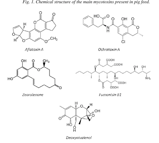
perspective, the most notorious mycotoxins (Fig.1) are aflatoxins (AF), ochratoxin A (OTA),
42
fumonisins B (FB), zearalenone (ZEN), and trichothecenes, especially deoxynivalenol (DON)
43
(CAST 2003).
44
This review will summarize the effect of mycotoxins on the intestine and analyze the
45
consequences in terms of pig health.
46 47
I- Toxicity of the main mycotoxins in pig feed
48
The toxicity of mycotoxins varies according to several parameters such as the dose, the duration of 49
exposure, the age and the sex of the animal, as well as nutritional factors (Andretta et al. 2012; Bryden 50
2007; Wild 2007). For example, the effects of AF, FB or DON on performance are greater in males 51
and young pigs (Andretta et al. 2012; Marin et al. 2006). In the European Union, only AFs are 52
regulated in animal feed; recommendations exist for OTA, DON, T2 and HT-2 toxins, FB1, FB2 and 53 ZEN (Tab. 1). 54 55 56 57 58 59 60 61 62 63
64
Fig. 1. Chemical structure of the main mycotoxins present in pig feed. 65
66
The AFs are rapidly absorbed and metabolized in the liver (Haschek et al. 2002); they
67
are hepatotoxic, and have some impacts on growth and on the immune response of the pig
68
(Meissonnier et al. 2006). OTA is nephrotoxic and hepatotoxic and its oxydative metabolites
69
are genotoxic (Aish et al. 2004; Pfohl-Leszkowicz and Manderville 2007). Among
70
mycotoxins, pigs are very sensitive to DON, the most common mycotoxin of the type B
71
trichothecene. Short exposure to high doses of DON induces vomiting and lower doses cause
72
feed refusal (Haschek et al. 2002); chronic exposure is associated with weight loss, anorexia,
73
immunomodulation and alteration of intestinal barrier functions (Haschek et al. 2002; Pestka
74
2010; Pinton and Oswald 2014). Type A trichothecene T2 and HT2 toxins have similar but
75
more pronounced effects than DON. They also induce irritation of the intestine and the skin
76
and increase the sensibility of pigs to diseases (Bryden 2012).
77 78 79 80 81 82 83 84
Table 1. Regulation and recommendations for the main mycotoxins present in pigs feed and feedstuffs. 85
(EC Directive 2002/32/EC, and EC Recommendations 2006/576/EC and 2013/165/EU) 86
87
Mycotoxins Pig feeds Max. content mg/Kg (ppm)
AFB1+ B2 Cereals 60
Complete and complimentary feeding stuffs for pigs, horse, rabbit and pets
0.5
OTA Complete and complimentary
feeding stuffs for pigs 0.05 DON Cereals
(without maize by-products)
8 (12) Complete and complimentary
feedstuffs for pigs 0.9 ZEN Cereals
(without maize by-products)
2 (3) Complete and complimentary
feeding stuffs: -for piglets and gilts -for sows and fattening pigs
0.1 0.25
FB1+FB2 Cereals 60
Complete and complimentary feeding stuffs for pigs, horse and rabbit
5
T2+HT2 Complete and complimentary feeding stuffs for animals -Oat milling products (husks) -Other cereals products -Compound feed, with the exception of feed for cats
1 0.5 0.25 88
FB1 is the most prevalent toxin of the fumonisin family. It has a carcinogenic effect in
89
humans and induces multiple toxic effects in different animal species. In pigs, this toxin
90
induces pulmonary oedema (Haschek et al. 2002) and alters the immune response with a
91
dysregulation of the T helper lymphocytes TH1/TH2 balance (Marin et al. 2006; Taranu et al.
92
2005). The last mycotoxin with a recommendation for pig feed is ZEN. This toxin has an
93
impact on pig fertility and reproduction. ZEN and its principal derivatives, α-zearalenol
(α-94
ZEL) and β-zearalenol (β-ZEL) (more toxic than the other two), are non-steroidal oestrogens
inducing an oestrogenic response in animals (Fink-Gremmels and Malekinejad 2007). In pigs,
96
especially young sows, ZEN induces red patching and tumefaction of the vulva, a prolapse of
97
the vulva and sometimes of the rectum (Gaumy et al. 2001).
98
In terms of intestinal toxicity, the effects of DON and FB have been studied in detail in pigs;
99
by contrast only few papers are concerned with the effect of OTA or AF on this organ.
100
101
II- Effects of mycotoxins on the pig intestine
102
The intestinal tract is the first target for mycotoxins following ingestion of
103
contaminated feed. The intestinal epithelium is a single layer of cells lining the gut lumen that
104
acts as a selective filter, allowing the translocation of dietary nutrients, essential electrolytes,
105
and water from the intestinal lumen into the blood circulation. It also constitutes the largest
106
and most important barrier to prevent the passage of harmful intraluminal substances from the
107
external environment into the organism, including foreign antigens, microorganisms, and their
108
toxins. Following the ingestion of mycotoxin-contaminated feed, intestinal epithelial cells
109
may be exposed to high concentrations of toxins, potentially affecting intestinal functions
110
(Alassane-Kpembi and Oswald 2015; Ghareeb et al. 2015; Grenier and Applegate 2013).
111 112
A. Effect on intestinal histomorphology
113
Consumption of mycotoxin-contaminated feed induces histological damage on
114
intestinal tissue. Epithelial lesions in the intestine of pigs fed with a diet naturally
115
contaminated with DON were observed (Bracarense et al. 2012; Eriksen and Pettersson
116
2004). Jejunal lesions, including shortened and coalesced villi, lysis of enterocytes, and
117
edema, were also observed in an ex-vivo model of intestinal tissues after exposure to DON
118
(Lucioli et al. 2013; Pinton and Oswald 2014). Exposure to FB also induces changes in
119
intestinal villi morphology such as reduced villi height and villi fusion and atrophy
120
(Bracarense et al. 2012).
121
A study on pigs showed that low doses of ZEN do not impair the morphology and
122
ultrastructure of the small intestine (Obremski et al. 2005), in contrast to what has been
123
observed in rats (Liu et al. 2014).
124
As far as AFB1 is concerned, no data on the effect of this toxin on the histomorphology of the
125
pig intestine are available. Nevertheless, exposure of broiler chicken to AFB1 induced a
decreased jejunal villus height, villus height/crypt ratio, and shedding of epithelial cells on the
127
tip of jejunal villi (Zhang et al. 2014).
128 129
B. Effect on intestinal digestion and nutrient absorption
130
The regressive intestinal lesions observed upon exposure to mycotoxins may explain,
131
at least in part, the reduced absorption of nutrients and the impaired digestion observed after
132
ingestion of mycotoxins. Pigs consuming corn culture extracts containing FB showed a
133
markedly lowered activity of aminopeptidase N (Lessard et al. 2009). Likewise, exposure to
134
1.5mg/kg b.w. FB1 has been shown to induce sphingolipid depletion in pig intestinal
135
epithelium, which can result in a deficiency of folate uptake (Grenier and Applegate 2013;
136
Loiseau et al. 2007). The sodium-glucose dependent transporter (SGLT-1) activity is
137
particularly sensitive to DON inhibition. SGLT-1 is the main apical transporter for active
138
glucose uptake in the small intestine. Inhibition of SGLT-1 has nutritional consequences and
139
could explain diarrhea associated with DON ingestion, since this transporter is responsible for
140
daily absorption of water in the gut (Maresca 2013). Conversely, sodium-dependent glucose
141
absorption might be up-regulated in pigs after acute or long term exposure to the mycotoxin
142
FB1 (Lessard et al. 2009).
143 144
C. Effect on barrier function
145
Several mycotoxins are able to alter intestinal barrier functions (Ghareeb et al. 2015;
146
Grenier and Applegate 2013). They affect the intestinal epithelium permeability through
147
modulation of the tight junction complexes. A defective expression of occludin and
E-148
cadherin has been observed in the ileum of piglets fed low doses of FB1 (Lucioli et al. 2013).
149
The FB-induced alteration of the sphingolipid biosynthesis pathway and the associated lipid
150
rafts could also contribute to impairing the establishment and maintenance of tight junctions.
151
Likewise, the activation of MAP kinases by DON affects the expression and cellular
152
localization of proteins forming or being associated with tight junctions such as claudins and
153
ZO-1, which results in increased intestinal paracellular permeability (Pinton and Oswald
154
2014). Similarly to DON, T2-toxin, FB1 and ZEN have been shown, in vitro and in vivo, to
155
impair the pig intestinal barrier function and to promote oral absorption of antibiotics such as
156
doxycycline, chlortetracycline and paromomycin (Goossens et al. 2012; Goossens et al.
157
2013).
D. Intestinal immune system
159
Some mycotoxins impact the systemic and/or the local immune response. At the
160
intestinal level, they decrease the immunity leading to enhanced intestinal infections. They
161
also have a direct or indirect proinflammatory effect (Cano et al. 2013; Maresca 2013).
162
Indeed, the intestine is a major source of cytokines and chemokines, molecules involved in
163
the regulation of the immune system. Among cytokines, IL-8, which is a chemoattractant
164
cytokine, is of particular interest because it is involved in the recruitment of
165
polymorphonuclear neutrophils at the site of infection, mediating a non-specific acute
166
inflammatory response.
167
Ingestion of FB1 specifically decreases expression of IL-8 mRNA in the ileum of
168
exposed piglets while expression of other inflammatory cytokines is not affected. This
169
decrease of IL-8 caused by FB1 may lead to reduced recruitment of inflammatory cells in the
170
intestine during infection, and may contribute to the observed increased susceptibility of FB1
-171
treated piglets to intestinal infections (Bouhet and Oswald 2007).
172
DON modulates intestinal innate immunity both directly (through activation of signal
173
pathways) and indirectly (through crossing of luminal bacterial antigens, which was observed
174
together with bacterial translocation following mucus layer alteration and tight junction
175
opening) (Maresca et al. 2008). DON affects expression of proteins involved in epithelial
176
innate immunity, including inflammatory cytokines, COX-2 and β-defensins (Cano et al.
177
2013; Lessard et al. 2015). Numerous studies have demonstrated that DON stimulates
178
expression and secretion of IL-8 and thus potentially participates indirectly in the central
179
effects of DON in terms of feed refusal and emesis. As described for immune cells (Pestka
180
2010), DON has a biphasic effect on the secretion of IL-8 by intestinal epithelial cells: Low
181
doses of toxin cause a massive increase in secretion of IL-8, whereas higher doses inhibit it.
182
Such a biphasic effect explains why DON acts: (i) as a proinflammatory toxin leading to
183
intestinal inflammation at low doses; and (ii) as an inhibitor of intestinal immunity leading to
184
higher susceptibility of animals to intestinal infections at higher doses (Maresca 2013).
185
The ability of ZEN to interact with the pig immune system has been poorly investigated.
186
However, it is known that exposure to high concentrations of ZEN (5-250mg/Kg feed or
200-187
1000 µg/Kg b.w./day) induces chronic inflammation of the genital tract in females pigs
188
(EFSA 2011; JECFA 2011). In vitro analyses also show that ZEN and its metabolites have
differential effects on synthesis of the inflammatory cytokines IL-8 and IL-10 in swine
190
intestinal epithelial cells (Marin et al. 2015).
191
There is no report of OTA- induced impairment of local immunity. However, this
192
mycotoxin decreases the level of inflammatory cytokines (TNF-alpha and IL-10) in the
193
plasma of exposed pig (Bernardini et al. 2014).
194 195
E. Intestinal microbiota
196
The gut hosts an important microflora. Surprisingly, the impact of mycotoxins on the
197
intestinal microflora has been poorly investigated. As far as pigs are concerned, only two
198
studies have investigated the impact of mycotoxins on the intestinal microflora (Burel et al.
199
2013; Wache et al. 2009). The first study indicates that consumption of feed naturally
200
contaminated with DON (2.8 mg/kg) for four weeks had a moderate effect on cultivable
201
bacteria in the pig intestine, but changed the microflora (Wache et al. 2009). In the second
202
study, pigs received feed contaminated with 12 mg FB/kg feed for 63 days. This diet
203
transiently affected the balance of the digestive microbiota during the first four weeks of
204
exposure; a co-infection with Salmonella typhimurium amplified this phenomenon (Burel et
205
al. 2013).
206
Two recent studies performed on rats have also demonstrated an effect of OTA and
207
AF on the intestinal microbiota (Guo et al. 2014; Wang et al. 2016). The effects of
208
mycotoxins on the intestinal microbiota are not surprising; indeed other secondary metabolites
209
produced by the same fungi, antibiotics, are well known for their effect on the gut flora.
210
Recent advances in next-generation sequencing technologies and metagenomics should give
211
us a comprehensive analysis of the effect of mycotoxins on the structure and function of gut
212
microbial ecosystem in the near future.
213 214
III- Consequences of intestinal toxicity of mycotoxins for pig health
215
A. Impairment of zootechnical performance
216
All damage induced by mycotoxins on the intestine level and on the different
217
functions lead to different symptoms expressed by the pig. Such symptoms are either directly
218
associated with local toxicity in the intestine, or indirectly with a systemic effect, and with
219
visible impact on the overall health of the pig.
The colloquial name of DON, vomitoxin, refers to its emetic effect observed both in
221
field reports and in experimental intoxications where high doses of the toxin were given orally
222
or intravenously to pigs. Complete feed refusal was observed at levels of 12 and vomiting at
223
20 mg DON/kg feed. Pig feeding trials with naturally or artificially contaminated diets have
224
shown decreased feed consumption and weight gain at doses from 0.6 to 3mg DON/kg feed
225
(Bracarense et al. 2012). A meta-analysis showed that deoxynivalenol reduced feed intake and
226
weight gain by 26%; the same analysis also demonstrated a 16% reduction of feed intake in
227
response to AFB1(Andretta et al. 2012).
228
Consumption of pure FB1 or FB1-contaminated feed also induces a slight reduction of
229
body weight in piglets. Although FB are poorly absorbed and metabolized in the intestine,
230
they induce intestinal disturbances (abdominal pain or diarrhea) and cause extra-intestinal
231
organ pathologies (pulmonary edema in pigs, leukoencephalomalacia in horses, or neural tube
232
defects in rodents).
233
Ingestion of ZEN and OTA doesn’t alter zootechnical performance (Bernardini et al.
234
2014; Schoevers et al. 2012). However ZEN can induce a decrease in reproductive
235
performance with a reduction of healthy follicles leading to premature oocyte depletion in
236
adulthood and so leading to abortion (Schoevers et al. 2012).
237 238
B. Bacterial translocation
239
The intestinal disturbance induced by mycotoxins may lead to increased bacterial
240
translocation across the intestine and increased susceptibility to enteric infections. The loss of
241
tight junction integrity and resulting increased paracellular permeability can lead to entry of
242
bacteria that are normally restricted to the gut lumen. Such an increase in bacterial passage
243
through intestinal epithelial cells after mycotoxin exposure has major implications for pig
244
health in terms of sepsis, inflammation and enteric infection.
245
Porcine ileal loops were used to reproduce Salmonella typhimurium induced intestinal
246
inflammation. Co-exposure to bacteria and DON dramatically enhances the inflammatory
247
response to S. typhimurium in the ileal loops, with a clear potentiation of expression of IL-1β,
248
IL-8 or IL-6 (Vandenbroucke et al. 2011). It has been suggested that this potentiation
249
coincided with significantly enhanced Salmonella invasion in and translocation over intestinal
250
epithelial cells.