DOI: 10.1126/science.1210240
, 1203 (2011);
334
Science
, et al.
Melanie Boly
Top-Down Processes in the Vegetative State''
Response to Comment on ''Preserved Feedforward But Impaired
This copy is for your personal, non-commercial use only.
clicking here.
colleagues, clients, or customers by
, you can order high-quality copies for your
If you wish to distribute this article to others
here.
following the guidelines
can be obtained by
Permission to republish or repurpose articles or portions of articles
):
February 3, 2012
www.sciencemag.org (this infomation is current as of
The following resources related to this article are available online at
http://www.sciencemag.org/content/334/6060/1203.5.full.html
version of this article at:
including high-resolution figures, can be found in the online
Updated information and services,
http://www.sciencemag.org/content/334/6060/1203.5.full.html#related
found at:
can be
related to this article
A list of selected additional articles on the Science Web sites
http://www.sciencemag.org/content/334/6060/1203.5.full.html#ref-list-1
, 7 of which can be accessed free:
cites 14 articles
This article
http://www.sciencemag.org/cgi/collection/neuroscience
Neuroscience
subject collections:
This article appears in the following
registered trademark of AAAS.
is a
Science
2011 by the American Association for the Advancement of Science; all rights reserved. The title
Copyright
American Association for the Advancement of Science, 1200 New York Avenue NW, Washington, DC 20005.
(print ISSN 0036-8075; online ISSN 1095-9203) is published weekly, except the last week in December, by the
Science
on February 3, 2012
www.sciencemag.org
Response to Comment on
“Preserved
Feedforward But Impaired Top-Down
Processes in the Vegetative State
”
Melanie Boly,1,2* Marta Isabel Garrido,2Olivia Gosseries,1Marie-Aurélie Bruno,1Pierre Boveroux,3 Caroline Schnakers,1Marcello Massimini,4Vladimir Litvak,2Steven Laureys,1Karl Friston2 Kinget al. raise some technical issues about our recent study showing impaired top-down processes in the vegetative state. We welcome the opportunity to provide more details about our methods and results and to resolve their concerns. We substantiate our interpretation of the results and provide a point-by-point response to the issues raised.
W
e thank Kinget al. (1) for deconstruct-ing our paper (2) showing impaired top-down processes in the vegetative state (VS). We hope our responses provide some useful clarifications.(i) Regarding the number of patients, it would have been disappointing not to have found a common abnormality in eight well-defined VS
patients. If we had needed 50 patients to obtain significant differences, we would probably end up reporting quantitatively trivial effects that had little diagnostic value [a well-known fallacy of classical inference (3)]. We consider the hetero-geneity as a strength of our cohort selection (2): We discovered a common mechanism underlying impaired consciousness, irrespective of its distal
causes and subsequent clinical course. The ability to generalize our finding would have been com-promised had we studied a more homogenous VS group.
(ii) Previous studies have provided incon-sistent results concerning the presence of mis-match negativity (MMN) in VS. Faugeraset al. (4) did not investigate the presence of a MMN (local effect) but rather show a global effect in 2 VS patients out of 27. Bekinschteinet al. (5) studied only 4 VS patients and failed to detect a MMN in some. References (6, 7) report consid-erable variability across studies, with a MMN in about 10 to 25% of patients. In short, a significant MMN, based on some threshold criteria, is not a generic characteristic of event-related potentials
TECHNICAL COMMENT
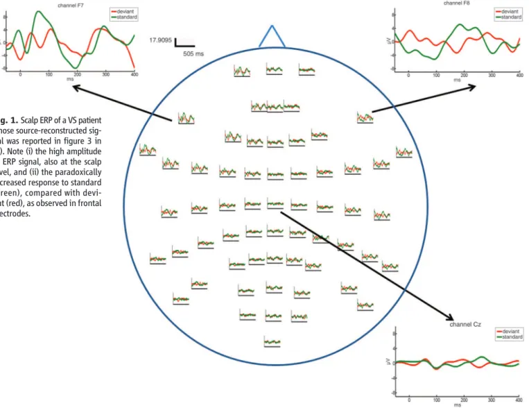
Fig. 1. Scalp ERP of a VS patient whose source-reconstructed sig-nal was reported in figure 3 in (2). Note (i) the high amplitude of ERP signal, also at the scalp level, and (ii) the paradoxically increased response to standard (green), compared with devi-ant (red), as observed in frontal electrodes.
1
Coma Science Group, Cyclotron Research Centre and Neu-rology Department, University of Liège and CHU Sart Tilman Hospital, Liège, Belgium.2Wellcome Trust Centre for
Neuro-imaging, Institute of Neurology, University College London, UK.3Anesthesiology Department, University of Liège and CHU Sart Tilman Hospital, Liège, Belgium.4Department of Clinical
Sciences,“Luigi Sacco,” University of Milan, Milan, Italy. *To whom correspondence should be addressed. E-mail: [email protected]
www.sciencemag.org SCIENCE VOL 334 2 DECEMBER 2011 1203-e
on February 3, 2012
www.sciencemag.org
(ERPs) in VS and is not a valid criterion for evaluating ERP data quality. Rather than assess-ing the presence of a threshold-based MMN, we examined correlations between ERP amplitude and the level of consciousness. We used a sum-mary statistic (random effects) approach in all our analyses, ensuring that group results could not be explained by a strong effect in a minority of subjects (8).
(iii) With regard to ERP components and their latencies, we analyzed the whole peristimulus time window and indeed observed an ERP com-ponent corresponding to P50 in VS. We were not modeling the MMN per se (i.e., the difference waveform) but used the roving paradigm to char-acterize network responses to all stimuli. Wave-form component latencies are defined using an ad hoc threshold on noisy time series, whereas dy-namic causal modeling (DCM) looks for differ-ences in the form of ERPs over all peristimulus time. To identify the MMN and reify it with a “latency” is not considered useful, necessary, or good practice in DCM.
(iv) It is not surprising that ERP topography is different in controls and VS patients, who are severely brain damaged. We used individual pa-tient anatomy to account for possible differences in head conduction when performing DCM source reconstruction. Worries about signal-to-noise ra-tios can be discounted because differences were significant at the between-subject level using classical inference. If the data were just random fluctuations, these tests would not be significant. Differences between our data and Kinget al.’s results (1) might be due to differences in the stimuli [see (9)].
DCM source reconstruction provides a rea-sonable account of the scalp ERP data of the VS patient displayed in figure 3 in (2). In particular, the amplitudes of both scalp (Fig. 1) and source-reconstructed ERPs are bigger than typically ob-served in controls [figure 1 in (2)]. At both levels, the patient’s frontal response to a “standard” is also bigger than the response to a“deviant.” Mere-ly observing ERP source reconstructions is insuf-ficient to assert anything about backward versus forward connections; this is the role of DCM. To test models with and without laterality differ-ences is another interesting issue, but not one that we have addressed.
(v) DCM implicit source reconstruction can efficiently reconstruct sources that are close to-gether (10, 11). Bayesian model selection (BMS) established that the use of five sources was the most appropriate for our data. ECD source re-construction using 64 electrodes has been shown to be as accurate as an extended setup (12), es-pecially when the data’s signal-to-noise ratio is low (13). Finally, DCM uses the whole ERP time window to optimize its source reconstruc-tion (10): Reconstructing only early components would not constitute a formal measure of inver-sion performance.
(vi) Our claim about preserved forward processes in VS was based on the involvement of frontal cortex in the generation of responses, as evidenced by BMS. At the level of quantitative parameter analyses, we can only reject the null hypothesis of no differences in the backward connections (because we used classical inference). This means that we can say nothing about the forward connections. We performed an addition-al anaddition-alysis of variance for repeated measures, searching for an interaction between forward and backward frontotemporal connection strength in VS patients compared with controls. This interaction did not reach statistical significance (P > 0.05). A failure to demonstrate a significant difference can, however, not be taken as evidence for no difference (2). A BMS analysis on the VS subjects alone showed that model 9 (with pre-served frontal forward connections but without backward connection) had more evidence than fully connected model 11 (with an 80% posterior confidence). Ideally, one would use BMS to ask about between-group differences in forward con-nections. However, hierarchical (between-subject) Bayesian models do not exist at present (for DCM).
Positron emission tomography measurements may fail to pick up the brief (subsecond) bottom-up afferents from auditory to frontal areas de-tected by ERP. Reduced frontal activation in VS could also reflect the pervasive effect of recurrent processing in the response to external stimuli (14). Several studies have established the impor-tance of backward connections (10, 15) and cog-nitive top-down processes (16) in long-latency component (such as P3) generation. An absence of P3 is therefore likely to reflect a disruption of
backward rather than forward connections. It is probable that both forward and backward con-nections are important for consciousness. Our analysis suggests that backward connectivity from frontal to temporal cortex is the most con-sistent mechanistic abnormality underlying im-paired consciousness in VS; however, this does not preclude a more widespread pathophysiology in any given patient.
We look forward to working with our peers to replicate our findings using other ERP para-digms. We would be glad to provide our help if needed.
References
1. J. R. King et al., Science 334, 1203 (2011); www. sciencemag.org/cgi/content/full/334/6060/1203-d (2011). 2. M. Boly et al., Science 332, 858 (2011).
3. K. Friston, W. Penny, in Human Brain Function, R. S. J. Frackowiak et al., Eds. (Academic Press, London, ed. 2, 2004), pp. 911–970.
4. F. Faugeras et al., Neurology 77, 264 (2011). 5. T. A. Bekinschtein et al., Proc. Natl. Acad. Sci. U.S.A.
106, 1672 (2009).
6. J. Daltrozzo, N. Wioland, V. Mutschler, B. Kotchoubey, Clin. Neurophysiol. 118, 606 (2007).
7. C. Fischer, J. Luauté, P. Adeleine, D. Morlet, Neurology 63, 669 (2004).
8. W. Penny, A. Holmes, in Human Brain Function, R. S. J. Frackowiak et al., Eds. (Academic Press, London, 2003), pp. 843–851.
9. J. Polich, K. L. Herbst, Int. J. Psychophysiol. 38, 3 (2000). 10. M. I. Garrido, J. M. Kilner, S. J. Kiebel, K. J. Friston,
Proc. Natl. Acad. Sci. U.S.A. 104, 20961 (2007). 11. J. Daunizeau, S. J. Kiebel, K. J. Friston, Neuroimage 47,
590 (2009).
12. F. Vatta, P. Bruno, P. Inchingolo, Biomed. Sci. Instrum. 38, 423 (2002).
13. O. Ryynänen, J. Hyttinen, J. Malmivuo, Conf. Proc. IEEE Eng. Med. Biol. Soc. 6, 4409 (2004).
14. J. J. Fahrenfort, H. S. Scholte, V. A. Lamme, J. Vis. 8, 12, 1 (2008).
15. J. J. Fahrenfort, H. S. Scholte, V. A. Lamme, J. Cogn. Neurosci. 19, 1488 (2007).
16. J. Polich, H. K. McIsaac, Int. J. Psychophysiol. 17, 25 (1994). Acknowledgments: This work was supported by the Belgian Fonds National de la Recherche Scientifique (FNRS), European Commission, Mind Science Foundation, McDonnell Foundation, French-Speaking Community Concerted Research Action (ARC 06/11-340), Fondation Léon Frédéricq, and National Institutes of Health. M.-A.B. and O.G. are Research Fellows, M.B. and C.S. Postdoctoral Fellows, and S.L. Senior Research Associate at the FNRS. M.I.G., V.L., and K.F. are supported by the Wellcome Trust. 23 June 2011; accepted 7 October 2011
10.1126/science.1210240
2 DECEMBER 2011 VOL 334 SCIENCE www.sciencemag.org
1203-e