CHEMOREFLEXES IN NEWBORN LAMBS
Marie St Hilaire, Charles Duvareille, Olivier Avoine, Anne-Marie Carreau, Nathalie Samson, Philippe Micheau, Alexandre Doueik, Jean-Paul Praud
From the Neonatal Respiratory Research Unit Departments of Pediatrics and Physiology
Université de Sherbrooke
Running title: Laryngeal chemoreflexes and smoke exposure
Address for correspondence Jean-Paul Praud MD PhD
Departments of Pediatrics and Physiology Université de Sherbrooke
J1H 5N4 - QC – Canada
Email : [email protected] Fax: 1 (819) 564-5215
ABSTRACT
Laryngeal chemoreflexes (LCR), which are elicited by the contact of liquids such as gastric refluxate with laryngeal mucosa, may trigger some cases of sudden infant death syndrome. Indeed, while LCR in mature mammals consist in lower airway protective responses, previous animal data have shown that LCR in immature newborns can include laryngospasm, apnea, bradycardia and desaturation. The present study was aimed at testing the hypothesis that postnatal exposure to cigarette smoke is responsible for enhancing cardiorespiratory inhibition observed with LCR. Eight lambs were exposed to cigarette smoke (20 cig/day) during 16 days and compared to seven control lambs. Urinary cotinine/creatinine ratio was measured at a level relevant to previously published levels in infants. On D15-D16, 0.5 ml of HCl (pH 2), milk, distilled water or saline was injected onto the larynx via a chronic supra-glottal catheter during sleep. Results showed that exposure to cigarette smoke significantly enhanced apneas and bradycardias during LCR while significantly decreasing swallowing and arousal. These results were observed independently of the state of alertness and of the experimental solution tested. In conclusion, 15 day-postnatal exposure to cigarette smoke increases cardiorespiratory inhibition and decreases lower airway protective mechanisms in non-sedated, full-term lambs.
KEYWORDS : Laryngeal chemoreflexes, cigarette smoke, lambs, sleep, sudden infant death syndrome
INTRODUCTION
In the mature organism, stimulation of the laryngeal mucosa receptor by liquids triggers highly protective reflexes, termed laryngeal chemoreflexes (LCR), which consist of coughing, arousal, and swallowing to prevent tracheal aspiration. In the newborn mammal however, LCR are quite different and are associated with vagal components such as laryngospasm, apnea, bradycardia and desaturation (48). The hypothesis that LCR can lead to apneas of prematurity, apparent life threatening events and sudden infant death syndrome stems from both daily clinical experience and experimental results (31, 37, 49). Personal results on LCR in full-term lambs during quiet sleep have shown that the LCR are consistently similar to the mature LCR reported in adult mammals and characterized by lower airway protective mechanisms with no significant apneas or bradycardias (42). These observations led us to hypothesize that the development of life-threatening cardio-respiratory events during LCR is linked to certain predisposing neonatal conditions, such as preterm birth (43).
Following worldwide “Back to sleep” campaigns, perinatal environmental tobacco smoke
(
ETS) exposure is now considered as the most important cause of SIDS, and especially due to ETS during pregnancy (1, 2, 3, 8, 21, 22, 34, 41). While SIDS has been reported to be increased 2 to 3 fold by postnatal ETS in some studies (4), partitioning the effects of post- from prenatal ETS is not straightforward in a clinical setting, most infants being exposed to both. Postnatalinfusion of nicotine has previously been shown to significantly increase cardio-respiratory inhibition during LCR in newborn lambs, suggesting that postnatal exposure to cigarette smoke may predispose to SIDS via enhancement of LCR. However, the clinical relevance of the high dose of nicotine used was questioned (46). In addition, studies on nicotine alone are likely insufficient to unravel all of the potential effects of ETS, given that cigarette smoke in itself contains at least 4000 different chemical compounds. Hence, the present study was aimed at testing our hypothesis that postnatal ETS, at a nicotine level equivalent to that reported in human infants, enhances cardio-respiratory inhibition during LCR. We also aimed at assessing whether the state of alertness or type of solution used to trigger the LCR has an effect on the observed responses.
MATERIALS AND METHODS
Animals
Experiments were performed in 15 lambs aged 15-16 days and weighing 3.1 (0.5) kg for the Control group and 3.6 (0.6) Kg for the Exposure group at time of experimentation. All lambs were born at term by spontaneous vaginal delivery and housed in our animal quarters (day light cycle 6:00-18:00) during the days of the experimentation. Lambs were bottle-fed ad libitum with ewe milk three times a day at 8 am, noon and 5 pm. The protocol of the study was approved by the Ethics Committee for Animal Care and Experimentation of our institution.
Secondary tobacco smoke exposure
Eight lambs were exposed to secondary tobacco smoke (smoke group; 20 cigarettes/day) during the first 16 days of life. The 8-hour daily exposure consisted in two periods of 4 hours separated by a 30 min pause at noon to bottle-feed the lamb and collect a urine sample for cotinine measurements using a urine bag (U-Bag 24-Hour for newborn Libertyville, IL, USA). Before and after exposure, the lamb was bottle-fed, its body temperature and weight measured and a blood sample was collected for arterial gas measurements. A cigarette-smoking machine was built in collaboration with the department of mechanical engineering of the Université de Sherbrooke. This custom-made equipment, which was fully programmable via a user-friendly interface, smoked cigarettes automatically according to preset parameters, and produced a mixture of
mainstream and sidestream smoke. The latter was vented into a 1.2 m x 1.2 m Plexiglas exposure chamber, where the non-restrained lambs stayed for the duration of the exposure. Preliminary experiments had shown that smoking 20 cigarettes / day induced levels of urinary cotinine at the upper limits of the range reported in infants exposed to cigarette smoke postnatally at home. Results obtained in the eight lambs exposed to secondary tobacco smoke were compared to a Control group (seven lambs), which were exposed to air in the same exposure chamber, for 8 hours daily and during 16 days.
Instrumentation of the lambs and recording equipment
Surgery was performed on the thirteenth day of smoke exposure (2-3 days before the first recording) under general anesthesia (2% isoflurane, 30% N2O
and 68% O2). Atropine sulfate (0.1 mg/kg), ketamine (5 mg/kg) and ketoprofen (3
mg/kg) were injected intramuscularly prior to anesthesia, and 10 ml/kg lactate Ringer were infused intravenously throughout the surgical procedure. Antibiotics (2.5 ml/50 kg duplocilline and 5 mg/kg gentamicin) were also injected intramuscularly prior to anesthesia and once daily throughout the study. Bipolar, gold-plated stainless steel electrodes were inserted into the thyroarytenoid muscle (Ta; a glottal adductor) through the lateral aspect of the thyroid cartilage for recording Ta electrical activity (Ta electromyogram; EMG). The electrode was then glued on the external surface of the thyroid cartilage. One bipolar electrode was usually placed in each Ta muscle to ensure that at least one Ta could be recorded in case of electrode damage. Two right-angled, platinum needle
electrodes (E7-12, Grasse Instruments, Quincy, MA) were inserted into the parietal cortex directly through the skull, at the level of the lambda suture, 1 cm from the midline, for electroencephalogram (EEG) recording. A platinum needle electrode (E2-12, Grass Instruments) was also inserted under the scalp as a ground. For electrocardiogram (ECG) recordings, two additional needle electrodes (F-E2M, Grass Instruments) were inserted under the periosteum of the 5 th rib, on both sides of the thorax, and directly glued on the rib. Leads from
all electrodes were subcutaneously tunneled to exit on the back of the lambs. Correct positioning of the electrodes was systematically verified at autopsy. In addition, a supra-glottal catheter was inserted transcutaneously to allow injection of liquids onto the larynx. The catheter was adapted from an infusion catheter and positioned such that its tip was 5-7.5 mm above the anterior part of the glottis. A plastic tubing (internal diameter 1 mm) was subcutaneously tunneled in the neck of the lamb and connected to the external part of the catheter, which protruded 15-20 mm at the level of the anterosuperior aspect of the thyroid cartilage (15). Correct catheter positioning above the glottis was monitored throughout the insertion procedure by direct laryngoscopy and was systematically confirmed during autopsy. Finally, an arterial catheter was inserted into the carotid artery for recording systemic arterial pressure.
Lamb instrumentation was completed immediately before recording in non-sedated lambs. Nasal airflow was recorded by use of the thermocouple wire (iron/constantan, type J; Omega Engineering, Stamford, CT) glued to the side of
one nostril. Two platinum needle electrodes (E2-12, Grass Instruments) were placed subcutaneously into the outer upper region and the inner lower region of the right eye socket for electrooculogram (EOG) recording. Thoracic and abdominal elastic bands were placed for recording respiratory inductance plethysmography (Respitrace, NIMS, Miami Beach, FL, USA) and to provide qualitative assessment of lung volume variations with respiration. A pulse oximeter sensor (Masimo Radical, Masimo, Irvine, CA) was attached at the
base of the tail for continuous monitoring of SpO2 and pulse wave. Finally, the
subcutaneous plastic tubing connected to the supraglottal catheter was attached to a custom-designed, radiotelemetry-driven injector, where the liquid to be injected was warmed and maintained at the lamb’s body temperature. Leads from the EMG, EOG, EEG, and ECG electrodes and the nasal thermocouple were connected to a transmitter attached to the lamb’s back immediately before the experiment. Raw EMG, EOG, EEG, and ECG signal, as well as nasal flow, were transmitted by a custom-made radiotelemetry equipment (32). The raw EMG signals were moving time averaged (100 ms). All parameters were continuously recorded using an acquisition software (Acknowledge version 3.2, Biopac System, Santa Barbara, CA). Collected data were stored on compact disk for further analysis.
Design of the study
The study was designed to allow for simultaneous recording of Ta EMG, ECG, EEG, EOG, nasal flow, sum of thoracic and abdominal movements, and SpO2,
while triggering laryngeal chemoreflexes (LCR) by injection of liquids onto the larynx. Recordings were performed in non-sedated lambs on postnatal day 15 and 16 in the evening, following smoke exposure. The lambs were comfortably positioned on a mattress without contention. Baseline values of heart rate, respiratory rate, mean arterial pressure, SpO2 and temperature were first logged.
Each recording day consisted of a random sequence of 0.5 ml injections via the supra-glottal catheter of saline (0.9% NaCl, pH = 5.5, osmolarity = 326 mosM, [Cl-] = 181 mmol/l), distilled water (pH = 5.7), hydrochloric acid (HCl) diluted in
saline (pH 2, osmolarity = 295 mosM, [Cl-] = 181 mmol/l), and ewe’s milk [pH =
6,4 (SD 0,2), osmolarity = 336 (SD 92) mosM, [Cl-] = 38 (SD 11) mmol/l]. The
number of solutions studied was limited by the necessity to repeat injections in two states of sleep and by the design of both our protocol (random sequence) and the telemetry injector, which cannot be loaded with two solutions at the same time. All solutions were injected once in both quiet sleep (QS) and active sleep (AS) (total of 8 injections per each experimental day). In addition, the catheter was systematically flushed with 1 ml of saline between 2 injections of test solutions. Each animal was given at least 15 min of recovery time between two injections. Events such as agitation, cough, and arousal or full awakening were noted by an observer throughout the recording sessions.
Data analysis
The main objective of the study was to assess the effect of postnatal cigarette smoke exposure on LCR. In addition, we queried whether this effect was different
in QS vs. AS, and from one experimental solution to another. Analysis of the LCR was performed as described previously (42). First, the cardiorespiratory responses to each laryngeal injection were assessed as follows. The percentage of decrease in heart rate (HR) [%dec HR = (HRBL – HRmin)* 100/ HRBL] was
calculated, with HRBL representing the baseline HR value averaged over 1 min
before challenge, and HRmin representing the minimal HR value observed during
the first min after the challenge. Moreover, any presence of bradycardia was noted, as defined by a %dec HR > 30%, as well as the number of bradycardias and total summed duration of bradycardias. The percentage of increase in mean arterial pressure (MAP) [%inc MAP = (MAPmax – MAPBL)* 100/ MAPBL] was
calculated with MAPBL representing the baseline MAP averaged 1 min before
challenge, and MAPmax representing the maximal MAP value observed during the
first min after challenge. The percentage of decrease in respiratory rate (RR) [%dec RR = (RRBL – RRmin)* 100/ RRBL] was also calculated, with RRBL
representing the baseline RR averaged 1 min before challenge, and RRmin
representing the minimal RR value observed during the first min after challenge. The presence of apneas (defined as at least two missed breaths relative to baseline breathing) was assessed within 1 min after laryngeal stimulation. The number of apneas and the total summed duration of apneas were recorded. Moreover, the presence of any apneas longer than 5, 10, and 20 s was also noted. Respiratory LCR duration was measured as the time duration between the onset of the LCR and resumption of three consecutive breaths (50). Moreover, the percent of desaturation in the minute following laryngeal injection was noted,
as defined by [%decSpO2 = (SpO2BL – SpO2min) ∗ 100/SpO2BL], SpO2BL being
SpO2 averaged over 1 min before challenge. The time spent with SpO2 below
90% and 85% was calculated, as well as the area under the same SpO2 values.
In addition to cardiorespiratory responses, the number of swallows (defined as a brisk, high-amplitude and short-duration Ta EMG) (36) occurring within the first minute after laryngeal stimulation was tallied. In addition, total summed duration of Ta EMG (total Ta EMG duration, indicating laryngeal closure) was calculated within 1 min after laryngeal stimulation for each stimulus. The presence of coughing was also noted (number of lambs which coughed/total number of lambs). Standard electrophysiological and behavioral criteria were used to define QS and AS from EEG, EOG and continuous visual observation (38). Cortical arousal from QS was defined by the association of a change in EEG (decrease in amplitude + increase in frequency) for 3 s or more, with at least two of the following modifications: a 10% increase in HR, a change in RR or a body movement (24). Arousal from AS was recognized by direct observation of the lamb and disappearance of intense EOG activity. Full awakening was defined when the lamb was still awake after 1 min (18). Finally, the number of stimulations with arousal or full awakening was documented.
Urinary cotinine was measured using an Elisa immunoassay kit (Bio-Quant COTININE Direct Elisa San Diego USA). Collected samples (3 ml) were stored at - 20°C until analysis. Cotinine measurement was preferred to nicotine in this study because of its longer half-life (15-20 h vs. 30 min-2h respectively), its slow
renal elimination and high urinary concentration (6- to 25-fold nicotine concentration). Creatinine was assayed in the Department of Biochemistry of the Université de Sherbrooke Hospital using their standard protocol. Cotinine / creatinine ratio was then calculated.
The larynx was immediately removed after euthanasia and placed in a formaldehyde solution (1/10). Specimens were cut and placed in a cassette for dehydration and fixation in paraffin. Paraffin blocks were cut using a microtome and slices were placed on a microscope slide for eosin-hematoxylin staining and subsequent observation. Laryngeal inflammation was assessed in 3 Control lambs and 4 Exposure lambs by a pathologist expert in larynx pathology, using a previously described inflammation score (28). This score (15-point scoring system) is based on epithelial changes (intraepithelial inflammation, squamous metaplasia, epithelial attenuation, erosion, superficial ulcer, deep ulcer) and subepithelial changes (stromal inflammation, periglandular inflammation, granulation/fibrosis) observed following laryngeal injury.
Statistical analysis
Each stimulation was considered as an independent observation. A total of 95 stimulations for the Control group and 116 stimulations for the Exposure group were performed. Values were then expressed as means (± standard deviation, SD). For all variables, except for binary variables, the potential effect of exposure to smoke was assessed by the Student’s t test or the Mann-Whitney U test,
according to the presence or not of a normal distribution. In addition, the potential effect of solution or sleep state was assessed by one-way ANOVA or the Kruskal-Wallis test, according to the presence or not of a normal distribution. A post-hoc test was performed using the Bonferroni correction for multiple comparisons. For binary variables (cough, arousal and awakening), analyses were performed using the Chi-square test or the Fisher’s exact test. Statistical analysis was carried out with the SPSS version 16.0 software. For all parameters, a p < 0.05 was considered as statistically significant, and a p value between 0.05 and 0.1 as indicative of a trend.
RESULTS
A total of 95 stimulations were performed in the Control group [QS: 14 saline, 14 distilled water, 13 HCl, 14 milk. AS: 12 saline, 10 distilled water, 9 HCl, 9 milk] and 116 stimulations in the Exposure group [QS: 16 saline, 16 distilled water, 16 HCl, 16 milk. AS: 12 saline, 11 distilled water, 15 HCl, 14 milk].
Passive smoking lamb model
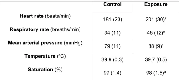
All lambs tolerated the 16 day-exposure to cigarette smoke without any apparent problems. No differences in arterial blood gases were apparent between the Control and Exposure groups (results not shown). However, although not quantified, an increase in spontaneous activity was observed in lambs from the Exposure group, including head movements to verify surroundings and multiple jumps in the exposure chamber during long periods (20-30 min). Overall, mean weight increase was similar between both groups from the first post-natal day [Control group: 3.1 (0.5) Kg, Exposure group: 3.6 (0.6) Kg] to post-natal day 16 [Control group: 5.6 (1.7) Kg, Exposure group: 6.4 (1.4) Kg]. Baseline parameter values obtained in each group are detailed in Table 1. Values for baseline cardio-respiratory parameters (heart rate, cardio-respiratory rate and mean arterial pressure) in the Exposure group were significantly higher than in the Control group. However, while a statistically significant SpO2 was observed in the Exposure group, the
mean difference between groups (1%) was of no physiological significance. No differences in temperature were observed between the two groups. The cotinine/creatinine ratio was significantly higher in the Exposure group throughout the 16 days of exposure [1116 (832) ng/mg in the Exposure group vs. 15 (12) in Control group, p< 0.0001], as well as during the last two recording days [891(506) ng/mg vs. 11 (11) mg/mg, p< 0.0001].
Effects of cigarette smoke exposure on laryngeal chemoreflexes
Results obtained during LCR are detailed in Table 2. Following two weeks of passive smoking, laryngeal stimulations (all solutions and both sleep states taken together) elicited significantly greater cardiorespiratory inhibition compared to the Control group. This inhibition included %decrease in heart rate, number of bradycardias, bradycardia duration, %decrease in respiratory rate, number of apneas, apnea duration and respiratory LCR duration. In addition, the % increase in mean arterial pressure (p = 0.08) tended to be lower in the Exposure group. No differences were observed for SpO2 variables (p = 0.49 to 0.93). Moreover,
certain parameters measuring lower airway protective mechanisms were significantly decreased in the Exposure group, including the number of swallows and arousals. No differences were noted however for the number of coughs (p = 0.27), total duration of active glottal closure (measured by the thyroarytenoid muscle EMG, p = 0.74) and awakening (p = 0.77).
Detailed results on apnea duration show that apneas longer than 10 s were rare events in both the Exposure (3 apneas) and Control groups (2 apneas), with none above 20 s. However, apnea duration between 5 s and 9 s was more often observed in the Exposure group than in the Control group (48 vs. 21 apneas respectively). In addition, while bradycardia duration was statistically significantly longer in the Exposure group, all bradycardias (defined by % decrease in HR > 30%) but one were shorter than 5 s. One severe event was observed in one lamb
in the Exposure group following HCl injection in QS, with marked hypoventilation and a decrease in SpO2 down to 76% (figure 1).
Effects of the type of solution on laryngeal chemoreflexes
The effects of type on solution are reported in table 3, with both sleep states taken together. In the Control group (table 3), injection of 0.5 ml of saline,
water, HCl or milk onto the laryngeal mucosa elicited similar cardiorespiratory responses. However, the number of swallows triggered was greater after HCl comparatively to saline and water and greater after milk compared to saline. In the Exposure group, desaturation magnitude was larger after HCl than water
(4.2% vs. 1.7%), and respiratory LCR duration longer after HCl than with saline or milk. Swallowing was significantly more frequent with HCl than water and milk and more frequent with milk than saline. Arousal from QS or AS occurred more frequently after water (24 arousals/27 stimulations) than saline (15 arousals/28 stimulations). Finally, lambs were fully awakened more often after injection of water (17 awakenings/24 stimulations) and milk (19 awakenings/21 stimulations) than HCl (9 awakenings/23 stimulations).
Exposure group compared to Control group. Regardless of the solution
tested, triggered respiratory responses were more important in the Exposure group than in the Control group. These included the % decrease in respiratory rate, number of apneas and apnea duration (not shown in the table, saline p = 0.02; water p = 0.01; HCl p = 0.02; milk p = 0.004). In addition, water induced a
significantly higher % decrease in heart rate, number of bradycardias and bradycardia duration (not shown in the table, p = 0.05) in the Exposure group. No differences in % decrease in heart rate were observed for milk while responses tended to be more intense for saline and HCl (p = 0.1). Overall, the type of solution had no influence on differences observed in lower airway protective responses between the two groups.
Effects of the sleep state on laryngeal chemoreflex components
Table 4 depicts the effects of sleep states, all solutions taken together. In the Control group, while the cardiorespiratory responses appeared similar in both
sleep states, depressed lower airway protective responses were observed in AS compared to QS. This included coughing, arousal, awakening and swallowing (p = 0.1, indicative of a tendency). In the Exposure group, some differences in
cardiorespiratory responses were observed, including an increase in the number of apneas in AS and a heightened decrease in heart rate in QS, although no clear picture emerged. In addition, lower airway protective responses were identical in QS and AS.
Exposure group compared to Control group. Cardiorespiratory responses
were greater in the Exposure group compared to Control group in both QS (%decrease in heart rate; %decrease in respiratory rate; number of apneas; %increase in mean arterial pressure) and AS (%decrease in heart rate; %decrease in respiratory rate; number of apneas; respiratory LCR duration).
Swallowing and arousal were significantly decreased in the Exposure group in QS while no differences were observed in AS.
Effects of passive smoking on laryngeal mucosa
There was no observable effect of cigarette smoke exposure on laryngeal inflammation in the few lambs studied. Indeed, histological analysis showed the presence of a moderate inflammation in both the Control (score 9/15, n = 3 lambs) and Exposure (score 8/15, n = 4 lambs) groups.
DISCUSSION
The present study provides novel findings on laryngeal chemoreflexes triggered in non-sedated, full-term lambs following exposure to passive smoking in the early postnatal period. Overall, our findings reveal that laryngeal chemoreflexes after postnatal exposure to environmental tobacco smoke are characterized by enhanced cardiorespiratory inhibition and decreased lower airway protective reflexes. In addition, the absence of any significant laryngeal inflammation suggests that the effect is mainly at the level of the central nervous system.
Passive smoking lamb model
Most studies on the effect of ETS exposure have focused on nicotine alone, despite the fact that tobacco smoke contains at least 4000 different chemical compounds, many of which are known to be harmful to human health. We have recently built a new automatic smoking machine to study the effect of both mainstream and sidestream cigarette smoke (secondary smoke) (13). In the present study, we used our custom-built smoking machine to mimic postnatal parental smoking. Dosage of urinary cotinine ensured that the level of ETS exposure was relevant to previously published cotinine levels in infants (5, 6, 23, 27). The increase in baseline heart and respiratory rates following ETS is in agreement with previous results in both the human fetus and newborn (19). Absence of any increase in heart and respiratory rates following postnatal infusion of nicotine in newborn lambs (46) suggests that the effect on cardio-respiratory control may be shared with other constituents of cigarette smoke.
Effect of passive smoking on LCR
To our knowledge, despite its clinical importance, only one study has assessed the effect of ETS on LCR: passive smoking during gestation was shown to induce longer apneas during LCR in newborn rats only if combined with hyperthermia (51). Many methodological differences (prenatal exposure, sedation, injection of distilled water via tracheal catheter), in addition to species differences, can explain discrepant results with the present study. In keeping with our results however, the authors observed no life-threatening events, even when ETS was added to hyperthermia. The effect of nicotine alone on the cardiorespiratory components of the neonatal LCR has been addressed in three studies. In two studies conducted in sedated, newborn piglets, the effect of one injection of nicotine on LCR was inconsistent, either showing no effect (16) or an enhanced cardio-respiratory inhibition (17). In the third study in non-sedated, newborn lambs injected with IV nicotine during several days, an enhanced cardio-respiratory inhibition was reported during LCR in both room air and mild hypoxia (46). However, it was suggested that the high nicotine dose, which was more representative of that experienced by a light smoker than an infant exposed to passive smoking, was less clinically relevant with regards to post-natal infant exposure in the latter study.
Interestingly, aside from cardio-respiratory components, LCR in lambs elicit lower airway protective mechanisms, including swallowing and coughing. Our present observations that swallowing and coughing were prominent during LCR in Control lambs are in agreement with our previous results in healthy, full-term lambs (42). Conversely, swallowing was significantly blunted in Exposure lambs, a likely central effect, given the absence of significant laryngeal inflammation. While active smoking has been previously shown to decrease reflexive
pharyngeal swallowing (10, 11), this is the first observation of a decrease in swallowing induced by passive smoking. Decreased swallowing activity during LCR may lead to increased contact time between stimulating liquid and the laryngeal mucosa and, in turn, to enhanced LCR and deleterious cardio-respiratory events in the infant. Although we did not observe any differences in “coughing” in Exposure lambs, we must recognize that our study did not allow to fully assess coughing. Indeed, as we only counted the numbers of coughs and were not able to discriminate between “real” coughs and laryngeal expiratory reflexes (= brisk expiration not preceded by a deep inspiration), we cannot dismiss a decrease in cough efficiency. Quantification of coughing efficiency during LCR would be an interesting addition in future studies.
Our observation of decreased arousal during LCR following post-natal ETS may also be very relevant with regards to the increased in the occurrence of SIDS after ETS. Indeed, arousal plays a critical role in homeostasis, by increasing heart rate, blood pressure and respiratory rate, among others, (21) and arousal deficiency is likely present in a number of SIDS victims (26). Both maternal smoking and nicotine during pregnancy have been shown to affect arousal threshold (39), including after obstructive apnea, hypoxia and auditory stimuli (19). The present results represent the first account that post-natal passive smoking decreases arousal induced by laryngeal stimulation. Ability to arouse appears essential during some LCR, by relieving cough suppression (30, 45), promoting swallow reflexes (40) and cessation of apnea and bradycardia (47).
While the design of the present study does not allow us to comprehensively infer the mechanisms involved in the effects of post-natal ETS on LCR, the absence of significant laryngeal inflammation, which is in agreement with previous reports in
adult smokers (12), does suggest that these effects are centrally mediated. Of note, brainstem nicotinic receptors are involved in the control of cardio-respiratory integration, arousal, REM sleep and somatic motor control (14, 25). In addition, exposure to nicotine increases GABAa receptor density and increases
GABA release, thus favoring breathing inhibition following LCR (33, 51). Aside from nicotine however, the potential role of the many constituents of cigarette smoke remains unknown.
Influence of various solutions or sleep states on laryngeal chemoreflexes Conversely to previous results in healthy, full-term lambs aged 4-5 days, cardio-respiratory responses induced during LCR by water and HCl in 15 day-old Control lambs were identical to those observed after saline stimulation (42). This result is likely related to post-natal maturation of LCR, as previously reported (7). In agreement with our previous studies in 4-5 day-old full-term lambs and in preterm lambs (42, 43), the present results confirm that HCl triggers more swallowing activity than the other solutions in both Control and Exposure lambs. Results herein also show that ewe's milk can trigger cardio-respiratory events identical to other solutions, which is in agreement with previous reports in lambs, piglets and puppies (7, 9, 29, 22, 44) and provides clinical relevance with regards to LCR triggered during bottle-feeding or non-acid pharyngo-laryngeal reflux. Overall, no distinctive pattern emerged with regard to a potential effect of one solution over the other on LCR in the Exposure vs. Control group. This lack of effect may however be related to the small number of lambs in each group. In this respect, the fact that the only severe desaturation event was observed in one lamb from the Exposure group is noteworthy.
Interestingly, AS was responsible for blunting lower protective mechanisms in the Control group. This effect was no longer observed in the Exposure group, which may be related to the blunting influence of post-natal ETS in itself. In addition, blunting of lower airway protective mechanisms in the Exposure group were present in QS, but not in AS. No systematic effects of the states of alertness on the differences on cardio-respiratory events in the Exposure vs. Control groups were observed, as previously reported in the preterm lamb (43).
Clinical implications
Clinical evidence strongly suggests that LCR can be triggered by numerous liquids, including upper airway secretions, milk bottle-feeding and both acidic and non-acidic laryngopharyngeal reflux (37). LCR are one of the mechanisms involved in apneas of prematurity, apparent life threatening events and probably some cases of the sudden infant death syndrome (26, 31, 37). Healthy, full-term lambs at birth already have mature-type LCR with efficient lower airway protective mechanisms and an absence of clinically significant apneas-bradycardias (42). Many conditions, such as hypoxia, anemia, hyperthermia, respiratory syncytial virus infection, and preterm birth (43, 48, 51) have been shown to enhance cardio-respiratory inhibition during LCR. The present findings show that post-natal ETS, at a level consistent with clinical observations, can also contribute in altering LCR toward more cardio-respiratory inhibition and less airway protective mechanisms. However, our results suggest that, at least in lambs, post-natal ETS is most often insufficient in itself to trigger potentially dangerous reflexes. Nevertheless, it is important to emphasize that most infants exposed to passive smoking have additional risk factors, which are likely synergistic with LCR for explaining the failing chain of events ultimately leading to SIDS. These include preterm birth, pathological laryngo-pharyngeal reflux, prone
position, respiratory viral infection and/or hyperthermia. Future studies will need to consider the effects of these multiple factors.
ACKNOWLEDGMENTS
The authors gratefully acknowledge the expert technical assistance of Jean-Philippe Gagné and Nathalie Samson, and Nathalie Carrier for statistics. The study wassupported by grants from the Canadian Institutes for HealthResearch and the Foundation of Stars (Quebec) allocated to J-P Praud. A-M Carreau was MD-MSc scholar of the Fonds de la recherche en santé du Québec (FRSQ). Jean-Paul Praud is a member of the FRSQ-funded Clinical Research Center Étienne-Le Bel, Sherbrooke University Hospital.
REFERENCES
1. Adgent MA. Environmental tobacco smoke and sudden infant death syndrome: a review. Birth Defects Res B Dev Reprod Toxicol 77: 69-85, 2006.
2. Alm B, Milerad J, Wennergren G, Skjaerven R, Oyen N, Norvenius G, Daltveit AK, Helweg-Larsen K, Markestad T, Irgens LM. A case-control study of smoking and sudden infant death syndrome in the Scandinavian countries, 1992 to 1995. The Nordic Epidemiological SIDS Study. Arch Dis
Child 78: 329-334, 1998.
3. Alm B, Norvenius SG, Wennergren G, Skjaerven R, Oyen N, Milerad J, Wennborg M, Kjaerbeck J, Helweg-Larsen K, Irgens LM. Nordic Epidemiological SIDS Study. Changes in the epidemiology of sudden infant death syndrome in Sweden 1973-1996. Arch Dis Child 84: 24-30, 2001.
4. Anderson HR, Cook DG. Passive smoking and sudden infant death syndrome: review of the epidemiological evidence. Thorax 52: 1003-1009, 1997.
5. Anuntaseree W, Mo-Suwan L, Ovatlarnporn C, Tantana C, and Ma-a-Lee A. Exposure to environmental tobacco smoke among infants in southern Thailand: a study of urinary cotinine. Bull Environ Contam Toxicol 80: 34-37, 2008.
6. Blackburn CM, Bonas S, Spencer NJ, Coe CJ, Dolan A, Moy R. Parental smoking and passive smoking in infants: fathers matter too. Health Educ Res 20: 185-194, 2005.
7. Bogg DF, Bartlett JR. Chemical specificity of a laryngeal apneic reflex in puppies. J Appl Physiol 53: 455-462, 1982.
8. DiFranza JR, Aligne CA, Weitzman M. Prenatal and postnatal environmental tobacco smoke exposure and children's health. Pediatrics 11: 1007-1015, 2004.
9. Downing SE, Lee JC. Laryngeal chemosensitivity: a possible mechanism for sudden infant death. Pediatrics 55: 640-649, 1975.
10. Dua K, Bardan E, Ren J, Sui Z, Shaker R. Effect of chronic and acute cigarette smoking on the pharyngo-upper oesophageal sphincter contractile reflex and reflexive pharyngeal swallow. Gut 43: 537-541, 1998.
11. Dua K, Bardan E, Ren J, Sui Z, Shaker R. Effect of chronic and acute cigarette smoking on the pharyngoglottal closure reflex. Gut 51: 771-775, 2002.
12. Duarte JL, Cardoso de Faria FA, Ceolin DS, Cestari TM, Francisco de Assis G. Effects of passive smoke inhalation on the vocal cords of rats. Rev
Bras Otorrinolaringol 72: 210-216, 2006.
13. Duvareille C, Beaudry B, St-Hilaire M, Boheimier M, Brunel C, Micheau P, Praud J-P. Validation of a new automatic smoking machine to study the effects of cigarette smoke in newborn lambs. Lab Animals Submitted.
14. Dwyer JB, Broide RS, Leslie FM. Nicotine and brain development. Birth
15. Fortier PH, Reix P, Arsenault J, Dorion D, Praud J-P. Active upper airway closure during induced central apneas in lambs is complete at the laryngeal level only. J Appl Physiol 95: 97-103, 2003.
16. Froen FJ, Akre H, Stray-Pedersen B, Saugstad OD. Adverse effects of nicotine and interleukin-1b on autoresuscitation after apnea in piglet: implications for sudden infant death syndrome. Pediatrics 105: e52-e57, 2000.
17. Froen JF, Akre H, Stray-Pedersen, Saugstad OD. Prolonged apneas and hypoxia mediated by nicotine and endotoxin in piglets. Biol Neonate 81: 119-125, 2002.
18. Goto K, Mirmiran M, Adams MM, Longford RV, Baldwin RB, Boeddiker MA, Ariagno RL. More awakening and heart rate variability during supine sleep in preterm infants. Pediatrics 103: 603-609, 1999.
19. Hafstrom O, Mileread J, Sandberg KL, Sundell HW. Cardiorespiratory effects of nicotine exposure during development. Respir Physiol Neurobiol 149: 325-341, 2005.
20. Harding R, Johnson P, McClelland ME. Liquid-sensitive laryngeal receptors in the developing sheep, cat and monkey. J Physiol 277: 409-422, 1978. 21. Horne RS, Franco P, Adamson TM, Groswasser J, Kahn A. Influences of
maternal cigarette smoking on infant arousability. Early Hum Dev 79: 49-58, 2004.
22. Hunt CE, Hauck FR. Sudden infant death syndrome. CMAJ 174: 1861-1869, 2006.
23. Joseph DV, Jackson JA, Westaway J, Taub NA, Petersen SA, Wailoo MP. Effect of parental smoking on cotinine levels in newborns. Arch Dis Child 92: F484-8, 2007.
24. Kato I, Franco P, Groswasser J, Scaillet S, Kelmanson I, Togari H, Kahn A. Incomplete arousal processes in infants who were victims of sudden death.
Am J Respir Crit Care Med 168: 1298-1303, 2003.
25. Kinney HC, O’Donnal TJ, Kriger P, White WS. Early development changes in (3H) nicotine binding in human brainstem. Neuroscience 55: 1127-1130, 1993.
26. Kinney HC, Thach BT. The sudden infant death syndrome. N Engl J Med 361: 795-805, 2009.
27. Kott KS, Salt BH, McDonald RJ, Jhawar S, Bric JM Joad JP. Effect of secondhand cigarette smoke, RSV bronchiolitis and parental asthma on urinary cysteinyl LTE4. Pediatr Pulmonol 43: 8: 760-766, 2008.
28. Koufman JA. The otolaryngologic manifestations of gastroesophageal reflux disease (GERD); a clinical investigation of 225 patients using ambulatory 24hrs pH monitoring and an experimental investigation of the role of acid and pepsin in the development of laryngeal injury. Laryngoscope 101: 1-78, 1991. 29. Lee JC, Stoll BJ, Downing SE. Properties of the laryngeal chemoreflex in
neonatal piglets. Am J Physiol 233: R30-36, 1977.
30. Lee KK, Birring SS. Cough and sleep. Lung 2009. Epub ahead of print. 31. Leiter JC, Böhm I. Mechanisms of pathogenesis in the Sudden Infant Death
32. Letourneau P, Dumont S, Kianicka I, Diaz V, Dorion D, Drolet R, Praud J-P. Radiotelemetry system for apnea study in lambs. Respir Physiol 116: 85-93, 1999.
33. Luo Z, Costy-Bennett S, Fregosi RF. Prenatal nicotine increases the strength of GABA receptor mediated inhibition of respiratory rhythm in neonates. J Physiol 561: 387-393, 2004.
34. McDonnell M, Mehanni M, McGarvey C, Oregan M, Matthews TG. Smoking: the major risk factor for SIDS in Irish infants. Ir Med J 95: 111-113, 2002.
35. Moon RY, Horne RS, Hauck FR. Sudden infant death syndrome. Lancet 4: 1578-1587, 2007.
36. Reix P, Fortier PH, Niyonsenga T, Arsenault J, Letourneau P, Praud J-P. Non-nutritive swallowing and respiratory coordination in full-term newborn lambs. Respir Physiol Neurobiol 134: 209-218, 2003.
37. Reix P, St-Hilaire M, Praud J-P. Laryngeal sensitivity in the neonatal period: from bench to bedside. Pediatr Pulmonol 42: 674-682, 2007.
38. Renolleau S, Letourneau P, Niyonsenga T, Praud J-P. Thyroarytenoid muscle electrical activity during spontaneous apneas in preterm lambs. Am J
Respir Crit Care Med 159: 1396-1404, 1999.
39. Richardson HL, Walker AM, Horne RS. Maternal smoking impairs arousal patterns in sleeping infants. Sleep 32: 515-521, 2009.
40. Sato K, Nakashima T. Sleep-related deglutition in children. Ann Otol Rhinol
41. Slotkin TA. If nicotine is a developmental neurotoxicant in animal studies, dare we recommend nicotine replacement therapy in pregnant women and adolescents? Neurotoxicol Teratol 30: 1-19, 2008.
42. St-Hilaire M, Nsegbe E, Gagnon-Gervais K, Samson N, Moreau-Bussière F, Fortier PH, Praud J-P. Laryngeal chemoreflexes induced by acid, water, and saline in non-sedated newborn lambs during quiet sleep. J Appl Physiol 98: 2197-2203, 2005.
43. St-Hilaire M, Samson N, Nsegbe E, Duvareilles C, Moreau-Bussiere F, Micheau P, Leblond J, Praud J-P. Postnatal maturation of laryngeal chemoreflexes in the preterm lamb. J Appl Physiol 102: 1429-1438, 2007. 44. Storey AT, Johnson P. Laryngeal water initiating apnea in the lambs. Exp
Neurol 47: 42-55, 1975.
45. Sullivan CE, Murphy E, Kozar LF, Phillipson EA. Waking and ventilatory responses to laryngeal stimulation in sleeping dogs. J Appl Physiol 45: 682-689, 1978.
46. Sundell HW, Karmo H, Milerad J. Impaired cardiorespiratory recovery after laryngeal stimulation in nicotine-exposed young lambs. Pediatr Res 53: 104-112, 2003.
47. Tirsoh E, Libon D, Bader D. The effect of maternal smoking during pregnancy on sleep respiratory and arousal patterns in neonates. J Perinatol 16: 435-438, 1996.
48. Thach BT. Maturation and transformation of reflexes that protect the laryngeal airway from liquid aspiration from fetal to adult life. Am J Med 111: 69S-77S, 2001.
49. Thach BT. Some aspects of clinical relevance in the maturation of respiratory control in infants. J Appl physiol 104: 1828-1834, 2008.
50. Van der velde L, Curran AK, Filiano JJ, Darnall RA, Bartlett D Jr, Leiter JC. Prolongation of the laryngeal chemoreflex after inhibition of the rostral ventral medulla in piglets: a role in SIDS? J Appl Physiol 94: 1883-1895, 2003.
51. Xia L, Crane-Godreau M, Leiter JC, Bartlett D. Gestational cigarette smoke exposure and hyperthermic enhancement of laryngeal chemoreflex in rat pups. Respir Physiol Neurobiol 28: 161-166, 2009.
FIGURE LEGEND
Figure 1Cardiorespiratory reflexes triggered in a lamb at post-natal day 15 after two weeks of exposure to cigarette smoke, following instillation of 0.5 ml hydrochloric acid onto laryngeal mucosa during quiet sleep. From top to bottom: EEG, electroencephalogram; EOG, electrooculogram; Ta, electrical activity of the thyroarytenoid muscle; Nasal Flow, nasal airflow; Lung volume, sum signal of the respiratory inductance plethysmograph, allowing qualitative measurement of respiration (inspiration upward); ECG, electrocardiogram; HR, heart rate (beats/min); AP, arterial pressure; SpO2, oxygen hemoglobin saturation
TABLES
Table 1: Baseline values for each group
Control Exposure
Heart rate (beats/min) 181 (23) 201 (30)a
Respiratory rate (breaths/min) 34 (11) 46 (12)a
Mean arterial pressure (mmHg) 79 (11) 88 (9)a
Temperature (oC)
39.9 (0.3) 39.7 (0.5)
Saturation (%) 99 (1.4) 98 (1.5)a
Values are expressed as means (Standard Deviation).
Control Exposure %dec HR 21 (11) 28 (14)c Nb. of bradycardias 0.2 (0.7) 0.5 (1)a Bradycardia duration (s) 0.2 (0.7) 0.3 (0.7)a %inc MAP 23 (16) 18(10) %dec RR 51 (18) 67 (15)c Nb. of apneas 1.1 (2.7) 2.2 (4.7)c Apnea duration (s) 4.5 (8.3) 7.8 (8.5)c Respiratory LCR duration (s) 13.2 (10) 14.8 (8.8)a %dec saturation 3 (3) 3 (3)
Saturation; Area under 90% 0.04 (0.2) 0.06 (0.3) Saturation; Area under 85% 0.01 (0.08) 0.02 (0.14)
Nb. of swallows 12 (10) 8 (7)a
Total Ta EMG duration (s) 2.2 (2.9) 2.5 (2.9) Coughs (nb. of LCR with cough/total nb. of LCR) 14/94 11/116 Arousal (nb. of LCR with arousal/total nb. of LCR) 76/89 83/116a
Awakening (nb. of LCR with awakening/total nb. of LCR) 48/89 55/116
Values are expressed as means (Standard Deviation). The results are means from all laryngeal stimulations (all solutions and all states of alertness) for each group. HR, heart rate; RR, respiratory rate; MAP, mean arterial pressure; %dec HR, RR or saturation, percentage decrease in HR, RR or O2 saturation respectively; %inc MAP,
percentage increase in MAP; LCR, laryngeal chemoreflexes; total Ta EMG, total duration of electrical activity of the thyroarytenoid muscle; nb, number. a p < 0.05 vs. Control. b p < 0.01 vs. Control. c p < 0.001 vs. Control
Saline H2O HCl Milk %dec HR Control 18 (9) 21(11) 22 (22) 23 (13)
Exposure 33 (54) 30 (13)f 31 (28) 28 (13)
Nb. of bradycardias Control 0.1 (0.6) 0.1 (0.3) 0.4 (0.4) 0.3 (0.9) Exposure 0.2 (0.5) 0.4 (0.6)e 0.9 (1) 0.3 (0.8)
%inc MAP Control 15 (10) 18 (9) 29 (20) 25 (8) Exposure 16 (14) 18 (8) 19 (6) 25 (19) %dec RR Control 47 (18) 54 (14) 50 (11) 51 (19) Exposure 59 (19)a 71 (12)g 68 (10)l 69 (12)p
Nb. of apneas Control 0.7 (2) 2 (5) 1 (7) 1 (1) Exposure 2 (5)a 4 (8)f 1 (5)j 2 (1)o
Respiratory LCR duration (s) Control 10.4 (8) 12.4 (7) 16.3 (13) 13.2 (10) Exposure 12.1 (8) 15 (9) 19.5 (10)c q 12.9 (6)
%dec saturation Control 1.6 (1) 3.4 (4) 4.1 (2) 2.4 (2) Exposure 2.6 (3) 1.7 (1) 4.2 (2)h 2.9 (3)
Nb. of swallows Control 6.1 (4) 12 (9) 19.1 (17)b e 12 (7)a
Exposure 4.4 (2) 5.7 (2) 14.7 (12)I q 8.3 (4)d
Coughs (nb. of LCR with cough/total nb. of LCR) Control 5/26 2/23 4/22 3/23 Exposure 2/28 3/27 5/31 1/30 Arousal (nb. of LCR with arousal/total nb. of LCR) Control 18/22 19/23 20/22 19/22
Exposure 15/28 24/27c 23/31 21/30
Awakening (nb. of LCR with awakening/total nb. of LCR) Control 13/22 13/23 11/22 11/22 Exposure 10/28 17/27m 9/31 19/30n
Values are expressed as means (SD). See table 2 for abbreviations. a p < 0.05 vs. saline in Control. b p < 0.001 vs. saline in Control. c p <
0.01 vs. saline in Exposure. d p < 0.001 vs. saline in Exposure. e p < 0.05 vs. H2O in Control. f p < 0.01 vs. H2O in Control. g p < 0.001 vs.
H2O in Control. h p < 0.01 vs. H2O in Exposure. I p < 0.001 vs. H2O in Exposure. j p < 0.05 vs. HCl in Control. k p < 0.01 vs. HCl in Control. l
p < 0.001 vs. HCl in Control. m p < 0.05 vs. HCl in Exposure. n p < 0.001 vs. HCl in Exposure. o p < 0.01 vs. milk in Control. p p < 0.001 vs.
Quiet sleep Active Sleep
%dec HR Control 22.5 (9) 20 (13)
Exposure 29 (12)b g 27 (16)e
Nb. of bradycardias Control 0.2 (0,5) 0.3 (0,7)
Exposure 0.6 (1) 0.3 (0.8)
%inc MAP Control 26.5 (17) 17 (12)
Exposure 16.5 (10)a 19 (9)
%dec RR Control 49 (19) 53 (17)
Exposure 65.5 (16)c 69 (13)f
Nb. of apneas Control 0.9 (1) 1.3 (4)
Exposure 1.7 (4)b 2.9 (6)d f
Respiratory LCR duration (s) Control 15 (12) 11 (6) Exposure 15.9 (10) 13.5 (7)e
%dec saturation Control 3 (4) 2.5 (2)
Exposure 3 (3) 2.5 (3)
Nb. of swallows Control 13.5 (12) 9 (6)
Exposure 9 (8)a 7.5 (5)
Coughs (nb. of LCR with cough/total nb. of LCR) Control 12/55 2/41a
Exposure 8/64 3/50
Arousal (nb. of LCR with arousal/total nb. of LCR) Control 47/51 28/37a
Exposure 49/64a 34/52
Awakening (nb. of LCR with awakening/total nb. of LCR) Control 35/47 13/28b
Exposure 33/49 22/34
Values are expressed as means (SD). See table 2 for abbreviations. a p < 0.05 vs. Control in quiet sleep. b p < 0.01 vs. Control
in quiet sleep. c p < 0.001 vs. Control in quiet sleep. d p < 0.05 vs. Exposure in quiet sleep. e p < 0.05 vs. Control in active sleep. f p < 0.001 vs. Control in active sleep. g p < 0.05 vs. Exposure in active sleep.