Open Archive TOULOUSE Archive Ouverte (OATAO)
OATAO is an open access repository that collects the work of Toulouse researchers and
makes it freely available over the web where possible.
This is an author-deposited version published in :
http://oatao.univ-toulouse.fr/
Eprints ID : 18535
To link to this article : DOI:10.1007/s00167-015-3707-4
URL :
http://dx.doi.org/10.1007/s00167-015-3707-4
To cite this version : Chotel, Franck and Raux, Sébastien and
Accadbled, Franck and Gouron, Richard and Pfirrmann, Clémence and
Bérard, Jérôme and Seil, Romain Cartilaginous tibial eminence
fractures in children: which recommendations for management of this
new entity ? (2016) Knee Surgery, Sports Traumatology, Arthroscopy,
vol. 24 (n° 3). pp. 688-696. ISSN 0942-2056
Any correspondence concerning this service should be sent to the repository
administrator:
[email protected]
Cartilaginous tibial eminence fractures in children: which
recommendations for management of this new entity?
Franck Chotel1,2 · Sébastien Raux1,2 · Franck Accadbled3 · Richard Gouron4 · Clémence Pfirrmann1,2 · Jérôme Bérard1,2 · Romain Seil5,6
were, respectively, 97.7 ± 2.6 and 97 ± 3.4. The median residual laxity was 2 mm (range 0–4). Non-operative treat-ment lead to 2 failures: intermeniscal ligatreat-ment entraptreat-ment and combined avulsion fracture at the femoral site. Suture fixation of the avulsed fragment allows regularly good results when performed acutely or even 4 years after the injury. The hypothesis that primary treatment gives better result than delayed treatment tends to be wrong as 2 fail-ures were reported in each group. An ACL reconstruction was performed in 3 out of the 4 treatment failures. Progres-sive resorption of the avulsed fragment was noticed in 3 of the 4 failures suggesting an associated ACL resorption. Conclusion CTEF has a good prognosis even after mis-diagnosis and treatment at the time of non-union; this could be due to low-energy mechanism of injury and low rate of associated lesion. Orthopaedic treatment for acute minimally displaced fractures is only indicated under strict MRI control, and suture fixation is the recommended strategy in other situations. Conservative management of non-union could expose to ACL involution and cannot be recommended.
Level of evidence Retrospective case series, Level IV. Keywords Anterior cruciate ligament · Tibial eminence fracture · Children · Misdiagnosis · Cartilaginous avulsion
Introduction
Tibial eminence fracture is a frequent pattern of ACL rup-ture in skeletally immarup-ture patients [5, 6, 21]. Traditionally, it occurs between 8 and 14 years of age and is rare under the age of 8 [2, 16, 21].
A new entity has been recently reported in very young children, with purely cartilaginous avulsions of Abstract
Purpose Cartilaginous tibial eminence fracture (CTEF) is a new pattern of ACL rupture in children under the age of nine. MRI signs have been recently reported, but no series gave information about outcomes. It was hypothesized that primary treatment gave better results than delayed manage-ment due to frequent misdiagnosis.
Method This retrospective study focused on 15 patients, managed acutely (n = 7) or delayed (n = 8). The patients’ median age at the time of initial injury was 6.5 years (range 5–9). Lysholm, IKDC 2000 subjective scores, and the measurement of the residual laxity by a side-to-side differ-ence with a KT-1000 junior arthrometer were used at the time of revision.
Results After a mean follow-up of 9.8 years (range 1–18.5), the mean Lysholm and IKDC subjective scores
* Franck Chotel
1 Department of Pediatric Orthopaedic Surgery, Lyon
University Hospital for Mother and Children, 59 Boulevard Pinel, 69677 Bron, France
2
Claude Bernard University Lyon I, Lyon, France
3 Department of Pediatric Orthopaedics, Hôpital des Enfants,
330, Avenue de Grande Bretagne, 31059 Toulouse Cedex 9, France
4
Department of Paediatric Orthopaedic Surgery, Jules Verne University of Picardie and Amiens University Medical Centre, Amiens, France
5 Department of Orthopaedic Surgery, Clinique d’Eich,
Centre Hospitalier de Luxembourg, 78, rue d’Eich, 1460 Luxembourg, Luxembourg
6
Sports Medicine Research Laboratory, Public Research Centre for Health, 78, rue d’Eich, 1460 Luxembourg, Luxembourg
the ACL insertion on either the femoral [4, 12, 17] or more commonly on the tibial site [7, 18, 28]. The latter are frequently misdiagnosed even after MRI examina-tion because classical primary and secondary MRI find-ings after ACL injury can be negative [7]. A fluid signal underneath the cartilaginous fragment reaching up to the ossified epiphysis on T2 sequences or a double PCL sign may orient the diagnosis [7]. Despite a better knowl-edge about the diagnostic principles, no series reported the management and results of these rare injuries. The aim of this study was to focus on these 2 items through a multicentre series. The authors hypothesized that pri-mary treatment of cartilaginous tibial eminence fractures (CTEFs) gave better results than delayed management due to misdiagnosis.
Materials and method
Out of 161 consecutive tibial eminence fractures treated in our department between 1994 and 2012, eleven patients with CTEF of ACL insertion were identified. Seven chil-dren were referred to our department as a tertiary referral hospital because of misdiagnosed injury, and 4 had primary treatment in our hospital. After a survey in the French Soci-ety of Paediatric Orthopaedics (SOFOP), 4 more patients were diagnosed and treated in two other institutions.
This retrospective study focused on 15 patients (8 right and 7 left knees) who had different regime of manage-ment: acute management (n = 7), delayed management <6 months (n = 2), and delayed management >6 months (n = 6). The patients’ median age at the time of initial injury was 6.5 years ± 1.4 (range 5 to 9). The main cause of injury was a low-energy domestic accident (n = 7), bicy-cle fall (n = 3), or sports accident (n = 4). All patients had immediate joint effusion at the time of injury. Other indi-vidual findings at the time of injury are summarized in Table 1 and Fig. 1 shows their management algorithm.
A retrospective analysis of the patients’ chart, traumatic history, conventional radiograph, and MRI was performed. Lesions were classified according to Meyers and McKeever as well as Zifko and Gaudernak [24, 31]. ACL reconstruc-tion was considered as a management failure of CETF.
In the acute management group, the diagnosis was radi-ologically suspected after careful examination by a sen-ior paediatric orthopaedic surgeon due to the presence of a very thin and hardly visible bone lamella. Five of these patients had an orthopaedic treatment with long leg cast. A simple cast in slight flexion without reduction was per-formed for patient no. 1, and cast in extension for fracture reduction was performed for the others. Non-weight bear-ing durbear-ing 4–6 weeks was recommended.
In the delayed management group <6 months, the ini-tial diagnosis of midsubstance ACL tear performed on MRI was revised during radiographic follow-up because of ossi-fications of the avulsed fragment. One patient had suture fixation 2.5 months after injury. The other underwent a non-operative management chosen at the time of referral 4 months after injury because of moderate functional disa-bility (limitation of hyperextension and occasional but rare instability) and non-acceptance of surgery by the parents (patient no. 7).
In the delayed management group >6 months, the 6 patients were referred because of functional disability due to extension deficit and/or instability and a past history of knee injury. The median delay for referral after the acci-dent was 30 months ± 27.6 (6–72). Initial knee radiographs were interpreted as normal from the referring centres as well as after a retrospective second lecture. Despite MRIs, the diagnosis of CTEF was only suspected in 1 patient. Patient no. 9 had misinterpretation of MRI that conducted to erroneous diagnosis of an avulsed lateral discoid menis-cus which conducted to inappropriate partial meniscectomy 9 months following the accident.
Management at the time of referral included 5 cases of suture fixation. In 4 cases, a mixed arthroscopic and mini-open reduction with a modified Ahn pull-out suturing tech-nique of the tibial eminence with transphyseal absorbable sutures was performed [1, 9]. After debridement of the fracture bed and the fragment, care was taken to optimize the ACL tension and compensate plastic deformation of the ligament by countersinking the tibial fragment moder-ately [21, 26]. For patient no. 9, a pull-out reattachment of the medial meniscus posterior root using a modified Kim method with transphyseal absorbable suture was associ-ated after arthroscopic shaving of the posterior ossification [19, 23] (Figs. 2, 3). Patient no. 14 had an epiphyseal suture without countersinking through an open approach. Because of good tolerance of the non-union, patient no. 6 had a con-servative treatment between the ages of 6 and 12.
Evaluation
At the final follow-up, all patients were evaluated with the Lysholm and IKDC 2000 subjective scores. The resid-ual laxity was assessed by a side-to-side difference with a KT-1000 junior arthrometer (MEDmetric, San Diego, CA) [14]. Clinical final results were scored according to the IKDC classification. A specific attention was given to detect growth disturbances clinically (leg discrepancy using the graduated blocks method and axial deformity assess-ment) and radiologically. Growth arrest, but also over-growth was assessed according to standard recommenda-tions [10].
T
able 1
Detail of indi
vidual patient’
s feature at the time of injury
a P atients included in a pre vious study [ 7 ]. M Male, F female, L left, R right, delay for MRI is gi v en in weeks or months (W and M); # classification according to [MM][Zifk o]; GA general anaesthesia, N A not a v ailable P atient no. Se x/age at injury T ype of initial accident/mechanism First radiograph
Misdiagnosed injury/ presumed diagnosis MRI (delay after accident)
Retrospecti
v
e
diagnosis/type of displ# Initial acute management Management in referred centre
1
M/6.5
F
all from bic
ycle Thin ossification – – I-A or II-A Cast in fle xion 5 weeks Acute 2 F/5.5 F
all from bic
ycle Ossification – – ll-A Cast in e xtension/GA Acute 3 F/5 C ru sh in g l eg ( m o to r-cy cle) Thin ossification – – IV -B
Suture fixation/tibia nailing
Acute
4
a
M/8.5
F
all from his height
Thin ossification
–
M
+
6
III-A and femur
Cast in e xtension/ needle aspiration/GA Acute 5 F/6.5 F
all from his height
Thin ossification – – II-A Cast in e xtension Acute 6 F/6 Ski twist Normal Sprain W + 6 III-A Splint Non-union 7 a M/6 F
all from his height
Normal Midsubstance A CL tear W + 4 IV -B Splint Delayed M + 4 8 a F/5 F
all from his height
Thin ossification Midsubstance A CL tear W + 3 III-A Splint Delayed M + 2.5 9 a M/6 F
all from his height
Normal
Desinserted lateral discoid meniscus?
W + 6 IV -B No immobilisation Non-union 10 F/7.5 F
all from bic
ycle Thin ossification – W + l IV -B Cast in e xtension 5 weeks Acute 11 a M/8.5
Ski/jump bad reception
Normal Sprain/suspected CETF W + 2 IV -B No immobilisation Non-union 12 M/8 F
oot ball/knee hypere
xtension Thin ossification – – III-B Suture fixation Acute 13 a M/8.5 Judo twist Normal Midsubstance A CL tear/
lateral meniscus tear?
W + 4 IV -B Splint Non-union 14 M/5.5 N A Normal Sprain M + 9 III-A Splint Non-union 15 M/9 T rampoline N A Sprain M + 20 III-B No immobilisation Non-union
Due to the limited number of cases, no statistical analy-sis was performed.
Results
After a mean follow-up of 9.8 years ± 5 from the initial acci-dent and 4.6 years (range 1–18.5) from the definitive treat-ment, the results of the series are presented in Table 2. The
mean age at the time of last revision was 15 years (±5). A total of four failures out of the fifteen managed cases were noticed (Fig. 1). No failure was reported after suture fixations (n = 8).
In the acute management group, non-surgical treatment failed in patient no. 4 and no 10 despite cast immobilization in extension for 5 weeks. Patient no. 4 had a combined second avulsion fracture at the femoral site that was diagnosed retro-spectively. Complete resorption of the ossified fragment on the tibial side was noticed after a few months of conservative
Fig. 1 Management algorithm
of the 15 patients with CTEF
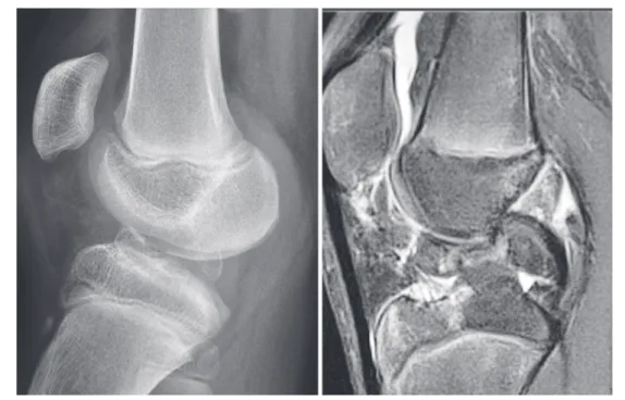
Fig. 2 Patient no. 9: MRI at the time of injury with purely cartilaginous avulsion (black arrows) (left) and double PCL sign due to posterior
treatment. Surgery, performed after skeletal maturity at the age of 15 because of instability, revealed an empty notch making suture fixation impossible, and an ACL reconstruc-tion was performed. An associated stable and partial longi-tudinal tear of the posterior segment of the medial meniscus was treated by stimulation alone. Patient no. 10 had an inter-meniscal ligament entrapment diagnosed retrospectively on radiographs under cast and MRI (Fig. 4). After cast immo-bilization, a rapid resorption of the ossified fragment was noticed. At the latest follow-up 2 years later, a non-operative treatment was still ongoing (stable knee after physiotherapy).
The 2 patients treated by suture fixation in emergency had an excellent result at follow-up.
In the delayed management group, 2 patients were man-aged by longstanding non-operative management. Patient no. 6 had a deficit of hyperextension due to avulsed ossified fragment during 3 years and a ski accident conducting to complete osseous resorption and required an ACL recon-struction with a modified Anderson procedure after 6 years [3]. Patient no. 7 had interstitial ACL fibres damage during specific ACL reconstruction [8] because of instability after 4 years of non-operative management.
Discussion
The most important finding of the present study was that suture fixation of the avulsed fragment allows regularly
good results when performed acutely or even 4 years after the injury. At the contrary, the orthopaedic treatment without MRI support could expose to failure. The hypoth-esis that primary treatment gives better result than delayed treatment due to misdiagnosis tends to be wrong as 2 fail-ures were reported in each group.
Cartilage tibial eminence fracture is a recent entity unknown before the 2 first cases reported in 2002 and 2008 [18, 28]. It should not be confounded with midsubstance ACL tears or peel-off injuries which are ligamentous soft tis-sue avulsions without any associated bony or cartilaginous fragment [13, 20]. CETF is involving children under the age of 9 and is usually due to domestic or low-energy injuries [7]. This may explain the low prevalence of associated bone bruises and meniscal tears in this series in opposition to clas-sical bony tibial eminence fractures [27, 30]. Because tradi-tional primary and secondary MRI findings after ACL injury are negative in CETF [7], the diagnosis is easily missed unless a thin bone lamella can be suspected on initial X-rays after careful examination. Unlike bony tibial eminence frac-tures, CTEF often extends far posteriorly lifting the entire intercondylar surface (9 types B of Zifko out of 15) giving a double PCL aspect on MRI. It could be argued that dur-ing this type of avulsion, the posterior hdur-inge is preserved and that this lesion should be classified as a Meyers and Mc Keevers type II. This highlights the difficulties of using the latter classification for CTEF as it is exclusively based on the radiologic appearance of the bone fragment.
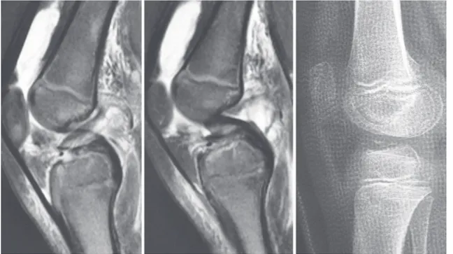
Fig. 3 Patient no. 9: 45 months after the injury, the lateral
radio-graphs view in extension before suture fixation surgery reveals 2 separated ossifications (left). MRI with enlarged ossifications on
pos-terior medial meniscus root avulsion under the PCL (right). The later avulsion was reinserted
T
able 2
Detail of indi
vidual patient’
s feature at the time of definiti
v
e treatment and results at longer follo
w-up
LMAH
lateral meniscus anterior horn,
PMMR
posterior medial meniscus root
P
atient no.
Age at the time of definiti
v e management Definiti v e management/
delay after accident
Intraoperati v e observ ation F ollo w-up/ definiti v e TT/ year L ysholm score IKDC 2000 subjecti v e score Observ ation/ complain
Residual laxity dif
ferent KT max a IKCD grade Le g discrep-anc y (mm) 1 6.5 Cast in fle xion/acute – 18.5 100 100 0 0 A No 2 5.5 Cast in e xtension/acute – 13.5 94 91
Occasional slight pain
1 A − 10 3 5 Suture fixation/acute
Anterior medical meniscus entrapment
7 100 100 0 2 A No 4 15 Secondary A CL after Y + 6.5
Empty notch/stable medical meniscus tear
5.5 95 100 Pi v ot shift: glide 4 F ailure IKDC B No 5 6.5 Cast in e xtension/acute – 1 100 100 0 2 A No 6 12 Secondary A CL after Y + 6 N A 1.5 N A N A 0 4 F ailure IKDC B N A 7 10 Secondary A CL after Y + 4 Interstitial A CL lesion b ut no manical tear 3.5 94 92
Lachman grade 2/pi
v ot shift: grade 1 4 F ailure IKDC C + 5 8 5
Delayed suture fixation/ after M
+ 2 No 3.5 99 97 0 1 A – 9 10
Secondary suture fixation/after
Y
+
4
LMAH posteriorly displaced –PMMI avulsion
2
99
98
Slight deficit of full fle
xion/ difficult squat 3 B + 7 10 7.5 Cast in e xtension/f ailure conserv ati v e TT w ait A CL r – – Restricted acti vities/w aiting period – F ailure – 11 11.5
Secondary suture fixation after M
+
30
LMAH on the fragment (discoid aspect)
1.5
95
94
V
ery occasional slight pain
1 A + 10 12 8
Suture refixation/ acute
LMAH on the fragment
2.5 100 100 0 0 A + 5 13 9
Secondary suture fixation/after M
+
6
Meniscal entrapment LMAH on the fragment
3
95
95
V
ery occasional slight pain
2 A + 7 14 6.5
Secondary suture fixation after M
+ 9 No 1 N A N A N A N A A N A 15 11
Secondary suture fixation after M
+ 24 – 1 W ait further N A W
ait further evaluation
W
ait further evaluation
N
An absence or an inappropriate immobilization of this lesion conducted regularly to delayed or non-union. The indication of non-surgical treatment with a long leg cast should be based on a careful MRI analysis allowing for a proper staging of the lesion and looking after associated lesions. If this treatment is initiated, a new MRI with the knee immobilized in the cast is recommended in order to assess the quality of closed fragment reduction and the absence of soft tissue interposition. As reported for osse-ous tibial eminence fracture [22], insertion of the anterior horn of the lateral meniscus on the avulsed fragment is a very consistent feature in CTEF (4 cases in this series). In 2 cases, it led to a confusing diagnosis of discoid lateral meniscus due to the posterior displacement of the anterior part of the meniscus.
When displacement is complete, arthroscopic suture fix-ation of CTEF is recommended. The management is sim-ilar to bony tibial eminence fractures, especially if a thin osseous layer is observed in the acute phase or at the time of secondary enlargement of the fragment. There are no data in this study suggesting that delayed treatment for few weeks is worth than acute management, and that is why the strategy of a new clinical examination with radiography after 2 or 3 weeks is a good alternative to MRI in all cases in the acute phase. In the case reported by Kim [18], a 5-year-old girl had successful treatment after a non-delayed pull-out suture under arthroscopy for a purely cartilaginous avulsion fracture, and there is no similar case in this series.
Long delay before surgical correction of CTEF with non-union is sometime justified in patients nearly asymp-tomatic, but parents must be aware that this option is poten-tially deleterious. The 2 patients in this series managed by conservative treatment for non-union failed after 4 and 6 years and eventually need ACL reconstruction. Vocke et al. [28] reported a good result in a 9-year-old boy treated conservatively after misdiagnosis, but follow-up was only limited to 1 year. During the waiting period, a progressive osseous resorption of the fragment avulsed is possible. It was noticed in 3 of the 4 failures reported in this study and could be an indirect sign of ACL involution. This is a plea to avoid prolonged conservative management of non-union.
Suture fixation procedures must be encouraged as it bears a low complication risk and good final results (7 IKDC A and 1 IKDC B). These very good results contrast with some moderate objective results reported after long term of displaced osseous tibial eminence fracture [15, 29]. This difference could be attributed to higher-energy mecha-nism and higher rate of associated ACL fibre damage at the time of injury for bony avulsions compared to CTEF [26, 30].
This study highlights arthroscopic transphyseal pull-out method of suture fixation as it is the technical option of our institution, but open and epiphyseal pull-out suture was also performed with success. Because the cartilagi-nous fragment is fragile and sometimes very thin, a suture of through the ACL could be more appropriate than screw
Fig. 4 Intermeniscal ligament entrapment seen on MRI could explain failure of orthopaedic treatment (patient no. 10). Double PCL sign is
or wire. Prior to fixation, curettage of the fracture bed is encouraged to facilitate the best placement of the fragment and to obtain countersinking and restitute appropriate ten-sioning of the ACL. No ACL retraction was reported nei-ther bone graft required when fixation was performed for non-unions, not even with long surgical delays after injury. The anterior fixation of the fragment must be the priority even with a CTEF extending posteriorly (Zifko type B) (Fig. 5). But care must be taken because a posterior ossifi-cation can be due to tibial avulsion of the medial meniscus posterior root and can require a specific associated proce-dure for meniscal fixation.
Growth arrest after transphyseal screw fixation for osse-ous tibial eminence fracture was reported [25]. This com-plication was not noticed in our series despite transphyseal fixation. The material used (thread) and the young age of patients could be argued, as the risk of growth arrest (type A according to Chotel’s classification) is lower for very young children compared to adolescents [10, 11]. On the contrary, a slight overgrowth process (type B) was regu-larly noticed (Table 2). Similar observations were reported by Ahn following pull-out arthroscopic treatment for tibial osseous eminence fractures in an 11-year-old male and a 6-year-old female. Both patients displayed a 1-cm length increase in the affected limb at follow-up [1].
To our knowledge, this retrospective multicentre study reports the largest series of CTEF so far, but the number of
patients is still low. The management of this rare injury was very heterogeneous which do not provide strong evidence-based treatment guidelines, but only recommendations based on a single-centre experience. Further multicentre studies on this probably underreported entity are necessary.
The clinical relevance of this study is to recommend the suture fixation of the avulsed fragment as the gold standard in the management of displaced CTEF.
Conclusion
Because of diagnosis difficulties for half of the patients, the management of CTEF was heterogeneous in this series. CTEF has a good prognosis even after misdiagnosis and treatment at the time of non-union. This could be due to low-energy mechanism of injury and low rate of associ-ated lesion. Orthopaedic treatment for acute minimally dis-placed fractures is only indicated under strict MRI control, and suture fixation is the recommended strategy in other situations. Conservative management of non-union could expose to ACL involution and cannot be recommended.
Compliance with ethical standards
Conflict of interest The authors declare that they have no conflict
of interest.
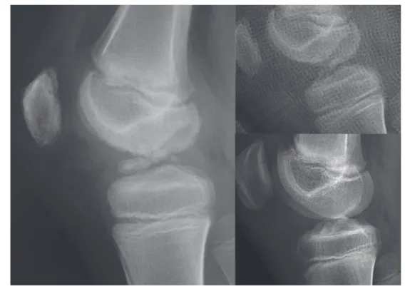
Fig. 5 Suture refixation procedure mainly involves the anterior part of the avulsed fragment (left and up). Remodeling and healing of the
References
1. Ahn JH, Yoo JC (2005) Clinical outcome of arthroscopic reduc-tion and suture for displaced acute and chronic tibial spine frac-tures. Knee Surg Sports Traumatol Arthrosc 13:116–121 2. Albright JC, Chambers H (2006) Tibial eminence fractures. In:
Micheli LJ, Kocher MS (eds) The pediatric and adolescent knee. Saunders, Philadelphia, pp 400–420
3. Anderson AF (2003) Transepiphyseal replacement of the anterior cruciate ligament in skeletally immature patients. A preliminary report. J Bone Joint Surg Am 85:1255–1263
4. Bengtson H, Giangarra C (2011) Osteochondral avulsion frac-ture of the anterior cruciate ligament femoral origin in a 10-year-old child: a case report. J Athl Train 46:451–455
5. Bonnard C, Chotel F (2007) Knee ligament and meniscal injury in children and adolescents. Rev Chir Orthop Reparatrice Appar Mot 93(Suppl):95–139
6. Bradley GW, Shives TC, Samuelson KM (1979) Ligament inju-ries in the knees of children. J Bone Joint Surg Am 61:588–591 7. Chotel F, Seil R, Greiner P et al (2014) The difficult diagnosis of
cartilaginous tibial eminence fractures in young children. Knee Surg Sports Traumatol Arthrosc 22:1511–1516
8. Chotel F, Chaker MM, Brunet Guedj E et al (2012) ACL recon-struction in children: an original technique. Tech Knee Surg 11:46–56
9. Chotel F (2012) ACL ruptures in children. In: Bonin M, Amen-dola A, Bellemans J, Mc Donald S, Ménétrey J (eds) The Knee joint: surgical techniques and strategies. Springer, Berlin, pp 291–323
10. Chotel F, Henry J, Seil R et al (2010) Growth disturbances with-out growth arrest after ACL reconstruction in children. Knee Surg Sports Traumatol Arthrosc 18:1496–1500
11. Chotel F, Seil R (2013) Growth disturbances after transphyseal ACL reconstruction in skeletally immature patients: who is more at risk? Young child or adolescent? J Pediatr Orthop 33:585–586 12. Corso SJ, Whipple TL (1996) Avulsion of the femoral
attach-ment of the anterior cruciate ligaattach-ment in a 3-year-old boy. Arthroscopy 12:95–98
13. Difelice GS, Lissy M, Haynes P (2012) Surgical technique: when to arthroscopically repair the torn posterior cruciate liga-ment. Clin Orthop Relat Res 470:861–868
14. Flynn JM, Mackenzie W, Kolstad K et al (2000) Objective evalu-ation of knee laxity in children. J Pediatr Orthop 20:259–263 15. Gans I, Baldwin KD, Ganley TJ (2013) Treatment and
manage-ment outcomes of tibial eminence fractures in pediatric patients: a systematic review. Am J Sports Med 42:1743–1750
16. Grönkvist H, Hirsch G, Johansson L (1984) Fracture of the ante-rior tibial spine in children. J Pediatr Orthop 4:465–468
17. Kawate K, Fujisawa Y, Yajima H et al (2004) Avulsion of the car-tilaginous femoral origin of the anterior cruciate ligament in a three-year-old child. A case report with a thirteen-year follow-up. J Bone Joint Surg Am 86:1787–1792
18. Kim JR, Song JH, Lee JH et al (2008) Cartilaginous avulsion fracture of the tibial spine in a 5-year-old girl. Skeletal Radiol 37:343–345
19. Kim YM, Rhee KJ, Lee JK et al (2006) Arthroscopic pullout repair of a complete radial tear of the tibial attachment site of the medial meniscus posterior horn. Arthroscopy 22:795
20. Kim SJ, Jo SB, Kim SG et al (2010) Peel-off injury at the tibial attachment of the posterior cruciate ligament in children. Am J Sports Med 38:1900–1906
21. Kocher MS (2006) Intra-articular injuries of the knee. In: Beaty JH, Kasser JR (eds) Rockwood and Wilkins’ Fractures in children, 6th edn. Lippincott Williams & Wilkins, Philadelphia, pp 985–995 22. Lowe J, Chaimsky G, Freedman A et al (2002) The anatomy of tibial eminence fractures: arthroscopic observations following failed closed reduction. J Bone Joint Surg Am 84:1933–1938 23. Matava MJ, Kim YM (2011) Tibial avulsion fracture of the
pos-terior root of the medial meniscus in a skeletally-immature child: a case report. Knee 18:62–65
24. Meyers MH, McKeever FM (1970) Fracture of the intercondylar eminence of the tibia. J Bone Joint Surg Am 52:1677–1684 25. Mylle J, Reynders P, Broos P (1993) Transepiphysial fixation
of anterior cruciate avulsion in a child. Report of a complica-tion and review of the literature. Arch Orthop Trauma Surg 112:101–103
26. Noyes FR, DeLucas JL, Torvik PJ (1974) Biomechanics of ante-rior cruciate ligament failure: an analysis of strain-rate sensitiv-ity and mechanisms of failure in primates. J Bone Joint Surg Am 56:236–253
27. Shea KG, Grimm NL, Laor T et al (2011) Bone bruises and meniscal tears on MRI in skeletally immature children with tib-ial eminence fractures. J Pediatr Orthop 31:150–152
28. Vocke AK, Vocke AR (2002) Cartilaginous avulsion fracture of the tibial spine. Orthopedics 25:1293–1294
29. Willis RB, Blokker C, Stoll TM et al (1993) Long-term fol-low-up of anterior tibial eminence fractures. J Pediatr Orthop 13:361–364
30. Yoon KH, Yoo JH, Kim KI (2011) Bone contusion and associ-ated meniscal and medial collateral ligament injury in patients with anterior cruciate ligament rupture. J Bone Joint Surg Am 93:1510–1518
31. Zifko B, Gaudernak T (1984) Problems in the therapy of avul-sions of the intercondylar eminence in children and adolescents. Treatment results based on a new classification. Unfallheilkunde 87:267–272
![Table 1 Detail of individual patient’s feature at the time of injury a Patients included in a previous study [7]](https://thumb-eu.123doks.com/thumbv2/123doknet/3113868.88450/4.892.196.681.124.1127/table-individual-patient-feature-injury-patients-included-previous.webp)