s—
Université de Montréal
Characterization of plasmacytoid dendritic celis in the CD4CIfflV transgenic mouse mode]
Par
Soheila Aflhami-Dastjerdian
Sciences biomédicales Faculté de médecine
Mémoire présenté à la Faculté des études supérieures en vue de l’obtention du grade de maîtrise
en sciences biomédicales
Avril 2006
L)5?n
(__\J ttcJ
Université
1411
de Montréal
Direction des bibliothèques
AVIS
L’auteur a autorisé l’Université de Montréal à reproduire et diffuser, en totalité ou en partie, par quelque moyen que ce soit et sur quelque support que ce soit, et exclusivement à des fins non lucratives d’enseignement et de recherche, des copies de ce mémoire ou de cette thèse.
L’auteur et les coauteurs le cas échéant conservent la propriété du droit d’auteur et des droits moraux qui protègent ce document. Ni la thèse ou le mémoire, ni des extraits substantiels de ce document, ne doivent être imprimés ou autrement reproduits sans l’autorisation de l’auteur.
Afin de se conformer à la Loi canadienne sur la protection des renseignements personnels, quelques formulaires secondaires, coordonnées ou signatures intégrées au texte ont pu être enlevés de ce document. Bien que cela ait pu affecter la pagination, il n’y a aucun contenu manquant.
NOTICE
The author of this thesis or dissertation has granted a nonexciusive license allowing Université de Montréal to reproduce and publish the document, in part or in whole, and in any format, solely for noncommercial educational and research purposes.
The author and co-authors if applicable retain copyright ownership and moral rights in this document. Neither the whole thesis or dissertation, nor substantial extracts from it, may be printed or otherwise reproduced without the author’s permission.
In compliance with the Canadian Privacy Act some supporting forms, contact information or signatures may have been removed from the document. While this may affect the document page count, it does flot represent any loss of content from the document.
Faculté des études supérieures
Ce mémoire intitulé
Characterization of plasmacytoid dendritic celis in the CD4CIIIW transgenic mouse model
Présenté par:
Soheila Aflhami-Dastjerdian
a été évalué par un jury composé des personnes suivantes:
Dr Andrew P. Makrigiannis Président-rapporteur
Dr Paul Jolicoeur Directeur de recherche
Dre Nathalie Labrècque Membre de jury
TABLE 0F CONTENTS
ABSTRACT iii
FIGURES LIST iv
TABLES LIST vi
ABREVIATIONS LIST viii
CHAPTER 7: ACKNOWLEDGMENTS ix
CHAPTER 1: INTRODUCTION 7
1. Innate and adaptive immune responses f
2. Characterization of dendritic cells (DCs) and their role in the
immune sytem 2
3. DC classification in mouse and human 4
4. Conventional DC (CDC) functions 5
5. Plasmacytoid DC (PDC) functions 5
6. PDC and myeloid DC (MDC) migrational features 9 7. TLR definition and roles in the immune system 17 8. Interferon (IFN)-alpha description and functions 72 9. Human immunodeficiency virus (HIV) infection 14
9.1. HIV description 14
9.2.The clinical course and immunopathogenesis of HIV infection 15
9.3. DC defects and implication in HIV 78
9.4. PDC defects and implication in HIV infection 79
9.5. The use of CpG ODNs in HIV infection 22
70. The biological system used for this study 22 10.1.CD4C/HIV transgenic (Tg) mouse description 23 70.2.AIDS-LIKE Disease and immune system dysfunctions 23
10.3.HIV-7 protein Nef 24
11. Rational and objectives of this study 25
CHAPTER 2: MATERIALS AND METHODS 28
1. Mice 28
2. Antibodies and reagents 28
3. Purification of pDCs from spleen and Iymph nodes 29 4. Generation of bone marrow (BM)-derived pDCs 29
5. Immunofluorescence 30
7. Flow cometry . 37
8. In situ hibridisation (ISH) 31
9. Giemsa staining of purified pDCs 32
70. Cytokines and stimulants of pDCs 32
11. Fetal liver transplantation: Chimera mouse model 32
72. PCR techniques for transgene detection 33
73. Statistics 34
CHAPTER 3: RESULTS 35
7. Identification and morphology of pDC derived from Iymphoid
organs 35
2. The biological system 41
3. Transgene expression in pDC 42
4. PDCs frequency, distribution, phenotype and in vitro survival 47
4.1. Frequency and distribution 47
4.2. In vitro survival 47
4.3. Phenotype 48
5. PDCs maturational state and response to in vitro stimulation 53 6. PDCs derived from FIt3L-enriched BM precursor cells and their
response to CpG 59
7. Transplantation chimera mouse model used for the study of
pDC 66
8. PDC Iocalization 71
9. PDC ïntracellular content in IFN-a 77
10.The effect of in-vivo CpG stimulation on immune ceil
populations 81
o
CHAPTER 4: DISCUSSION 87
CHAPTER 5: CONCLUSION 93
CHAPTER 6: REFERENCES 94
111
CHARACTERIZATION 0F PLASMACYTOID DENDRITIC CELLS IN THE CD4C/HIV TRANSGENIC MOUSE MODEL
Plasmacytoid dendritic ceNs (pDCs), or interferon (IFN) producing ceils (IPCs), participate in innate and adaptive immune responses against viral infections. Type I interferons exert antiviral effects by direct inhibition of viral replication and stimulation of a variety of immune ceil types. Murine pDCs are CD1 1 cb0B220+CD1 1 bGrlI0IL3RI0LY6Ch1 while human pDCs are CD4+IL3RhiCD45RA+(B220)HLADR+. They are both identified as cells that show plasmacytoid morphology, and reside in T cell zones of peripheral lymphoid organs and in blood at a low frequency.
To study HIV-1-mediated pathogenesis, the CD4C/HlVmutG transgenic (Tg) mice have been constructed in our Iaboratory. In this model, no viral infection and replication occurs but the expression of HIV-1 nef gene in CD4T celis and in macrophages/DC lineages induces an AIDS-like disease with several hallmarks of AIDS. These common features include CD4 down-regulation on T cells, depletion of of CD4 T cells, Iymphoid tissue atrophy and some non-lymphoid organ pathologies. The immune system in this model is impaired in many aspects and is under investigation. DCs other than pDCs display functional, maturational and homing impairments in these Tg mice.
Here, we show that 25-40% ot pDCs express HIV-1 gene nefin Tg mice. Compared to non-Tg, Tg-derived pDC show alterations in their frequency, maturation, localization and in vitro response to stimulation. Also, based on preliminary results splenic pDCs f rom Tg mice produce more IFN-a than their equivalents from nTg mice.
Considering the major immunoregulatory functions of pDCs, the observed changes in their characteristics suggest their implication in the pathogenesis of the AIDS-like disease in CD4C/HIV Tg mice.
Key words: AIDS, IFN-Œ, Antiviral immunity, Dendritic ceils, Nef, Lymphoid organs, CpG, FLT3L.
Caractérisation des cellules dendritiques plasmacytoides chez la souris transgénique CD4C/HIV
Les cellules dendritiques plasmacytoides, aussi appelées cellules productrices d’interferon, participent dans les réponses immunitaires innées et acquises contre les infections virales. Les interferons de type I accomplissent des fonctions antivirales par l’inhibition directe de la réplication virale et par la stimulation de plusieurs types de populations cellulaires du system immunitaire. Le phénotype des cellules dendritiques plasmacytoid chez la souris est CD1 1 cb0B220+CD1 1 bGrlIolL3RIoLy6ChI alors que chez l’homme leur phénotype est CD4lL3RCD1 1cB22OHLA-DW. Les cellules dendritiques plasmacytoid sont identifiées par leur morphologie similaire à celles des plasmocytes autant chez la souris que chez l’homme. Elles représentent un faible pourcentage de la population totale de cellules dendritiques et sont localisées dans les zones de cellules T des organes lymphoïdes périphériques et dans le sang.
Afin d’étudier les mécanismes impliqués dans la pathogenèse du virus d’immunodéficience humaine (VIH), la souris transgénique (Tg) CD4C,HlVmutG qui exprime le gène nef du VIH, a été mise au point dans notre laboratoire. Dans ce modèle murin, il n’y a ni l’infection ni la réplication virale, cependant une maladie ressemblante au SIDA est induite par l’expression du gène nef du VIH-1 dans les cellules T CD4 et les cellules des lignées macrophage/dendritique. Cette maladie et le syndrome d’immunodéficience humaine ont plusieurs caractéristiques communes incluant la régulation négative de l’expression de la molécule CD4 à la surface des cellules T, la déplétion des cellules T CD4, l’atrophie des tissus lymphatiques ainsi que des maladies des organes non-lymphoïdes. Dans ce modèle, le système immunitaire est défectueux à plusieurs niveaux et ces altérations sont actuellement sous investigation. Une étude récente a démontré que chez la souris Tg, les cellules dendritiques excluant les cellules dendritiques plasmacytoides, ont des anomalies au niveau de leur fonction, état de maturation et localisation.
La présente étude démontre que 25-40% des cellules dendritiques plasmacytoides chez la souris Tg expriment Nef. Les cellules dendritiques plasmacytoides chez la souris Tg présentent des altérations dans leur fréquence, maturation, localisation et leur réponse in vitro aux stimulants. De plus, selon des résultats préliminaires, les cellules
ivB
dendritiques plasmacytoides issues de la rate de la souris Tg produisent plus d’IFN-Œ que leur équivalentes chez la souris non-Tg.
En considérant d’une part les multiples fonctions immuno-régulatrices que possèdent les cellules dendritiques plasmacytoides et d’autre part les changements observés dans leur caractéristiques principales chez la souris Tg, on peut suggérer que ces cellules sont impliquées dans la pathogenèse de la maladie ressemblante au SIDA.
Mots clés: SIDA, L’immunité antivirale, Cellules dendritiques, Nef, Organes lymphoïdes, CpG, FIt3L.
FIGURES LIST
Fïgure 1: PDC identification and morphology 36
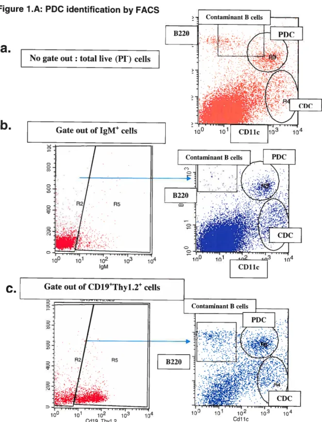
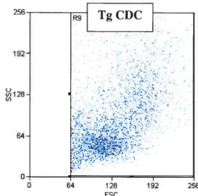
A. PDC identification by FACS 36
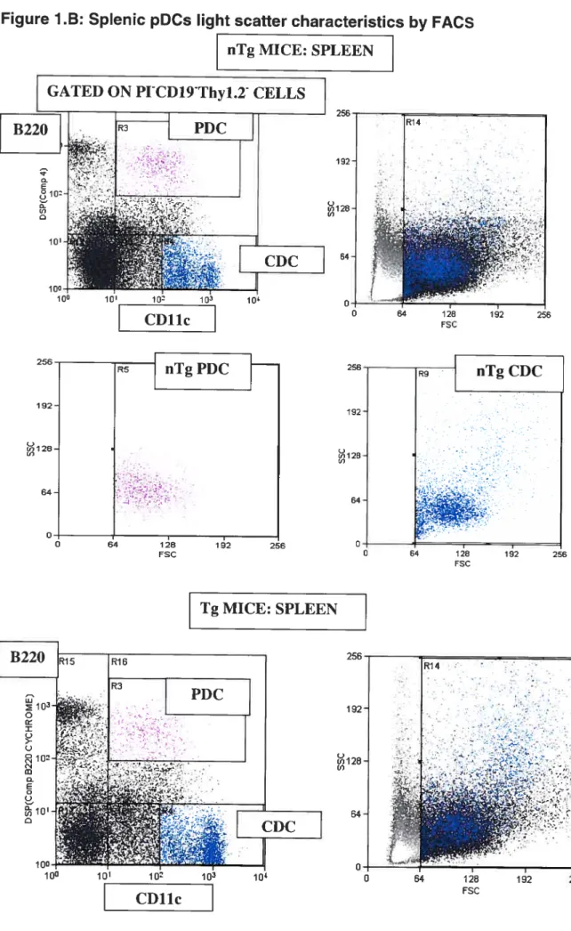
B. Splenic pDC Light scatter characteristics by FACS 37
C. Spienic pDC morphology: Giemsa staining 38
D. PLN pDC Light scatter characteristics by FACS 39
Figure 2: The biological system 41
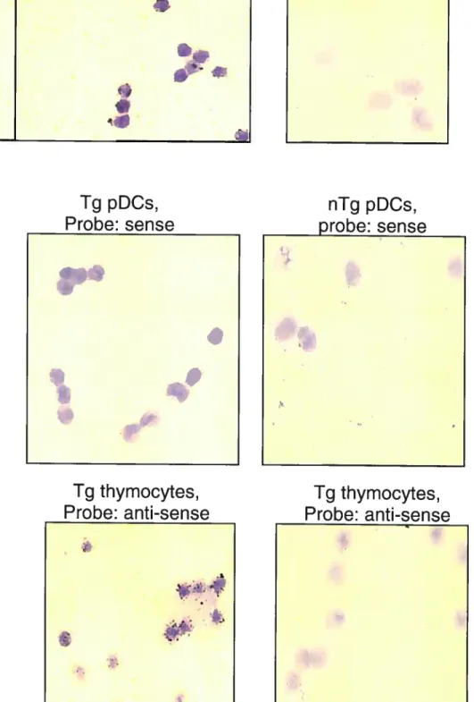
Figure 3: Transgene expression in pDC 43
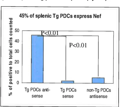
A. PDC transgene expression by ISH 43
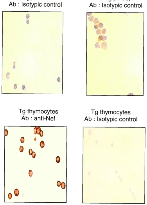
B. Transgene expressing pDC frequency in spleen (by ISH) 44 C. PDC transgene expression by immunohistochemistry (IHC) 44
D. PDC transgene expression by FACS 45
Figure 4: PDC frequency in blood 50
Figure 5: Phenotype 51
A. PLN pDC 51
B. Splenic pDC 53
Figure 6: In vitro FIt3L-enrïched BM-derived PDC and further CpG stimulation 60 A. FACS analysis: BM ex-vivo and post enrichment with FIt3L 60
B. FIt3L enrichment: PDC yield 61
C. FIt3L enrichment: PDC maturational state (MHCII) 61 D. FIt3L enrichment: PDCs maturational state (CD86) 61
E. Flt3L enrichment: PDC phenotype (Ly49Q) 62
F. CpG stimulation: FACS profiles 63
G. CpG stimulation: PDC maturational state (MHCII%) 64 H. CpG stimulation: PDC maturational state (MHCII MFI) 64
Figure 7: Chimeric mouse model 68
A. Percentage of chimerism in spleen 68
B. FIt3L enriched BM-derived pDC 69
V
B.2. FIt3L enrichment: PDC maturational state 69
Figure 8: PDC localization 71
A. Spleen white pulp 71
A.1 .Structure of periarteriolar lymphoid sheet (PALS) in nTg mice 71
A.2 PALS in nTg and Tg mice: PDC visualization 72
B. Spleen T cell and T-B celi transitional zone 73
C. Spienic T zone: pDCs count by IHC 74
D. Splenic T-B transitional zone: pDCs count by IHC 74 E. Spienic marginal zone (MZ), pDCs count by IHC 74
F. PLN T zone: pDCs count by IHC 75
G. PLN T-B transitional zone, pDCs count by IHC 75 H. PLN subcapsular (SC) zone: pDCs count by IHC 75
I. PLN subcapsular zone (confocal microscopy) 76
Figure 9: PDC intracellular content in IFN-a 78
A. Splenic PDC intracellular content in IFN-Q (graph) 78 B. Splenic PDC intracellular content in IFN-a (pictures) 78
C. Negative controls: non-specific staining 78
Figure 10: The effect0f in-ViVO CpG stimulation on immuneceil populations 81
A. PDC mobilization into blood and their maturation in response to CpG 81 B. NK cells frequency and expression of the activation marker CD69 82
C. PLNs mDC maturation 83
D. CD4 T cells in peripheral lymphoid organs 83
D.1 PLNs CD4 T cells 83
D.2 Splenic CD4 T ceils 84
E. PLNs CD4CD25 regulatory T celis 85
Supplementary Figures xi
Figure 11: IFN-a tunctions xi
Figure 12: PDC and the pathogenesis 0f AIDS xii
Figure 13: Description of CD4C/HIVmtG transgene xiv
A. BM-derived DCs by GM-CSF .xv
B. BM-derived pDCs by FIt3L xvi
vil
TABLES LIST
Table I: PDC frequency and distribution in lymphoid organs 48
Table Il: PDC in vitro survival 49
Table Il: PDCs maturational state and response to in vitrostimulation 55
A. Bone marrow 55
B. Spleen 55
ABBREVIATIONS LIST
Ab.: Antibody Ag.: Anti gen
AIDS: Acquired immunodeficiency syndrome BM: Bone marrow
BrdU: Bromodeoxyuridine labeling
CC: Chemokine
CCR: Chemokine receptor
CDC: Conventional dendritic celis: lymphoid DCs (CD8DEC2O5CD1 1 cB22OE) and myeloid DCs
CD17cb0t: CD11c expressed at lowto intermediate level
CD4OL: CD4O ligand
CpG: Unmethylated, phosphorylated oligodeoxynucleotide rich in CG motifs CTL: cytotoxic T lymphocyte
ER: Endoplasmic reticulum FDC: Follicular dendritic ceil
FIt3L: Fms-like tyrosine kinase Ligand GC: Germinal center
GM-CSF: Granulocyte-monocyte colony stimulating factor hIPC: Human interteron producing cell
HIV: Human immunodeficiency virus
IDC: lnterstitial dendritic celis (DCs residing in ail peripheral tissues except in the skin)
IFN-a: Interferon alpha
IHC: lmmunohistochemistry IL: Interleukin
ix
IPC: Interferon producing celi ISH: In situ hybridization Kda: Kilo-dalton
LC: Langerhans ceils (DCs found in stratified epithelia such as skin)
LN: Lymph node
MA: Medium alone MC: Monoclonal
MDC: Myeloid dendritic ceils (CD1 1 bCD1 1 cB22O) MFI: Mean fluorescence intensity
MHC: Major histocompatibility complex MIPC: Mouse interferon-producing ceils MLR: Mixed leucocyte reaction
MZ: Marginal zone
NIPC: Natural interferon producing cell NK: Natural killer cells
nTg: Non-transgenic, normal mice ODN: Oligodeoxynucleotide
O.N.: Overnight
PALS: Periarteriolar lymphoid sheets (in white pulp of spleen) PAMP: Pathogen associated molecular paffern
PBMC: Peripheral blood mononuclear cells PDC: Plasmacytoid dendritic cells
PDGF: Platelet derived growth factor PG: Proteoglycan
PKR: Protein kinase R pLN: Peripheral lymph node
PRR: Pathogen recognition-receptor RBC: Red blood cells
rIL-3: Recombinant interleukine-3
SC: Subcapsular (in this texte refers to pLN subcapsular zone) Ss: Single-stranded
TCR: T-ceII receptor
Tg: Transgenic, can refer toCD4C,HIVmUtG or CD4CIHIVm mice
Th: Helper T ceil
TLR: Toli-like receptor
TNF: Tumor necrosis factor
xB
ACKNOWLEDGMENTS
I thank Stéphanie Lemay and Eve-Lyne Thivierge for animal care assistance; Lin Jia
and Ginette Massé for technical assistance, Martine Dupuis and Eric Massicotte for f Iow
cytometry sevices. I am grateful to Dr Johanne Poudrier for teaching me ail the techniques
of DC enrichment and culture and helping me to analyze and interpret data obtained on
PDC. I thank Dr Pavel Chrobak for giving me access to the chimeric mouse construct in
order to study pDCs. Finally, I thank Dr Paul Jolicoeur, my director, for making this
research work possible and his intellectual assistance as weIl as Dr Zahar Hanna for
7. INNATE AND ADAPTIVE IMMUNE RESPONSES
The immune system utilizes innate and adaptive responses in order to recognize and clear pathogens from the host [1]. Innate immunity, which is antigen (Ag) non-specific, includes natural barriers to pathogens such as skin and mucous membranes and specialized effector celis such as phagocytes and natural killer (NK) celis [1]. Macrophages, neutrophils and dendritic celis (DCs) at certain stage of their differentiation, show phagocytic functions. NK celis are cytotoxic celis participating in innate immunity because of their ability to lyse virus infected and tumor ceNs [1, 2, 3]. Phagocytes and cytotoxic NK celis, as the main players of innate immunity, can discriminate between pathogens and self by utilizing signais upon ligation of their ceN suriace pathogen recognition receptors (PRRs) [4, 5]. Innate immunity is closely linked to and influences adaptive responses [6]. For example, proinfiammatory cytokines produced by cells involved in innate immunity, enhance the Ag-presenting capacity of DCs [6]. Adaptive immunity refers to Ag-specific responses including humoral and cell-mediated defense mechanisms, which depend on B ceNs and T celis respectively [1]. The acquired immune response has also a memory component that recognizes a pathogen that has previously infected the host [1].
In adult, B and T ceNs develop from pluripotent stem cells in bone marrow (BM). Whiie B ceils mature in BM, T ceNs complete their maturation in thymus where after undergoing a selection process, only 5% of them survive and leave this organ. Negative selection induces central tolerance through deletion of self-reactive T cells in order to avoid immune response against self-tissues [1].
DCs process Ags and present them to T cells, in association with peptide-binding proteins i.e. major histocompatibility molecule (MHC) for recognition by T cell receptors (TORs). Intracellular Ags, processed into peptides in the cytosol of the antigen-presenting ceil (APC), bind to MHC class I while extracellular Ags that have entered the endocytic pathway of the APC associate with MHC class Il molecules [7]. CD4 T cells, also called helper T cells, recognize Ag presented by MHC Class II and upon activation have profound immune regulatory effects on processes such as antibody (Ab) responses, cytotoxic T lymphocytes (CTL) responses and generation of memory cells. CD8 T ceNs are mainly CTLs that recognize Ags presented by MHC class I. These cells participate in cell-mediated immune defense [1, 8]. However, particular subsets of CD4 and CD8 T cells have also been
2
attributed with major roles in inducing peripheral tolerance that together with central tolerance taking place in thymus maintain the homeostasis of the organism [9, 10].
When CD4 T cells are primed upon Ag recognition, they become activated, expand and release cytokines [1]. At this stage, helper T ceils are categorized based on the cytokine profile they secrete [11]. CD4 T celis polarized as Thl produce interleukin IL-2 and IFN-y stimulating ceil-mediated immune responses [1, 72]. Type 2 helper T cells (Th2) secrete IL-4, IL-5, IL-6 and IL-10 and induce Ab secretion by B cells [11, 12]. Elaboration of each class of cytokines, Thl or Th2, inhibits the production of the other class of cytokines [12]. Previously, it was believed that each DC subset was specialized for priming either Thl or Th2 adaptive responses (DC1 for Thi inducers versus DC2 for Th2 inducers) [13]. However, ail DC subsets can initiate adaptive Th1-Th2 ceil responses and dictate the ciass of T ceil immunity through the production of Th polarizing cytokines [11]. lndeed, the cytokine microenvironment is central in T helper cell polarization toward Thl or Th2 celI type during immune responses [14].
DCs can direct a Thi response, whenever the microbial stimuli that drive their maturation elicit IL-12 production [11, 15J. IL-12 is a cytokine produced by mononuclear phagocytes, DCs and activated NK celis inducing adaptive cell-mediated responses by promoting the generation of Thl cells [1]. Also ail DCs can suppress Thi development and allow Th2 polarization upon exposure to IL-7 0 [1]. IL-70, produced by activated macrophages and Th2 polarized T cells down-regulates MHCII and costimulatory molecules on APCs [16]. It conters tolerogenic properties to DCs and drives the generation of mouse CD4 Tr cells [76]. IL-10 inhibits cell-mediated immunity favoring humoral responses [1]. Overall, a Thl or Th2 response develops depending on antigen dose, the state of maturation of DCs together with parameters such as kinetics 0f activation and the nature of the maturational stimulus of
DCs [11, 17,18].
2. CHARACTERIZATION 0F DCS AND THEIR ROLE IN THE IMMUNE SYTEM
DCs are a migratory group of BM-derived leukocytes with generally a short haif-Iife and a fast turnover regenerating continuously f rom BM precursor cells through multiple developmental stages. In BM, DC progenitors develop into immature precursors that circulate in blood and home into various tissues throughout the organism. DCs represent a network of APCs specialized in the capture, processing, transport and presentation of Ag to T cells. They play a wide range of roles in initiation and regulation of immune responses against pathogens and in homeostatic regulation. They are implicated in innate and adaptive
immune responses and participate in the establishment of central and maintenance of peripheral seif-tolerance [17, 19]. Central toierance is established by DCs presenting self Ags to T ceNs that undergo the selection process in the thymus [9, 20]. Moreover, in the absence of inflammation, DCs do not become fully activated and induce tolerance rather than immunity [6, 9]. Indeed, contrary to activated DCs that produce immunity, immature or quiescent DCs, i.e. mature DCs that are not fuily activated, induce peripheral tolerance through multiple mechanisms. These inciude kiiling of T cells, paralyzing them (anergy) or/and generating regulatory T celis [20, 21]. The tolerogenic potential of DCs correlates with the absence of activation by proinflammatory signais [9, 21]. These signais are responsible for the switch from a quiescent to an activated state in some DC lineages [20].
Proinflammatory signais reieased by infectious agents attract DCs to the site of inflammation where they undergo a process caiied maturation that is required for their Ag presenting function and for their migration into lymphoid organs. Once in lymphoid organs, matured DCs induce T cell activation and initiate Ag-specific acquired immune responses. Upregulation of ceii-surface MHC class Il and costimulatory moiecules i.e. CD8O, CD86 and CD4O characterize DC maturation [21]. This process also causes the secretion of cytokines and chemokines. MHC Il and costimulatory molecule expression on APCs provide the two prerequisite signais to activate naïve T celis [1, 7]. Upon maturation, MHCII mainly residing in intraceilular compartments, relocate from lysosomes to the celi surface [7]. Matured DCs upregulate celi surface chemokine receptors (CCRs), which play a role in their migration to T cell areas of iymphoid organs. Matured DCs also produce chemokines, which attract various subsets of T ceils promoting DC-T celi interactions [18]. Thus, DC maturation is a key control point in converting an antigen into an immunogen [21, 22]. This centrai role of DC maturation in defense mechanisms is iiiustrated by the exampie 0f some tumor celis, which secrete factors inhibiting DCs maturation in order to escape the immune responses [6].
The interaction between NK celis and DCs may also be important in defense mechanisms. DCs acquire the ability of regulating NK celis function early post-infection allowing the production of cytotoxic and inflammatory responses that participates in the eradication of the pathogen [3]. In fact, cytokines produced by different DC subsets enhance NK ceii activation and cytotoxic function. These eftects are dependent on cytokines such as IFN-a and IL-12 for NK ceil cytotoxicity and IFN-y secretion while tumor necrosis factor (TNF)-a s required for CD69 expression. In addition, IL-2 activated NK cells interact with DCs and induce their maturation [2, 3, 23].
4
3. DC CLASSIFICATION IN MICE AND HUMANS
DOs are a heterogeneous population of cells sharing major functional and phenotypic features [17, 18]. Different DC subpopulations are distinguished based on ontogeny, phenotype, function, localization and migrational characteristics and accomplish complementary and overlapping functions in the immune system [18]. DCs can be obtained in large number, both in vitro and in vivo, in the presence of Fms-like tyrosine kinase-3 ligand (FIt3L) that is a hematopoietic growth factor [24, 25]. This is because most DCs develop from Flt3, hematopoietic progenitor cells in BM regardless ot their myeloid or lymphoid origin [25, 26].
Mouse DCs are classitied into three subsets. The majority of mouse DCs are caIled myeloid DCs (mDCs) because they originate from common myeloid precursors in BM. Lymphoid DCs derive from common Iymphoid precursors, as do T cells. The myeloid DCs have a characteristic celI surface phenotype, namely CD8aCD1 1 bCD1 1c, they are widely distributed in Iymphoid and non-lymphoid tissues. Lymphoid DCs are CD8aCD1 1 bCD1 1 c cells residing mostly in the thymus, but are also present in the periphery at a lower frequencythan mDCs [15, 27].
More recently, plasmacytoid DCs (pDCs) with a particular morphology mimicking immunnoglobulin (Ig)-secreting plasma ceNs have been characterized as a distinct DC subset in mice and humans. Contrary to what was believed previously, mouse pDCs develop efficiently f rom both Iymphoid and myeloid-committed progenitors in BM [9, 28]. PDCs are localized in peripheral and cord blood, in the thymus as well as in T celi areas of secondary Iymphoid organs including peripheral iymph nodes (pLNs), spleen and Peyer’s patches [11, 29]. They are identitied by their celi surface phenotype as CD11cI0B22O+CD11bGr1I0IL3RI0LY6ChI. In contrast to other DCs that show a fast turnover, pDCs in uninfected mice are Iong-lived and their frequency varies between different strains and organs [30].
Human DCs are subdivided into two main groups including CD11c mDCs and CD11c pDCs. Human pDCs phenotype is slightly different from mouse pDCs, since they are CD4iL3RhCD45RA±(B220)HLADR+. In humans as in mice, mDCs constitute the majoritary of DCs and include skin DCs called Langerhans cells (LC5) and interstitial DCs (intDCs) residing in ail other tissues [31]. PDCs in human and mouse are equivalent in their main function as they are both identified as the natural interferon-aipha (IFN-a) producing ceNs (NIPCs) and they both can be mobilized by in vivo treatment with FIt3L [32].
4. CON VENTIONAL DENDRITIC CELL (CDC) FUNCTIONS
In this text the term cDC is used to designate ail mouse DCs including myeloid and Iymphoid DCs, but excluding pDCs.
Various DC subsets difterentially control immune responses. There is a complementarity in recognition as well as in effector functions among DC subpopulations [15]. CDCs Iink innate and adaptive immune responses by activating both B ceils and NK cells [33]. Different patterns of cytokine production by DC subsets may occur in response to particular microbes. Also, different functions of Ag-presentation can be assigned to DC subsets depending on the conditions cf stimulation [34]. Mouse Iymphoid DCs (CD8aCD11b), produce IL-12 and prime naïve CD4 T lymphocytes to secrete Thi cytokines, whereas mDCs (CD8c(CD11b) induce both Thi and Th2 cytokines following in vivo immunization [15, 27]. However, only Iymphoid DCs possess the ability to cross-present Ag in association with MHCI [34]. While mDC induce a strong allogeneic mixed-Iymphocyte reaction (MLR) response, Iymphoid DCs induce a comparatively low MLR response and express Fas-Iigand [35]. In fact, the limited CD4 T ceIl response induced by lymphoid DCs is associated with T celI death that is caused by the interaction cf Fas on T cells and Fas-ligand on DCs. Because of these characteristics, lymphoid DCs seem to play a regulatory function while mDCs are stimulatory and induce a strong immune response [35, 36].
In humans, mature monocyte-derived DCs induce a Thi response causing the differentiation cf naive CD8 T ceNs into cytotoxic T lymphocytes (CTL5) [10, 36]. Apart f rom their role in T cell priming, cDCs activate NK cells and control B celI growth and differentiation [30, 37]. They also participate in innate responses through functions such as pathogen recognition and phagocytosis [2, 3, 4]. In addition, follicular DCs (FDC5) reside in follicles cf lymphoid organs and retain Ag in a native state for presentation to germinal center (GC) B cells [1]. FDCs are responsible for B celI activation and selection through the process of affinity maturation [1]. On the other hand, thymic DCs derive from an intrathymic precursor, develop and die within the organ. They show a non-migratory behavior and participate in the induction cf central tolerance [20].
5. PLASMACYTOID DC
f
PDC) FUNCTIONSPreviously, pDCs were called plasmacytoid T cells or monocytes based on their tissue localization in T celI rich area of lymphoid organs, their round morphology with a smooth plasma membrane and an eccentric nucleus resembling plasma cells [38]. Studies showed
6 that non-monocytes, non-T and non-B plasmacytoid lymphocytes can activate NK cells, in vitro [39]. Later, pDCs were classified as a distinct subset of DCs because of their lack of lineage markers, the expression of MHCII and the capacity to stimulate naive T ceNs [40, 41]. lndeed pDCs do not express typical ceIl surface markers of T celis, such as CD3, B cells, such as CD19, monocytes, such as CD14 or NK ceils, such as CD16 and CD56 [42]. Currently, they are recognized as IPCs, which upon microbial and especially viral infection are able to activate NK ceils as weIl as regulate the immune responses. It is now known that IPCs are necessary for NK celi activation partly because IFN-u enhances NK celi cytotoxicity [2, 43].
PDCs are implicated in innate and adaptive immune responses to viruses [11, 31]. Upon microbial infection, pDCs accomplish their functions partly through the secretion of IFNa. PDCs produce up to 1000 times more IFN-a in response to viral stimulation than any other peripheral blood mononuclear celis (PBMCs) [32]. Apart from NK cell activation that was originally attributed to pDCs, IFN-a plays multiple immunoregulatory functions including differentiation of monocytes into dendritic-like celis, maturation of mDCs and pDCs, Thi polarization of unpolarized Ag-experienced CD4 T cells and the differentiation of B cells into plasma cells [44, 45, 46]. Since DCs are the most potent APCs but are at 10w frequency in the organism, the ability of pDCs to promote monocyte-derived DCs amplifies total Ag presenting function [7, 47]. This IFN-a-mediated effect of pDCs on monocytes and mDCs indirectly regulates T celI responses [47]. Type I IFNs are also important for pDC maturation since their maturation is drastically impaired in IFNo—receptor (IFNAR)-deficient mice [39]. In the context of viral infection, pDCs sense the presence of the virus through TLR7 and TLR-9 expressed in their endosomal compartment [48]. Upon viral recognition, pDCs control innate immune celI functions as they activate macrophages and NK cells through release of cytokines. In fact, pDCs responses to viruses include the production of IFN-a, IL12, chemokines
f
CC5) such as CXCL9 and CXCL1 O and chemokine receptors (CCR5) such as CCR3, CCR4 and CCR5 [44]. AIl these factors affract and activate NK ceils and T cells and modulate the function of mDCs by inducing their maturation [45, 46, 49].The production of type I IFNs is exclusively reserved to immature pDCs. In fact, as pDCs mature, they ose the ability to produce IFN-a and acquire the ability to present Ag to T cells [42]. Thus, pDCs show a dual Ag-presenting and IFN-producing function that links innate and adaptive immunity [50]. However, pDCs possess a weaker ability to stimulate naive CD4 T cells than mDCs. PDCs are less potent APCs than mDCs because they express Iower celI surface MHCII and costimulatory molecules, do not endocytose Ag as efficiently
as mDCs and Iack the expression of lysosomal proteases involved in Ag processing [42, 44]. PDC and mDC differ also based on various sets cf PARs that they express (see section on TLRs). Moreover, while immature as weIl as mature mDCs morphology includes cellular protrusions called dendrites, immature pDCs are round and Iack dendrites. PDCs can deveiop dendritic protrusions only upon maturation and transformation into APCs [27, 35]. It is possible that pDCs induce the differentiation cf unpoiarized Ag-experienced T celis that have previously been expanded by mDCs [44].
Despite the difficulty to study pDCs because cf their low frequency in blood and secondary lymphoid organs cf both human and mouse, different research groups have investigated the APC function cf pDCs [51]. These experiments include in vivo and in vitro assays using different stimulants such as CD4O ligand (CD4OL), IL-3 or viruses. CD4OL is a weIl-known DC activator expressed on many ceIl types including helper T cells and seems to amplify activation of DC previously stimuiated by infectious agents [52]. IL-3 s a potent maturation and survival factor for human pDCs [53]. However, it shouid be noted that IL-3 and CD4OL are factors produced by T celis atter their activation and in early responses te viruses, these signais cannot dictate DC differentiation [54].
In mouse, upon microbiai, IL-3 or CD40L stimulation, pDCs differentiate into CD8DEC205 DCs distinct from Iymphoid CD8DEC205 DCs [50]. Microbial infection can thus induce the generation cf a new DC subtype flot present in uninfected organism. Although the complete functional implication cf this shift in the DC network remains te be determined, it is known that mature CD8DEC205 pDCs can efficiently present Ags to T celis and to a lesser extent than mDCs induce Ag-specific adaptive responses [50]. Mutine pDCs generally promote Thi responses at high and Th2 responses at low antigen dose. It has been shown that viral stimulation cf pDCs induces IFN-a production and their differentiation into Th1 polarizing DCs [12, 55, 56]. Other experiments in mouse show that influenza virus stimulation cf pDCs in vivo results in their differentiation into APCs. These matured pDCs induce effector/memory CD8 T celI responses that could be recalled 4 weeks later [43].
In human, pDCs difterentiate into CD11c mature DCs when cultured with IL-3 and CD4OL. These CD4OL and IL-3-treated pDCs have been shown to stimulate CD4 helper T cells, which become Thl-polarized in vitro due te the synergistic effect cf IL-12 and IFN-a [56]. However, prier te these findings, human pDCs were called precursors cf DC2 because upon stimulation with CD4OL or viruses in vitro, they induced an IL-4-independent Th2 polarization cf naive T cells [57]. In contrast to DC2s, monocyte-derived DCs were termed DC1s because they induce Th polarization and pro-inflammatory responses [57]. However,
8
since type I IFNs promote a Thi response in humans, this view of pDCs as precursors of DC2s does not correlate with their ability to produce type I IFNs [42, 57]. The discrepancy between these resuits could reflect various conditions of in vitro pDCs exposure to CD4OL and methods used to culture them [45, 57]. In vivo, DCs shouid receïve signais with a different strength in a sequentiai fashion, preserving the plasticity of the DC’s Th polarization function until the final stages of their maturation in the iymph node [57]. Accordingly, in order to accomplish aN these diverse functions, pDCs must migrate from biood into inflamed LNs [49].
Other in Wtro studies on human pDCs show that they influence the humoral immune responses. When activated T ceNs secrete IL-2 and express CD4OL, these factors signai pDCs to secrete IL-6. PDC-derived IFN-a/r3 induces B ceils ta differentiate into plasma biasts whiie pDC-derived IL-6 is responsible for inducing their Ab secretion [58]. Thus, in vitro, virus-activated pDCs induce the differentiation of CD4O-activated B celis into plasma cells producing virus specific Abs [43, 58].
On the other hand, a role for the induction of tolerance was attributed to pDCs. In vitro studies show that immature human pDCs can differentiate CD8 Tr ceNs that suppress Ag specific T celi proliferation [31, 58]. These Tr cells produce IL-10, which is implicated in immuno-suppression [10, 43, 59]. IL-10 producing CD8 Tr cells inhibit Ag-specific proliferation of naive CD8 T ceNs in a primary MLR [59]. Another study reported, that CD40L-activated pDCs similar to immature mDCs have the ability to induce primary Tr-cell differentiation permitting peripheral toierance [59]. One study on human pDCs demonstrates that TLR-9 stimulation of pDCs promotes the generation of CD4CD25 Tr celis in vitro [60]. Studies in mice also support that pDCs play a raie in the generation of CD4 Tr cells [60].
Overall, the production of IFN-a by pDC initiates innate immune responses Iimiting the spread of pathogens and promotes adaptive immune responses by enhancing the function and maturation of APCs including monocytes and mDCs [47]. Also, when matured, pDCs participate directly as APCs in adaptive responses. Thus, pDCs initiate an immune response through the secretion of cytokines and link innate and adaptive immunity upon activation [42]. PDCs show great flexibiiity in their functions and depending on the stimuli they receive, they can induce Thi, Th2 or regulatory (Tr) responses [31, 50, 55].
Several studies have shown heterogeneity in pDC population [50, 51]. PDCs phenotype can be used ta identify their subtypes since pDCs have been ciassified on the basis of celi sur[ace CD4 expression. CD4 pDCs have been shown ta be functionaily distinct from CD4
pDCs. CD4 pDCs do flot produce IFN-Πalthough they show similar MHCI1 expression to CD4 cells [50]. Kinetic studies have shown that CD4 pDCs are precursors of CD4 pDCs. Moreover, blood CD4 pDCs are the precursors of spienic CD4 pDCs, which are the precursors of splenic CD4 pDCs [50].
In addition, BM pDCs f rom mouse could be classified based on the expression of Ly49Q [51]. Ly49Q, a type II C-lectin membrane-associated polypeptide, is a member of Ly49 NK receptor subfamily that binds to MHCI and regulates NK ceil activation. However, Ly49Q is expressed on ail peripheral pDCs but flot on NK or NKT cells [51]. Ly49Q is flot exclusively expressed by pDCs since Gr1 myeloid Iineage celis and activated macrophages have been reported to express this celI surface molecule [61]. In BM, the expression of Ly49Q defines two subsets of pDC [51]. Different expression level of Ly49Q defines sequential deveiopmental stages of pDCs derived from BM by using cytokines such as FIt3L [62]. One study showed that Ly49Q pDCs respond less to infectious agents than Ly49Q pDCs possibly because they have flot yet acquired the full sets ot ceil surface and/or signaling molecules necessary for their functions against microbes [51]. Accordingly, a correlation was found between Ly49Q expression on pDCs and their maturational state [61]. In fact, Ly49Q pDCs are mostly CD4 and upon activation they become Ly49Q and concomitantly upregulate celi surface expression of CD4 and MHCII [61, 62].
6. PDCS AND MYELOID DCS (MDCS) MIGRATIONAL PROFILES
DC development includes distinct stages. In BM, first DC progenitors proliferate and give tise to DC precursors, which circulate in blood. Blood DCs migrate to sites of inflammation in response to chemotactic stimuli and from there they are recruited into secondary lymphoid organs to prime T cells. Thus, DCs migration is critical for optimal immune responses [56]. Migration of leukocytes is a complex process to which CCs contribute [44, 63]. Pathogens induce local production of TNF-a, which once in circulation promotes systemic inflammation and up-regulates CCL3 causing mobilization of DC precursors into the blood [64, 65]. These precursor ceils develop into immature DCs that localize in various non-lymphoid tissues in order to capture Ags upon invasion of the host by foreign agents. In general, immature DCs express receptors for inflammatory CCs, which facilitate their migration to sites of inflammation [11, 15]. Local TNF-a accelerates influx of mDC precursors to sites of inflammation via CCL3 and their efflux via CCL21 [64]. Once there, they respond to inflammatory cytokines secreted by innate immune celis that promote their maturation after capturing Ags [11, 15]. Upon maturation, DCs show a CCR switch, down regulating inflammatory CCRs (CCR5, CCR2 and CCR1) and up-regulating CCR7 [16]. The
10
interaction of pathogens with innate immune celis aiso induces the release of proinflammatory signais that cause DC migration to T ceil rich zones of Iymphoid organs (spleen and LNs) for Ag presentation and T ceil priming [11, 63, 66]. in fact, upon maturation DCs up-regulate cell surface CCR7, a receptor for SLC/CCL21 and ELC/CCL1 9, which are constitutively produced in T ceH areas of secondary lymphoid organs and drives DC migration to these sites [11, 15].
Serum TNF-a also up-regulates CXCL9 and E-selectin on LN high endothelial venules (HEVs), which attracts pDCs to cluster around HEVs. HEVs are speciaiized post-capillary blood vessels with a cuboidal endothelium, which constitute one of the entry routes to T cell zone of LNs for biood lymphocytes [1, 67]. It is now known that it is around HEV5 where pDCs produce large amounts of type I IFN [56, 64, 65]. CXCR3 is required for this migration, because in CXCR3-deficient mice, trans-HEV migration of pDCs into LNs from biood is impaired, [56, 64]. In contrast to pDCs, mDC precursors as well as monocyte derived DCs that have captured Ags migrate into the draining LNs through afferent iymphatics [11, 64, 65].
in human blood, pDC and mDC express similar sets of CCRs with the exception of CCR7 and CXCR3. Expression of CXCR3 and CCR7 is low on mDCs and high on pDCs [13]. Following maturation, both pDCs and mDCs downregulate CCR1, CCR2, CCR5, CCR6 and CXCR1 and strongly upregulate CCR7. However, in contrast to mDCs, most CCRs expressed on pDCs are flot functional on circulating cells [13, 66]. in fact, only after maturation induced by stimulants, such as CD4OL, are the receptors for inflammatory chemokines downregulated, and CCR7 on pDCs becomes functional and induces migration [13]. The differences between mDC and pDC migration include the expression of CCRs such as CCR7 and CXCR3, direct entry of pDCs to LNs from blood while mDCs enter LNs through afferent lymphatics and the fact that on some pDC, CCRs are flot functional when they circulate in blood [13]. These differences in migration between pDCs and mDCs, suggests a distinct role for each subset in the induction and regulation of the immune responses [13]. Accordingly, PDCs could be more involved in homeostatic control of immune responses since they can induce Thi, Th2 and possibly Tr cells depending on the conditions [13]. In addition, virally stimulated pDCs have been shown to produce CCs such as CCL4 and CXCL1 O that attract NK and activated T cells to pDCs [65].
7. TLR DEFINITION AND ROLES IN THE IMMUNE SYSTEM
The innate immune system is the first une of defense against pathogens and allows the initiation and regulation of adaptive immune responses for effective clearance cf infectious agents [54]. To detect the presence of infection, the innate immune system f irst recognizes conserved molecular structures that are predominantiy found in microorganisms but not in vertebrates [5, 68, 69]. Pathogen associated molecular patterns fPAMP5) are recognized by various types of PARs expressed on innate immune ceils including monocytes, macrophages, DCs, B celis and NK cells [5]. Among these cells, effector ceils, such as neutrophils and macrophages, exert phagocytic and/or cytotoxic functions upon binding cf PAMP5 te PRRs [5, 68]. A major part of PRRs is represented by Toli-Like receptors (TLR5), which constitute a group of transmembrane signaling receptors [70]. Ten different TLRs in human and twelve in mice have been described [70]. They belong to phylogeneticaily conserved factors that generate selective immune responses upon ligation by various PAMPS [70, 71]. TLRs ligation activates NFKB transcriptional factor and induces the expression of many genes involved in immune responses [5, 72]. The responsiveness to a given PAMP depends on the set of TLR expressed by the ceil. Ail TLRs permit to mount a f irst set of stereotyped responses such as inflammation. Individual TLRs induce distinct set of immune responses necessary for host defense. For example, intracellular pathogens require Thi polarization of CD4 T cells [5, 15, 73].
It is known that TLR1 is required for the response to imiquimod, TLR2 to peptidoglycan (PG) and mycobacteria, TLR3 to double stranded fds)-RNA (viruses), TLR4 to LPS, TLR5 to flagellin, TLR7 and TLR8 to G/U rich single-stranded (ss) RNA or imidazoquinoline, TLR9 to intracellular bacterial and virai DNA or CpG ODN [3, 68]. In humans TLR7 and TLR 9 are expressed by B ceNs and pDCs, while TLR2, TLR3, TLR4 and TLR8 are expressed by mDCs and monocytes [3, 68]. In the mouse, expression of TLR9 is not exclusive to pDCs and B cells and other DCs as well as macrophages express TLR9 [68].
MDC and pDC express complementary sets of TLR, providing them the capacity to respond to different PAMPs [11]. While TLR4 and TLR3-mediated mDCs activation by LPS and Poly(I:C) respectively, causes their IL-12 production, TLR9 ligation to CpG induces IFN-a production by pDCs [50, 74]. LPS is a major component of the ceil wall cf Gram-negative bacteria and PoIy(l:C) mimics viral dsRNA that is released after virally infected ceil lysis [75, 76]. The differential expression of TLRs by human DC subsets could explain selective immune responses to various pathogens. TLR9 signaling induces the maturation of pDCs among human DCs and of pDC and mDC among mouse DOs.
ut
aIse activates matured12
DCs to produce cytokines. Thus, TLR-signaling pathways iink innate and adaptive immunity by promoting Ag-presenting function of DCs that allows in turn the generation of adaptive T ceil responses [35].
In order to induce an efficient adaptive immune response, TLR signaling must also block the suppressive eftect of Tr cells on T celis. Indeed, as mentioned earlier, Tr ceNs prevent the activation of peripheral auto-reactive T ceils. It has been postuiated that TLR signaling induces the concomitant secretion of IL-6 by DCs. Since IL-6 renders pathogen-specific T ceils refractory to the suppressive effect of CD4CD25 Tr celis, TLR-signaling can overcome peripheral toierance and induce an immune response [77].
Pathogen-derived DNA recognition as non-self by mammalian immune ceNs through TLR9 signaling pathway is due to its high frequency of unmethylated CpG dinucleotides. In fact, mammalian-derived DNA has much lower frequency of these dinucleotides and they are mostly methyiated [63, 71, 78]. CpG ODNs are synthetic compounds that mimic microbial DNA and interact with TLR9 specifically, as shown by the absence of CpG effects in TLR9-deficient mice [71]. CpG effects include proliferation of spienocytes, inflammatory cytokine production and maturation of DCs [71]. In addition, TLR9-engaged pDC activate NK celis as indicated by CD69 upregulation and enhanced cytotoxicity. One study showed that IL-2 produced by CD4 heiper T cells enhances NK cells activation, while Tr ceils cancel this additive action by blocking CD4 T ceIi-mediated enhancement of NK ceil activation [79].
In mice, CpG ODNs promotes MHCI presentation of soluble Ags to CD8 T celis inducing Thi-type ceIl-mediated responses independently of CD4 T celi help. This characteristic of CpG could potentially be useful in the treatment of chronic viral infections such as HIV, where there is progressive Ioss of Thi immune responses along with depletion of CD4 helper T cells [47].
8. INTERFERON (IFN)-ALPHA DESCRIPTION AND FUNCTIONS
IFN-o and IFN-13, are classified as type I IFN5 whiie IFNy corresponds to type II [69]. IFN-y is an antiviral agent as weIl as an activator of CTLs and is one of the main cytokines produced by Thl-polarized CD4 heiper T ceils, CTL type I and NK cells [53, 58]. Type I and II IFN5 have non-redundant and complementary functions in the host response to viral infection [80]. Type I IFNs are inducible cytokines produced by different celi types in response to viral infection and exert an antiviral activity at ail stages of virus life cycle [80, 81, 82]. In the mouse, 1 IFN-3 and 13 IFN-a have been identified and are encoded by an
intronless multigene family [81]. Fibroblasts, T ceNs, macrophages, monocytes, DCs and NK cells secrete IFN-a [80]. However, pDCs identified as NIPCs produce up to 1000 times more of this cytokine than any other ceIl type [80]. Type I IFNs bind to a common receptor IFNAR that is expressed on a large variety of ceIl types [70].
Stimuli, such as viral components, endotoxin treatment of cells, UsRNA, poly (I: C) or CpG ODNs can induce IFNa production [80, 83]. IFNa/j3 acts directly on most ceIl types and turns on biochemical pathways, which induce an antiviral state. IFNaI3 antiviral effects include the restriction of viral replication and confer cellular resistance to further viral infection [58]. Various proteins are involved in IFNa/r3 induced antiviral state including PKR, ADAR (adenosine deaminase acting on RNA), CAS (2’,5’-oligoadenylate synthetase), RNase L and MX proteins [80, 84].
Protein kinase receptor (PKR) is a cytoplasmic serine-threonine kinase containing two conserved d5RNA-binding domains in its N-terminal region. PKR belongs to a class of more than 20 dsRNA-binding proteins, which recognize non-specifically dsRNA. Once activated, PKR phosphorylates the protein synthesis factor eIF-2a, which inhibits initiation of viral translation and consequently interteres with viral replication. Numerous viruses encode genes responsible for inhibiting PKR activity. ADAR inhibits ANA editing; CAS activates the endoribonuclease RNase-L to degrade s5RNA [80, 84] Human MxA protein and mouse Mxl are IFN-induced GTPases that inhibit replication of certain viruses by binding target viral proteins [74]. Many viruses have developed mechanisms to escape the inter[eron system by inhibiting its synthesis or blocking its action as for example through suppression of PKR activation [4, 74].
Moreover, lFNaIr3 regulates functions of immune cells, enhancing humoral and cellular antiviral responses in vivo [32, 82]. These regulatory functions on immune cells include the activation of NK ceNs by promoting their survival and proliferation through IL-15 production. IFNa/j3 activates CD8 and CD4 T cells and has an effect on proliferation of B celis. Type I IFN5 drive monocyte differentiation into DC-Iike ceNs, maturation of DCs as well as differentiation of pDCs into efficient APCs. IFN-a mediates its immunoregulatory functions in part through the modulation of the activity of other cytokines and CCs such as IFNy, IL-1, 1L2, IL-3, IL-8, IL-12, IL-13, IL-15, TNF-a, IP-l0, etc [68, 80]. IFNa/F3 also affects expression of ceIl surface cytokine and CC receptors. For example, it causes IL-12R up-regulation on CD4 T cells promoting Th-1 polarization of helper T cells [80]. Moreover, IFN-a as weII as IFN-y enhances immunoglobulin G (lgG) production and downregulation of IgE secretion by B cells, promoting humoral immunity [80]. IFNa43 is an important cytokine in innate immune
14
responses since it activates innate immune celis such as NK ceNs to kiil virally infected ceNs. This cytokine also causes up-regulation of MHCI and MHCII on APCs and promotes adaptive immune responses by CD8 and CD4 T celis respectively [85] Thus IFN-a bridges the innate to adaptive immune responses through the increase of total APC function [80] (See supplementary figure 11).
Studies investigating IFN-u production in response to pathogens show that this cytokine has very rapid kinetics and disappears after about 24 hours. This suggests that pDCs initiate an antiviral innate immunity early during viral infection. The capacity of pDCs to produce great amounts of IFN-a rapidly and effectively in response to viral pathogens is due at least in part to a specific genetic program [86]. IFN-a acts through multiple signaling pathways involving JAK kinases, STAT transcription regulators and IRF transcription factors [80, 84]. The induction of IFN-a secretion requires a positive feedback-loop as shown by IFNAR-deficient celis, which are inhibited in their production of IFN-a [83]. However, mouse and human pDCs rather than other ceil types can bypass the positive feedback loop that is essential in the production of IFN-a. It has been reported that IRF7, which interacts with the adaptor protein MyD88 is required for type I IFN production in response to TLR7 and TLR9 through the signaling pathway involved in the positive feedback Ioop [39, 86]. Both human and mouse pDCs constitutively express higher levels of IRE-7 than other celI types allowing them to bypass the positive feedback in IFNaI3 signaling that requires de novo IRE-7 synthesis [39, 86]. Moreover, the production of high-Ievel type I IFNs in response to TLR9 necessitates the retention of MyD88-IRF-7 complex in the endosomal compartment for an adequate lapse cf time and this spatiotemporal regulation is observed only in pDCs [39, 87].
9. HUMAN IMMUNODEFICIENCY VIRUS (HIV) INFECTION
9.1. HIV DESCRIPTION
Human immunodeficiency virus fHIV) is a retrovirus, a family member of lentiviruses that causes an infection cf the immune system leading to the acquired immunodeficiency syndrome (AIDS) [67, 88]. The HIV genome contains two ssRNA molecules, each 9.2 Kb long, bound to a molecule of reverse transcriptase. Following entry into host cells, the viral reverse transcriptase copies the RNA into d5DNA. This viral DNA integrates into the host cell DNA and becomes a provirus. HIV genome is composed cf nine genes flanked by non coding long terminal repeats (LTR5) that are necessary for the integration of the provirus into the host ceil genome. LTR5 also contain binding sites for gene regulatory proteins that control the expression cf the viral genes and are required for viral replication. HIV RNA is
within a core of viral proteins and surrounded by a nucleocapsid. The outer portion of the nucleocapsid consists of a phospholipid bilayer envelop derived from host celI containing virally encoded envelop glycoproteins, gpl2O and gp4l which are required for the infection of cells [67, 88, 89].
HIV-1 possesses three typical retroviral genes: the gag gene encodes core structural proteins, the env sequences encode gpl2O and gp4l of the viral envelop, mediating the membrane fusion required for the entry of the virus into host ceils and the p0! sequences encode reverse transcriptase, integrase, and viral protease enzymes necessary for viral replication. In addition, H IV-1 genome also includes six other regulatory genes, the tat, rev, vif, nef vpr, and vpu genes[1]. Vif enhances infectivity, Vpr promotes nuclear import of viral DNA and arrest ceIl cycle at G2 phase, Tat is necessary for elongation of viral transcripts and markedly augments viral transcription independently of the integration site, Vpu downregulates CD4 expression and augments viral release f rom ceNs. HIV-1 -protein Nef, also down-regulates MHCI. Tat and Rev are necessary for HIV replication. The Tat protein functions as a transactivator of the HIV promoter and Rev facilitates the cytoplasmic export of incompletely spliced transcripts. Vif, Vpr, Vpu and Nef are termed accessory because their inactivation does not totally block HIV replication in vitro. However, these genes are important virulence factors [89, 90].
Since the CD4 molecule functions as a receptor for the virus, HIV infects CD4 cells including CD4 T cells, monocytes and DCs [67, 88, 91]. Membrane fusion between the Iipid bilayers of the viral envelope and the host ceil membrane is required for viral entry to the cell. The gpl2O/gp4l -Env complex catalyses the membrane fusion by interacting with host cell CD4 molecule and coreceptors. [89]. Host celI coreceptors for viral membrane glycoproteins are CXCR4, expressed on macrophages, and CCR5 by T ceils. Certain HIV isolates, replicate efficiently in primary macrophage culture and are termed ‘TM”—tropic or X4 for CXCR4. Other isolates replicate in T cells but not in macrophages and are called “T” tropic or R5 for CCR5 [89].
9.2. THE CLINICAL COURSE AND IMMUNOPATHOGENESIS 0F HIV INFECTION
There are two strains of HIV, called HIV-1 and HIV-2. HIV-1 is the most common cause of AIDS. However, HIV-2, which differs in genomic structure and antigenicity, induces a similar clinical syndrome [92]. The clinical course of HIV infection includes an acute infection with high level of viremia, followed by a latent stage, where the immune response controls infection but the virus persists as a provirus in infected cells [92]. Initially, HIV infection
16
induces specific humoral and celI-mediated anti-HIV immune responses [91]. During the latent phase, LNs and spleen are sites of ongoing HIV replication and tissue destruction. HIV infection progresses to AIDS when destruction of lymphoid tissue is complete and blood CD4 T cells drops below 200 cells / mm3 [1]. AIDS includes various signs and symptoms such as weight Ioss, night sweats, fever and diarrhea. AIDS is a state of suppressed immunity mainly because of the loss of CD4 helper T cells and impairment of adaptive immune responses [93]. AIDS immunosupression underlies the development of opportunistic infections (Cl) and neoplasia [93].
The latent stage can vary f rom less than 1 year to 15 years. The outcome of HIV-1 infection depends on the host anti-viral immune response. Long-term non-progression (LTNP) status, defined by at least 10 years of HIV infection without the development of AIDS, is associated with the development ot effective and persistent T ceIl responses against HIV [24]. The clinical stages of HIV disease correlate with a progressive spread of HIV from the initial site of infection (i.e. blood or mucosa) to lymphoid organs. In early HIV-1 infection, gut associated lymphatic tissue (GALT) is a principal site of virus replication and depletion of primarily lumina propria memory CD4 T cells. The immune response controls temporarily the acute infection but a chronic infection is established when virus trapped in lymphoid tissues by FDCs persists [1].
The major hallmark of the suppression of immune responses in HIV-1 infected patients is the progressive decline in the number of CD4 T cells. In fact, the consequences of HIV infection of T cells are devastating because the CD4 helper T cells play major roles in shaping immune responses [24, 93]. In vitro and in vivo studies have shown that in AIDS patients, lymphocyte proliteration in response to defined Ags is reduced. This is referred to as an anergic state of the immune system [12]. In addition to CD4 T cell depletion, HIV induces a wide range of impairments in the regulation of immune responses. These include premature atrophy of the thymus and thymocyte depletion [82], loss of LN architecture, loss of CD4 T celI function, inversion of CD4/CD8 T-cell ratio, abnormal B celI functions despite their polyclonal activation and hypergammaglobulinemia, serum auto-Abs and circulating immune complexes [92, 93, 94]. Also, since cytokines control the homeostasis of the immune system, their dysregulation in HIV infection contributes to the pathogenesis ot AIDS [95].
Other defects, in the immune system of HIV infected patients, include a decrease in memory T cell responses to Ag, poor CTL responses to viral infection and lowered humoral immune responses to defined Ags [93, 94]. Some studies report a decrease in the
proportion of Thi cytokines-secreting T-cells in HIV-infected patients while Th2 secreting T ceNs are increased. MHC-restricted CD8 CTLs, found in large numbers during latent stage of HIV-1 infection, decrease with disease progression [96]. Since CD4 T ceNs are required at least partly for CTL activity, their depletion may explain the decline in CTLs as the disease progresses [93]. Also, since HIV is an intracellular pathogen, its clearance f rom the organism requires a Thi type cell-mediated immune response in host defense [15, 73].
The ability of HIV-1 to evade the host immune responses leads to the establishment 0f chronic infection [55]. The viral mechanisms of immune evasion include the following strategies. The high mutation rate of HIV that allows the virus to evade detection by Ab or T cells produced in response to viral proteins before mutation. Also, HIV-Nef mediates selective down-regulation of HLA-A and HLA-B, rendering infected cells undetectable by CTLs. Concomitantly, the normal cell surface expression of HLA-C and HLA-E allows the infected cells to evade NK cell-killing as these cells are inhibited by the expression of HLA-C and —E [55]. Finally, HIV infection preferentially inhibits cell-mediated immunity due to the immune deviation from Thi to Th2. This immune dysfunction increases the susceptibility to infection by intracellular pathogens including HIV itself [1]. In fact, since IFN-y activates and IL-4 inhibits macrophage-mediated killing of intracellular pathogens, the shift in Th responses may explain the host susceptibility to such microbes [1, 74].
The initial adaptive immune response to HIV infection corresponds to the expansion of CD8 CTLs specitic for HIV-derived peptides [12, 94]. CD8 T-ceII response to HIV infection is crucial for antiviral defense through direct destruction of virafly infected cells and/or through secretion of soluble antiviral molecules [91]. Thus, the partial control of HIV infection causing the transition of acute infection to the latent phase seems to be due to CD8 CTLs specific for viral peptides. On the other hand, Abs to a variety of HIV Ags are secreted by B cells within 6 to 9 weeks after infection. However, the beneficial effect of anti HIV Ab responses in limiting the disease s questionable [12]. While the most immunogenic viral proteins in the humoral responses are gp-1 20 and gp-41, anti-envelope-Abs inhibits poorly viral infectivity [12]. In addition, despite the established role of humoral immune response in many viral infections, Abs in sera against HIV-1 have only weak neutralizing activity and are mostly directed against virion debris [92]. However, high level of Abs to HIV in infected patients suggests an interaction between B cells and activated T ceNs in the development of immune activity [92]. Moreover, neutralizing Abs able to block the infectivity of virus may limit the replication of HIV during the asymptomatic stage of infection [93].
1$
HIV infection disrupts cell-mediated and humoral immunity and renders the host susceptible to CI and maiignancies [12, 93]. Highly active anti-retroviral therapy (HAART) suppresses viral replication and partially restores CD4 T celi counts, reducing AIDS-related mortality [12]. Some reports concluded that adaptive immunity is the most critical component of the immune system for control of H1V infection and that H1V-specific CD4 helper T cells and CTL responses may decide the rate of disease progression [93, 97].
9.3. DC DEFECTS AND IMPLICATION IN HIV
DCs are the first mucosai cells infected by the virus during oral and sexual transmission and participate in viral dissemination by transmitting the virus to CD4 T cells [98]. Mucosal DCs that have been infected process HIV-1 proteins and carry live HIV into the LNs where they present the viral Ags to T cells. In LNs, HIV-1 replication is the most active and thus DCs contribute to the efficient propagation of HIV by their migration that is mediated by CCR7 upregulation upon activation [98].
DCs send signais to T cells that promote their ability to replicate the virus [99]. The DC-T ceN microenvironment is an explosive site for HIV propagation because activated T cells become highly susceptible to HIV-1 and enhance viral repiication. HIV-1 preferentially infects CD4 T celis specific for HIV-1 Ags [97]. Viral replication is fast and efficient in activated T celis and macrophages, partly because cellular factors that positively regulate viral transcription are abundant in these celis [99]. HIV-infected DCs initiate rounds of T cell infection by transmitting HIV to resting T ceNs in LNs [92]. Some DCs show latent infection and become productiveiy infected after maturation. This shift of non-productive to productive infection is a way for DCs to facilitate the persistence of the virus in the organism [100]. This is because a productively infected cell produces many virions each capable of infecting cells and amplifying the infectious cycle [1]. Moreover, in the acute infection, virus particles are trapped in the FDC network and this is another important mechanism for DCs to maintain HIV infection over time [94].
DCs are flot only central to the pathogenesis of HIV-1 by serving as initial and continuous source of the virus, but paradoxically are also essential for the induction of effective anti-HIV immune responses involving both CD4 and CD8 T cells [92, 97, 101]. Carnage of virus by DC into LNs allows clustering and priming of T ceils and production of antiviral immune responses [100]. Therefore, DCs depletion during HIV infection is implicated in HIV-1 pathogenesis by reducing immune system capacity to prime T celis and to generate antiviral responses. However, the loss of DCs when they become overtly infected imitates H1V
dissemination [91]. Depletion of HIV infected DCs and T-cells, is due to either direct lysis by the virus or by HIV-specific CTLs. Also, in AIDS, DC development from CD34 stem cells is impaired [101]. Thus, DCs disappearance from blood of HIV-1 infected patients could be caused by various mechanisms. These events include failure of DC precursors to differentiate, their death due to infection or CTLs and their relocalization into the secondary Iymphoid tissues after maturation [55].
Since HIV-1-induced maturation of DCs may contribute to their decrease in the blood of patients with high viral Ioads, this process may have important consequences for HIV-1 cellular transmission and HIV-1-specific T-cefl responses [102]. MDC maturation depends on the stimuli produced in the local environment by HIV-1 activated pDCs [45]. PDCs’ IFN-a secretion in response to HIV-1 induces the maturation of bystander CD11c DCs (mDCs in human). MDCs maturation is characterized by the induction of CD83 and CCR7 expression and the upregulation of CD8O and CD86 allowing their migration to LN5 and Ag presentation to T cells [45]. However, it has been reported that the T-cell stimulatory function of DCs is impaired in HIV infection possibly due to defective co-stimulatory molecules expression [103]. Accordingly, viral proteins such as Nef and Tat selectively induce immature DC biology that allows DC-T celI interaction without up-regulation of co stimulatory molecules for effective priming of T cells [104]. Defects in DCs maturation can be beneficial to HIV-1 pathogenesis because immature DCs promote the induction of peripheral tolerance to seif-peptides, and HIV may use this function of DCs to inhibit immune responses [50]. In addition, IL-12 is a cytokine involved in the initiation of Thi type of immune responses and its production depends on DC5 maturational state. The altered production of IL-12 by DCs in HIV-1 infection could be involved in the loss of Thi type immune responses in HIV [15, 73].
Overali, DC abnormalities are important in the induction of immunosuppression caused by HIV [96]. The infection of DCs, their depletion and impaired signaling to T cells participates in shaping the pattern of immune responses in HIV-1 infected individuals. Treatment that reverses the defects caused by HIV in the DC population may improve cell-mediated immunity [92].
9.4. PDC DEFECTS AND IMPLICATION IN HIV INFECTION
The preservation of pDCs and mDCs is crucial for shaping anti-HIV immunity [45]. PDCs expressing CD4, CXCR4 and CCR5 (the major coreceptors of HIV-1) are susceptible to infection by the virus [32]. It is known that HIV infects pDCs as well as mDCs. But, it is flot
20
clear if HIV-1 stimulates pDC through TLRs or after direct entry into the cytoplasm [41, 105]. PDCs respond to HIV-1 infection by producing cytokines such as IFN-a, TNF-o and chemokines, up-regulating celi surface maturational markers, such as MHCII, CCR7 and CD83. Thus, HIV-activated pDCs acquire the ability to migrate in response to CCL1 9, which functions as the ligand for CCR7 and is expressed in Iymphoid organs [60].
Several differences have been reported between pDCs and mDCs in HIV infection. In the case of intravenous HIV infection, pDCs may be one of the f irst cells to transport virus into LNs. This is due to pDCs direct access f rom blood to pLNs by trans-HEV migration [56, 64]. Thus, pDCs are involved in rapid trafficking of the virus to LNs where viral spread and replication are most efficient [45, 60]. In addition, HIV-activated pDC migration to Iymphoid organs seems necessary for mDC maturation since HIV does not directly activate mDCs, but seem to be activated by cytokines produced by pDCs [45]. Another difference between pDC and mDC in HIV infection is that viral replication in pDC requires their activation by CD4OL, while mDCs replication is independent of CD4OL mediated maturation [106]. Moreover, HIV-activated pDCs acquire the ability to stimulate naïve CD4 T cells, but Iess efficiently than mDCs probably due to the lower upregulation of costimulatory molecules [45]. However, both DC subsets can efficiently transmit HIV to T ceNs [45, 97].
Clinical findings support pDCs implication in HIV-1 infection. In fact, the decrease of pDCs number in the blood of HIV-1-infected individuals correlates with disease severity as for the reduction in CD4 T celi counts [32]. This depletion of blood IPC in AIDS patients is not due to a generalized hematopoietic failure. It has been reported that elevated levels of IL-10 as observed in HIV infection could decrease IFN-a production and the frequency of pDCs because IFN-a is a survival factor for pDCs [74].
PDCs seem to play a particular role in the resistance to CI during the course of HIV infection. This is based on many findings that show an increase in pDC number in long-term non-progressor individuals compared to AIDS-developing patients [107, 108]. Moreover, during HAART, an increase in pDC frequency is associated with normalization of CD4 T ceil count, Cl resistance and overall clinically apparent immune reconstitution [108, 109]. Accordingly, one study has reported the existence of a correlation between pDC number after one month of HAART and viremia after interruption of HAART. In fact, HAART must be discontinued because of various complications that it induces in treated patients. This correlation suggests that pDC count can be used as a tool to identify individuals in whom HAART interruption can be beneficial [110]. Thus, IPC count is used as a new parameter to monitor the status of the immune system in HIV-infected subjects [11].