Université de Montréal Faculté de médecine
Département de microbiologie, infectiologie et immunologie
Molecular Characterization of Th17 Lymphocytes and
Monocyte-Derived Dendritic Cells in the Context of HIV-1 Infection
Présenté par:
Vanessa Sue WACLECHE
Sous la direction scientifique de la Dre Petronela Ancuta Laboratoire Chimiokines et VIH-1
Le centre de recherche du CHUM
Thèse présentée à la Faculté des études supérieures de l’Université de Montréal en vue de l’obtention du diplôme de doctorat en microbiologie et immunologie
Montréal, le 7 Décembre 2015
SOMMAIRE
Le virus de l’immunodéficience humaine de type 1 (VIH-1) altère les fonctions du système immunitaire pour promouvoir sa persistance. Les composantes de l’immunité ciblées par le VIH-1 incluent les lymphocytes Th17 et les cellules dendritiques dérivées des monocytes (CDDMs). Deux sous-populations de lymphocytes Th17, nommées Th17 et Th1Th17, ont précédemment été décrites avec des propriétés transcriptionnelles et des spécificités antigéniques distinctes. Les cellules Th17 et Th1Th17 sont hautement permissives à l’infection par le VIH et leur fréquence est diminuée chez les sujets chroniquement infectés sous trithérapie antirétrovirale. Toutefois, seulement une fraction des lymphocytes Th17 est infectée par le VIH, indiquant l’existence de Th17 résistants à la réplication virale. Également, il est connu que l’infection à VIH induit une altération de la fréquence des monocytes reflétée par l’expansion de la population monocytaire exprimant le récepteur Fcγ de type III/CD16. Les monocytes sont des précurseurs de cellules dendritiques et une altération de ratio entre les monocytes CD16+ et CD16- pourrait avoir des conséquences délétères sur la qualité des réponses immunitaires. Le rôle fonctionnel des CDDM exprimant ou non CD16 dans le contexte de la pathogénèse à VIH-1 demeure inconnu. Ce projet de thèse est divisé en 2 parties: 1) l’étude de l’hétérogénéité des cellules Th17 et 2) la caractérisation approfondie des CDDM CD16+ et CD16- dans le contexte d’homéostasie et de la pathogénèse de l’infection à VIH. Dans la première partie, nous avons fonctionnellement caractérisé deux nouvelles sous-populations de lymphocytes Th17 avec une expression différentielle des récepteurs de chimiokines CXCR3 et CCR4 : nommés CCR6+DN et CCR6+DP, exprimant toutes les deux CCR6, marqueur de lymphocytes Th17. Nous avons démontré que les cellules CCR6+DN et CCR6+DP partagent des caractéristiques biologiques communes avec les cellules Th17 et Th1Th17 incluant la permissivité au VIH. Nos résultats indiquent que les cellules CCR6+DN représentent un stade précoce de différentiation des lymphocytes Th17 et expriment des marqueurs de cellules T folliculaires. De plus, comparativement aux sous-populations Th17, Th1Th17 et CCR6+DP, la fréquence et le compte des CCR6+DN sont préservés au sein des sujets chroniquement infectés sous thérapie antirétrovirale. Nous proposons un modèle dans lequel les cellules CCR6+DN représentent des lymphocytes Th17 résistantes à l’effet cytopatique du virus
qui contribuent à la persistance virale par leur capacité de porter un virus compétent en matière de réplication. Dans la deuxième partie, nos résultats révèlent que les CDDMs CD16+ et CD16
distinctes. Les CDDMs CD16- détiennent un potentiel immunogène supérieur tandis que les
CDDMs CD16+ ont une meilleure capacité de transmettre le virus aux cellules T CD4+ au repos. Également, nous confirmons l’effet néfaste du VIH sur les fonctions immunologiques des cellules DC à stimuler la prolifération et la polarisation des cellules Th17 spécifiques à C. albicans et à S. aureus. En conclusion, les résultats inclus dans cette thèse fournissent une compréhension détaillée sur l’hétérogénéité présente au sein des lymphocytes Th17 et des CDDMs et révèlent de nouveaux déterminants moléculaires de l’immunité exploités par le VIH au profit de sa persistance.
Mots-clés : VIH-1, Th17, Th1Th17, CCR6, cellules T-CD4, mémoires, profil transcriptionel, monocytes, cellules dendritiques, CD16
ABSTRACT
The ultimate aim of immunity is to restrict the emergence of exogenous pathogens while providing immune tolerance to self-antigens. The human immunodeficiency virus type 1 (HIV-1) disrupts the functions of the immune system to promote its own dissemination and persistence. The components of the host immunity targeted by HIV-1 include the Th17 lineage and the monocytes. The Th17 lineage was previously reported to include two different populations referred to as the Th17 and Th1Th17 cells exhibiting different transcriptional profiles and antigenic specificities. Both Th17 and Th1Th17 cells are permissive to HIV and their frequency is reduced in the blood and gut mucosa of chronically HIV-infected subjects. Nevertheless, HIV-1 infects only a fraction of the Th17 pool, suggesting the existence of Th17 cells resistant to HIV. In addition, it well documented that HIV-1 infection alters the pool of peripheral blood monocytes and induces the expansion of a monocytic population expressing the Fcγ receptor III/CD16. Monocytes are precursors for dendritic cells (DCs) and an altered CD16+/CD16- monocyte ratio may have deleterious consequences on the quality of immune responses. The functional features of CD16+ versus CD16- monocyte-derived DCs (MDDCs) in the context of HIV infection remain to be elucidated. This thesis is divided in two parts: 1) the study of Th17 cell heterogeneity and 2) the in depth characterization of CD16+ and CD16- monocytes-derived DCs (MDDCs) at homeostasis and during HIV-1 infection. In the first part, we have identified and functionally characterized two new previously uncharacterized subsets of CCR6+ T-cells with differential expression of CXCR3 and CCR4, double negative CCR4-CXCR3- (CCR6+DN) and double positive CCR4+CXCR3+ (CCR6+DP) subsets. We demonstrated CCR6+DN and CCR6+DP share cytokine production, antigenic specificity, lineage plasticity and HIV permissiveness with the previously characterized Th17 (CCR6+CCR4+CXCR3-) and Th1Th17 (CCR6+CCR4-CXCR3+) subsets. Among these four Th17 subsets, CCR6+DN cells were found to represent an early stage of Th17 differentiation and expressed features of T follicular helper T-cells. Moreover, in contrast to Th17, Th1Th17 and CCR6+DP subsets, the frequency and counts of CCR6+DN cells was preserved in chronically HIV-infected subjects under antiretroviral treatments compared to unHIV-infected controls. Our results suggest that CCR6+DN represent long-lived Th17 cells contributing to HIV persistence by
carrying replication-competent virus. In the second part, our results reveal that CD16+ and CD16
-MDDCs represent two distinct populations with unique transcriptional programs and immunological functions. CD16- MDDCs displayed a superior immunogenic potential, whereas
CD16+ MDDCs exhibited a higher capacity to induce HIV replication in resting CD4+ T-cells.
Also, we confirmed the negative effect of HIV on DCs immunogenic function involving the stimulation of T-cell proliferation and Th17 polarization in response to pathogens such as C. albicans and S. aureus. Overall, in this thesis we provide a better understanding on Th17 and MDDC heterogeneity and reveal new molecular determinants of pathogenicity in immune cells that are exploited by HIV-1 to insure its persistence in the infected host.
Keywords: HIV-1, Th17, Th1Th17, CCR6, human CD4+ T-cells, memory, transcriptional profiling, monocytes, dendritic cells, CD16
TABLE OF CONTENTS
CHAPTER 1:INTRODUCTION ... 19 1.1 HIV INFECTION ... 20 1.1.1 HIV GENERALITIES ... 20 1.1.2. HIV STRUCTURE ... 21 1.1.3 HIV-1 GENOME ... 221.1.4 HIV TARGET CELLS ... 23
1.1.5 HIV-1 REPLICATION CYCLE ... 23
1.1.6 HIV-1 PATHOGENESIS ... 25
1.1.6.1 THE ACUTE PHASE ... 26
1.1.6.2 THE CHRONIC PHASE ... 27
1.1.6.3 THE AIDS PHASE ... 29
1.1.7 TRANSMITTER FOUNDER VIRUSES ... 29
1.1.8 RESTRICTION FACTORS ... 30
1.1.9 HIV PERMISSIVENESS FACTORS ... 33
1.1.10 FACTORS CONTRIBUTING TO HIV PERSISTENCE ... 34
1.1.11 HIV-1 TREATMENT AND CURRENT CHALLENGES ... 36
1.1.11.1 ANTIRETROVIRAL THERAPY ... 36
1.1.11.2 VACCINES ... 40
1.1.11.3 ALTERNATIVE HIV CURE STRATEGIES ... 41
1.2 BIOLOGY OF THE TH17 CELLS ... 44
1.2.1 HISTORY OF TH17 CELL DISCOVERY ... 44
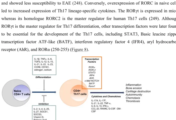
1.2.2 TRANSCRIPTIONAL REGULATION OF TH17 DIFFERENTIATION ... 47
1.2.3 SURFACE MARKERS DEFINING HUMAN TH17 SUBSETS ... 52
1.2.4 TH17 DIFFERENTIATION PROGRAM AND PLASTICITY ... 53
1.2.5 ROLE OF TH17 CELLS IN IMMUNITY AT BARRIER SURFACES ... 56
1.2.6 TH17 REGULATION MECHANISM ... 59
1.2.7 NATURAL TH17 CELLS ... 61
1.2.8 ROLE OF TH17 CELLS IN AUTOIMMUNITY ... 62
1.2.9 ROLE OF TH17 CELLS IN CANCER ... 65
1.2.10 ROLE OF TH17 CELLS IN HIV-1 PATHOGENESIS ... 66
1.2.11 PATHOGENIC VERSUS NON PATHOGENIC TH17 CELLS DURING HIV/SIV INFECTION .. 71
1.3 BIOLOGY OF MONOCYTE-DERIVED DENDRITIC CELLS ... 73
1.3.1 HERETOGENEITY OF MONOCYTES ... 73
1.3.2 MONOCYTES AND HIV-1 ... 78
1.3.3 DISCOVERY OF DENDRITIC CELLS ... 83
1.3.4 DENDRITIC CELLS TYPES AND ASSOCIATED FUNCTIONS ... 84
1.3.4.1 PLASMACYTOID DENDRITIC CELLS ... 84
1.3.4.2 MYELOID DENDRITIC CELLS ... 85
1.3.4.3 MONOCYTE-DERIVED DENDRITIC CELLS ... 87
1.3.5 ONTOGENY OF DENDRITIC CELLS ... 89
1.3.6 DENDRITIC CELL MATURATION ... 90
1.3.7 REGULATION OF TH17 DEVELOPMENT BY DENDRITIC CELLS ... 91
1.3.8 DENDRITIC CELLS AND HUMAN IMMUNODEFICIENCY VIRUS ... 95
CHAPTER 2: HYPOTHESIS AND OBJECTIVES ... 103
2.1 IDENTIFICATION OF TWO NEW TH17 SUBSETS WITH UNIQUE IMMUNOLOGICAL FEATURES AND CONTRIBUTION TO HIV-1 PATHOGENESIS ... 105
2.2 DISTINCT IMMUNOLOGICAL FEATURES OF DENDRITIC CELLS DERIVED FROM CD16+ AND CD16- MONOCYTES AND CONTRIBUTION TO HIV-1 PATHOGENESIS ... 110
CHAPTER 3: NEW INSIGHTS INTO THE HETEROGENEITY OF TH17 SUBSETS CONTRIBUTING TO HIV -1 PERSISTENCE DURING ANTIRETROVIRAL THERAPY
(MANUSCRIPT #1) ... 113
CHAPTER 4: MOLECULAR DETERMINANTS OF PATHOGENICITY IN DENDRITIC CELLS DERIVED FROM CD16+ MONOCYTES IN THE CONTEXT OF HIV-1 INFECTION (MANUSCRIPT #2) & HIV-1 IMPAIRS THE ABILITY OF MYELOID DENDRITIC CELLS TO PROMOTE CD4+ T-CELL RESPONSES AGAINST TH17-SPECIFIC PATHOGENS (MANUSCRIPT #3) ... 271
CHAPTER 5: DISCUSSION AND CONCLUSION ... 450
5.1 THE HETEROGENEITY OF TH17 CELLS AND ROLE IN HIV-1 INFECTION (MANUSCRIPT #1) ... 452
5.1.1 IDENTIFICATION OF TWO NEW TRANSCRIPTIONALLY DISTINCT TH17 SUBSETS WITH DIFFERENTIAL EXPRESSION OF CCR4 AND CXCR3 ... 452
5.1.2 TH17-SPECIFIC EFFECTOR CYTOKINES PRODUCED BY CCR6+DN AND CCR6+DP SUBSETS ... 456
5.1.3 THE POTENTIAL PATHOGENIC VERSUS NON-PATHOGENIC FEATURES OF TH17 SUBSETS IN THE CONEXT OF AUTOIMMUNITY ... 458
5.1.4 ANTIGENIC SPECIFICITY OF CCR6+DN SUBSETS ... 460
5.1.5 DIFFERENTIATION RELATIONSHIP BETWEEN THE FOUR CCR6+ SUBSETS ... 460
5.1.6 THE PERMISSIVENESS OF TH17 SUBSETS TO HIV INFECTION ... 462
5.1.7 THE ROLE THE TH17 SUBSETS IN HIV-1 PERSISTENCE ... 463
5.2 THE IMMUNOLOGICAL FUNCTIONS OF CD16+ AND CD16- MDDCS AT HOMEOSTASIS AND DURING HIV PATHOGENESIS (MANUSCRIPTS #2 AND #3) ... 467
5.2.1 FATE OF CD16+ & CD16- MONOCYTES DIFFERENTIATION INTO DENDRITIC CELLS ... 467
5.2.2 THE IMMUNIGENIC POTENTIAL OF CD16+ AND CD16- MDDCS ... 470
5.2.3 THE EFFECT OF HIV INFECTION ON DENTRITIC CELL IMMUNE RESPONSES ... 471
5.2.4 CONTRIBUTION OF MDDC SUBSETS TO HIV PERSISTENCE ... 472
5.3 CONCLUSIONS ... 475
5.3.1 TH17 HETEROGENEITY AND CONTRIBUTION TO HIV PATHOGENESIS ... 475
5.3.2 CHARACTERIZATION OF CD16+ AND CD16- MDDCS AT HOMEOSTASIS AND DURING HIV INFECTION ... 480
CHAPTER 6: PERSPECTIVE ... 483
6.1 UNDERSTANDING THE EXTENSIVE IMMUNOLOGICAL ROLE OF TH17 CELLS AT HOMEOSTASIS AND UNDER INFLAMMATORY CONDITIONS ... 485
6.1.1 DETERMINING THE OVERLAP BETWEEN CCR6+DN AND TFH CELLS ... 485
6.1.2 DETERMINING THE DIFFERENTIATION RELATIONSHIP BETWEEN THE FOUR TH17 SUBSETS ... 486
6.1.3 DETERMINING THE BIOLOGICAL FUNCTION OF IL-17F ... 487
6.1.4 IDENTIFYING FURTHER THE HUMAN PATHOGENIC AND NON PATHOGENIC TH17 CELLS IN THE CONTEXT OF AUTOIMMUNITY ... 488
6.1.5 DETERMINING THE FACTOR/MECHANISM INVOLVED IN THE PRESERVATION OF CCR6+DN CELLS DURING CHRONIC HIV INFECTION ... 489
6.1.6 FURTHER DEFINING THE CHARACTERISTICS OF PATHOGENIC AND NON-PATHOGENIC TH17 CELLS IN HIV PATHOGENESIS ... 489
6.2. FURTHER CHARACTERIZING THE IMMUNOLOGICAL ROLE OF CD16+ AND CD16- MDDCS AT HOMEOSTASIS AND IN HIV PATHOGENESIS ... 491
6.2.1 VALIDATION OF OUR RESULTS FROM THE TRANSCRIPTIONAL ANALYSIS AND FUNCTIONAL ASSAY ... 491 6.2.2 DETAILED CHARACTERIZATION OF MOLECULAR MECHANISMS FAVORING HIV
6.2.3 EVALUATING THE CONSEQUENCE OF CD16+ AND CD16- MONOCYTE DIFFERENTIATION INTO DCS IN HIV-INFECTED SUBJECTS ... 493
CHAPTER 7: APPENDIX ... 495 BIBLIOGRAPHY ... 497
LIST OF FIGURES
Figure 1: HIV-1 Structure. ... 21
Figure 2: HIV-1 Genome ... 22
Figure 3: The regulation of the HIV-1 replication cycle by host factors and antiviral drugs. ... 25
Figure 4: The clinic phases of HIV-1 pathogenesis ... 26
Figure 5: The effect in the absence or presence of ART ... 38
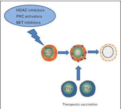
Figure 6: The “shock and kill” HIV eradication strategy ... 42
Figure 7: History of Th17 lineage discovery ... 47
Figure 8: The Th17 differentiation program ... 48
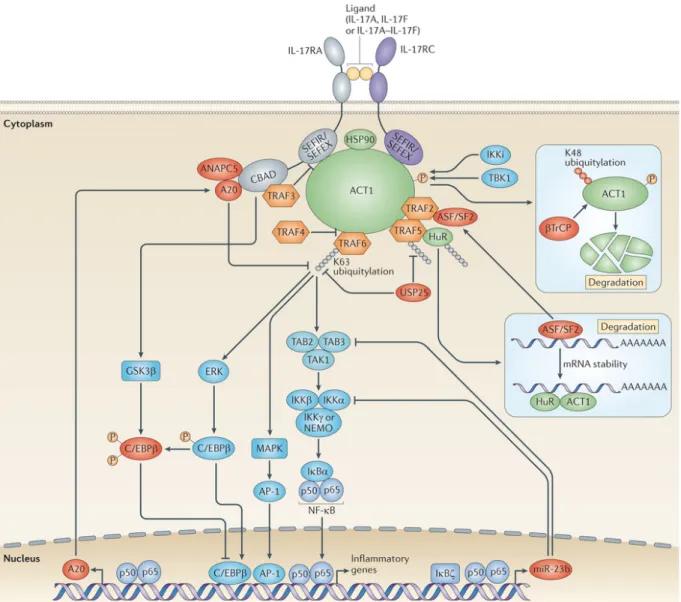
Figure 9: IL-17A and IL-17F signaling pathway ... 58
Figure 10: Mechanisms involved in the regulation of Th17 cell functions ... 61
Figure 11: Heterogeneity of human peripheral blood monocytes ... 74
Figure 12: Differential HIV permissiveness in Th1, Th2, Th17, and Th1Th17 ... 106
Figure 13: Heterogeneity of memory CD4+CCR6+ T-cells ... 107
Figure 14: The expression of RORγt mRNA by CCR6+DN and CCR6+DP subsets ... 108
Figure 15: CD16+ MDDC selectively promote HIV replication in autologous CD4+ T-cells ... 111
ABBREVIATION LIST
Abs, antibodies
ADCC, Antibody-Dependent Cell-Mediated Cytotoxicity AGs, Aicardi-Goutières syndrome
AhR, Aryl Hydrocarbon Receptor APC, Antigen-Presenting Cells
AIDS, Acquired Immune Deficiency Syndrome
APOBEC3G, Apolipoprotein B editing catalytic polypeptide-like 3G ART, Antiretroviral Therapy
ATP, Adenosine Triphosphate AZT, Zidovudine
BATF, Basic Leucine Zipper Transcription Factor ATF-like
BBB, Blood-Brain Barrier
BCA, Breast Cancer-Associated Gene 2
BDCA, blood dendritic cell antigen
bNABs, Broadly Neutralizing Abs
BRD4, Bromodomain Containing 4
BSA, Bovine Serum Albumin
BSF-1, B-Cell Stimulatory Factor-1
BST-2, Bone Marrow Stromal Cell Antigen 2 CD, Cluster of Differentiation
CDK, Cyclin-Dependent Protein Kinase cDNA, complementary DNA
CCL#, Chemokine (C-C motif) Ligand # CCR#, Chemokine (C-C motif) Receptor #
C/EBPs, CCAAT/Enhancer-Binding Proteins Transcription Factors CFSE, Carboxyfluorescein Succinimidyl Ester
CI on ART, Chronically Infected HIV Individuals on ART
CLA, Cutaneous Lymphocyte Associated Antigen
CLEC, C-type Lectin Domain Family
CPZ, Chimpazee
CTLA-4, Cytotoxic T-Lymphocyte Antigen 4 CXCL, Chemokine (C-X-C motif) Ligand CXCR, Chemokine (C-X-C motif) Receptor CypA, Cyclophilin A
DC, Dendritic Cells
DNA, Deoxyribonucleic acid
dNTP, Deoxynucleoside triphosphate dU, Deoxyuridine
DTH, Delayed-Type Hypersensitivity
EAE, Experimental Autoimmune Encephalomyelitis EGR2, Early Growth Response Gene
ELF4, E74-like factor 4
env , Envelop
ETS-1, E-Twenty Six 1
FACS, Fluorescence-Activated Cell Sorting FBS, Fetal Bovine Serum
FDA, Food and Drug Administration FOXO3a, Forkhead Box O3
FOXP3, Forkhead Box P3
GFI-1, Growth Factor Independent 1
GFRA2, Glial Cell Line-Derived Neurotrophic Factor Family Receptor-α2 GFP, Green Fluorescent Protein
GM-CSF, Granulocyte-Macrophage Colony-Stimulating Factor
gp, glycoprotein
gag, Group-Specific Antigen GR-1, Granulocyte 1
HAART, Highly Activated Anti-Retroviral Therapy HAD, HIV-Associated Dementia
HAND, HIV-Associated Neurocognitive Disorders
HDF, HIV-Dependency Factors
HIFα, Hypoxia-Inducible Factor Alpha HIES, Hyper IgE Syndrome
HIV, Human Immunodeficiency Virus
HLA, Human Leukocyte Antigen HPV, Human Papilloma Virus HSV, Herpes Simplex Virus
hs-CRP, High-Sensitivity C-Reactive Protein HVTN , HIV-1 vaccine trials network
IBD, Inflammatory Bowel Disease
ICAM-1 Intercellular Cell Adhesion Molecule-1
ID3, Inhibitor of DNA-binding 3
IDO1, Indoleamine 2,3-dioxygenase 1 i.e., id est (latin)
IFN, interferon Ig, Immunoglobulin
IKKα, Iinhibitor of nuclear factor-κB kinase-α IκBζ, IkappaB-zeta
IL, Interleukin
IRF4, interferon regulatory factor 4 iTreg, inducible Treg
LAG-3, Lymphocyte-Activation Gene 3 LAV, Lymphadenopathy Associated Virus
LDLR, Low Density Lipoprotein Receptor
LEDGF, Lens Epithelium-Derived Growth Factor LEF1, Lymphoid enhancer-binding factor-1
LFA-1, Leukocyte Function-Associated Molecule-1 LMNA, Lamin A/C
LPS, Lipopolysaccharide LTR, Long Terminal Repeat Ly-6C, Lymphocyte Antigen 6C
M, Main
MACS, Magnetic Associated Cell Sorting M-CSF, Monocyte/Macrophage Growth Factor mDC, myeloid DC
MDDC, Monocyte-Derived DC
MDR-1, Multi-Drug Resistance Type-1
MHC, Major Histocompatibility Complex MIP, Macrophage Inflammatory Protein mIR, micro-RNA
MOG, Myelin Oligodendrocyte Glycoprotein mRNA, messenger RNA
MS, Multiple Sclerosis
mTOR, Mammalian Target of Rapamycin M-tropic, Macrophage
N, New
Nef, Negative Factor Neg, Negative
NF-κB, Nuclear Factor Kappa Beta
NIAID, National Institute of Allergy and Infectious Diseases NIH, National Institutes of Health
NK, Natural Killer
NKT, Natural Killer –T-cell
NNRTI, Non- Nucleoside Reverse Transcriptase Inhibitor NO, Nitric Oxide
NRTI, Nucleoside Reverse Transcriptase Inhibitor nTh17, natural Th17
nTreg, natural Treg
O, Outlier p24, protein 24 P, Putative
PBS, Phosphate-Buffered Saline PCR, Polymerase Chain Reaction PD-1, Programmed Death 1 pDC, Plasmacytoid DC PGE2, Prostaglandin E2
PHAC, Public Health Agency of Canada PIC, Pre-integration Complex
PKC, Protein Kinase C
PLZ, Promyelocytic Leukemia Zinc Finger PMA, «Phorbol Myristate Acetate»
pol, Polymerase
PPARγ, Peroxisome Proliferator-Activated Receptor Gamma
PrEP, pre-exposure prophylaxis
PIAS3, Protein Inhibitor of Activated STAT3
PSGL, P-Selectin Glycoprotein Ligand-1
PTEFB, Positive Transcription Elongation Factor B
RA, Rheumatoid Arthritis
RANBP2, RAN Binding Protein 2
RANTES, Regulated upon Activation Normal T-cell Expressed and Secreted R4, CCR4
R5, CCR5 R6, CCR6
Rev, Regulator of Expression of Virion RNA, Ribonucleic Acid
RPMI, Roswell Park Memorial Institute
RORγt, Retinoic Acid Related Orphan Receptor gamma t ROS, Reactive Oxygen Species
RUNX1, Runt-Related Transcription Factor 1
sCD14, Soluble CD14
SAMHD1, Sterile alpha motif domain- and HD domain-containing protein 1
SEB, Staphylococcal Enterotoxin B SHIV, HIV-SIV Hybrid Virus siRNA, Small Interfering RNA
SIV, Simian Immunodeficiency Virus SLAN, 6-sulfo LacNAc
SLE, Systemic Lupus Erythematosus SM, Sooty Mangabey
STAB1, Stabilin-1
STAT, Signal Transducer and Activator of Transcription SOCS3, Suppressor of Cytokine Signaling-3
SOX5, SRY-related high-mobility-group-box 5 Tat, Trans-Activator of Transcription
Tbet, T-box transcription factor expressed in T cell TCF-1, T-Cell Factor 1
TCM, Central Memory T-cells
TCR, T-Cell Receptor TEM, T effector memory
TEM, Tie2-Expressing Monocyte Tfh, Follicular Helper T-cells
TGF, Transforming Growth Factor Beta 1
Th, T Helper
TLR, Toll-like-receptor TM, Transitional Memory TNF, Tumor Necrosis Factor TNPO3, Transportin 3
TR1, T Regulatory Type 1
Treg, Regulatory T-cell TRIM5, Tripartite Motif 5 tRNA, Transfer RNA TSCM, Stem Memory T cells
TTM, Transitional Memory T-Cells
TTE, Terminally effector T-Cells
TWIST1, Twist Family bHLH Transcription Factor 1 USA, United State of America
Vif, Viral Infectivity Factor Vpr, Viral Protein R
Vps, Vacuolar Protein Sorting Vpu, Viral Protein U
Vpx, Viral Protein X
YPD, Yeast Peptone Dextrose X3, CXCR3 X4, CXCR4 α, alpha β, bêta ºC, degré Celsius δ, delta γ, gamma µ, micro θ, Thêta %, percent
ACKNOWLEDGMENTS
I want to first thank God for the inspiration, passion and courage I was blessed with during the past 6 years. This has been an interesting journey that I will never forget as it irreversibly shaped my life. I have grown into a stronger person that is more eager to discover diverse facet of life. I want to thank my supervisor and mentor, Dr. Petronela Ancuta, for her guidance, her passion as a scientist and her confidence in me. It has been a privilege to be your first graduate student. Even in dark periods, you never ceased to believe in me. Thank you for the time invested on teaching me how to be a scientist and guiding me in becoming a stronger and more mature person. I will always be grateful for the opportunity you gave me as a master and a Ph.D. student, allowing me to develop my scientific creativity. Despite the end of this chapter of my life, you will always remain my mentor, the one I will seek advice when needed.
In addition to Dr. Petronela Ancuta, I want to thank the members of my jury: Dr. Nathalie Arbour, Dr. Rupert Kaul and Dr. Cheolho Cheong for their time spent in reviewing my thesis. It has been an honor for me to have you as members of my Ph.D. defense committee.
Special thanks to the present and past members of Dr. Ancuta’s lab, especially Dr. Patricia Monteiro and Dr. Marie-Claude Gaudreau who generated preliminary results that represented the prelude of my thesis. I would like to thank all of the colleagues and co-authors that helped me in generating results included in three manuscripts.
Special thanks to my colleagues of the CRCHUM from other labs for their scientific advise and for their time spent conversing with me. The list is long and I prefer not including names out of fear of forgetting one.
I want to thank my close friends, especially Maria Dimitropoulos, for being there for me when I needed the most. I don’t think I would have made it through without your support.
Lastly, I want to thank my entire family, especially my grandmother and mother for whom I have the upmost respect and admiration. You have been supporting and encouraging me ever since I shared with you my ambition to become a scientist. You have been my rock and anchor during this adventure. Thank you for believing in me. I love you.
CAPTATIO BENEVOLENTIAE
Since the establishment HIV pandemics, a total of 39 millions of individuals have died from AIDS, the final clinical stage of HIV-infection associated with the failure of the immune system to control HIV and other opportunistic pathogens. Considerable achievements were made in recent decades in the field of HIV research, resulting in the discovery of conventional antiretroviral therapy that increased the life expectancy of HIV-infected individuals. Nevertheless, the antiretroviral therapy does not cure HIV nor does it restore the function of the immune system. Furthermore, HIV-infected individuals under treatment experience co-morbidities including cardiovascular disease and cognitive impairment. Therefore, the search and development for a cure aimed at the HIV eradication is the current goal in the field of HIV research. One of the main priorities in the field is to determine the mechanisms that contribute to the establishment and maintenance of latent infection. Here, this thesis focuses on two components of the immune system that HIV targets: the Th17 lineage and the myeloid dendritic cells. The purpose of this thesis is to provide a further understanding on the biology of the adaptive and innate immunes system and on the mechanisms by which HIV modulates host defense to promote its dissemination and persistence. Our discoveries will orient novel therapeutic strategies for the development of an HIV cure.
CHAPTER 1:
1.1 HIV INFECTION
1.1.1 HIV GENERALITIES
The human immunodeficiency virus type 1 (HIV) (Figure 1) belongs to the retroviridae family, member of the genus lentivirus (1). HIV causes acquired immune deficiency syndrome (AIDS), a
disease associated to active viral replication, diminished T CD4 counts (<200 cells/µl), and
susceptibility to multiple opportunistic infections. HIV was isolated the first time in 1983 from a lymph node biopsy of a patient suffering from lymphadenopathy by the group of Dr. Luc Montagnier and so was referred to as Lymphadenopathy Associated Virus (LAV) (2). In 1986, the retrovirus was formally named HIV by the International Committee on Taxonomy of Viruses (3). According to the World Health Organization (WHO), 35 million people lived with HIV at the end of 2013, and 2.1 million new cases of infection were reported the same year (4). In Canada, the Public Health Agency of Canada (PHAC) estimated that 71,300 people were infected with HIV or were living with AIDS at the end of 2011 (5). The HIV-1 infection is most widespread in African countries. This virus is the most pathogenic and infectious of all lentiviruses. Two types of HIV exist: HIV type 1 (HIV-1) and HIV type 2 (HIV-2). HIV-2 is genetically similar to the simian immunodeficiency virus (SIV) that infects its natural hosts, the sooty mangabey (SIVSM), while
HIV-1 is similar to the SIV infecting the non-human primate, the chimpanzee (SIVcpz); both
HIV-1 and SIVcpz cause AIDS (6, 7). Worldwide, HIV-1 is the most pathogenic and predominant type
of this virus. In addition, HIV-1 is easily transmitted and is mainly responsible for the AIDS pandemic. This virus is classified into four groups: M (main), O (Outlier), N (New), and P (Putative) (8, 9). The M group is the most preponderant worldwide and is divided into nine sub-types (A, B, C, D, F, G, H, J, K). Sub-type B is the most prevalent in North America. The other sub-types are observed in infected individuals from African countries. The N and O groups are rare and found in infected individuals from central Africa. Recently in 2009, a study identified a new type of HIV in a woman from Cameroon and was designated as group P (10).
In the past 30 years, the HIV research field has been marked with successes and disappointments. Discoveries regarding the biology, transmission, and pathogenesis have led to a better understanding of virus interaction with the host cell and targets for the development of antiviral drugs. Currently, with the findings of efficient antiviral treatments, HIV is not considered a lethal
not achieved with current antiretroviral therapies and a life-long treatment is a heavy burden for both the patient and the healthcare system.
1.1.2. HIV STRUCTURE
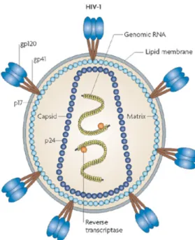
Retroviruses are positive-sense RNA viruses (80 to 120 nm diameter) that are generated from budding of infected cells (11). All retroviruses commonly encode for the enzyme reverse transcriptase, which is responsible for the generation of complementary DNA (cDNA), from an RNA template. HIV-1 particles are surrounded by a nucleocapsid and an external envelope constituted of phospholipid bilayer derived from the host cell membrane and viral glycoproteins gp120 and gp41. The gp41/gp120 complex forms a trimeric structure at the cell surface. The viral core encloses the matrix composed of viral protein p17. The matrix itself encloses the nucleocapsid constituted of viral protein p24 where two copies of positive-sense single-stranded RNA are located together with three virion-associated enzymes, reverse transcriptase, integrase and protease.
Figure 1: HIV-1 Structure. Shown are various components of the HIV-1 virions including the envelope proteins gp120 and gp41, the capsid protein p24, the two copies of single stranded RNA
and the two molecules of the reverse transcriptase enzyme. Figure reproduced with the permission
1.1.3 HIV-1 GENOME
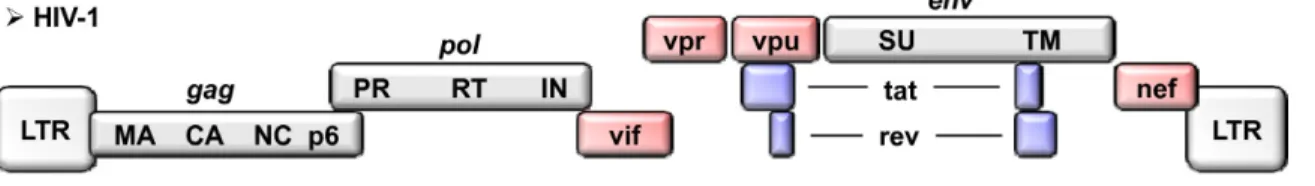
The HIV-1 genome (Figure 2) is of 9.2kb length and is encoded for structural, as well as accessory genes (13). The structural genes are Gag (group-specific antigen), Pol (polymerase) and Env (envelop). The accessory genes are Trans-Activator of transcription (Tat), Regulator of expression of virion (Rev), Negative factor (Nef), Viral Infectivity Factor (Vif), Viral Protein R (Vpr), and Viral protein U (Vpu). The proviral DNA, or cDNA, that is integrated in the host genome, includes an U3-R-U5 identical sequence referred to as Long Terminal Repeat (LTR) at the 5’ and 3’ extremities. These sequences allow the viral DNA to integrate into the host genome.
Figure 2: HIV-1 Genome. This figure illustrates the structural and accessory genes that compose
the HIV genome. Figure reproduced with the permission of Retrovirology (14), author copyrights
2010.
The structural proteins Gag, Pol and Env code for the nucleocapsid, viral enzymes and surface proteins, respectively (11, 13). The gag gene is originally transcribed into a Gag-Pol precursor of 55 kDa, which will be cleaved into the matrix protein p17 and the nucleocapsid protein p24. The pol gene encodes for the viral enzymes protease, integrase, RNase H, and reverse transcriptase. The protease is responsible for the cleavage of polyproteins into functional proteins. The integrase forms the pre-integration complex (PIC) and mediates the integration of the viral DNA into the host genome. All viral enzymes are generated following the cleavage of the Gag-Pol poly-protein. This later precursor is translated after a ribosomal frame shift signal allowing for not only Gag but also Pol expression. The env gene is translated into a gp160 poly-protein precursor before getting cleaved into gp120 and gp41 via host cell proteases such as Furin (15, 16).
The accessory proteins are non-structural proteins that are very important for viral replication. Vif has a role in viral assembly and maturation (17). Vpr is involved in the induction of cell apoptosis, the disruption of cell-cycle regulation, the nuclear transport of the viral pre-integration complex as well as in the suppression of immune activation (18). Vpr was also reported to facilitate reverse
transcription and to transactivate the viral LTRs. Tat is a regulatory protein that activates viral transcription (19, 20). Rev is another regulating protein and is essential for the transport, stabilization, and translation of messenger RNA (mRNA) from the nucleus to the cell cytoplasm (21, 22). Nef is the most immunogenic accessory protein as it is involved in enhancing viral infectivity and cell apoptosis (23). Vpu is exclusively specific to HIV-1 and has a role in CD4 degradation and release of viral particles from the surface membrane of infected cells (24).
1.1.4 HIV TARGET CELLS
The HIV-1 targets the cells of the immune system, mainly CD4+ T lymphocytes but also macrophages, monocytes, and dendritic cells (DCs) (25, 26). All these leukocytes express at their surface the transmembrane glycoprotein CD4which is used by HIV as the main receptor for entry (27). CD4+ T-cells have important roles in orchestrating adaptive immune responses against various pathogens, including viruses. Macrophages, monocytes and DCs are antigen-presenting cells (APCs) and act as bridges between the innate and adaptive immune system (28). Originally, the HIV-1 tropism was characterized based on the type of infected cells. The M-tropic strains referred to viruses infecting macrophages, whereas the T-tropic strains referred to viruses infecting T-cells (29). In 1996, the chemokine receptors CCR5 and CXCR4 were identified as major co-receptors for HIV entry (30-36). This discovery led to the current classification of viral strains based on the type of co-receptor used for entry. Viruses using CCR5 as a co-receptor are now termed R5 strains, whereas viruses using CXCR4 are termed X4 strains (37). This classification is more suitable as it is now well established that the R5 and X4 strains can infect both T and macrophages.
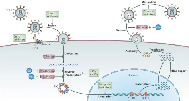
1.1.5 HIV-1 REPLICATION CYCLE
The HIV-1 replication cycle involves two main phases: early and late (38) (Figure 3). Each step within these two phases has been further studied to identify antiviral targets for therapies that focus on stopping the viral replication cycle. The early phase includes the steps from viral entry into target cells until integration into the human genome. The late phase involves all steps from post-integration including transcription and translation of viral protein, assembly, and budding.
First, the virus interacts with the target cells via the binding of the viral glycoprotein gp120 to the CD4 receptor located on the cell membrane (38, 39). This interaction induces a conformational change in gp120 allowing the glycoprotein to bind to chemokine receptors, CCR5 or CXCR4. The later step is followed by a second conformational change of the gp120, which allows the release and implantation of gp41 into the cell membrane. The fusion between the viral and the cellular membrane occurs and allows entry of the virus to its target cell. The nucleocapsid penetrates into the cytoplasm and is degraded, releasing the two copies of single-stranded RNA and associated viral enzymes known as the reverse transcription complex that includes reverse transcriptase, integrase, protease, and accessory proteins. Reverse transcription then ensues and the RNA is retro-transcribed into double stranded DNA. The initiation of this step requires the host transfer RNA (tRNA), which serves as primers. Upon viral cDNA synthesis, the PIC is formed, involving the integrase, Vpr and cellular proteins such as LEDGEF (40). The viral double-stranded DNA is transported from the cytoplasm into the nucleus where integration into the host genome occurs via the integrase activity. The period from integration to viral transcription is undetermined. The integrated viral DNA likely stays in a latent form for years until reception of a signal initiating transcription. Spliced mRNA are produced and exported from the nucleus to the cytoplasm where translation takes place. The first proteins to be synthesized are Nef, Tat and Rev. The unspliced mRNAs, including sequences coding for precursors Gag, Gag-Pol and Env, are later transcribed, and their export is dependent on Rev expression. The gp160 precursor is translated followed by the translation of poly-proteins Gag and Gag-Pol. Once the translation step is completed, viral assembly ensues. Viral proteins associate with viral genome to form virions. The enveloped proteins are inserted into host cell membrane. Viral particles are then ready for budding. Maturation is still underway after budding with the cleavage of precursors into functional proteins, which is performed by viral protease.
Figure 3: The regulation of the HIV-1 replication cycle by host factors and antiviral drugs.
Shown are the multiple steps of the HIV replication cycle that are interrupted by host restriction
factors and also steps targeted by current antiretroviral therapeutic strategies. Reproduced with the permission of Macmillan Publisher Limited: Nature Reviews in Immunology (41), author copyrights 2013
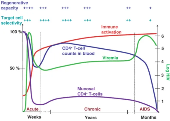
1.1.6 HIV-1 PATHOGENESIS
HIV-1 infection causes massive depletion of CD4+ T-cell, leading to an impaired immune system incapable of mounting appropriate adaptive responses against HIV-1 and various opportunistic pathogens (42). Infection with HIV-1 is divided in three phases: acute, asymptomatic, and AIDS (42, 43) (Figure 4). The infection starts with a symptomatic acute phase that may last a few weeks and is characterized by active viral replication, establishment of viral reservoirs, and important depletion of CD4+ T-cells in the peripheral blood as well as the intestinal mucosal tissues. At the end of the acute phase, the viral replication temporarily decreases as the specific immune response mediated by the cytotoxic CD8+ T-cells and B cells are developed, allowing a partial restoration of peripheral CD4+ T-cell counts. The asymptomatic phase, also referred to as the chronic phase, is
associated to persistent viral replication, chronic immune activation, and progressive depletion of CD4+ T-cells. The AIDS phase is declared when the CD4+ T-cell count is equal or lower than 200 cells/µl. This phase is associated with an increase in the viral load as well as the occurrence of
opportunistic infections and cancers. All these three phases are clinically distinct as detailed below.
Figure 4: The clinic phases of HIV-1 pathogenesis. Shown are the three major clinical phases of
HIV-1 disease progression. Changes in mucosal and blood CD4+ T-cell counts, as well as levels of
immune activation and viremia are illustrated. The figure is reproduced with the permission of
Macmillan Publisher Limited: Nature Medicine (43), author copyrights 2006. 1.1.6.1 THE ACUTE PHASE
The HIV-1 mainly targets for infection CD4+ T-cells located in the gastrointestinal and/or reproductive tract, leading to an important active viral replication in cells of these affected tissues (Figure 4). Active viral replication is generally associated with flu-like symptoms including fever, headaches and weight loss. The gastrointestinal tract is the lymphoid organ containing the majority (60%) of the T-lymphocytes of the body and is the main anatomic site of viral replication. During the acute phase, the infection initially affects activated CD4+ T-cells expressing CCR5 leading to their massive depletion with the transmitted viruses being reported to exhibit a CCR5 tropism (R5 strains) (44, 45). This depletion is also observed in the subsequent phases of infection. Indeed, the majority of CD4+ T-cells from mucosal tissues express CCR5 (>50%) with only ~15% of CD4+CCR5+ circulating in the peripheral blood (42). Viruses using CXCR4 as co-receptors
emerge later and expand the repertoire of target cells as the majority of CD4+ T-cells express this
chemokine receptor (46). The usage of CXCR4 is correlated to an accelerated progression to the AIDS phase. Variations in the peripheral blood CD4+ T-cells do not always accurately reflect the true proportion of cells depleted in mucosal tissues (42). This large depletion of CD4+CCR5+ cells in the mucosa occurs within the first three weeks following infection and is associated with viral cytopathic effects that subsequently cause the death of infected cells. An impaired functionality of the innate response is also observed during the early phases of HIV-1 infection. The cytokine production by DCs is altered and the phagocytic activity of macrophages is exhausted (47, 48). The structural aspect of the gastrointestinal tract is significantly impaired following HIV-1 infection. Apoptosis of enterocytes and the disruption of tight junction are observed during acute infection and throughout the course of HIV-1 disease progression (48, 49).
The activation state of the CD4+CCR5+T-cells makes them susceptible to infection. Indeed, these target cells express early tissue-resident marker CD69 (50, 51) as well as other activation markers such as CD25, and Human Leukocyte Antigen- DR (HLA-DR) (42, 52 , 53 , 54). Cellular activation may be due to the increased presence of pro-inflammatory cytokines in the local environment. Furthermore, Nef, Tat, Vpr and Rev have the capacity to activate cells independently of signals coming from the T-cell receptor (TCR) (42). In quiescent cells, the virus can establish latency thus creating long-lasting cellular reservoirs for HIV-1. The establishment of cellular reservoirs occurs within the first weeks of acute infection. Among total CD4+ T-cells, our group
demonstrated that long-lived central memory (TCM) and transitional memory (TTM) CD4+ T-cells
are major HIV-1 reservoirs in subjects receiving viral suppressive antiretroviral therapy (ART) (55). At the end of the acute phase, viral replication is controlled by the development of the specific immune responses involving the CD8+ T-cells and B-lymphocytes; this leads to a slight increase of the CD4+ T-cell counts in the peripheral blood but not mucosal tissues in the absence of treatment (43).
1.1.6.2 THE CHRONIC PHASE
The chronic phase is mainly characterized by a persistent viral replication, increased levels of CD4+ and CD8+T-cell activation, and large production of pro-inflammatory cytokines (42, 43)
(Figure 4). Despite the viral load being at lower magnitude compared to observations in the acute phase, the CD4+ T-cell count restoration is only partial in peripheral blood and in the mucosal tissues. The cellular activation level is thought to be a better forecast of disease progression rather than levels of viral replication (42, 56). Viral replication does not seem to be the sole responsible factor for cell depletion as only a small fraction of CD4+ T-lymphocytes are infected (estimated range: 0.01-1%). Indeed, most depleted cells are not productively infected. Studies by Greene et al. demonstrate that integrative infection is compatible with survival, while abortive infection leads to cell death by pyroptosis (57). In contrast, reports by Cooper et al. support the opposite concept that integrative infection causes cell death (58). Unpublished observations in our laboratory demonstrate that while a small fraction of CD4+ T-cells carry integrated HIV-DNA, a significant fraction of both naive and memory CD4+ T-cells carry early HIV reverse transcripts in aviremic subjects receiving ART (Niessl et al., manuscript in preparation). The later findings support the idea that abortive infection contributes to alterations in CD4+ T-cell counts and functions despite viral suppressive ART (59). In addition to infection per se, recent findings suggest that chronic activation of the immune system leads to rapid T-cell turnover, clonal exhaustion and cell death. Evidence showing that chronic immune activation is a key pathogenic factor of disease progression comes from findings in primate models of SIV pathogenesis (60-62). Infection with SIV in natural hosts, such as Sooty mangabeys, does not progress into AIDS as
opposed to the Asian or Indian rhesus macaques, known as non-natural hosts. Despite the observed
high viremia, the activation and inflammation level in SIV natural hosts is relatively low. The
CCR5+CD4+ T-cells are preserved. The integrity of mucosal barrier is maintained, preventing
microbial translocation. Furthermore, the viral burden in tissues is low. Microbial translocation
was designated as one of the key factors driving chronic immune activation (49, 56, 63). The increased enterocyte apoptosis, together with the disruption of the intestinal epithelial tight junctions and the loss of Th17 cells conferring protection against bacterial infections, were reported to promote microbial translocation in HIV-infected subjects. Several mechanisms leading to cell depletion involve: i) the gradual reduction of the T memory pool caused by the continual cellular activation, ii) the increased levels of broad naive cells differentiating into memory T-cells, iii) the decrease in the frequency of resting T-cells due to constant activation state, and iv) the reduced ability to generate naive T-cells pools from lymphopoietic sources (42). Moreover, HIV infection creates dysfunction in the thymus leading to a reduced output in the generation of
naïve CD4+ lymphocytes (64, 65)). Furthermore, viral replication and chronic immune activation
may be responsible for the observed abnormal structure in lymph nodes associated with the loss of CD4+ T-cells and collagen deposition (66, 67). Abnormal structures of the thymus and bone marrow were also documented (42). The CD4/CD8 ratio is altered as the CD8+ population is expanded and the frequency of CD4+ T-cells is diminished (42, 68). Indeed the CD4/CD8 ratio is a useful marker to predict disease progression, CD4+ T-cell depletion, and the size of HIV reservoirs (55, 69).
1.1.6.3 THE AIDS PHASE
The AIDS status is declared when the total CD4 count in the peripheral blood is lower than 200 cells/µl (68). This final phase is characterized by an increased viremia and massive depletion in peripheral blood CD4+ T-cells (Figure 4). The immune system becomes weak and susceptible to several pathogens that are normally contained under homeostatic conditions. The opportunistic infections related to AIDS include pulmonary and esophageal candidiasis by Candida albicans, pulmonary infections by Pneumocystis jirovecii, Pneumocystis carinii, Mycobacterium avium, Staphylococcus aureus, Escherichia coli and Pseudomonas aeruginosa, viral infection by Cytomegalovirus (CMV), Human papilloma virus (HPV) and herpes simplex virus (HSV),
encephalopathies by protozoan infections, and neoplasm such as cervical cancer and rectal Kaposi sarcoma (26). In the absence of antiretroviral treatment, the HIV-infected individuals develop AIDS and die within 10 years following primary infection.
1.1.7 TRANSMITTER FOUNDER VIRUSES
Transmission of HIV is mostly the result of heterosexual or homosexual intercourse. The establishment of primary infection is due to a single HIV-1 variant, referred to as transmitter/founder (T/F) viruses (70). During primary infection, the viral population is homogenous since the establishment of the infection results from one single T/F viral genotype. Once in the chronic phase, the HIV-infected individuals exhibit genetically heterogeneous viral population in their blood and tissues. The T/F viruses appear to have distinct features compared to the global viral population circulating in the periphery. The T/F viruses are thought to be
viruses differ in their level of glycosylation. The T/F HIV from subtypes A, C, and D has few N-linked glycosylation in their envelope (71-73). An interesting study from Ping et al. observed that weakly glycosylated viruses were frequent during female to male transmission (73). Highly glycosylated viruses are unlikely to be sexually transmitted because they can be trapped in the transmission fluid or can be inhibited by host factors (70). The T/F viruses appear to have less glycosylation in the highly variable region of Env and are thus sensitive like other HIV variants to a broadly neutralizing antibody that normally targets the conserved region of Env (73). Furthermore, T/F viruses are mostly R5 strains as they use exclusively CCR5 for viral entry (70). Also, T/F viruses require high expression of CD4, which is found in CD4+T-cells. In fact, T/F viruses are less capable of infecting monocytes/macrophages. Moreover, compared to viruses in the chronic phase of infection, T/F viruses express more than two fold of the Env protein, indicating their efficient capacity for transmission (74). The T/F viruses also exhibit an increased binding to DC and are thus transmitted efficiently to CD4+ T-cells. Selective pressure appears to induce T/F viruses that are highly replicative competent.
1.1.8 RESTRICTION FACTORS
During the course of viral replication, host innate immune defenses generate antiviral responses aimed at blocking critical steps of HIV-1 life cycle. These antiviral responses are mediated via specific proteins, referred to as restriction factors (75, 76). To date, four restriction factors have been reported and mechanistically characterized: Apolipoprotein B editing catalytic polypeptide-like 3G (APOBEC3G) (77), tripartite motif5 (TRIM5) (78), bone marrow stromal cell antigen 2 (BST-2) (79, 80), and Sterile alpha motif domain- and HD domain-containing protein 1 (SAMHD1) (81, 82) (Figure 3). Very recent studies identified a cellular membrane bound protein, SERINC5, as being a target for HIV Nef (83, 84), thus extending the panel of cellular restriction factors counteracted by viral proteins.
As part of an effort to determine the role of viral protein Vif, APOBEC3G was discovered (77) through experiments involving cell fusion of cells susceptible and resistant to HIV. The use of a cDNA screen database identified ABOBEC3G. The APOBEC3G is a cytidine deaminase targeting the reverse transcription step in resting CD4+ T-cells and macrophages (76, 85-87). The enzyme is
incorporated into virions and causes deamination of cytidine residues leading to the production and insertion of deoxyuridine (dU) in the first strand of cDNA. The reverse transcriptase misreads the insertion of dU for thymidine during the synthesis of the second strand of cDNA resulting in the incorporation of adenine instead of guanine. This mutation consequently reduces the viral fitness. The insertion of dU in the first strand of cDNA itself can also lead to the activation of the cellular DNA repair machinery, specifically the uracil inducing total destruction of viral DNA. Independent of its deaminase activity, several studies suggest that ABOBEC3G can still block viral replication as shown in catalytically inactive mutants (88, 89). Indeed, ABOBEC3G was reported to inhibit steps of reverse transcription and integration by: 1) decreasing the efficiency of viral RNA strand transfer; 2) decreasing the interaction between tRNA and viral RNA, representing the first step of reverse transcription; 3) preventing DNA elongation; and 4) interacting with integrase, thereby preventing viral integration (86). Vif prevents APOBEC3G encapsidation in virions thus allowing for normal replication to occur (76, 85-87). Precisely, Vif simultaneously interacts with the restriction factor and an E3 ubiquitin ligase complex resulting in the ubiquitination and the subsequent degradation of APOBEC3G. Vif preferentially acts on newly synthesized APOBEC3G as pre-existing APOBEC3G are insensitive to Vif due to complex formations with other cell host proteins (90).
The TRIM5α was discovered using cDNA screen assays (78). Prior to the discovery, studies by Hoffman et al. using a pseudotyped HIV-1 virus bypassing viral entry demonstrated a blocking of HIV-1 replication in Old World monkey. Also, SIV infection was restricted in New World monkeys. This restriction was originally defined as genetic barrier and was called lentivirus susceptibility factor 1. TRIM5 belongs to the TRIM family characterized for their conserved motif structured known as the RBCC motif (86). The TRIM5 can be expressed in many isoforms among which only TRIM5α and TRIM5-Cyclophilin A (TRIM5Cyp) exhibit antiviral activity. TRIM5Cyp was discovered after TRIM5α by Sayah et al. in Old World monkeys (91). These researchers wanted to identify the restriction factor blocking viral replication in monkeys and found that TRIM5 was fused with cyclophilin A. TRIM5 inhibits viral replication at the early stages of the viral life cycle. TRIM5 binds to the viral capsid and induces premature disassembly thereby preventing reverse transcription (78). In addition, TRIM5 functions as a pattern recognition receptor and senses the viral capsid then alerts the innate immune response (92). Both
TRIM5α and TRIM5Cyp contain a RING (for Really Interesting New Gene) domain with E3-ubiquitin ligase activity (85, 86). The C-lectin domain, conferring capsid binding and specificity, differs in the two isoforms. The cyclophilin A domain is expressed in TRIM5Cyp instead of the PRYSPRY found in TRIM5α. The TRIM5 is species-specific and is not efficient against viruses infecting their natural host (86). For example, primate TRIM5α inhibits efficiently HIV-1 infection but fails to block SIV replication in monkey cells. Human TRIM5α is efficient against N-tropic murine leukemia virus but poorly blocks HIV-1 (86). Engineering a human TRIM5α mimicking primate TRIM5α functions is an avenue for generating new therapeutic approaches against HIV-1. Nevertheless, the delivery of such genes into human cells remains a challenge.
Other members of the TRIM family were reported to exhibit antiviral functions, including TRIM1, TRIM19, TRIM22, TRIM32, and TRIM34 (93); however, TRIM15 was identified as an adhesion molecule (94).
The BST-2 (for Bone Marrow Stromal Cell Antigen 2) also known as tetherin or CD317 is a trans membrane protein and targets the end stage of viral replication (85, 86). It was discovered that BST-2 physically binds and retains virions at the host surface membrane thereby inhibiting viral budding. Tethered virions have the fate to remain at the cell surface or to be degraded by a series of process involving the E3 ubiquitin ligase, BCA2 (95). Similar to TRIM5, BST-2 also triggers the innate immune response by inducing activation of the NF-κB signaling pathway (96, 97). HIV-1 evades the host’s attempts to prevent viral release by expressing viral protein Vpu (85, 86). The accessory protein Vpu interacts with BST-2, leading to its degradation through proteosomal or lysosomal pathway (98-103).
The SAMHD1 is a Deoxynucleoside triphosphate triphosphohydrolase (dNTPase) that normally functions as a regulator of dNTP levels and DNA damage response (85, 86). SAMHD1 was discovered based on studies performed with SIV Vpx showing that expression of Vpx in human myeloid cells increased their permissiveness to HIV infection (82). Thus, Vpx, absent in HIV, was able to overcome a HIV restriction factor in these cells. In 2011, the teams of Benkirane and Skowronski discovered independently the HIV restrictive activity of SAMHD1 (81, 82). This restriction factor is ubiquitously expressed in various cell types including resting CD4+ T-cells,
macrophages, monocytes and DCs (86, 87). This restriction factor restricts viral replication by cleaving dNTPs into deoxyribonucleoside and triphosphate residues thus depleting the dNTP pools available for reverse transcription. Consequently, the HIV-1 genome is not reverse transcribed thus blocking viral replication. A second mechanism of action involves RNA degradation (76). The functions of SAMHD1 seemed to also evade the innate immune response involving the activation of IFN responses (86). Indeed, defects in SAMHD1 have been associated with Aicardi-Goutières syndrome (AGs), a genetic disorder characterized by the excessive production of type I interferon, which is responsible for the increase of unneeded immune responses (76). SAMHD1 is counteracted in HIV-2 by Vpx and in Rhesus macaque by Vpr. Of note, Vpx seems to be derived from Vpr gene duplication (76, 86). Vpx is also expressed in SIV strains infecting macaques (SIVmac) as well as red-capped (SIVrcm) and sooty mangabeys (SIVsm). The SIV Vpx is packaged into virions during assembly and interacts with SAMHD1 leading to its degradation by a process involving the E3 ubiquitin ligase complex and thereby allowing reverse transcription to occur. Vpx only acts on SAMHD1 in the nucleus but not in the cytoplasm (104). SAMHD1 is effective only in non-dividing cells such as macrophages, DCs and CD4+ T resting cells. Because SAMHD1 fails to restrict replication in activated CD4+T-cells (105), it has been suggested that the antiviral activity of SAMHD1 acts in combination with other unidentified cellular proteins or is regulated post-translation (87). The viral protein counteracting SAMHD1 during HIV-1 infection has so far not been identified. Studies indicate that HIV-1 uses host protein cyclin L2 to degrade SAMHD1 (106). Recently, Ruffin et al. demonstrated that the expression of SAMHD1 is decreased following activation of memory CD4+ T-cells, leading to an increase susceptibility to
infection in vitro (107).
1.1.9 HIV PERMISSIVENESS FACTORS
Similar to all viruses, HIV-1 exploits the host cell functions for its own survival. Several studies using multiple techniques (i.e., siRNA screens, bioinformatics analysis, cDNA cloning, etc.) identified host proteins essential for specific steps of HIV-1 replication cycle (108). Blocking the expression of these proteins, known as permissiveness factors (or HIV-dependency factors, HDFs), inhibits viral infection. First, HIV-1 depends on the cellular glycoprotein CD4 and the chemokine receptors CCR5 or CXCR4 for entry (40) (Figure 3). Fusion does not only depend on
viral glycoprotein gp41 but also requires Rab6, a regulator of retrograde transport of the Golgi apparatus (109). Other factors, including glycosphingolipids which function as mediators of cell adhesion, were also reported to participate in this step (110-112). Once fusion is completed, the reverse transcription complex is released into the cytoplasm and binds to actin (40). The transport of the pre-integration complex (PIC) to the nucleus requires interaction with microtubules. HIV also uses the nuclear import factors Transportin 3 (TNPO3) and the RAN (for RAs-related Nuclear protein) binding protein 2 (RANBP2), along with nuclear complex component Nup153 for entry of the pre-integration complex into the nucleus (109, 113). Interaction of viral integrase with host TNPO3 and lens epithelium-derived growth factor (LEDGF) is needed prior to integration (40). Proviral transcription requires the viral regulatory protein Tat, which will form a complex with host cyclin-dependent protein kinase (Cdk9) and HIV-1 Tat specific factor (40), positive transcription elongation factor B (PTEFB), and the nuclear factor of activated T cells (NFAT) (114). HIV transcription also depends on mediator complex component Med28, consistent with its functions as a regulator of the RNA polymerase II transcripts (109). Furthermore, NF-κB binding sites within the viral promoter appear to be essential for transcription. HIV interacts with host class E Vps proteins as well as the late endosome trans-Golgi network transport factor Rab9 p40 in order to bud from cell plasma membrane (115, 116). Other proteins including those associated to autophagy, NF-κB signaling, vesicular transport, DNA-damage response, energy metabolism, mitochondrial functions, ubiquitination/proteasome pathway, nucleic acid, and cytoskeleton proteins seem to be implicated in viral replication (108). Validation assays aimed at confirming the role of this protein during HIV replication cycle will need to be performed. All of these proteins may be used as targets for novel therapies to block HIV infection.
1.1.10 FACTORS CONTRIBUTING TO HIV PERSISTENCE
Despite the discovery and use of the anti-viral therapy, which reduces significantly the viral load, HIV-1 continues to persist during the chronic phase of viral infection in cellular and anatomic reservoirs that are not completely characterized (117). A relationship between HIV and the immune system is established once the virus infects its host. HIV reservoirs are established in the very early stages of the infection (118). Studies in SIV models of infection demonstrate that viral reservoirs into the brain are established a few days upon SIV exposure (119). By extrapolation, the
early establishment of HIV reservoirs into distal tissues represents a major challenge for HIV eradication (120). So far, there is no identified surface biomarker to distinguish infected cells from
uninfected cells (117). It is well established that HIV infection induces chronic immune activation
and dysfunction, which in turn will lead to viral persistence thus creating a vicious cycle that is
hard to break (42, 56, 117, 121). Several factors contributing to viral persistence have been
identified. HIV preferentially infects activated CD4+ T-cells as studies demonstrate that activated
cells harbor higher levels of HIV-DNA compared to resting cells (122). The continuous presence of viral antigens stimulates the generation of HIV-specific CD4+ T-cells that are prone to migrate into the replication site resulting in further infection. Indeed, Douek et al. demonstrated that HIV-specific CD4+T cells are susceptible to HIV infection (123). Interestingly, activated cells during HIV infection are mostly specific for herpes viruses (121). The frequency of CMV-specific CD4+ and CD8+T-cells was found to be superior in HIV-infected individuals compared to age-matched HIV-uninfected subjects (124). Of note, CMV-specific CD4+ T-cells were found to be relatively resistant to HIV infection as opposed to Mycobacterium tuberculosis in part due to the production of CCR5 ligands (125). Nevertheless, studies of PBMCs from untreated subjects with acute infection showed a positive association between CMV replication, immune activation, and HIV disease progression (126). Therefore, CMV infection is causing cell activation, immune dysfunction, and viral persistence. The damage in mucosal barriers induced by HIV is a source of inflammation that may also drive the migration CD4+ T-cells into inflamed tissues, allowing further viral replication. Resting CD4+ T-cells are poorly infected (121) probably due to SAMHD1
activity (105). HIV infection leads to increased production of pro-inflammatory cytokines and chemokines (i.e., IL-2, IL-6, IL-12, IL-18, TNF, CCL19 and CCL21), which can act in combination, engendering viral susceptibility in resting CD4+ T-cells and establishing latency (127-131). The production of anti-inflammatory cytokines such as IL-10 might also increase the establishment of latency (121). Known to restrain T-cell activation, IL-10 can act on activated cells that harbor HIV DNA and help cells return to a resting state thus contributing to viral persistence (132). Negative regulators of cell activation, including PD-1 and CTLA4, may also promote viral persistence by preventing cell apoptosis (121). PD-1 expression in CD4+ T-cells was observed to be positively correlated with HIV-DNA (55, 133). Indeed, follicular helper T-cells (Tfh) characterized by the expression of CXCR5 and PD-1 are present in increased numbers during HIV infection in lymph nodes and harbor HIV-DNA (134-136). The CD4+ T-cells,
identified as HIV cellular reservoirs, are the TCM and TTM (55). These cells have self-renewal
capacities upon antigenic exposure and are long lived (137). Signals transduction pathways involving STAT5 and FOXO3a as well as the IL-7 signaling are required for the survival of TCM.
Furthermore, IL-7 and its role in the maintenance of homeostatic proliferation were reported to be the main mechanism promoting latency in TTM and resting cells (121, 138). Effector memory cells
(TEM) of treated HIV-infected individuals are prone to harbor residual HIV-DNA. TEM represent
only 15% of total reservoir compared to TCM and TTM that comprise 85% of cells harboring
HIV-DNA (121) Other T-cell subsets including the newly described stem T-cell memory (TSCM), naive
cells, hematopoietic precursors, as well as the γδ T-cells, may also represent alternative viral reservoirs (117, 120, 121). In addition to T-cells, antigen-presenting cells (APCs) may also contribute to HIV-1 persistence. Although still controversial, macrophages may represent a long-lasting viral reservoir (139-141). Novel therapies aimed at addressing these different factors responsible for the maintenance of viral dissemination need to be found.
1.1.11 HIV-1 TREATMENT AND CURRENT CHALLENGES 1.1.11.1 ANTIRETROVIRAL THERAPY
The introduction of antiretroviral drugs as treatment against HIV was a major breakthrough in an attempt to stop the numerous deaths caused by the AIDS pandemic. At the time when HIV was first discovered (2), the only drug available was the nucleoside analogue acyclovir, which was used against the herpes simplex virus (142, 143). Once phosphorylated, nucleoside/nucleotide analogues resemble cellular nucleotides and can be incorporated into the ongoing synthesis of the DNA strand. They act as chain terminators of DNA synthesis, blocking viral replication, and were considered good candidates to prevent HIV infection. In 1987, zidovudine (also known as AZT) became the first nucleoside reverse transcriptase inhibitor (NRTI) approved by the USA regulatory authorities (FDA, Food and Drug Administration) as a treatment against HIV-1 (144, 145). Originally synthesized in 1964, AZT was used as an anti-cancer drug, but the research quickly ceased due to deleterious effects observed on mice (146, 147). Strong activism and increased pressure by the gay community on government, pharmaceutical companies, and research agencies resulted in the rapid approval of AZT for the treatment of AIDS patients (148). Indeed, the clinical
trial was prematurely terminated after the observed beneficial effect on the survival of patients treated with this drug. Unfortunately, the benefits of AZT were short lived with the emergence of viral resistance (149). Following AZT, other NRTIs, such as didanosine and zalcitabine, were approved, in 1991 and 1992, respectively (148, 150). The scientific community sadly did not take into consideration lessons obtained from the development of tuberculosis treatments, and the antiviral drugs were not given in combination but as monotherapy. After extensive studies showing better effects compared to immunotherapy, dual therapies of two NRTIs were adopted. Nevertheless, the effects were only temporary due to the development of viral resistance. The treatments became durable with the use of a triple antiretroviral drug regimen, known as highly active antiretroviral (HAART), which was introduced in 1996 (151, 152). This new therapeutic strategy was possible due to the appearance of new classes of drugs: the protease inhibitor (PIs) in
1995 and the non-nucleoside analogue reverse transcriptase inhibitor (NNRTIs) in 1996 (148).
Also, the discovery of new molecular technologies such as the polymerase chain reaction (PCR) served as a tool for the quantification of HIV-DNA and provided vital insights on viral dynamics (153). It was reported by using novel experimental techniques that HAART reduced the plasma viral load to undetectable levels. The HAART originally consisted of two NRTIs and one PI and was shown to prolong the lives of HIV infected subjects (151, 154, 155). The approval of non-nucleoside reverse transcriptase inhibitors (NNRTIs) offered an alternative option to PIs. In addition, NNRTIs production cost was less expensive than the one for PIs and could consequently be distributed in resource-poor countries (148). The entry of generics for antiretroviral drugs led to the availability of these drugs to African countries, where the disease burden is the greatest worldwide. The 2000 era brought new classes of antiretroviral drugs. The fusion inhibitor T20 was approved in 2003 (156, 157) and was followed by the introduction of Maraviroc, a CCR5-blocker inhibiting viral entry (158, 159) as well as the integrase inhibitor Raltegravir in 2007 (160, 161). The most recently released treatment, introduced in 2013, is another integrase inhibitor known as Dolutegravir (162, 163). The emergence of the new classes of drugs allowed for the maintenance of undetectable plasma viral loads in patients that developed a resistance to a
Figure 5: The effect in the absence or presence of ART. Shown in A and B are the dynamics of
HIV pathogenesis in terms of viral load, CD4 count and chronic activation of the immune system. Shown in C and D are changes in HIV pathogenesis in the presence of ART. This figure is reproduced with the permission of The Lancet (164), author copyrights 2014.
particular treatment (148). There are now a total of 25 antiviral drugs available for treatment against HIV, drugs of six distinct classes targeting each step of the HIV replication cycle (Figure 3). Over the years, only two drugs, including the NRTI zalcitabine, were withdrawn because the clinicians no longer used them. Due to the efficacy, safety profiles and fixed-dose combinations of the ARV, the investment of pharmaceutical companies in the development of new classes of drugs has slowed down. Developing treatments focusing on new targets seems much more difficult. For example, drugs targeting viral attachment to CD4 and the step of maturation were not efficient enough to generate viral susceptibility (148, 165). Still, GlaxoSmithKline is in the process of