Skeletal muscle disuse atrophy: implications OC) intracellular signaling pathways and mitochondrial permeability transition pore function
by
Kristina Csukly Département de kinésiologic
Tiiesis subnijtted to the Faculté des études supérieures in partial fulfiliment of tue requirements for the degree of
Philosophiae Doctor (Ph.D.) en sciences de l’activité physique
(o
ç.. .-..
Octobei 2006
© Kristina Csukly, 2006
Ail riglits reserved. This work may flot be reproduced in sIioIe or in part w’ithoutpermission of the author.
L)5
V. COR
Universif
de Montréal
Direction des bibliothèques
AVIS
L’auteur a autorisé l’Université de Montréal à reproduire et diffuser, en totalité ou en partie, par quelque moyen que ce soit et sur quelque support que ce soit, et exclusivement à des fins non lucratives d’enseignement et de recherche, des copies de ce mémoire ou de cette thèse.
L’auteur et les coauteurs le cas échéant conservent la propriété du droit d’auteur et des droits moraux qui protègent ce document. Ni la thèse ou le mémoire, ni des extraits sub5tantiels de ce document, ne doivent être imprimés ou autrement reproduits sans l’autorisation de l’auteur.
Afin de se conformer à la Loi canadienne sur la protection des renseignements personnels, quelques formulaires secondaires, coordonnées ou signatures intégrées au texte ont pu être enlevés de ce document. Bien que cela ait pu affecter la pagination, il n’y a aucun contenu manquant.
NOTICE
The author of this thesis or dissertation has granted a nonexclusive license allowing Université de Montréal to reproduce and publish the document, in part or in whole, and in any format, solely for noncommercial educational and research purposes.
The author and co-authors if applicable retain copyright ownership and moral rights in this document. Neither the whole thesis or dissertation, nor substantial extracts from it, may be printed or otherwise reproduced without the author’s permission.
In compliance with the Canadian Privacy Act some supporting forms, contact information or signatures may have been removed from the document. While this may affect the document page count, it does flot represent any loss of content from the document.
11 Université de Montréal
Faculté des études supérieures
Ttiis thesis eiititled
Skeletal muscle disuse atrophy: implications on intracellular signaling pathways and mïtochondrial permeability transition pore function
(Étude de l’inactivité musculaire sur les voies de signalisation intracellulaire et les fonctions du pore de transition de perméabilité mitochondriale)
presented by Kristina Csukly Département de kinésiologie
vas evaluated by tue following examining committee: Dr. Jean-i’4Iarc Lavoïe Cornmittee President Département de kinésiotogie Dr. Phillip Cardiner Thesis Advisor Département de kinésiologie Dr. Yan Burelle Thesis Co-advisor Département de kinésiologie Dr. François Péronnet Internai Examiner Département de kiriésiologie Dr. fan Noble External Examiner Depantment of Kinesiology University of Western Ontario
Dr. Louise Béliveau Representative of the Dea;i Département de kinésiologie
Sursi1ARY
In ail biological systems, a balance between ceil growth and death is required for
normal development as weli as for adaptation to a changing environment. Skeletai muscle is considered a mechanosensitive ceil type since mechanical forces such as stretch, tension, and loading have been shown to play a criticai role in regulating the dynamic processes of protein synthesis, degradation, ceilular proliferation and apoptosi s. Despite intensive investigation, the intracellular mechanisms by which muscle disuse is perceived and converted to biochemical responses ieading to muscle remodeling and atrophy have yet to be completely understood. Whereas it was appreciated a long time ago that muscle atrophy comes about via an increase in protein degradation concomitant with suppression oC protein synthesis, only recently have specific signaling pathways and celiular processes been identified. The identification of
a growing number of apoptosis-associated factors and events in disused muscle is
providing increasing evidence that apoptotic cell-death plays a role in muscle fiber atrophy dcte to a variety of causes.
This thesis is composed of three research studies developed with the objective of contributing to the present understanding of the intracellular processes regulating di suse muscle atrophy. The first study explores the intracellular signaling potential in disused muscle in response to a mechanical stimulus. The results of this study show that c—jun
NH2—terminai kinase (JNK) phosphorylation response to mechanicai stimulation is
decreased foilowing hindlimb suspension indicating that atrophic muscle tiuy iose the abiiity to transduce mechanical signais to the mitogen-activated protein (MAP) kinase pathways. However, we also show that basal JNK activity is increased in disused muscle. Since the contribution of cellular apoptosis bas been proposed as a possible mechanism regulating the loss of myofibers as a result of reduced mechanical loading, increased JNK-mediated regulation of celluÏar apoptosis may expiain the increased basal phosphorylation levels measured in muscle following hindlimb suspension. The second study investigates the effect of loss of neural input to muscle fibers on changes
in the sensitivity and modulation of the mitochondrial penieability transition pore
(PTP), a structure responsible for initiating the release ofpro-apoptotic factors from the mitochondrial intermembrane space and thought to be implicated in a fom of
iv
programmed ceil death, or apoptosis. We show that muscle atrophy caused by denervation (for 21 days) is associated with heightened sensitivity of the PTP to opening in response to progressive Ca2 loading. These flndings are substantiated by studies with cyclosporin A (CsA), a pharmacological agent which inhibits PTP opening by binding to cyclophulin D (CypD). We report that the inhibitory effects of CsA are significantly more potent in mitochondria from denervated muscle. The major finding
that the susceptibility to PTP opening is dramaticafly favored following denervation provides evidence Cor a previousÏy unreported mechanism that could at least partly
accocmt for the activation of the mitochondrial death pathway in denervation disorders in animal models and humans. The third study is a characterization ofthe properties and
function of the mitochondrial PTP in different skeletal muscles characterized by different fiber types. Considering the well-established metabolic differences between oxidative and glycolytic muscle fiber phenotypes, there exist surprisingÏy few studies which have investigated intrinsic mitochondrial properties in relation to fiber type. We demonstrate that basic PTP function and sensitivities are different depending on the
type of muscle fiber from which mitochondria are isolated. Our flndings support the emerging view that mitochondria display distinct properties that differ qualitatively according to muscle fiber type.
S0rsINJAIRE (FRANÇAIS)
Dans tous les systèmes biologiques, l’équilibre entre la croissance et la mort cellulaire est nécessaire au développement normal ainsi qu’aux adaptations face aux changements environnementaux. Les fibres du muscle squelettique sont considérées comme des cellules mécanosensitives puisque les forces mécaniques comme l’étirement, la tension et la charge jouent un rôle important dans la régulation des processus dynamiques
comme la synthèse et la dégradation de protéines ainsi que la prolifération cellulaire et l’apoptose. Malgré l’existence de plusieurs études sur le sujet, les mécanismes intracellulaires pal- lesquelles l’inactivité musculaire est convertie en réponse biochimique entraînant le remodelage et l’atrophie musculaire ne sont pas encore bien compris. Bien qct’il est accepté depuis longtemps que l’atrophie musculaire résulte d’une concomitance entre l’augmentation de la dégradation et la suppression de la synthèse protéique, ce n’est que récemment que des voies de signalisation et des processus cellulaires spécifiques ont été identifiés. L’identification de plus en plus de facteurs et d’événements associés à l’apoptose prenant place dans l’inactivité musculaire suggèrent que la mort cellulaire par apoptose joue un rôle dans l’atrophie des fibres musculaires induite par différentes causes.
La présente thèse comprend trois projets de recherche qui ont pour objectif de contribuer à l’avancement de la compréhension actuelle des processus intracellulaires
régulant l’atrophie musculaire. La première étude explore le potentiel de la signalisation
intracellulaire dans l’inactivité musculaire en réponse à un stimulus mécanique. Les résultats de cette étude montrent que la phosphorylation de c—jun NH2—terminal kinase
(JNK) en réponse à un stimulus mécamque est réduite lors de la suspension de l’arrière train, indiquant que le muscle atrophié peLit perdre l’habilité à traduire un signal mécanique à la voie des mitogen-activated protein (MAP) kinases. Nous démontrons également que l’activité basal de JNK est augmentée dans l’inactivité musculaire. Puisqu’il a été proposé que l’apoptose soit un mécanisme possible dans la régulation de la perte de myofibres résultant d’une diminution de charge mécanique, il est possible que l’augmentation de la régulation de l’apoptose par JNK explique la phosphorylation
vi de base plus élevée mesurée dans le muscle suivant une période de suspension de l’arrière-train. La deuxième étude examine l’effet de la perte d’innervation des fibres musculaires sur les changements dans la sensibilité et dans la modulation du pore de perméabilité transitionnelle (PTP), une strttcture responsable pour l’initiation de la relâche de facteurs pro-apoptotiques résidants normalement dans l’espace intermembranaire mitochondrial et impliqué dans la mort cellulaire programmé, oit apoptose. Nous démontrons que l’atrophie musculaire induite par dénervation (21 jours)
est associée à une sensibilité accrue de l’ouverture du PTP en réponse à un chargement
progressif de Ca2. Ces résultats sont appuyés par des expériences effectuées avec la cyclosporine A (CsA), un agent pharmacologique qui inhibe l’ouverture du PTP en se liant à la cyclophilin D (CypD). Nous rapportons que l’effet inhibiteur de la CsA est significativement plits élevé dans les mitochondries provenant de muscles dénervés. La découverte importante que la susceptibilité de l’ouverture du PTP est considérablement augmentée siti vant la dénervation suggère l’existence d’un mécanisme non identifié
jusqu’à présent et qui pourrait, dii moins en partie, être responsable de l’activation de la voie mitochondriale de mort cellulaire dans les désordres neurologiques observés dans le modèle animal et humain. La troisième étude porte sur la caractérisation des propriétés et des fonctions du PTP mitochondrial dans différents muscles squelettiques
se distinguant par leurs types de fibres. Considérant les différences métaboliques déjà
bien établies entre les phénotypes des fibres musculaires oxydatives et glycolytique, il existe étonnamment peu d’études qui ont investigué les propriétés intrinsèques mitochondriales en relation avec le type de fibre. La troisième étude présentée dans cette thèse examine certaines propriétés fonctionnelles du PTP dans des mitochondries isolées de muscles provenant des pattes postérieures de rat, qui sont caractérisés par différents types de fibres. Nous démontrons qtte les fonctions de bases et la sensibilité
du PTP sont différentes selon le type de fibres musculaires d’où proviennent les
mitochondries. Nos résultats supportent l’idée grandissante que les mitochondries ont des propriétés distinctes, qui diffèrent qualitativement selon le type de fibre musculaire.
KEYw0RDs
skeletal muscle atrophy
di suse
intracellular signaling
mitochondrial permeabi lity transition pore mitogen-activated protein kinase
cyclophilin D denervation hindlimb suspension MOTS CLÉs muscle squelettique atrophie inactivité
signalisation intraceil ulaire
pore de transition de perméabilité mitochondi-ial mitogen-activated protéine kinase
cyclophilin D dénervation
viii TABLE 0F CONTENTS Titie page Committee page ii Summary Sommaire y Keywords vii Mots clés vii
Table of contents Viii
List of tables xi
List of figures xii
Abbreviations xiv
Acknowledgements xv
Dedication xvi
Chapter 1: Introduction 1
1.INTRODUCTION 2
1.1 Presentation ofthe experimental studies 2
1 .2 Introduction to the review of literature 4
2. SKELETAL NIUSCLE FIBER TYPES 5
2. 1 Contractile properties 5
2.2 Proteins involved in excitation contraction coctpling and Ca 2+
handling 6
2.3 The myonuclear domain 7
2.4 Mitochondrial profiles 8
2.4.1 Mitochondria] volume density 9
2.4.2 Intrinsic properties ofmitochondria 9
2.4.2.1 En:vinoÏogvcind re.spiraton’capacity 10
2.4.2.2 Coupiing ejjicienci’,proton conchicîance anti
membrane properties 1 8
2.4.2.3 Sensitivitrofo.vidctth’ephosphorviation ta ADP
anti creatine 1 9
2.4.2.4 Production ofreactive oxygen species (‘ROS) 23
3.SIL.ETAi. MUSCLE ADAPTATIONS TO DISUSE 26
3.1 Models ofdisiise and overview ofmuscle response to disuse 26
3.1.1 Morphological and functional changes associated with
muscular inactivity 27
3.1.2 Changes in proteins that dictate phenotype in response to
muscle inactivity 28
3.1.3 The case ofmuscle denervation 30
3.1.3. 1 Muscle mass ancifimctionai properties 30
and contractflephenot)pe 31
3.1.3.3 Otïier changes u’ithin nuisclefibers 32 3.2 Molecular etiology ofdisuse muscle atrophy 32
3.2.1 The Akt/PkB pathway 33
3.2.2 The NFkB pathway 34
3.2.3 Role ofmechanical stretch and the MAP kinase pathway 35
4. CELLDEATII AND ITS ROLE IN MUSCLE DISUSE 3$
4.1 General distinctions between apoptosis atid necrosis 3$ 4.2 Overview ofapoptotic processes and known pathways 39
4.2.1 The caspase family 39
4.2.2 Extrinsic pathway . 40
4.2.3 Intrinsic pathway 40
4.2.3.1 Caspase—dependent uiechctnisuis 40
4.2.3.2 Cctspctse—indepencÏent mecÏiaiusins 41
4.2.4 Mechanisms of release of mitochondrial pro-apoptotic
proteins 42
4.2.4.] The mie of the Bel-2 fiimiÏy ofproteins 42 4.2.5 The pemieability transition pore 45
4.2.5. 1 The phenomenonofperinectbiflti’ transition 45
4.2.5.2 Regiilation ofthe PTP 46
4.2.5.3 Moiecidar iclentitr ofthe PTP 47
5. Ev1DENCE FOR THE ROLE 0F APOPTOSIS IN MUSCLE DISUSE ATROPHY 50
5.1 Regulation ofmyonuclear domain size 50
5.2 Loss ofmuscle fibers in various modets ofdisuse 51
5.2.1 Ageing 51
5.2.2 Denervation 53
6. PRESENTATION 0F TuE MANUSCRIPTS 54
6.1 Summaryofstudy I 55
6.2 Summary ofstudy11 55
6.3 Summary ofstudy 111 56
Chaptet 2: Otiginal Research Article 1 58
“SENsmvITY 0F RAT SOLEUS MUSCLE TO A MECHANICAL STIMULUS IS
DECREASED FOLLOWING HINDLIMB UNWEIGHTING”
Titie page 58 Abstract 59 Introduction 60 Methods 62 Results 66 Discussion 6$ Acknowledgements 72 References 73
X
Table(s) 79
figure legends 80
Figure(s) 82
Chapter 3: Original Research Article II $8
“MUSCLE DENERVATION INCREASES THE EXPRESSION 0F MITOCHONDRIAL
CYCLOPHILINDAND PROMOTES OPENING 0F THE PERMEABILITY TRANSITION PORE”
Titie page $8 Abstract $9 Introduction 90 Methods 93 Results 9$ Discussion 101 References 105 Acknowledgements 112 Table(s) 113 Figure tegends 114 figure(s) 116
Chapter 4: Original Research Article III
“MIToCHoNDRIA FROM SOLEUS MUSCLE EXHIBIT INCREASED SENSITIVITY TO CA2-INDUCED OPENING 0F THE PERMEABILITY TRANSITION PORE: EVIDENCE FOR PHENOTYPE-SPECIFIC PORE REGULATION
Title page 121 Abstract 122 Introduction 1 23 Methods 125 Results 130 Discussion 134 Acknowledgements 140 References 141 Table(s) 148 Figure Iegends 151 figure(s) 153 Chapter 5: Conclusion 158
5. CoNccusio, AND GENERAL DISCUSSION 159
5.1 General discussion 160
5.2 Possible altemate functions ofPTP 162
5.3 Study limitations 164
[JST 0F TABLES
Chapter I
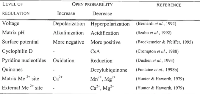
Table 1-l: Comparison ofMHC isoforms in selected muscles ofthe rat and human Table 1-2: Modulators ofthe mitochondrial pemieability transition pore
Chapter 2
Table 2-1: Effects ofHLS on soleus muscle mass, isometric twitch force and titanic force
Chapter 3
Table 3-1: Morphometric data and mitochondrial yield in muscle from sham and denervated animais
Chapter 4
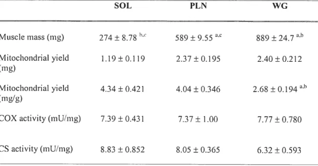
Table 4-1: Muscle mass and mitochondrial isolation yield
xii
LIST 0F FIGURES
Chapter I
Figure 1-1: Respiratory properties measured in mitochondria isolated from skeletal muscles with oxidative and glycolytic phenotypes.
Figure 1-2: Activity or content ofenzymes ofthe respiratory chain, TCA cycle and -oxidation pathway in mitochondria isolated from fish and rat muscles. Figctre 1-3: Activity or content of enzymes of the respiratory chain, TCA cycle and
-oxidation pathway in mitochondria isotated from rabbit muscles. figure 1 -4: Respiratory properties measured in mitochondria isolated from skeletal
muscles with oxidative and glycolytic phenotypes in rat and rabbit. figure 1-5: P/0 ratio at different respiration rates with pyruvate and
palmitoylcamitine in soleus and EDL.
Figure 1-6: Respiration rates of sapouin permeabilized libers from rat heart, soleus,
and white superficial portion of the gastrocnemius as a ftmction ofADP concentration in the incubation medium.
Figure 1—7: Schematic representation or the phosphocreatine shuttle network in muscle cells.
Figure 1-8: Production of reactive oxygen species (expressed as a % of 02 consumed) from complex I and III of the respiratory chain in saponin
pemieabilized liber bundies from gastrocnemius and soleLis muscles.
Figure l-9: The proposed molecular structure of the mitochondrial perneability transition pore (PTP).
Chapter 2
Figure 2-l: Effect ofl-ILS on total muscleJNK content in soleus muscle.
Figure 2-2: Effect ofHLS on baseline .INK phosphorylation in non-stimulated soleus
muscle.
figure 2-3: JNK phosphorylation in response to isometric contractile activity.
figure 2-4: JNK phosphorylation perunit titanic force capacity ofsoleus mLlscle.
Figure 2-6: Immunohistochemical analysis of pJNK in soleus muscle sections foilowina muscle stimulation via the sciatic nerve.
Chapter 3
Figure 3—1: Respiratory function in mitochondria from sham and denervated animais. Figure 3-2: Response to Ca2 challenge in mitochondria from sham and denervated
animais.
Figure 3-3: Effect of denervation on endogenous mitochondrial and whole muscle Ca2 content in sham and denervated animais.
Figure 3-4: Total Ca2 retention capacity and effect of PTP inhibitors in mitochondria from sham and denervated animais.
Figtire 3-5: Cyciophilin D, VDAC and ANT content ofmitochondriai fraction from sham and denervated animal s.
Chapter 4
Figctre 4-1 : Calcium uptake kinetics during Ca2 challenge experiments in mitochondria froiii SOL, PLN, and WG muscle.
Figure 4-2: Mitochondriai 112O2 production.
Figure 4-3: Endogenous Ca2 content of mitochondria isolated from SOL, PLN, and WG muscles.
Figure 4-4: Effect of substrate and the PTP inhibitor CsA on calcium retention capacity in mitochondria from SOL, PLN, and WG muscles.
Figure 4-5: Cyclophilin D, VDAC and ANT content of mitochondrial fraction from SOL, PLN, and WG muscles.
xiv
AB B R EV I ATI ONS
ADP adenosine diphosphate
AIF apoptosis-inducing factor
ANOVA analysis of variance
ANT adeni ne nucleotide transiocator
Apaf-1 apoptosis protease activating factor-1
ATP adenosine triphosphate
BSA bovine serum albumin
Ca2 calcium
COX cyclooxygenase
CRC calcium retention capacity
CsA cyclosporin A
CTL control
CypD cyclophilin D
DISC death-inducing signaling complex
DEN denervated
EDL extensor digitorum longus
EGTA ethylene gI ycol-bis(13-aminoethyl ether) tetraacetic acid
EndoG endonuclease G
FOXO forkhead box O
HLS hindÏimb suspension
IAP inhibitor of apoptosis protein
JNK c—jun NH2—terminal kinase
kDa kilo Dalton
MAPK mitogen-activated protein kinase
MiCK mitochondrial isoform ofcreatine kinase
MG medial gastrocnemius
MHC myosin heavy chain
PAGE polyacrylamide gel electrophoresis
PLN plantaris
PPlase pepidyl propyl-cis, trans-isomerase
PTP pemeability transition pore
ROS reactive oxygen species
SOL soleus
TUNEL terminal deoxyribonucleotidyl transferase-mediated dUTP-biotin nick-end labeling
AcKNowLEDcI ENTS
I acknowledge NSERC and the Canadian Space Agency for funding the studies presented in this thesis.
I thank the Iaboratory personnel, professorial staff, fellow graduate students, and administration staff ofthe depailment ofKinesiology at the Université de Montréal for their contributions (intellectual, technical, administrative) to my formation and
academic development.
xvi
f INTRODUCTION
I.] Presentcition oftÏie experiniental stiiclies
The purpose of the work presented in this thesis was to broaden our understanding of the cellular processes involved in mediating the adaptations of skeletal muscle to disuse. Thus, this thesis investigates two major axes of research relating to the disuse atrophy of skeletal muscle.
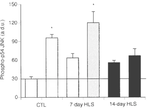
The flrst area of investigation extends on the research interests ofDr. Phil Gardiner and pertains to the effects ofreduced weight-bearing on the sensitivity of mechanically responsive intracellular signaling pathways in muscle. Mechanical forces play an important role in the regulation of muscle size (Vandenburgh, 1987). Since there appears to exist a relationship between muscle unloading and atrophy, the flrst study presented in this thesis investigates the sensitivity of tiiechanical ly-responsive intracellular signaling pathways in order to establish the extent to which a given mechan ical stimulus can influence the trophic response of muscle following atrophy. We focused on c-jun NH2-tei-minal kinase (JNK), a member of the mitogen-activated protein (MA?) kinase family that is activated by phosphorylation in skeÏetal muscle in response to a number of cellular stresses including changes in loading conditions. JNK activation was also repoiled to activate programmed cell death (i.e. apoptosis) (Davis, 2000; Papadakis et al., 2006), a process that is activated in the disused muscle (Kandarian et ctl., 2006) and may account for the loss of entire myofibers and/or of some nuclei within remaining myofibers. The resuits of this study showed that basal JNK activation state (i.e. phosphorylation) is increased in response to muscle ati-ophy, which may reflect activation of cellular apoptosis. On the other hand, we reported that the capacity to activate JNK by phosphorylation in response to an acute mechanical challenge is reduced following hindlimb unloading. These results lcd us to propose that atrophic muscle may lose the ability to transduce mechanical signaIs to the MA? kinase pathways.
Yan Burelle pertaining to the role of mitochondrial plasticity in heart and skeletal muscle under physiological and pathological conditions. A particular focus of this second research area relates to the investigation of the mitochondrial permeability transition pore (PTP), a structure identified as a key player in signaling necrosis and apoptosis in several tissues and ceil types (Zoratti & Szabo, 1995; Hengartner, 2000). Despite a large number of experimental studies providing evidence implicating the PTP
as a trigger for ceil death, little is known regarding the regulation and behavior of the
PTP in skeletal muscle and whether or flot it is affected in the process of severe mLtscle atrophy.
In the second study presented in this thesis we determined whether the sensitivity
or occurrence of PTP opening is altered in response to muscle denervation, a model of severe muscle disuse that mimics several denervation disorders observed in humans (Tews, 2002). The resuits from this study showed that a Ioss of innervation for 21 days
Ied to a dramatic increase in the vulnerabiÏity of isolated mitochondria to opening of the
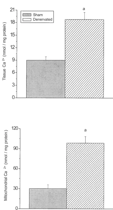
PTP. This phenomenon was at least partly caused by a significant increase in the endogenous Ca2 content of both myoflbers and mitochondria in response to denervation. In addition, we made the novel observation that cyclophilin D (CypD), a matrix protein that was recently shown to act as a regulator by sensitizing the PTP to Ca2-induced opening (Baines et al., 2005; Basso et ctl., 2005; Nakagawa et cd., 2005; Schinzel et cii., 2005) was upregulated compared to several other mitochondrial proteins including other putative PTP component, namely the adenine nucleotide translocator (ANT) protein and the voltage-dependent anion channel (VDAC), and enzymes of the respiratory chain (i.e. cytochrome oxidase). To our knowÏedge, this study provides the first evidence that changes in the expression of CypD could play a role in a non-genetic model of disease and suggests that opening of the PTP could be involved in the activation ofapoptosis generally observed in response to disuse atrophy.
Another question that remains unanswered with regards to the PTP is whether its regulation varies across muscle fiber types. This question is pailicularly relevant as the appearance and progression ofseveral neuromuscular disorders is very heterogeneous in muscles with various phenotypes (Tews, 2002), which may in part reflect a different
4 vulnerability of mitochondria to PTP opening and activation of mitochondrial death pathways across fiber types. In the third study presented in this thesis, we isolated mitochondria from muscles displaying different fiber type compositions and determined their sensitivity to PTP opening in vitro and examined a selected number of
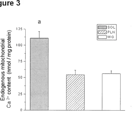
physiological regulators of pore opening at the mitochondrial level incÏuding endogenous Ca2 levels, production of reactive oxygen species (ROS) and content of putative PTP components including CypD. The resuits from this study indicated that mitochondria fiom the slow-twitch soleus composed predominantly of type I fibers, displayed a significantly greater vulnerability to PTP opening compared to mitochondria isolated from the plantaris and white gastrocnemius muscles, composed primarily of type II fibers. ROS production, which is a weII-known PTP inducer, was lower in mitochondria from soÏeus compared to the other muscles implying that factors other than ROS were involved. One of these factors appeared to be the greater endogenous Ca2 content within mitochondria from the soleus as compared to plantaris and white gastrocnemius. However, this could not entirely account for the differences in vulnerability to pore opening among muscles. We observed that the expression ofANT and VDAC, two putative PTP components, vas significantly greater in mitochondria from soleus compared to mitochondria frotii the other muscles, which may increase the likelihood that these proteins undergo conformational changes under conditions that favor PTP opening. On the other hand, pharmacological and molecular evidence indicated that CypD expression was similar across muscle types suggesting that this pmtein was not involved, This leU us to speculate that the existence of a fiber type specificity in the regulation oC PTP opening could at least partly account for the heterogeneous progression of neuromuscular disorders across muscles.
1.2 Introchiction to tue re‘icii’ of Ïiterctture
The review of literature is divided into four main sections. The objective of the first section is to provide the reader with an overview of skeletal muscle fiber types and some of the particular aspects that are pertinent to the understanding of disuse atrophy. In particular, the literature available on the differences in mitochondrial profile that exist between muscles with different metabolic and contractile phenotypes wiII be reviewed.
The second section ofthe review will focus on muscle disuse. First, an overview of the experimental models from which much of our understanding with regards to how muscles react to disuse was obtained will be presented and some of the classic changes that occur with disuse will be reviewed. As one of the important mechanisms responsible for the ioss of muscle mass involves an mci-case in protein degradation and
a reduction in protein synthesis, some of the recent advances in the identification of
signaling pathways that could mediate these effects, including the Akt/PkB, nuclear factor-kB (NFkB) and MAP kinase pathways and highlight the t-ole for these pathways
in apoptotic signaling will then be presented.
In the third section, an overview of the apoptotic machinery, inciuding the intrinsic
pathway of ccli death, where mitochondria are believed to play a pivotai role, is presented. Given the nature of the experimental work presented in this thesis, the consequences of PTP opening as weB as the structure of this pore and its regulation are covered in more detail. finally, the fourth section of the review vil1 focus particularly
on ccii death in models of muscle disuse and on the evidence supporting a role for
mitochondria in this process.
2 SKELETAL MUSCLE FIBER TYPES
2. 1 Contractileproteins
Skeietal muscle tissue is composed ofheterogeneous muscle fibers with different contractile and metabolic profiles. This fiber diversity confers, to a certain extent, the propetly of adaptability to skeletal muscles. Mammalian skeletal muscle is a multinucieated, highly specialized tissue made up of fibers with a diverse range of properties (Schiaffino & Reggiani, 1996; Bottinelli & Reggiani, 2000). The general properties of a given muscle typically resuit from the distinct properties of the represented fiber types combined with their proportions. The classification of muscle fibers into slow and fast ‘types’ is based primarily on the kind ofmyosin heavy chain
(MHC) isoform predominantly expressed (Schiaffino & Reggiani, 1996). Myosin is
considered the main structural and reguiatory protein in skeletai muscle and plays a prominent i-ole in dictating skeletal muscle functional and contractile properties. in the
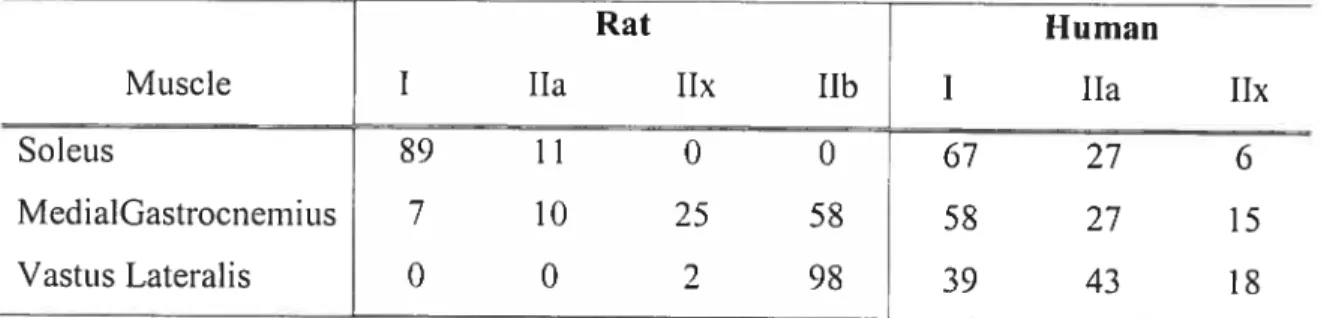
6 limb muscles of the rat, the MHC protein exists as four main isoforms: the slow type I and the fast types lia, lix, and lIb. Table I summarizes the proportions of MHC isofoms in selected muscles ofhumans and rats. Interestingly, protein expression ofthe 11h MHC isoforni is lacking in humans (Pereira Sant’Ana et aI., 1997).
Table 1-1: Comparison ofMNC isoforms in selected muscles of the rat and human
Rat Human
Muscle I lia lix llb I lia lix
Soleits 89 1 1 0 0 67 27 6
MedialGastrocnemius 7 10 25 58 58 27 15
VastusLateralis 0 0 2 98 39 43 18
Values are in percent(%). Table adapted from Talmadge, 2000.
In addition to the foctr major MHC isoforms already mentioned, other isoforms
have been identi lied including embryonic, neonatal, extraocular, and laryngeal-specific isofbrms (Pette et cii., 2000), but their expression levels in 11mb muscles are negligible and therefore, will not be further discussed. Siiice the contractile properties of a muscle liber are in large part determined by the MHC composition (Bottinelli & Reggiani, 2000; Reiser et al., 1985; Schiaffino & Reggiani, 1996; Fitts et al., 1998; Widrick et cil., 1999), the nomenclature for the four major liber types identified in Iimb muscles suitably follows the type ofMHC isofomi present (i.e. liber types are designated as type I, liA, I1X, or IIB) (Rivero et ctÏ., 1998). Presently, there is some debate as to whether or not discrete’ fiber types even exist as it is becoming increasingly evident that liber types might actually exist on a continuum of contractile velocities and metabolic properties rather than representing discrete entities (Talmadge et aï., 1993; Pette et aï., 2000).
2.2 Proteins invoÏved in excitcttion-contraction coupÏing and Ca hanclÏiiïg
Although slow and fast-twitch skeletal muscles are used to perform distinct functions, ah vertebrate skeletal muscles have the same basic structure and use the same basic contractile system (Rome & Lindstedt, 1998). It is the specific modifications of the proteins involved in the contractile system that allow the muscles to perform these
diverse tasks. Both qualitative and quantitative modifications of several muscle proteins seem to underlie the physiological differences between slow and fast twitch libers. For instance, slow-twitch libers, compared with fast-twitch libers, contain siower MHC isoforms, which have a reduced maximal velocity of shortening (Reiser et al., 1985), a decreased content of sarcoplasmic reticulum (SR) (Appeit et cil., 1989), with a siower isoform ofthe Ca2 pump (SERCA2 in slow-twitch vs. SERCAI in fast-twitch) (Lytton
et cii., 1992), and a lower concentration of the cytoplasmic Ca2 buffering protein
parvalbumin (Heizmann et cii., 1982). Calsequestrin, a Ca2 binding protein located in the lumen of the SR, also bas a unique isoform expression pattet-n in slow-twitch and fast-twitch muscles of rabbit and rat (Damiani & Margreth, 1994). Finally, the ryanodine receptors are also considered a part of this molecular excitation-contraction associated machinery that varies across liber types and molecular evidence indicates that distinct isoforms of ryanodine proteins are expressed in tuna slow- and fast-twitch skeletal muscle (Franck et ciL, 1998). During transitions in muscle liber type, these proteins are co-regulated with the expression ofspecilic MHC isoforms.
2.3 The nivonucÏear domain
A special feature of skeletal muscle libers compared to most other ceils is that they are multinucleated and successive muscle liber segments are controlled by individual nuclei. Mammalian skeletal muscle libers have been shown to maintain a relatively limite, liber type-specilic relationship between myofiber size and myonuclear number (Hikida et cii., 1 997). In fact, the relationship between myonuclear number, ceil size, succinate dehydrogenase activity (a marker of mitochondrial density), and MHC type bas previously been examined in single liber segments mechanically dissected
from soleus and plantaris muscles of rats (Tseng et cti., 1994). The resuits from this study show that cytoplasmic volume per myonucleus is higher in fast and slow plantaris libers (112 vs. 34 x microns3) than fast and slow soleus libers (40 vs. 30 x i03 microns3), respectively. The authors report that slow libers always had srnall cytoplasmic volumes per myonucleus, regardless of liber diameter, succinate dehydrogenase activity, or muscle oforigin. Slow soletis libers had signilicantly greater numbers of myonuclei/mm than did either fast soleus or fast plantaris libers (116 vs. 55
8 more limited in slow than fast fibers, and in the fibers with a high, compared to a low, oxidative metabolic capacity (Tseng et aÏ., 1994).
Interestingly, the myonuclear domain maintains a degree of plasticity by adapting
to changes in muscle fiber size, and likely phenotype. The atrophic response due to
unloading on the number of myonuclei, and size of the myonuclear domain in fibers of rat hindlimb muscles bas been studied. It would appear that as a muscle undergoes a disuse-induced phenotypic transition from slow-to-fast or from fast-to-faster, existing myonuclei are eliminated to support not only the reduction in fiber size, but also the inherent change in phenotype. Although intuitively sound, this rationale implies that it
is fiber phenotype and size that dictate myonuclear number. The question bas been raised whether the loss of myonuclei in response to muscle disuse is the early event which causes muscle atrophy and slow-to-fast directional transitions in fiber type. Most studies have shown that the loss of muscle mass becomes significant between 3 and 7 days following the onset ofdisuse (Allen et al., 1997; Krawiec et ctÏ., 2005) even though protein syntbesis is suppressed as promptly as 6 hours after muscle disuse (Watson et al., ] 984; Thomason et cil., 1989). However, a very recent study shows that in response
to unloading of the rat hindtimb muscle, loss of myonuclei is already increased at a time
when no measurable loss of muscle mass or cross sectional area has occulTed, suggesting that myonuclear loss is an early, and possibly the causative event in the process of muscle atrophy (Dupont-Versteegden et cii., 2006). The significance of this observation needs further investigation since it is still unclear whether loss ofmyonuclei initiates, or occurs as a consequence ofthe Ioss of muscle mass during atrophy.
2.4 Mitochondrictl profiles
Beside substantial differences in contractile proteins, components involved in excitation-contraction coupling, and size of myonuclear domains, muscle fibers also dispÏay dramatic differences in their metabolic profile. Given that in the present thesis a main focus is on mitochondria, this section examines differences in the mitochondrial profile that occurs across muscle fiber types including quantitative differences in mitochondrial volume density as well as qualitative differences that may exist in terms of ultra—structure, function, and regulation of respiration.
2.4. 1 MitochoncÏrictÏ voïmne densitv
The most important difference in mitochondrial profile that can lie observed across fiber types is the volume of muscle ceils occupied by mitochondria, which lias been termed mitochondrial volume density. Morphometric analysis of electron micrographs obtained from muscles with different fiber type compositions indicate that mitochondrial volume density is approximately 6 ¾ in soleus, a muscle composed predominantly of type I oxidative libers (Schwerzmann et cii., 1989). In type lia fibers,
which have a high capacity for oxidative phosphorylation as well as for glycolysis, mitochondrial volume density is similar and in rat specifically, may even reach higher values than type I libers (this is not the case in humans where type 1 lIa (Gardiner, 2001)). 0f note however, mitochondria in lia fibers appear smaller and more numerous compared to type I libers in which mitochondria are less numerous but larger (Shah &
Sahgal, 1991). Finally, type Ilb fibers have the lowest mitochondrial density at 2-3 % of liber volume (Schwerzmann et ai., 1 989), which reflects their high dependence on
glycolysis for ATP productiori.
2.4.2 Intrinsic properties ofmitochoncÏria
Although it is clear that skeletal muscle exhibits considerable variation in
mitochondrial volume density among liber types, it is less clear whether the mitochondria also present specific functional characteristics and/or distinct regulatory properties that allow fine adjustments of mitochondrial performance to the conditions and needs encountered in various libers. The few studies available on this topic have used mainly mitochondria (or less frequently saponin-permeabilized libers) isolated
from fast and slow muscles from rabbits (Jackman & Willis, 1996; Howlett & Willis,
1998; Gueguen et cii., 2005a; Gueguen et aï., 20051), cats (Schwerzmann et aÏ., 1989),
pigs (Gueguen et ai., 2005e), and rats (Pande & Blanchaer, 1971; Yajid et aï., 199$;
Capel et ai., 2004; Mogensen & Sahlin, 2005 Anderson & Neufer, 2006), which
express predominantly type 11h and 1 libers, respectively. Fish white and red muscles have also been used because they offer the unique advantage of having muscle compartments with very homogeneous liber types (Leary et ai., 2003). As discussed
10 below, properties that have been compared include mitochondrial enzyme content, respiratory capacities, coupling efficiency between oxidation and phosphorylation, proton conductance of the inner membrane (the so—called proton leak), membrane
fluidity, and production ofROS.
2.4.2.1 En:’inoÏogvctnc/ respiraton’ capacitv
The measurement of maximal respiratory capacity in the presence of various respiratory substrates has been measured in several studies. The group of Weibel in Switzerland (Schwerzmann et cii., 1989) reported that maximal ADP-stimulated
respiration vas similar in mitochondria isolated from the cat soleus and gracilis muscles when energized with a combination of substrates feeding the respiratory chain at the level of complex I (pyruvate-malate, glutamate-malate), complex II (succinate), as well as complex IV (reduced cytochrome c after rupture of the outer membrane) (Figure I
-lA). Similarly, Yajid et al. (Yajid et ciL, 1998) observed no significant difference in maximal state 3 respiration of mitochondria isolated from several rat muscles (soleus, extensor digitorum lougus (EDL), tibialis anterior (TA), gastrocnemi us) nei ther in the presence of glutamate-malate nor succinate (Figure l-lB). This lack of difference also appeared in mitochondria isolated from red and white muscles in fish (Figure 1-1 C), which displayed nearly identical rates of state 3 respiration in the presence ofpyruvate malate (Figure 1—1 D). Finally, in mitochondria from rabbit muscle, state 3 respiration in the presence of pyrctvate-malate xvas repoiÏed to be significantly (25 %) higher in mitochondria from the soleus compared to the fast gracilis muscle (Jackman & Willis, 1996), while no difference vas observed with the respiratory substrate 2-oxoglutarate. In general, the available data suggest that the respiratory capacity per milligram of mitochondria! protein is fairly constant act-oss fiber types and that upregulation of mitochondrial volume density is probably the main mechanism by which the oxidative potential ofa fiber can be increased.
This relative similarity in respiratory capacity in mitochondria across fiber types is in general agreement with the available data regarding enzyme content. Indeed, Leary et al. (Leary et cii., 2003) reported that the activity ofthe respiratory chain complexes (I, 11, 1+111 and IV) as well as that ofthe ATP synthase (complex V) and citrate synthase
C
Leary et aI. (2003).Figure 1-1:
E fish red muscle fish white muscle
Respiratory properties measured in mitochondria isolated from skeletal muscles with oxidative and glycolytic phenotypes in rabbit (panels A and D), rat (panel B), and fish (panel C). Respiratory rates are expressed per mg of mitochondrial protein and were determined in the presence of various substrates: substrates for complex I (pyruvate (Pyr) + malate (Mal), glutamate (Glut) + Mal,
2-oxoglutarate (2-0G)); complex II (succinate); and exogenous cytochrome c (Cyt C) (following rupture of the outer membrane). Abbreviations for panel B: Sol: soleus; EDL: extensor digitorum longus; TA: tibialis anterior; Gastroc: gastrocnemius.
A Schwerzmann et aI. (1989). B Yajid et aI. (1998).
E Soleus • Gracilis 1000 o I. o b) 800 600 400
o
) 200 o E Glut+ Mal • SuccinatePyr+Mal Glut ÷ Mal Succinate
Cyt
C Sol EDL TA Gastroc 600 :;:;- 500 o I— o 400 300 200 100 OD Jackman and WiIIis (1996).
35° — 300 o 250 b) 200 E 150 o o-b) o E C c’J
o
-: 120 100 80 60 40 20 0•12
vas similar overafl in mitochondria isolated from the white and red muscles ofrainbow
trout (Figure 1-2A). However, the activity of aconitase and 2-oxoglutarate dehydrogenase, two other enzymes of the TCA cycle, was significantly lower in mitochondria from white compared to red muscle, which lcd this group to suggest that subtie differences in the stoichiometry of TCA cycle enzymes cou Id occur across fiber types (Figure l-2A). Schwerzrnann et al. (Schwerzmann et aÏ., 1989) also reported that the activity of complex IV of the respiratory chain was similar in mitochondria isolated
from cat soleus and gracilis. Similarly, Philippi and SiÏlau (Philippi & Sillau, 1994)
reported that the content ofcytochromes c+cj and ct+a3, which provide an indication of
the content of cytochrome c as well as complexes III and 1V, was similar in mitochondt-ia isolated from the white gastrocnemius and soleus muscles in rats. 0f note,
this study analyzed the subsarcolemmal (SSM) and intemiyofibrillar (IMF) mitochondria separately and found no difference between the two populations of mitochondria neither within nor between muscles (Philippi & SiIlau, 1994) (Figure 1— 23 and C).
In stark contrast with these studies, Jackman and Willis (Jackman & WilÏis, 1996) reported that in rabbit muscle, the activities of several complexes of the
respiratory chain were 1.6 to 2.0 fold greater in mitochondria from the soleus compared
to the gracilis (Figure I -3A). Although the differences were more modest, they also
observed higher activities for citrate synthase and malate dehydrogenase. On the other hand, isocitrate dehydrogenase activity was two-fold higher in the gracilis compared to soletis (Figure 1—33). These data are somewhat surprising given that in this study, maximal state 3 respiration was unchanged (in the presence of 2-oxoglutarate) or only 25 % higher (in the presence of pyruvate + malate) in the mitochondria from the soleus
compared to the gracilis (figure 1 -4). Except for this apparent discrepancy, the data available in the literature would sciggest that mitochondria from different liber types do
not substantially differ with respect to the maximal capacity ofthe respiratory chain.
However, under some conditions, differences do appear between mitochondria of slow and fast mctscles. This is the case when mitochondria are energized with lipid
E Fish ted muscle • Fish white muscle
A
(Q Q) C u, w (u > 4-o > > 4-, o Q) E > N C w * 100 50 0 lI 11+111 IV V CS OGDH Aconitase HOAD 0,4 0,3 0,25B
o cL o) E o E C o + C w E o L. -c C o >‘ o 0,2 E a+a3 Soleus• a+a3 White gastroc
E c+cl Soleus
• ci-cl White gastroc
--0,15 0,1 0,4: 0,35: o -cL o 032 E 0,25: 0,2 0,15 Q) E B 0,1 0,05. o-j II 0,05
il
]
o SSM Figure 1-2:Activity or content of enzymes of the respiratory chain, TCA cycle and
f3
-oxidation pathway in mitochondria isolated from fisli and rat muscles. Abbreviations in panel A are : HOAD: hydroxyacyl-CoA dehydrogenase; OGDH: 2-oxoglutarate dehydrogenase, CS: citrate synthase, I 1V respiratory chain complexes; V: ATP synthase. Abbreviations in panels B and C: SSM: scibsarcolemmal mitochondria; IIVIF: intemiyofibri Ilar mitochondria.Activity or content of enzymes of the respiratory chain, TCA cycle and -oxidation pathway in mitochondria isolated from rabbit muscles. Data ai-e from Jackrnan and Willis (1996). In panel A, numbers on the x-axis refer to the respïratory chain complexes. In panel 3, abbreviations are the following: CS: citrate synthase; MDH: malate dehydrogenase; IDH: isocitrate dehydrogenase.
14
j-
E• GracilisSoleusA
1,1 10 1 10 9000 8000 7000 6000 5000 4000j
C D, D E >‘ >B
2,5 i04 2 10 1,5 1 2500 2000 1500 1000 C D) Dg
>-‘ > 4-’ o 800 600 400 200 o E Soleus I Gracilis * IDHj
* * * I II I—i—III II—I—III 500 O Figure 1-3: OSA
Mogensen and Sahlin (2005)B
Jackman and WiIIis (1996)Figure 1-4:
Respiratory properties measured in mitochondria isolated from skeletal muscles with oxidative and glycolytic phenotypes in rat (panel A) and rabbit (panel B). Respiratory rates are expressed
per mg of mitochondrial protein and were determined in the presence of lipid substrates
païmitoyl camitine+ malate (PC+M) or glycerol-3-phosphate (G3P). D Soleus • EDL * D Soleus • GraciIs .4-., o I— D-b)
n
100 80 60 40 20 0 * 400 350 300 o I— D- 250 E 200 C 150 100 50 PC+ M o PC÷M G3P16
substrates or with glycerol-3-phosphate, a glycolytic intemiediate involved in the shuttling ofcytosolic reduced equivalent to mitochondria. Indeed, both in rat and rabbit, mitochondria from slow oxidative muscles (soleus) display a higher state 3 respiration
in the presence ofpalmitoylcarnitine compared to mitochondria from glycolytic muscles
(EDL or gracilis) (Figure I -5A and R). These data are consistent with the fact that the activities of enzymes of the -oxidation pathway such as hydroxyacyl-CoA dehydrogenase are 1 .6 to Ï .9-fold higher in mitochondria from oxidative vs glycolytic
muscles (Figure I -4A and B). This difference also appears consistent with the fact that oxidative muscles derive a significant portion of their energy from the oxidation of circulating fatty acids and express much higher levels of proteins involved in sarcoiemmal fatty acid transport (FAT/CD36, FATP, FABP1) and fatty acid
intracellular binding (fABP) (Bonen et ci!., 2002).
Conversely, because in type IIb fibers the high glycolytic rates achieved during short burst contractions are likely to resuit in the accumulation of cytosolic reducing
equivalent and glycolytic intermediates, mitochondria in these fibers may have an increased capacity to shuttle cytosolic reducing equivalent into mitochondria for oxidation. The only available evidence to support this possibility cornes from the study by Jackman and WiIlis (Jackman & WiIlis, 1996) which showed that compared to mitochondria from the soleus, mitochondria from the rabbit gracilis muscle had a ten— fold higher state 3 respiration when glycerol-3-phosphate was the respiratory substrate (Figure 4B). They showed that this was probably due to the fact that mitochondria from the gracilis muscle expressed more glycerol-3-phosphate dehydrogenase and were thus able to produce mitochondrial FADH2 at a much higher rate. Therefore, at least in the
rabbit, glycolytic muscles may depend more on the Œ-glycerolphosphate shuttie compared to other muscles in which the malate-aspartate shuttle may predominate
(Jackman & Willis, 1996).
Finally, it should be mentioned that although several mitochondrial properties appear similar in mitochondria across muscle fibers at steady state, oxidative capacity
(a)
Pyruvate • J . II I ____ -2 .2/
- I I2
û. DI Û—I 0 20 40 60 60 100 120 Respiration (pmol 02 C min U CS)(b)
P&mitoyl-C-carnitine IIi
—-— •I -D 2 O I/Q IJn I, I. o I 0 20 40 60 80 100 120 Respiration (pmol O L’ niin’ U’ CS)Figure 1-5:
P70 ratio at different respiration rates with pyruvate (a) and palmitoylcarnitine (b) in soleus (open circles) and extensor digitorum longus (dark squares). P/0 ration at Vrnax is shown as the mean +1- SEM. The mean state 4 respiration is shown on the x-axis when
‘s change acutely in response to signais that alter mitochondrial biogenesis. For example, mitochondrial enzymes increase signiflcantly during myogenesis (Moyes et aÏ., 1997)
and during electrical stimulation of skeletal muscle (Henriksson et cii., 1986;
Henriksson et aI., 1989; Reichmann et ai., 1991), btit individual respiratory chain
complexes and TCA cycle enzymes do not change in parallel. This phenomenon is probably related to the extreme complexity of the mitochondrial biogenesis program, which involves several stimuli and transcription factors, the coordination of transcription of genes located in two separate genomes (nuclear and mitochondriai), importation of pre-proteins into mitochondria, and assembly of multi-complex enzymes and holoenzymes (Hood, 2001; Moyes & Hood, 2003). It is thus possible that at least
transi torily, mitochondria undergo changes in their normai composition.
2.4.2.2 Coupling efficiencv, proton concÏuctctnce cinci mcm brune properties
Very few studies have investigated whether variations across fiber types exist with respect to the coupiing efficiency of oxidative phosphorylation. This parameter, known as the P10 ratio, is conventionally measured by adding a quantity of ADP to
isolated mitochondria and measuring the amount of oxygen constimed to phosphorylate ADP into ATP. It is well estabiished that the P10 ratio decreases as respiration is
progressively decreased from maximal ADP-stimulated respiration to submaximal respiration i-ates (Guaiger et cii., 2000). The reasons for which mitochondrial efflciency is progressively lowered as respiration approaches resting values are complex.
However, one ofthe main factors appears to be that the proton Ieak, i.e. the passive re entry of protons into the mitochondrial matrix, accounts for an increasing fraction of
respiration as it approaches resting values (Brand et aÏ., 1994), thereby increasing 02
consumption that is not devoted to ATP synthesis.
[n one ofthe earliest studies comparing P/0 ratios in mitochondria isolated from different skeietal muscle fibers in the rat, Pande & Blanchaer (1 971) wcre unable to find diTerences when using pyruvate or palmitoyl-camitine as suhstrate. 0f note, in this
study P/0 values were only measured at maximal respiration rates in the presence of saturating amounts of ADP, which does not allow one to exciude the existence of differences when mitochondria are respiring at submaximal rates (as is usually the case
in vivo). However, Mogensen and Sahlin (Mogensen & Sahiin, 2005) recently
compared the P/O ratios over the entire range of respiratory capacity in mitochondria isolated from the rat soleus and the fast EDL. These authors reported no significant
difference in the P/O ratios between mitochondria from the two muscles, both in the
presence of pyruvate or palmitoylcamitine (figure i-5). Direct measurement of proton leak also revealed an absence of significant difference in mitochondria isolated from red and white fish muscle (Leary et aï., 1998). However, these authors noted that when
proton leak vas expressed per unit of complex IV instead of per mg of total mitochondrial proteins, the leak appeared greater in mitochondria from white compared
to the heart or red muscle. The attthors argued that ctnder certain circumstances,
normalization against a marker of the respiratory chain capacity cotild be more appropriate than total protein to express mitochondrial parameters. Leary et aï. (Learyet ciL, 2003) also determined the fluidity of mitochondrial membranes since local Iipid environment can affect structure and function of mitochondrial proteins. These authors observed that the membrane fluidity of mitochondria in red muscles was significantly greater than in white muscles. This phenomenon could be due to many factors including variations in phospholipid profiles (i.e. chain length, saturation, or cardiolipin content). However no information is available regarding the phospholipid profile in mitochondria across fiber types and it also remains unclear how this could affect the in vivo activity of membrane-bound proteins. Taken as a whole, these data suggest that although some membrane properties and the proton leak may slightly differ, the coupling efficiency of mitochondria appears to be relatively constant across fiber types.
2.4.2.3 Sensitivitv of oxidcitive pïiosphor lcttion to ADP cmctcreatine
One of the most striking and systematically repotted differences between mitochondria from oxidative and glycolytic muscles relates to the mechanisms by which changes in the concentration of cellular adenylates regtilate the rate of oxidative phosphorylation. The maj ority of the experimental evidence in favor of such di fferences
has been obtained using saponin permeabilized fibres, which allows for the
investigation of mitochondrial function within a relatively preserved cyto-architectural environment. Several stctdies have shown that the apparent K11 of mitochondrial respiration for ADP is several-fold higher in slow oxidative muscles predominantly
20
composed of type I fibers (200-300 iM for the heart and soleus) compared to muscles expressing mainly type II fibres (10-30 iiM in EDL and white gastrocnemius) (Kay et
eT, 1997; Saks et aï., 1998; Braun et uT, 2001; Saks et cd., 2001; Seppet et al., 2001)
(Figure i-6). This apparently low sensitivity of oxidative fibers to ADP was shown to be at least pailly due to the fact that the porin pore VDAC, which is responsible for the transport of ADP across the outer membrane, has a low conductance for ADP in slow compared to fast muscles (Saks et ctl., 1998). Indeed, if the mitochondrial outer membrane is disrupted by a carefuiiy controlied osmotic shock, the K1, foi- ADP becomes similar in both types ofmitochondria (20-30 j.tM) (Saks et aï., 1993).
Another important difference is that in mitochondria from oxidative muscles, the phosphocreatine shuttie appears to play a predominant role in the control of respiration compared to that observed in mitochondria from glycolytic muscles (Kay et aÏ., 1997; Saks et ctÏ., 199$; Braun et cd., 2001; Saks et ctï., 2001; Seppet et al., 2001). Indeed,
mitochondria from oxidative muscles express high levels of the mitochondriai isoforni
of creatine kinase (MiCK). MiCK is located in the intermembrane space where it
associates wjth VDAC in the outer mitochondrial membrane and the ATP/ADP exchanger (i.e. ANT) of the inner membrane (Brdiczka et cd., 199$). In contrast to ADP, the condtictance of VDAC for creatine coming from the cytosol is reiativeiy high
in ail muscles. Therefore in oxidative fibers, the presence of creatine allows MiCK to
preferentially use the ATP exiting the ANT exchanger to directly regenerate ADP in the mitochondria thus acting as a powerfui stimulator of oxidative phosphorylation (Figure
1-7; scheme ofthe CK shuttie).
Finally, recent evidence also suggests that subtle differences in the regulation of respiration by the ATP/ADP ratio exist between mitochondria isolated from oxidative and glycolytic muscles, independent ofthe phosphocreatine shuttie. Indeed, Gueguen et
al. (Gueguen et aï., 2005e) showed that in mitochondria from glycolytic muscle,
respiration was more sensitive to inhibition by ATP compared to mitochondria from oxidative muscle. Given that the ANT exchanger is known to exert a significant amount ofcontrol over respiration (Groen et cd., 1982), the authors posttilated that the content
10
CE
E
6
—1 C2,5
Km G0,2
03
tO
[ADP], mM
Figure 1-6:Respiration rates of saponin permeabilized fibers from rat heart, soleus, and white superficial portion ofthe gastrocnemius (Gw) as a function ofADP concentration in the incubation medium.
Dotted unes indicate the [ADP] required to elicit haif of the maximal respiratory rate (K11). Respiration is expressed in nmol/min/mg dry weight. Adapted fromKay et aL (1997).
Hea
Km heartSoleus
K soleusO
011
22
ATP \ ATPase AIPaç \
(N
(N
H(N
AIP AP AP J AP A[P \v!
DPK AIP FK rVA
PCr Figure 1-7:Schematic representation of the phosphocreatine shuttie network in muscle celis. In
mitochondria, die specific isofomi ofcreatine kinase (MiCK) form an octamer that is structurally and functionally coupled to porin pores (P) in the outer membrane and the ATP/ADP exchanger (AT) of the inner membrane. Cytosolic creatine generated by CK isozymes located at sites of ATP consumption enter mitochondria through porin pores and stimulates MiCK. MiCK through a preferential access to mitochondrial ATP vill generate ADP at the vicinity of AT exchangers resulting in a powerful stimulation of respiration. This system is predominant in oxidative muscles such as the heart and soleus. On the other hand, it is virtually absent in mitochondria from fast muscle. Instead, in these muscles mitochodondrial respiration is directly regulated by changes in cytosolic ADP. Diagram originally pubhshed by WalÏirnann et al. (1992).
oJ
Mt
9Frpbfl
or the regulation ofANT could be different in both types ofmitochondria.
In general, the literature available on the regulation of respiration indicates that in glycolytic muscles, respiration is predominantly regulated by changes in cytosolic
[ADP] and ATP/ADP ratio, while in oxidative muscles, respiration is predominantly regulated by the phosphocreatine circctit which couples sites of ATP consumption to sites ofATP production.
2.4.2.4 Procluction ofrectctive ox’‘gen species (R OS,)
It is well established that the mitochondrial respiratory chain is one of the main
source of ROS in ceils. ROS production arises mainly from the Ieak of electrons at complex I and III of the respiratory chain, which then react with 02 to produce the superoxide anion (Brand et al., 2004; Muller et al., 2004; Andreyev et al., 2005). Electron leaks at these sites are favored by conditions such as high membrane potential, high levels of reduction of respiratory chain complexes, or physical damage to the respiratory chain (Nicholls, 2004; Brand, 2005). Not surprisingÏy, mitochondria have an elaborate antioxidant system composed of enzymatic and non-enzymatic mechanisms. The main enzymatic system involves the mitochondrial isoform of superoxide dismtitase (MnSOD) which converts the highly toxic superoxide anion into the somewhat less reactive F1202 (Fridovich, 1995). Other enzymatic systems discovered more recently involve specific mitochondrial isofomis of thioredoxin (TRX-2) and peroxyredoxin (Prx-3) which work in concert to scavenge H202 (Tanaka et ctÏ., 2002; Yamawaki & Berk, 2005; Matsushima et cii., 2006). finally, the main non-enzymatic system is the mitochondrial reduced gluthatione pool and the enzymes associated glutathione metabolism (glutathione peroxidase and reductase) which are also involved
in H202 scavenging.
Only three studies have determined whether mitochondrial ROS production varies across muscle fiber types. Recently, Anderson et al. (Anderson & Neufer, 2006) measured rates of H202 production by mitochondria in situ in saponin pemieabilized fiber bundies from muscles with distinctive fibers types including the soleus (type I), the red gastuocnemicts (type lIA) and the white gastrocnemiLls (type IIB). If H202
24
production was soiely a function of mitochondrial content, then tiber bundies from soletis and red gastrocnemius muscles would be expected to generate the highest levels ofH2O2 production. Surprisingly, these authors observed that mitochondrial free radical leak (II2O2 produced/02 consumed) was two- to three-fold higher in white gastrocnemius (type 113) than in red gastrocnemius (type lIA) or soleus (type I) muscle libers during basal respiration supported by complex I or complex II substrates (Figure l—8) despite the fact that the number of mitochondria in white gastrocnemius was 50 % less. When normalized for mitochondrial content, total H202 scavenging capacity was lower in red gastrocnemius and white gastrocnemius libers, whereas glutathione peroxidase activity, which is largely responsible for H20? removal in mitochondria, was similar in ail three muscle types indicating that factors other than the activity of this enzyme were responsible for the lower scavenging capacity in white gastrocnemius. The fact that mitochondrial H202 production observed among the three types of muscle libers did not mirror diffetences in respiratory capacity or mitochondrial content suggest that mitochondria possess distinct features that affect their ROS production and/or removal. It would appear that type Il muscle fibers, particularly type 113, possess unique properties that potentiate mitochondrial H202 production and/or release (Anderson & Neufer, 2006). Similarly, the results of another study measuring mitochondrial ROS production in various muscles of the rat show that in soleus muscle, glutamate/malate (complex I) supported mitochondrial H202 release vas lower than in tibialis anterior muscle (Capel et cii., 2004), consistent with the observations reported by
Anderson et al. (Anderson & Neufer, 2006). Leary et al. (Leary et cd., 2003) made a similar observation in fish when mitochondria isolated from red and white muscles were compared. 0f note, in this study, Leary et al. repoiled that the activity ofthe TCA cycle enzyme aconitase was significantly lower in mitochondria from white muscle. This phenomenou could be at least paiÏly due to the greater amounts of ROS produced in these mitochondria as it is weIl known that aconitase is very sensitive to inactivation in the presence ofoxidative stress (Benderdouret cii., 2004).
As a general conclusion to this section, it appears that despite the fact that
mitochondria from different liber types may not differ substantially in terms of maximal respiratory capacity, they do have distinct properties at least in terms ofregulation of
W
ÀW
ÀW
Àr
Pyr/M alA
H202
H202
Pyrf
Mal
Succinate
*
+Jvp
*
I
*
*N0.4
c3
Q ka,ï
B
8
6
4
2
0—
cc
r’l*
I
*
I
140
120
190
D
89
69
40 g,
20
RSol
RG
WG
Sol
RG
WG
Figure 1-8: ‘JSol RG WG
IISol RG tVG
Panel A shows the production of reactive oxygen species (expressed as a % of°2 consumed) from complex I and III of the respiratory chain is significantly higher in saponin permeabilized liber bundies from the white gastrocnemius (WG) compared to more oxidative muscles such as the soleus (Sol) or the
red
portion of the gastrocnemius (RG). This phenomenon is observed both inthe
presence of substrates feeding complex I (pyrctvate
+ malate (Pyr/Mal)) and Il (succinate) ofthe respiratory chain. Panel B shows total ROS scavenging capacity in liberbundles from the three muscles expressed per mg
of
dry liberweight
or per unit of citrate synthase (CS) to take into account differences in mitochondrial content. Data from Andersen and Neufer (2006).26
respiration and production of ROS. It is entirely possible that other differences exist as
weÏI, however, further studies ai-e needed to explore this possibility. 0f note, there is
apparently no study availabie regarding possible variations across fiber types in the
mitochondriaÏ ptoperties that pertain to their t-ole in the initiation of ccli death. For
example, the question of whether differences exist in the mitochondrial content of several proteins involved in the regulation of apoptosis remains to be established. In addition, there are currently very few studies available on the mitochondrial PTP in skeletal muscle (Fontaine et ctL, 199$a; Irwin et al., 2003) and none have investigated
whether its regiLlation varies across fiber types. Cleariy, these questions need to be addressed especially in the context of understanding the response of muscle fibers to dis use.
3 SKELETAL MUSCLE ADAPTATIONS TO DISUSE
In this section of the literature review, the response of muscle fibers to distise will be discussed. First, an overview of the experimental models from which much of our understanding of how muscles react to disuse vas obtained will be presented, followed by an overview of some of the classic changes that occur in muscle in t-esponse to disctse. The molecular etiology of muscle disuse will then be discctssed. As one of the
important mechanisms responsible for the loss of muscle mass involves an increase in protein degradation and a reduction in protein synthesis, this section will present some of the recent advances in the identification of factors that couid mediate these effects, including stimuli such as mechanical stretch and signaling pathways such as the Akt/PkB, nucleat--factor-kB (NFkB) and MAP kinase pathways. The t-ole that these pathways play in apoptotic signaling will also be highlighted.
3. 1 MocÏeÏs ofclisztse ancÏ overview ofintisele response to disuse
Since it is difficult to investigate the mechanisms responsible for disLise muscle
ati-ophy in humans, most investigations have used laboratory animais (namely mice, rats, rabbits, cats, and guinea pigs) to understand the underlying causes and ceilular
pt-ocesses implicated in muscle remodeling following disuse. These models vary in their degree of invasiveness and extent of muscle inactivation ranging from essentially