Université de Montréal
Occlusion of patients with Osteogenesis Imperfecta:
A comparison with the severity of the syndrome
par Cynthia Carbone
Université de Montréal Faculté de Médecine Dentaire
Mémoire présentée à la Faculté de Médecine Dentaire en vue de l’obtention du grade de M.Sc.
en médecine dentaire option orthodontie
[Mars, 2016]
Université de Montréal
Faculté des études supérieures et postdoctorales
Ce mémoire intitulé:
Occlusion of patients with Osteogenesis Imperfecta A comparison with the severity of the syndrome
présenté par: Dre Cynthia Carbone
a été évalué par un jury composé des personnes suivantes :
Dre Clarice Nishio président-rapporteur
Dre Nelly Huynh directeur de recherche
Dre Basma Dabbagh codirectrice Dre Frank Rauch
codirecteur Dr Jean Rizkallah
Résumé
IntroductionL’ostéogenèse imparfaite (OI) est un désordre de collagène héréditaire caractérisé par du tissu conjonctif défectueux et dont l'incidence est de 1 sur 20 000 naissances. Il y a une surreprésentation marquée de malocclusion de Classe III et d'occlusion croisée antérieure et postérieure dans la population d’OI. L’objectif principal de cette recherche est d’évaluer si la sévérité des malocclusions présentes chez les patients atteints d’OI est proportionnelle à la gravité du syndrome. L’objectif secondaire de cette recherche est d’évaluer si la sévérité de la malocclusion augmente avec l’âge.
Matériels et méthodes
Cette étude rétrospective observationelle fut effectuée par calcul du Discrepancy Index (DI) de 56 modèles dentaires de patients atteints d’OI. Les résultats du DI ont été comparés à trois variables qui caractérisent la gravité du syndrome: le type de OI, le type génétique et le z-score de la grandeur de chaque patient. En outre, l’analyse longitudinale d’un sous-ensemble de 20 modèles a été faite pour déterminer si la sévérité de la malocclusion augmente avec le temps.
Résultats
La médiane du DI était de 33,5 [1, 109]. Le DI est plus augmenté chez les patients atteints d’un type de OI plus sévère (p = 0,001) ainsi que chez les patients avec un z-score de grandeur plus petit (p <0,0001). L'analyse longitudinale a démontré une augmentation statistiquement significative du DI au fil du temps (p=0.05).
Conclusion
La malocclusion des patients atteints d’OI semble liée à la gravité de ce syndrome. En outre, la sévérité de la malocclusion semble augmenter avec l’âge.
Abstract
Introduction: Osteogenesis imperfecta (OI) is an inherited collagen disorder characterized by defective connective tissue with an incidence of 1 in 20,000 births. There is a marked over-representation of Class III malocclusion, negative overjet and lateral openbite in the OI population.
Objectives: Primary objective is to evaluate whether the severity of the malocclusions present in OI patients is proportional to the severity of the syndrome. Secondary objective is to evaluate whether the malocclusion severity increases with age.
Methods: Retrospective observational study performed by calculating the Discrepancy Index (DI) of 56 dental casts of patients with mild to severe OI. DI scores were compared to three variables that characterize the severity of the syndrome: OI type, genetic type and height z-score of each patient. In addition, longitudinal analysis of a subset of 20 OI casts was done to determine whether the malocclusion increases in severity with time.
Results: The median DI score was 33.5 [1, 109]. The DI score increased with increasing severity of OI type (p=0.001) and decreasing height z-score (p<0.0001). In addition, longitudinal analysis of 20 OI patients demonstrated a statistically significant increase in DI over time (p=0.05).
Conclusion: The malocclusion characteristic of OI patients seems linked to the severity of the syndrome. In addition, the malocclusion severity seems to increase with age.
Table of Contents
Résumé ... 4
Abstract ... 5
Table of Contents ... 6
List of Tables ... 8
List of Figures ... 9
Remerciements ... 12 Introduction ... 13 Osteogenesis Imperfecta ... 13 Overview ... 13 Pathogenesis ... 13 Inheritance ... 15 Clinical manifestations ... 16 Diagnosis ... 21 Classification ... 24 Treatment ... 25 Conclusion ... 27 Malocclusion ... 28 Overview ... 28 Classification ... 28 Terminology ... 30 Etiology ... 33 Severity ... 33 Treatment ... 34
Malocclusion in patients with osteogenesis imperfecta ... 35
Overview ... 35
Conclusion ... 43
Hypotheses and study aims ... 44
Purpose ... 44
Study aims ... 44
Hypothesis ... 45
Materials and methods ... 46
Study design and setting ... 46
Subjects ... 46
Variables ... 47
Statistical analysis ... 50
Results ... 51
Malocclusion Severity ... 53
DI score vs OI Type ... 56
DI score vs. Height z-‐score ... 57
DI scores vs. Genetic mutation ... 58
Longitudinal Analysis ... 62 Discussion ... 66 Overview ... 66 Study limitations ... 70 Difficulties encountered ... 71 Future studies ... 71 Conclusion ... 74 Bibliography ... 75
List of Tables
Table 1: Differential diagnosis of osteogenesis imperfecta [2, 9] ... 23
Table 2: Expanded Sillence classification of osteogenesis imperfecta [2-4, 6, 9] ... 24
Table 3: Expanded Angle Classification [35] ... 32
Table 4: Descriptive characteristics of OI study sample (n = 56) ... 52
Table 5: DI scores of OI study sample (n = 56) ... 54
Table 6: Spearman correlation between DI score and individual components ... 55
Table 7: Descriptive characteristics of OI longitudinal study sample (n = 20) ... 63
List of Figures
Figure 1: Mechanisms contributing to autosomal dominant osteogenesis imperfecta bone
dysplasia: from mutant type I collagen gene to bone defect. [4] ... 14
Figure 2: “Summary of histological bone abnormalities in OI : Osteogenesis imperfecta bone has a smaller than normal external size (bone thickness) because of sluggish periosteal bone formation. Trabeculae are reduced in number and are abnormally thin. Although individual osteoblasts produce less bone than normal, the overall bone formation rate in the trabecular compartment is amplified, because the number of osteoblasts is raised. However this increase does not lead to a net gain in trabecular bone mass, because the activity of bone resorption is also enhanced.” [2] ... 17
Figure 3: “Bowing of the radius (a) and tibia (b) in a baby with OI type III” [10] ... 18
Figure 4: Female with short stature and scoliosis; height = 93cm. [19] ... 19
Figure 5: Blue Sclera [14] ... 20
Figure 6: Dentinogenesis imperfecta of primary dentition (courtesy of the Montreal Children’s Hospital) ... 21
Figure 7: OI patient exhibiting Class III malocclusion, anterior openbite, lateral posterior crossbite and opalescent teeth [27] ... 21
Figure 8: “Bone lamellation pattern as seen under polarized light (A) Healthy control. (B) OI type I; lamellae are thinner than normal, but lamellation is smooth. (C) OI Type III; lamellation is slightly irregular. (D) OI type IV; lamellation is similar to type III disorder. (E) OI type V; mesh-like pattern. (F) OI Type VI; fish-scale pattern.” [2] ... 22
Figure 9: Mesiobuccal cusp of upper first molar occludes with buccal groove of lower first molar [34] ... 28
Figure 10: Upper and lower lines of occlusion [34] ... 28
Figure 11: Class I malocclusion [34] ... 29
Figure 12:Class II malocclusion [34] ... 29
Figure 13: Class III malocclusion [34] ... 30
Figure 14: Overjet between the maxillary and mandibular incisors [34] ... 31
Figure 15: Overbite between the maxillary and mandibular incisors [34] ... 31
Figure 17: Patient with OI exhibiting a triangular shaped face, broad forehead and midface
hypoplasia [45] ... 36
Figure 18: Pre-orthognathic surgery photos in a woman with OI and a Class III malocclusion [46] ... 41
Figure 19: Post-orthognathic surgery photos in a woman with OI and a Class III malocclusion [46] ... 42
Figure 20: DI scoring sheet [38] ... 49
Figure 21:Inclusion and exclusion criteria for sample size ... 51
Figure 22: Discrepancy Index vs. OI Type ... 56
Figure 23: Discrepancy index (DI) vs. height z-score ... 57
Figure 24: Discrepancy index vs. genetic mutation ... 58
Figure 25: Discrepancy index vs. type of genetic mutation ... 59
Figure 26: Discrepancy index vs. position of genetic mutation ... 60
Figure 27 : Discrepancy index vs. position of amino acid substitution ... 61
Figure 28: Inclusion and exclusion criteria for longitudinal analysis ... 62
Figure 29: Change in DI correlated vs Age at T1 ... 65
Figure 30: Anterior crossbite and class III malocclusion in an OI patient (courtesy of the Montreal Children’s Hospital) ... 67
Figure 31: Severe lateral openbite in an OI patient (courtesy of the Montreal Children’s Hospital ... 68
To my family, thank you for your constant support and unconditional love.
Remerciements
Merci à ma directrice de recherche, Dre Nelly Huynh, d’avoir accepté de superviser mon projet et pour son support pendant les trois années de mon programme.
Merci à mon co-directeur, Dr Frank Rauch, de m’avoir acceptée comme étudiante de recherche et pour son aide pendant la prise de données de l’étude, ainsi que la rédaction de ce mémoire.
Merci à ma co-directrice, Dre Basma Dabbagh, pour son mentorat tout au long de mon projet et son aide pendant la rédaction de ce mémoire.
Merci à Dre Stéphane Schwartz, pour tout ce que vous m’avez appris pendant ma résidence à la clinique dentaire à l’Hôpital de Montréal pour Enfants et pour m’avoir sensibilisée au syndrome de l’ostéogenèse imparfaite. Merci également de m’avoir permis d’utiliser votre base de données pour mon étude. À vous, je serai toujours reconnaissante.
Merci à la clinique dentaire à l’Hôpital de Montréal pour Enfants et l’Hôpital Shriners à Montréal.
Merci à mon président-rapporteur, Dre Clarice Nishio, et mon membre du jury, Dr Jean Rizkallah, pour votre temps dans la correction de ce mémoire et pour votre mentorat pendant les dernières années.
Merci à Dr Claude Remise, directeur du programme d'orthodontie, pour votre dévouement et générosité à vouloir former les meilleurs orthodontistes.
Un grand remerciement à mes collègues de classe, Caroline, Natasha et Pascale. Vous êtes devenues des amies au fil des années et vous m'avez appris beaucoup pendant les trois dernières années. Merci pour tout!
Introduction
Osteogenesis Imperfecta
Overview
Osteogenesis Imperfecta (OI) is a complex genetically inherited disorder characterized by defective connective tissue with an incidence of 1 for every 20 000 births. [1] This heterogeneous disorder has been categorized into types from I to VII, based on clinical, radiographic and genetic criteria. [2, 3] Within these seven types, the pattern of heredity can be autosomal dominant or recessive; however, the genes that cause this syndrome always affect the primary structure of Type I collagen or alter the pathway of its production. [4] Consequently, individuals born with OI have increased bone fragility and low bone mass. These patients are susceptible to spontaneous bone fractures and may show a number of other defects, such as joint laxity, hearing impairments, malformed long bones, growth deficiency, muscle weakness, blue sclera, dentinogenesis imperfecta and/or dental malocclusion. The severity of these clinical manifestations is widely variable, ranging from mild forms with no apparent features and perinatal death. [2]
Pathogenesis
OI is a connective tissue disorder characterized by a hereditary defect in the production of Type I collagen. [2, 5] Collagen is the most abundant protein in the body and serves as the major extracellular component of bone. [6] It is also found in the skin, connective tissue, blood vessel walls, sclera, cornea of the eye, etc. [7] Although this protein is found throughout the body, there are different types of collagen and their structural differences are based on their role in each organ. [7, 8]
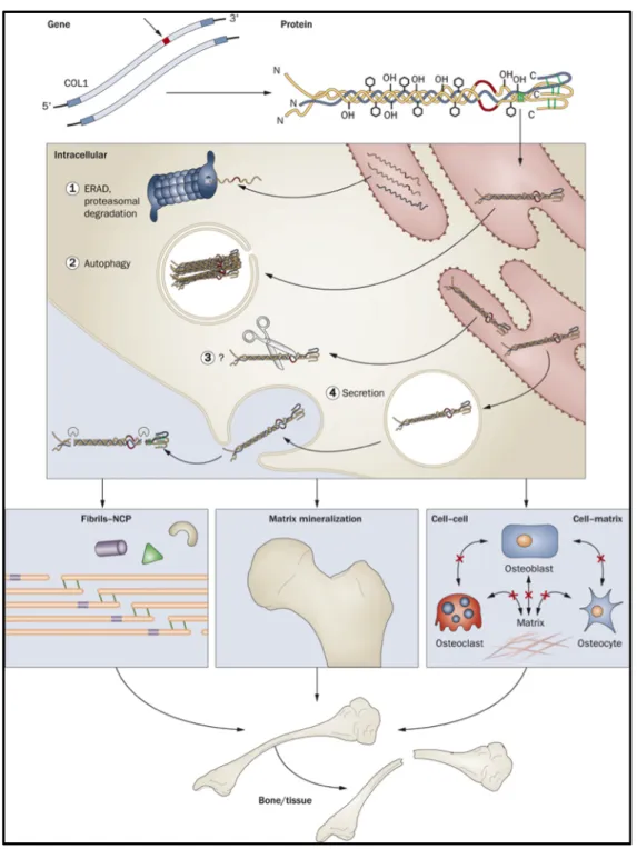
Type I collagen has a particular structure that is extremely important for proper functioning. This fibrous protein is initiated as procollagen, a triple helical molecule of three intertwined polypeptide chains, two pro-α1 chains and one pro-α2 chain. [6, 9] (Figure 1) These chains are composed of a repeating amino acid sequence, with the most important feature being a glycine molecule in every third position. Glycine, the smallest amino acid, is
essential since its small shape allows for the three pro-α polypeptide to join together as a well bound pro-collagen triple helix. [2, 5, 7, 10-12]
This triple helix is then exported to the endoplasmic reticulum where it is modified extensively by hydroxylation and glycosylation. [6] For example, cartilage-associated protein (CRTAP), cyclophilin B (CyPB) and prolyl hydroxylase (P3H1) come together as collagen 3-hydroxylation complex in order to hydroxylate the proline amino acid in each polypeptide chain and adequately fold the pro-collagen helix. [4, 9] The N- and C- propetides are then cleaved in the extracellular space, which then allows the mature tropocollagen molecules to assemble together and form collagen fibrils. [6]
Since type I collagen is the main protein in bone extracellular matrix, any aberrations in its synthesis can create an OI phenotype. [2, 6] Once a deficient collagen molecule is produced, it will either be degraded or incorporated into the body’s structures. The former will create a quantitative defect, while the latter will create a qualitative one. Defective tropocollagen protein secreted into the cell matrix not only affects fibrillogenesis and bone mineralization, but also cell-to-cell communication. [4, 11] In fact, the OI phenotype created by the various genetic mutations will depend on which protein structures are affected and which organs harbor these proteins. [13]
Inheritance
Over 800 mutations in the Type I collagen genes have been linked to the OI syndrome. [5, 11] The majority of patients (90%) with OI have an autosomal dominant mutation in the COL1A1 gene on chromosome 17 or COL1A2 gene on chromosome 7. [6, 9, 14] These genes encode the amino acid sequence in the pro-α1 chains and pro-α2 chains respectively. [2, 4, 6, 9, 10, 15] DNA mutations at these sites alter the structure and/or quantity of Type I collagen in the affected individual and the severity will range from clinically undetectable to lethal. [4] In addition to autosomal dominant forms of OI, there are autosomal recessive inheritance patterns that have also been discovered. [4, 6]
Type I OI, the mildest and most common form of the disease, is produced via a premature stop codon in the COL1A1 gene. The mRNA produced by the mutated allele will be degraded by nonsense-mediated decay, which leads to a haploinsuffuciency and diminishes the total amount of Type I collagen in the body. [2, 5, 11, 12] The result is a mild phenotype of the syndrome characterized by minimal bone fragility, blue sclera and hearing loss. [5, 9, 10]
Types II, III and IV, are caused by mutations leading to a distorted three-dimensional structure of type I procollagen. [5] The most common type of DNA mutation changes the amino acid sequence by substituting the obligatory glycine molecule for a bulkier amino acid. [2, 12] This substitution will impair the triple helical folding mechanism, as well as post-translational hydroxylation and glycosylation. In addition, improper splicing, deletions and insertions have also been discovered as other types of possible mutations. [6] Consequently, the amino acid sequence of the procollagen protein is altered which produces a less functional Type I collagen molecule. Mutations in the COL1A1 genes are more commonly lethal, whereas mutations in COL1A2 genes are 80% non-lethal. [5, 11]
In recent years, more types of OI have been discovered and added to the classification system. OI Types V, VI and VII are typically recessive in autosomal inheritance and usually produce severe phenotypes of the disease. [4, 6]
Clinical manifestations
The OI syndrome has a very broad phenotypic range. Patients with the same type of OI will present with different clinical manifestations and varying degrees of severity.
Bone Fragility
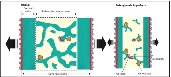
Bone fragility is the principal clinical characteristic in the OI syndrome, with its severity increasing in the following order: Type I < Type IV, V, VI, VII < Type III < Type II. [2, 16] Due to the abnormal collagen production, there are several disturbances in the organic and mineral compounds of the body, and these changes have a deleterious effect on bone mass, strength and stiffness. [2, 4, 16] The bone that is formed has an abnormal and irregular morphology, as well as an increased mineral density. [2, 4] Consequently, OI bone breaks much more easily when deformed even though the increased mineralization makes it harder in consistency. [2, 17] Furthermore, histomorphometric analysis of bone from OI patients revealed an increased number of osteoclasts and osteoblasts, an overall increase in the rate of
number of osteoblasts, each OI osteoblast secretes less bone than normal and bone resorption is amplified by the simultaneous increase in osteoclast numbers. [2, 4, 12, 16]
Figure 2: “Summary of histological bone abnormalities in OI : Osteogenesis imperfecta bone has a smaller than normal external size (bone thickness) because of sluggish periosteal bone formation. Trabeculae are reduced in number and are abnormally thin. Although individual osteoblasts produce less bone than normal, the overall
bone formation rate in the trabecular compartment is amplified, because the number of osteoblasts is raised. However this increase does not lead to a net gain in trabecular bone mass, because the activity of bone resorption
is also enhanced.” [2]
Consequently, the skeleton is more fragile and it fractures much more easily. There is an increase in number of lower limb fractures, deformity and bowing of long bones and scoliosis caused by vertebral crush fractures. [2, 9] (Figure 3)
Figure 3: “Bowing of the radius (a) and tibia (b) in a baby with OI type III” [10]
Growth
A decrease in height is one of the chief clinical characteristics in patients with OI. [4, 6, 18] Even children with OI Type I, who seem to be in the normal range, are often below the 50th percentile range. [6, 9] Patients with the more severe Type III OI have a very short stature, typically within 90 to 120 cm. [10] (Figure 4) In fact, Jensen & Lund have demonstrated that height is significantly reduced in patients with the more severe phenotypes of the disease, which suggests that height is a predictor of disease severity in the OI syndrome. [18]
Figure 4: Female with short stature and scoliosis; height = 93cm. [19]
Hearing Loss
Hearing loss is a common secondary feature in individuals with an autosomal dominant form of OI, which affects about 50% of adult patients. [20] This condition is caused by a combination of conductive and sensorineural defects and usually manifests itself between the second and fourth decade of life. [4, 6, 20] However, about 5% of children with OI have been found to show signs of early-onset hearing loss. Kuurila et al. recommend an audiometrical analysis be performed in children with osteogenesis imperfecta even without symptoms of hearing loss starting at the age of 10 years, with a repetition every 3 years. [20] No cases of hearing defects have been reported in patients with a recessive form of OI. [6]
Blue Sclera
Blue sclera is caused by abnormal collagen fibers in the sclera, which create the characteristic blue hue of the cornea. [21] Not all patients with OI present with blue sclerae. In cases of those who do, the scleral hue is variable. In Type I and Type III OI, blue sclerae are present at birth and persist throughout life, whereas in Type IV, the scleral hue becomes progressively normal with age. (Figure 5) In the perinatal-lethal form of OI, the sceral hue is often the darkest, sometimes even reaching a shade of black. [9]
Figure 5: Blue Sclera [14]
Oral Manifestations
There are several oral manifestations within the OI syndrome, the most widely reported feature being dentinogenesis imperfecta. Dentinogenesis imperfecta is a dental pathology characterized clinically by an amber-brown to blue grey opalescent hue of the teeth, thin/cracking enamel and severe attrition. (Figure 6) The radiographic findings include short roots, bulbous crown structure, obliteration of the pulp chamber and frequent peri-radicular radiolucencies. [22-24] The primary dentition is often affected more severely than the permanent dentition. [23, 25] Approximately 80% of patients with autosomal dominant OI have dentinogenesis imperfecta, but it is extremely rare in autosomal recessive types of OI. [6, 23, 26]
Figure 6: Dentinogenesis imperfecta of primary dentition (courtesy of the Montreal Children’s Hospital)
Many authors have described dentinogenesis imperfecta in detail, but very few have documented another important facet of the OI syndrome: the dental malocclusion. A study by Rizkallah et al. in 2012 revealed that the malocclusions present in the OI population are more severe than those present in the general population. They confirmed that there is a distinct over-representation of Class III malocclusion, negative overjet and lateral openbite in patients with OI. [27] (Figure 7)
Figure 7: OI patient exhibiting Class III malocclusion, anterior openbite, lateral posterior crossbite and opalescent teeth [27]
Diagnosis
The diagnosis of OI is usually done on a clinical basis. It is often clear in individuals with a positive family history in whom several cardinal manifestations are present. However, it can be quite difficult when other family members are unaffected and bone fragility is the only
indication of the syndrome. In addition, there is no agreed minimum number of criteria that can establish a clinical diagnosis of the syndrome. [2] Thus, practitioners typical rely on a complete clinical examination as well as genetic work-up to establish a diagnosis of the disease. DNA analysis of the genes involved in the biosynthesis of Type I collagen can be performed and are highly sensitive. [2, 9] However, results that do not detect a genetic mutation in COL1A1, COL1A2, CRTAP and SERPINF1 do not rule out a diagnosis of OI. [2]
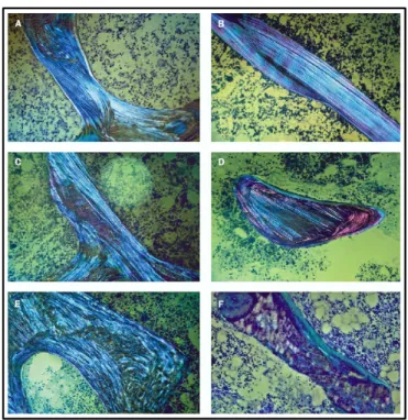
If a diagnosis of OI seems possible but is inconclusive with a mere clinical exam and genetic analysis, other analyses can be done. Radiographs can detect bowing of the long bones, presence of crush vertebral factures and scoliosis. [9] Bone histomorphometry can help distinguish OI from other osteoporotic conditions. In addition, when polarized light microscopy is used, OI types V and VI can be diagnosed. [2, 9] (Figure 8) However, this invasive method is usually avoided whenever possible since it depends upon a bone biopsy retrieved under general anesthesia. [9]
Figure 8: “Bone lamellation pattern as seen under polarized light (A) Healthy control. (B) OI type I; lamellae are thinner than normal, but lamellation is smooth. (C) OI Type III; lamellation is slightly irregular. (D) OI type IV;
There are a variety of differential diagnoses available for OI, which depend on the age of presentation, clinical manifestations and the severity of the signs and symptoms. [9] (summarized in Table 1)
Table 1: Differential diagnosis of osteogenesis imperfecta [2, 9]
Condition Childhood phenotype Inheritance Pathophysiology
Non-accidental
injury Multiple unexplained childhood fractures -- Pathologic fractures Bruck syndrome Moderate to sever. Congenital joint
contractures; scoliosis; white sclerae AR Deficiency of telopeptide lysyl hydroxylase Cole-Carpenter syndrome
Severe. Normal at birth; short stature; osteoporosis; diaphyseal fractures; hydrocephalus; ocular proptosis; distinctive facial features
Uncertain No abnormality of type I collagen
Hypophosphatasia Mild to severe. Low alkaline phosphatase activity; very variable clinical expression; early loss of teeth
AD/AR Mutation in ALPL
Idiopathic
hyperphosphatasia or juvenile Paget disease
Severe. Raised alkaline phosphatase activity; very variable phenotype; thickened skull; widened diaphysis; progressive deformity; scoliosis; deafness AR Osteoprotegerin deficiency due to mutation in TNFRSF11B in the majority of cases Panostotic fibrous dysplasia
Sever. Characteristic lesions in all bones.
Somatic Somatic mutation in GNAS
Osteoporosis pseudoglioma syndrome
Moderately severe. Congenital blindness; torus palatinus
AR Mutation in LRP5
Idiopathic juvenile
osteoporosis Mild to moderately sever. Transient osteoporosis; prepubertal presentation; metaphyseal fractures; neo-osseous osteoporosis; no extraskeletal manifestations
Uncertain Unknown etiology in the majority of cases; sometimes associated with heterozygous mutation in LRP5
Classification
The classification of OI has proven to be quite difficult, given that the syndrome is highly heterogeneous. In 1979, Sillence et al. categorized OI patients into four types based on clinical presentation. [28] In recent years, there has been further subdivision of these four types according to genetic factors and bone histology techniques. (Table 2) [2, 3] Presently, eleven types of OI have been described. [4, 9] These subdivisions are mainly based on differences in the genetic factors leading to the disease, even though the clinical phenotype may be very similar to the pre-existing OI types. In other words, OI Type V, VI and VII are very often clinically undistinguishable from OI Type IV. [29] Some authors have stated that this intermingling of genetic and clinical classification is very confusing and problematic. [6, 29] For the purpose of simplicity, the classification system used in this paper will be from The Lancet’s 2004 seminar on osteogenesis imperfecta. [2]
Table 2: Expanded Sillence classification of osteogenesis imperfecta [2-4, 6, 9] Type Clinical
severity
Mode of inheritance
Typical features Typically
associated mutations* I Mild non-deforming osteogenesis imperfecta
AD Normal height or mild short stature; blue sclera; no dentinogenesis imperfect Premature stop codon in COL1A1 II Perinatal lethal
AD Multiple rib and long-bone fractures at birth; pronounced deformities; broad long bones; low density of skull bones on radiographs; dark sclera
Glycine substitutions in COL1A1 or COL1A2
III Severely
deforming AD Very short; triangular face; severe scoliosis; greyish sclera; dentinogenesis imperfect Glycine substitutions in COL1A1 or COL1A2 IV Moderately
V Moderately deforming
AR? Mild to moderate short stature; dislocation of radial head;
distinctive histology; mineralised interosseous membrane;
hyperplastic callus; white sclera; no dentinogenesis imperfecta
Unknown
VI Moderately to severely deforming
AR Moderately short; scoliosis; accumulation of osteoid in bone tissue, fish-scale pattern of bone lamellation; white sclera; no dentinogenesis imperfecta
Homozygous SERPINF1 mutations
VII Moderately
deforming AR Mild short stature; short humeri and femora; white sclera; no dentinogenesis imperfecta
Homozygous CRTAP mutations *May or may not be detectable in a given patient
Treatment
The medical management of patients diagnosed with OI is a multidisciplinary approach that combines physical therapy, orthopedic surgery and pharmacotherapy. Treatments are usually focused on improving functional ability by decreasing bone fragility and increasing mobility in patients with more severe OI phenotypes. [30, 31] The key to successfully treat these patients is to combine a multidisciplinary approach with early diagnosis. [5, 31]
Physical Therapy
The primary goal of physical therapy is to improve motor function and reduce immobility-induced bone-loss. [30] Many children with severe phenotypes of OI have very limited mobility and are often wheelchair-bound. These patients benefit greatly from physical therapy, especially in the lower extremities.
Orthopedic surgery
Orthopedic surgery is very common in the treatment of severe OI phenotypes. Surgical intervention often consists of the placement of intramedullary rods in the long bones to correct
deformities and stabilize the bone. [4, 30] This kind of corrective surgery enables walking and improves overall mobility in OI patients. [4]
Pharmacotherapy
Pharmocotherapy is currently the most widely used medical intervention for children with moderate to severe OI. [10] The ultimate goal is to reduce fracture numbers, prevent long bone deformities and increase functional mobility. [2] The most popular medication used today are bisphosphonates, but new advances in the use of recombinant human growth factor have also been made in recent years. [13]
Bisphosphonates have an inhibitory effect on osteoclasts and therefore, decrease bone resorption. [2, 10, 13, 31] When treated with this medication, the quality of the new bone formed does not improve, but the skeleton does benefit from an increase in bone volume and an overall increase in mechanical strength. [10, 31] The most popular bisphosphonate used today is cyclic intravenous pamidronate, given in cycles of three days every two to four months. [2, 10] In a study by Glorieux et al. in 1998, more than 50% of the OI patients treated with cyclic pamidronate had an improvement in mobility, as well as a decrease in overall fracture rate. [31] In the moderate to severe OI phenotypes, bisphosphonate therapy should be started as early as possible in order to benefit from the growth process. On the other hand, mild forms of OI should not receive bisphosphonate therapy as the negative side effects (abnormal bone metabolism, risk of osteonecrosis, etc.) may outweigh the benefits of this treatment modality. [2, 10, 13]
Growth hormone has also been studied in the past as a treatment for the OI syndrome, given that short stature is one of its more common features. [32] In a study in 2003, Marini et al. revealed that the baseline growth rate of about 50% of the OI patients treated with recombinant human growth hormone doubled during the first year of treatment. [4, 33] This study also showed that bone turnover rates were increased, a secondary effect that is potentially harmful to OI patients. [33] For this reason, it is probably beneficial to combine recombinant human growth hormone with bisphosphonate therapy, but this treatment still
Conclusion
Osteogenesis imperfecta (OI) is a complex genetically inherited disorder characterized by defects in biosynthesis of Type I collagen. Consequently, individuals affected by OI are susceptible to spontaneous bone fractures, long bone deformities, short stature, hearing impairments, muscle weakness, blue sclera, dentinogenesis imperfecta and/or dental malocclusion. [2] It is well known that the OI syndrome is caused by alterations in the collagen type I biosynthesis pathway, but the exact mechanism by which the genetic defects cause abnormal bone formation have not been described. [16] A better understanding of the disease pathway and the resulting phenotypic expression can aid in the diagnosis, classification and the efficient treatment of this debilitating disease. Great advances have been made in the understanding of the OI syndrome and the help that is now available to these patients is extensive; however, the morbidity of the more severe phenotypes is still astounding. No cure for the disease has been found, so more must be done in order to optimize their mobility, autonomy and quality of life. [10, 13]
Malocclusion
Overview
The study of orthodontics and the treatment of malocclusion have become increasingly popular in the last century. However, malocclusion has been a part of human history since antiquity and attempts at correcting misaligned, protruding, and irregular teeth have been documented since 1000 B.C. Dentistry has come a long way since the 18th and 19th centuries, and the same is true for orthodontics, the branch of dentistry focused on correcting malocclusion. In the 1890’s, Edward H. Angle, the so-called “father of orthodontics”, subdivided the major types of malocclusion and proposed a classification system that is still widely used today. [34]
Classification
A classification system proposed by Angle was based on the position of the upper first molar. Angle believed that in order to have a normal occlusion, two cardinal features must be present. First, the mesiobuccal cusp of the upper first molar must occlude with the buccal groove of the lower molar (Figure 9) and second, all the teeth must be arranged on a standard smoothly-curving line of occlusion. [34] (Figure 10)
Figure 9: Mesiobuccal cusp of upper first molar occludes with buccal groove of lower first molar [34]
Angle then described three distinct classes of malocclusion based on aberrations in the two previous features.
Class I Malocclusion
Mesiobuccal cusp of upper first molar occludes with buccal groove of the lower first molar, but the teeth are not well aligned on the line of occlusion. (Figure 11)
Figure 11: Class I malocclusion [34]
Class II Malocclusion
Mesiobuccal cusp of upper first molar positioned mesial relative to the lower first molar; teeth may or may not be well aligned on the line of occlusion. (Figure 12)
Class III Malocclusion
Mesiobuccal cusp of upper first molar positioned distal relative to the lower first molar; teeth may or may not be well aligned on the line of occlusion. (Figure 13)
Figure 13:Class III malocclusion [34]
Class I malocclusions (69.7%) are the most common, followed by Class II malocclusions (23.8%) and finally, a small number of the population possess a Class III malocclusion (6.5%). [34-36] Many studies have been done on the incidence of malocclusion in different populations. The previous percentages are limited to the American Caucasian population. [36]
Terminology
Other important features of a malocclusion that are not part of Angle’s classification system are as follows: anterior and posterior crossbites, anterior and posterior openbites, increased overbite and increased overjet.
Overjet
Overjet is the horizontal measurement between the maxillary and mandibular incisors. (Figure 14) A normal overjet is typically 2mm when measuring from the labial surface of the mandibular incisor to the labial surface of the maxillary incisor. A negative overjet or anterior crossbite will result if the mandibular incisors are anterior to the maxillary incisors. [34]
Figure 14: Overjet between the maxillary and mandibular incisors [34]
A posterior crossbite would result if the posterior overjet is reversed. More specifically, to have a posterior crossbite, the maxillary posterior teeth would be positioned more lingual than the mandibular posterior teeth. A posterior crossbite can be unilateral or bilateral.
Overbite
Overbite is the vertical measurement between the upper and lower incisors. (Figure 15) Normally, the maxillary incisors overlap the mandibular teeth by 2mm. [34]
Figure 15:Overbite between the maxillary and mandibular incisors [34]
When a space exists between the maxillary and mandibular teeth, this is referred to as an openbite. (Figure 16) An openbite can be anterior, posterior, unilateral or bilateral.
Figure 16: The presence of an anterior openbite [34]
In order to make Angle’s classification more complete, Mills suggested the addition of more information as an aid in describing the various malocclusions that exist. (Summarized in Table 3) [35]
Table 3: Expanded Angle Classification [35]
Class Type Clinical Characteristics
I I Crowded incisors; the canines are frequently labial II Protrusion or labioversion of the maxillary incisors III Presence of anterior crossbite
IV Presence of posterior crossbite
V Mesial drifting of molars resulting from premature loss of teeth
II Division 1 Proclined maxillary incisors (labioversion) Division 2 Retroclined maxillary central incisors
III I Maxillary and mandibular teeth in good alignment, but incisors in edge-to-edge occlusion
II Maxillary teeth in good alignment, mandibular teeth crowded; mandibular incisors lingual to maxillary teeth
Etiology
As previously stated, Angle’s classification of the 3 types of malocclusion is highly simplified. He fails to take into account several important components critical in assessing the etiology of the malocclusion, which are: “(a) the size of the maxilla, (b) the size of the mandible, body and ramus, (c) the factors that determine the relationship between the maxilla and mandible, which are genetic and environmental, (d) the arch form, (e) the size and morphology of the teeth, (f) the number of teeth present, and (g) the soft tissue morphology.” [37]
A malocclusion can have a skeletal etiology, a dentoalveolar etiology, or a combination of the two. A skeletal malocclusion is one in which there is an aberrant relationship between the maxilla, mandible and/or cranial base. A dentoalveolar malocclusion can be due to an abnormal arch form, abnormal tooth size and/or missing teeth. All malocclusions result from a combination of genetic and environmental factors which affect craniofacial growth, arch size, tooth size, tooth loss, etc. [34, 37] Diagnosing the etiology of the malocclusion is essential in providing the best treatment.
Severity
Angle’s “normal occlusion” is actually quite rare in the human population and should be considered as the ideal. [34] Furthermore, great variability exists when measuring the incidence of malocclusion in our society. There are several reasons for the lack of consensus; there is a wide divergence in the definition of a normal and abnormal occlusion, the presence of diagnostic error and a lack of a universal index. [35] In recent years, several indices have been developed to assess the presence and severity of the malocclusions in the general population. The American Board of Orthodontics uses the discrepancy index. [38]
The discrepancy index (DI) is used as a means of quantifying the severity of malocclusions. Normally, assessing a malocclusion can be very subjective. With the use of the discrepancy index, various easily measured clinical components are tabulated to give an overall severity score. Cephalometric radiographs, panoramic radiographs and dental casts with a proper bite registration are needed in order to take measurements of the following: overjet, overbite, open bite, crowding, occlusal relationship, crossbite, presence of missing
teeth, and several cephalometric values. The more the measurements differ from ideal in each component category, the greater the complexity and severity of the overall malocclusion. [38]
Treatment
The presence of a malocclusion does not necessarily mean that the individual requires treatment. When assessing malocclusion, the discrepancy index can be very useful in quantifying the severity and difficulty expected for the orthodontist. An orthodontic treatment is usually reserved for the more severe malocclusions or when patients are not content with their oral functions and/or dental esthetics. There are several treatment modalities available when correcting a malocclusion based on its severity and etiology. The three main options are: traditional orthodontics, orthognathic surgery and a combination of the two. Osteodistraction can also be used in severe craniofacial anomalies when surgical movements will be extreme.
Malocclusion in patients with osteogenesis imperfecta
Overview
Osteogenesis imperfecta is a syndrome that has been characterised with a high prevalence of malocclusion. [26, 39] Several authors have reported an increased development of Class III molar relationships, posterior and anterior openbites, crossbites and impacted teeth. [25-27] The precise etiology of the malocclusion is not yet known; however, it seems that the abnormal craniofacial growth present in OI patients results in aberrations in the relationship of the upper and lower jaws, dental arches and teeth. [40-43] Several authors have reported that the more severe phenotypes of OI seem to produce the more severe craniofacial abnormalities, but this hypothesis does not always seem to be true and merits an investigation. [18, 25, 40] Mild forms of OI are often very hard to diagnose and an investigation of the dental malocclusion may prove to be a cardinal diagnostic aid. [23, 44]
Clinical characteristics
Considerable variation exists in the expression of the dental phenotype within the different types of OI. [25] A different combination of dental characteristics and severity will be present in each individual and the reasons for these differences are still unknown. Although, there is considerable variation, there are several craniofacial and dentoalveolar features that have a high prevalence in patients with OI.
Craniofacial features
Patients with OI usually have a triangular-shaped face, a broad forehead, macrocephaly and basilar invagination. [19, 26, 45] (Figure 17)
Figure 17: Patient with OI exhibiting a triangular shaped face, broad forehead and midface hypoplasia [45]
Due to these changes in growth and associated abnormal posture, panoramic and cephalometric radiographs may be very difficult to obtain. [46] There seems to be a higher incidence of craniofacial disproportion in Types III and IV than in Type I. [18, 43, 45] Nevertheless, facial bones were found to be smaller than normal in all types of the syndrome. [43] More specifically, vertical facial dimensions, maxillary and mandibular lengths and the anterior/posterior cranial base lengths are all significantly shorter in OI patients. [41] The high proportion of Class III skeletal malocclusion is characterized primarily by midface hypoplasia. [25, 41-43] According to Waltimo-Siren et al, “the growth deficiency was more pronounced in the severely affected patients than in those with type I OI”. [43]
Dentoalveolar characteristics
Dental malocclusion is very common in patients with OI and has proven to be significantly more severe than that found in the general population. [27] There is an increased presence of Class III malocclusions, further compounded by a negative overjet, posterior crossbites and lateral openbites. [25-27]
have found the incidence of Class III malocclusion to be closer to 62.5%-80%, which is significantly greater than the 6.5% previously found in the general population. [25, 26, 35, 36, 41] The only study with a 9.6% incidence of Class III malocclusion was composed of a sample with 79% of the milder type I form of OI. [47] Other studies that found a higher incidence of malocclusion, also had a higher percentage of the more severe OI phenotypes in their sample. [25, 26, 41] These results support the theory that the more severe phenotypes of OI seem to produce more severe craniofacial abnormalities.
In addition to the high proportions of Class III malocclusion in the OI population, there is also a high degree of lateral openbite and posterior crossbite. [27] Interestingly enough, lateral openbites are extremely rare in the general population. [26, 27] However, O’Connell and Marini found a 27% incidence of lateral openbite in Type III OI patients and 33% in patients with type IV. He also observed that the severity of the lateral openbite seems to increase with age. Within the 40 patient sample, none of the children younger than 9 years of age experienced posterior openbites whereas 46% had a either unilateral or bilateral posterior openbite after that age. [26]
Other features are also commonly found in the OI population. Posterior crossbites were present in 65% of a heterogeneous OI sample by Schwartz & Tsipouras, whereas O’Connell and Marini found 38% of Type III OI patients and 47% of Type IV OI patients possessed this debilitating feature. [25, 26] OI patients also have an increased number of impacted teeth, permanent tooth agenesis and a high prevalence of ectopic eruption of first and second molars. [23, 25-27, 39, 41, 47]
Etiology
The same bony defects that cause malformed long bones in OI patients seem to also affect the craniofacial complex. There is an aberrant growth pattern of the upper and lower jaws that results in abnormal facial characteristics and severe malocclusion. [42] The typical craniofacial features observed are: triangular face, maxillary hypoplasia, mandibular prognathism, basilar invagination and broad forehead. [18, 26, 43] The etiology of these facial characteristics seems to be the increased bone fragility, which changes bone morphology, function and subsequently the growth pattern in these patients. [43]
The inability of the poor-quality bone to withstand the weight of the brain and/or head causes changes in bone morphology and its growth pattern. [18] According to Moss’ functional matrix theory, growth is highly dependent on function and any change in the latter will have considerable consequences on the former. [48] In OI patients, the weight of the head on the osteoporotic bone of the cervical area creates a basilar invagination, which will change the patient’s posture and functional capacities. Subsequently, growth in the craniofacial complex will be altered. [18, 26, 43] The result will depend on the phenotypic severity of the OI syndrome. [17, 39, 40, 49, 50]
OI patients develop more severe malocclusions than the general population. [27] Researchers claim that the typical malocclusion associated with the syndrome is primarily caused by maxillary hypoplasia, mandibular protrusion or a combination of the two. [19, 25, 26, 39, 41, 42, 51] When examining dental casts and cephalometric radiographs of OI patients, the maxilla is usually short in length and the mandibule is either normal in length or slightly shorter. These findings lead to the assumption that the Class III skeletal pattern is primarily due to a midface hypoplasia. [42, 43] Other features of the craniofacial growth include: a strong closing growth rotation of the mandible, a short condyle, underdeveloped alveolar bones in both jaws, and a decrease in vertical lower face height. The decrease in vertical growth seems to be the most pronounced growth abnormality in the craniofacial aspect of the syndrome. [43] Therefore, the Class III dentoskeletal malocclusion present in OI patients seems to be caused by a smaller than normal upper and lower jaw, an increased closing growth rotation of the mandible and a decreased vertical facial development. [41, 43]
In addition to a Class III malocclusion, there is a high incidence of lateral openbites, anterior/posterior crossbites and impacted teeth. Lateral openbites are extremely rare in the general population, and can be particularly debilitating in OI patients. The high incidence of this feature in the OI population could be explained by an abnormal vertical dentoalveolar development and a lack of dental compensation. [26] The high incidence of anterior and posterior crossbites seems to be created by the disharmony resulting from a hypoplastic maxilla opposed by a normal mandible. [42]
the variable craniofacial phenotypes of this syndrome. Longitudinal prospective studies in this area of the OI syndrome are needed. [26]
Treatment
Despite the frequent functional and esthetic dental aberrations in OI patients, major treatments such as bone augmentation, orthodontics and orthognathic surgery are extremely rare in this population. [51] Very little has been published about orthodontic treatment in these patients and the only type of data available is in case report format. There are no controlled trials researching the outcomes of orthodontic treatments with or without orthognathic surgery in this patient population, nor is there any focusing on the variability in orthodontic outcomes in OI patients treated with bisphosphonates. [46] Nevertheless, the severe malocclusions seen in patients with this syndrome usually requires a combined orthodontic/surgical approach. [45] Furthermore, some authors have recently succeeded in doing osteodistraction and bone augmentation in these patients despite the morbidity of osseous surgery associated with the OI syndrome. [51]
Traditional orthodontics
Orthodontic therapy seems to be possible in the OI population, but there are only a few documented cases in the literature. More knowledge on this subject could be very useful because there are many facets of the OI syndrome that can become problematic during an orthodontic treatment. More specifically, the poor quality of the bone, the presence of dentinogenesis imperfecta and treatment with bisphosphonates may make traditional orthodontics very difficult.
Successful orthodontic therapy is dependent on the bone remodeling process. In order for tooth movement to be effective, osteoblasts and osteoclasts need to perform their role adequately around the periodontal ligament. Osteoblasts are the bone-forming cells, while osteoclasts are the bone-resorbing cells. The cycle of bone formation and resorbtion provides the very important balance usually seen in bone remodeling. [34] As previously stated, osteoblasts in OI patients do not function normally and the quality of the bone formed is poor. [2] In addition, in patients treated with bisphosphonates, osteoclast activity is inhibited. [31,
34] Thus, the bone remodelling process is disrupted on many levels in the OI patient treated with bisphosphonates. Since bone remodeling is essential for orthodontic therapy, the changes in bone metabolism can decrease the amount and rate of tooth movement achieved in these patients. [52] The magnitude and duration of the orthodontic forces may have to be adjusted in order to counterbalance the deficiency in collagen production created by the syndrome and the decreased bone resorption caused by bisphosphonate therapy. [39]
Patients with OI combined with dentinogenesis imperfecta may also present another challenge for the orthodontist. Due to a qualitative abnormality in the dentin, the enamel in patients with a dentinogenesis imperfecta phenotype usually fractures very easily. [23, 26] These fractures can cause significant amounts of attrition and can lead to early tooth loss. [26] The adhesive forces of orthodontic bracketing may not be strong enough to withstand normal intra-oral forces and regular debonding can cause significant damage to these teeth. Bands with welded brackets can be used when the enamel is not strong enough for regular bracketing procedures. [46] Dentinogenesis imperfecta may complicate orthodontic therapy, but it is definitely still possible in these patients.
In the future, researchers should document more cases of orthodontic therapy in this patient population. Recent functional therapy with a Frankel Type III appliance has shown promising results. [43] If appliance therapy is successful and started at the right time, the need for orthognathic surgery may decrease in patients with OI.
Orthognathic surgery
Orthognathic surgery, such as maxillary advancement and mandibular set-back, has been successful in many patients with OI and is often combined with traditional orthodontics. A diagnosis with this syndrome does not seem to be a contraindication for a surgical intervention; however, great care must be taken when treatment planning, explaining the procedure to the patient and obtaining informed consent. [46, 53] Several authors have written case reports detailing maxillofacial surgery on OI patients, and claim to have achieved acceptable results, improving both function and esthetics. [40, 46, 49, 50, 53, 54] (Figure 18
and respiratory distress. [44, 46, 49] Remarkably, the most severe sequelae do not seem to correspond to the most severe clinical phenotypes of the syndrome. [46, 49, 50] Complications seem to arise more when performing a Le Fort I maxillary osteotomy when compared to mandibular procedures. To help prevent a negative surgical experience, it is very important to properly diagnose a patient with OI, especially with a mild phenotype, and incorporate this
Figure 19: Post-orthognathic surgery photos in a woman with OI and a Class III malocclusion [46]
diagnosis in the treatment planning process. [53] Great care must be taken to minimize the tendency for unfavourable fractures due to poor bone quality and thin maxillary and mandibular walls. [53, 55]
Despite the increased bone fragility in OI patients, previous studies have shown that there is adequate bone healing after orthognathic surgery. [19, 40, 51] However, Rauch et al. have demonstrated that abnormal bone remodelling exists in all OI patients. [16] Consequently, Tashima et al. recommend intermaxillary fixation for up to 5 to 6 weeks to prevent fracture during the healing period. [53] Successful surgery in OI patients is definitely possible, but the surgeon must keep in mind that the chances of encountering complications are more common in this subset of patients. [17, 53, 54]
[51] Since most complications occur during the down-fracture of a Le Fort I osteotomy, osteodistraction has been proposed to replace this part of the surgery. [51, 53] Bone biopsy 6 months after osteodistraction and 4 months after augmentation showed adequate callus formation and good bone healing. These results are very promising when trying to diminish complications observed during orthognathic surgery. [51]
Conclusion
Patients with OI have been identified as a “high-risk group for the development of malocclusion”. [26, 39] There is a higher prevalence of Class III molar occlusion, posterior and anterior openbite, crossbite and impacted teeth in these patients. [25-27] In addition, considerable variation exists in the expression of the dental and craniofacial phenotype within the different types of OI as well as in patients with the same collagen type I abnormality. [18, 25] Due to the extreme variability in the phenotype of the syndrome, every time an OI patient is treated, he/she should be viewed as a unique case with a wide range of possible treatment options. [46] Whatever treatment is chosen, a multidisciplinary approach should be adopted in order to optimize function and esthetics, as well as minimize any adverse consequences. [45] More importantly, diagnosis of the milder OI type I patients must be done prior to any decision with regards to treatment, in order to prevent any avoidable adverse consequences. In the future, the characteristic malocclusion present in OI patients may be used as an additional diagnostic feature.
Hypotheses and study aims
Purpose
Osteogenesis imperfecta is a connective tissue disorder characterized by a hereditary defect in the production of Type I collagen. [2, 5] Patients affected by OI are susceptible to spontaneous bone fractures and may show a number of other defects, such as joint laxity, hearing impairments, malformed long bones, growth deficiency, muscle weakness, blue sclera, dentinogenesis imperfecta and/or malocclusion. The syndrome is extremely heterogeneous, producing a variety of phenotypes ranging from clinically undetectable to perinatal lethal. [2] In fact, the milder forms of the syndrome are sometimes extremely difficult to diagnose; thus, more must be done to aid the diagnosis and classification of this debilitating disease.
It seems that the same disease pathway that creates the severe skeletal deformity in these patients, also creates growth disturbances in the craniofacial complexe. [18, 39] Consequently, there is an increased incidence of Class III molar occlusion, posterior and anterior openbite, and posterior and anterior crossbite. [25-27] The aim of this study is to evaluate whether the severity of the malocclusion present in the OI population is proportional to the severity of the systemic bone disease. If so, the malocclusion present may serve an important diagnostic role when trying to identify and classify the multiple forms of this syndrome.
Study aims
The primary aim of this study is to compare the severity of the malocclusion present in OI patients with the severity of their systemic bone disorder. The study aim will be achieved by fulfilling the two following objectives:
1. The malocclusion observed in a retrospective examination of OI patients will be quantified using the Discrepancy Index at one point in time.
2. The severity of the malocclusion quantified with the DI will be compared to other variables that characterize the severity of the OI syndrome (OI type, height z-score, type of genetic mutation)
The secondary aim of this study is to evaluate whether the severity of the malocclusion present in OI patients changes with time. The secondary study aim will be achieved by fulfilling the following objective:
1. The discrepancy index for a subgroup of OI patients will be tabulated at several points in time in order to determine the longitudinal progression of the malocclusion.
Hypothesis
The hypothesis for the primary study aim is as follows:
Research Hypothesis: The severity of the malocclusion observed in OI is related to the
severity of the systemic bone involvement. As the severity of the malocclusion increases, there will be :
• A decrease in height
• An increase in the presence of the more severe types of OI (Type I < Type IV, V, VI, VII < Type III < Type II)
• An increase in the more severe genetic mutations
Null Hypothesis: The severity of malocclusion in OI is unrelated to the severity of the
systemic bone involvement.
The hypothesis for the secondary study aim is as follows:
Research Hypothesis: The severity of malocclusion in OI increases as the child ages. Null Hypothesis: The severity of malocclusion in OI does not increase with time.
Materials and methods
Study design and setting
A retrospective observational study was conducted involving 3 administrative facilities: the Montreal Shriners Hospital, the Montreal Children’s Hospital, and the University of Montreal. The physicians at the Shriners Hospital follow the largest cohort of patients with OI in North America. Consenting patients seen at the Shriners are referred to the Montreal Children’s Hospital dental clinic, where complete dental examination is done and required treatment is provided. Consequently, over the past decade, the dental clinic has documented oral records for over 70 OI patients. Whenever possible and/or appropriate, intra-oral and extra-oral findings, panoramic and cephalometric radiographs, intra- and extra-oral photos, and dental casts with bite registration were obtained.
The ethics review board of the Montreal Children’s Hospital approved the study (study 13-486-PED). Scientific merit and approval was given by the review board at the University of Montreal. Informed consent was obtained from the legal guardians and/or patients when age-appropriate.
Subjects
The study population consists of 73 patients with varying types of OI who were referred to the Montreal Children’s Hospital by the Shriners Hospital between the period of July 2006 and July 2013.
Inclusion criteria for the study sample were defined as:
• definite diagnosis of OI
• regular follow-up at the Montreal Shriners Hospital • adequate casts and radiographs available
Exclusion criteria were set as:
• a history of orthodontic treatment • a history of orthognathic surgery
• presence of an inadequate bite registration • several missing teeth (>8 missing teeth)
All OI patients had received or were still receiving intravenous bisphosphonate therapy.
Variables
Information regarding the following variables was collected: • Age
• Gender
• Presence of dentinogenesis imperfecta • Height z-score
• OI type • Mutated gene
• Type of genetic mutation
• Position of amino acid substitution • Molar relation
• Discrepancy Index (DI)
All information, except for the molar relationship and DI score, was collected through chart review at the Shriners Hospital in Montreal. Height measurements had been converted to age- and sex-specific Z-scores on the basis of reference data published by the Centers for Disease Control and Prevention. [12, 56] For the genetic mutation type, genomic DNA had been isolated in the laboratory of Dr. Frank Rauch at the Montreal Shriners Hospital from either blood or saliva using standard extraction methods. [12] The types of genetic mutations identified in this sample were: triple helical defect, protein complex defect, splice mutation,
stop codon, or was unidentified. Sequencing had been done with a polymerase chain reaction using primers. Helical mutations were numbered according to the position of the mutated amino acid within the triple helix of each alpha chain. [12] The mutated genes identified in this sample were: COL1A1, COL1A2, CRTAP, SERPINF1 or was unidentified. The OI classification by type had been done by the Montreal Shriners Hospital based on clinical, radiographic and genetic information. The types of OI present in this sample were: type I, III, IV, VI.
Molar relationship was determined while calculating the DI of the malocclusion using each patient’s dental cast at the Montreal Children’s Hospital. The severity of the malocclusion was assessed using the DI scoring system by evaluating the overjet, overbite, open bite, crowding, occlusal relationship, crossbite and presence of missing teeth. [38] The cephalometric component of the DI was not included in the analysis as lateral cephalograms were not possible for patients with severe craniofacial deformations. The DI scores for each component were measured by one investigator, and then tabulated to give a final DI score of the malocclusion. (Figure 20) Measurements were taken using a Carrera Precision CP7908 8-inch fractional digital LCD caliper from Whitworth.
Statistical analysis
Descriptive statistics are presented for demographic and clinical characteristics. DI measurements in 56 OI patients at one point in time are presented as medians for continuous variables and proportions for categorical variables. Statistical significance was established as p < 0.05.
For the primary aim, DI measurements were compared with height z-score, genetic mutation and classification type for univariate analysis with one-way ANOVA if the data followed a gaussian distribution and via Spearman correlation, Mann Whitney U test and Kruskal-Wallis test for non-normally distributed data. Bonferoni adjusted p-values were calculated when indicated.
For the secondary aim, Spearman correlations for repeated measures were used to characterize the change over time in a sample of 20 OI patients measured at two points.
The intra and inter reliability of the quantified malocclusion (Discrepancy Index) were assessed by calculating the Kappa score. Fifteen subjects were measured twice by the same rater (intra-rater reliability) and twenty-nine subjects were measured once by another rater (inter-rater reliability).
All calculations were performed using Statistical package for social sciences (SPSS, version 20.0, Chicago, IL, USA)
Results
Of the 73 OI patients referred to the Montreal Children’s Dental Clinic between July 2006 and July 2013, 56 fulfilled the eligibility criteria. (Figure 21)
Figure 21:Inclusion and exclusion criteria for sample size
73 patients
accepted to have a dental examination
and consented to participate in the
study 13 patients = inadequate bite registration 1 patient = > 8 missing teeth 2 patients = inadequate follow-‐up 1 patient = mistaken diagnosis Sample size =
![Figure 3: “Bowing of the radius (a) and tibia (b) in a baby with OI type III” [10]](https://thumb-eu.123doks.com/thumbv2/123doknet/11590671.298693/18.918.233.688.115.594/figure-bowing-radius-tibia-baby-oi-type-iii.webp)
![Figure 4: Female with short stature and scoliosis; height = 93cm. [19]](https://thumb-eu.123doks.com/thumbv2/123doknet/11590671.298693/19.918.138.783.117.586/figure-female-short-stature-scoliosis-height-cm.webp)
![Figure 5: Blue Sclera [14]](https://thumb-eu.123doks.com/thumbv2/123doknet/11590671.298693/20.918.302.620.371.592/figure-blue-sclera.webp)
![Figure 7: OI patient exhibiting Class III malocclusion, anterior openbite, lateral posterior crossbite and opalescent teeth [27]](https://thumb-eu.123doks.com/thumbv2/123doknet/11590671.298693/21.918.263.655.598.820/figure-exhibiting-malocclusion-anterior-openbite-posterior-crossbite-opalescent.webp)
![Table 2: Expanded Sillence classification of osteogenesis imperfecta [2-4, 6, 9]](https://thumb-eu.123doks.com/thumbv2/123doknet/11590671.298693/24.918.122.783.582.1064/table-expanded-sillence-classification-osteogenesis-imperfecta.webp)
![Figure 9: Mesiobuccal cusp of upper first molar occludes with buccal groove of lower first molar [34]](https://thumb-eu.123doks.com/thumbv2/123doknet/11590671.298693/28.918.573.751.693.1002/figure-mesiobuccal-upper-molar-occludes-buccal-groove-lower.webp)
![Figure 13: Class III malocclusion [34]](https://thumb-eu.123doks.com/thumbv2/123doknet/11590671.298693/30.918.300.619.255.422/figure-class-iii-malocclusion.webp)
