Science Arts & Métiers (SAM)
is an open access repository that collects the work of Arts et Métiers Institute of
Technology researchers and makes it freely available over the web where possible.
This is an author-deposited version published in:
https://sam.ensam.eu
Handle ID: .
http://hdl.handle.net/10985/19057
To cite this version :
Ayman ASSI, Ziad BAKOUNY, Aren Joe BIZDIKIAN, Joeffroy OTAYEK, Fares YARED, Virginie
LAFAGE, Nour KHALIL, Abir MASSAAD, Ismat GHANEM, Wafa SKALLI - Variation of the sagittal
vertical axis during walking and its determinants - Gait & Posture - Vol. 65, p.68-70 - 2018
Any correspondence concerning this service should be sent to the repository
Administrator :
[email protected]
O 034
– Variation of the sagittal vertical axis during walking and its
determinants
A. Assi
a,b,⁎, Z. Bakouny
a, A.J. Bizdikian
a, J. Otayek
a, F. Yared
a, V. Lafage
c, N. Khalil
a,
A. Massaad
a, I. Ghanem
a, W. Skalli
baFaculty of Medicine, University of Saint-Joseph, Laboratory of Biomechanics and Medical Imaging, Beirut, Lebanon bArts et Métiers ParisTech, Institut de Biomécanique Humaine Georges Charpak, Paris, France
cHospital for Special Surgery, Spine Division, New York, USA
Keywords: Sagittal vertical axis Gait analysis Biplanar X-rays Image registration
1. Introduction
Patients with adult spinal deformities (ASD) are known to have altered postural alignment affecting their quality of life and activities of daily living, especially gait. The Sagittal Vertical Axis (SVA), a postural parameter calculated as the distance between the posterior corner of the sacrum and the C7-plumbline on full-body sagittal radiographs [1], has been shown to be highly altered in ASD. Even though this parameter is positional and could vary during gait, no studies have investigated its variation during walking even in asymptomatic subjects.
2. Research question
Is SVA modified during gait in asymptomatic subjects? 3. Methods
75 asymptomatic adults (age: 28 ± 11years [18–59],32 F) under-went gait analysis with reflective markers on the pelvis, lower limbs(LL) [2] and the C7 spinous process. Kinematics of the pelvis and LL were extracted. Subjects then underwent low-dose full-body biplanar radio-graphs in standing position, with the reflective markers still in place. Subject-specific 3D spino-pelvic, acetabular and LL parameters were calculated as well as the 3D radiological SVA (rSVA). Then, the 3D bones were registered on each frame of the gait cycle [3] (Fig.1). A new
technique developed for the purpose of this study, utilizingfinite ele-ment modeling, was used to reduce soft tissue artefacts. The SVA was then computed during the gait cycle, using the 3D registered bones, at each time frame: average (av_SVA), range of motion (ROM_SVA) and the difference between dynamic and static SVA (ΔSVA = av_SVA- rSVA) were calculated. In order to investigate the determinants of static and dynamic SVA parameters, a univariate analysis (Pearson’s correlations) followed by a multivariate analysis (stepwise-multiple-linear regres-sions) were performed; the dependent variables were demographics, 3D kinematics and 3D skeletal parameters (spino-pelvic, acetabular and LL).
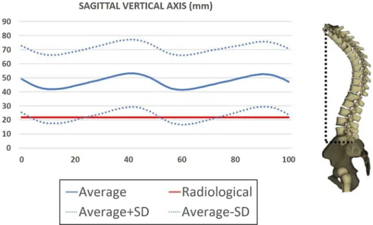
4. Results
The rSVA ranged from -66 to 38 mm with a mean of−9 mm. The av_SVA (ROM_SVA) during gait ranged from 9 to 108 mm (9 to 45 mm) with a mean of 46 mm (23 mm, respectively).ΔSVA ranged from 16 to 120 mm with a mean of 55 mm.Fig.2shows the SVA variation during gait relative to its radiological value. The multivariate analysis showed (Fig.3) that: rSVA (R2 = 0.2) was solely determined by sex (F:-20 mm vs M:1 mm,p < 0.001); av_SVA (R2 = 0.4) during gait was determined by weight (β = 0.97,p < 0.001) and rSVA (β = 0.18,p < 0.001); ROM_SVA (R2 = 0.2) was determined by acetabular abduction ( β=-0.52,p = 0.038) and ROM hipflexion/extension during gait (β = 1.48, p < 0.001); ΔSVA was solely determined by lumbar lordosis (r = 0.33,p = 0.004).
E-mail address: [email protected] (A. Assi).
Fig. 1. Registration of 3D skeletal reconstructions from biplanar X-rays with 3D gait analysis.
5. Discussion
This is thefirst study to evaluate SVA during walking in asympto-matic subjects. In general, subjects tended to bend their trunk forward during gait (positiveΔSVA), especially those with higher lumbar lor-dosis and higher rSVA. Patients with ASD, who are known to have an increased rSVA, might show a more forwarded trunk during gait which
could possibly increase their risk of falling. References
[1] Jackson (1994). [2] Davis (1990). [3] Söderkvist (1993).