Development and application of sensitive genome-wide
platforms to study the genetic and epigenetic (DNA
methylation) makeup of gametes and early bovine
embryos
Thèse
Habiballah Shojaeisaadi
Doctorat en sciences animales
Philosophiae doctor (Ph.D.)
Québec, Canada
Development and application of sensitive genome-wide
platforms to study the genetic and epigenetic (DNA
methylation) makeup of gametes and early bovine
embryos
Thèse
Habiballah Shojaeisaadi
Sous la direction de:
Directeur de recherche: Claude Robert
Codirecteur de recherche: Marc-André Sirard
iii
Résumé
Pour ce projet, nous avons développé une plateforme pour l’analyse pangénomique de la méthylation de l’ADN chez le bovin qui est compatible avec des échantillons de petites tailles. Cet outil est utilisé pour étudier les caractéristiques génétiques et épigénétiques (méthylation de l'ADN) des gamètes soumis aux procédures de procréation médicalement assisitée et des embryons précoces.
Dans un premier temps, une plateforme d’analyse de biopuces spécifiques pour l’étude de la méthylation de l’ADN chez l’espèce bovine a été développée. Cette plateforme a ensuite été optimisée pour produire des analyses pangénomiques de méthylation de l'ADN fiables et reproductibles à partir d’échantillons de très petites tailles telle que les embryons précoces (≥ 10 ng d'ADN a été utilisé, ce qui correspond à 10 blastocystes en expansion). En outre, cet outil a permis d’évaluer de façon simultanée la méthylation de l’ADN et le transcriptome dans le même échantillon, fournissant ainsi une image complète des profils génétiques et épigénétiques (méthylation de l’ADN). Comme preuve de concept, les profils comparatifs de méthylation de l'ADN spermatique et de blastocystes bovins ont été analysés au niveau de l'ensemble du génome.
Dans un deuxième temps, grâce à cette plateforme, les profils globaux de méthylation de l'ADN de taureaux jumeaux monozygotes (MZ) ont été analysés. Malgré qu’ils sont génétiquement identiques, les taureaux jumeaux MZ ont des descendants avec des performances différentes. Par conséquent, l'hypothèse que le profil de méthylation de l'ADN spermatique de taureaux jumeaux MZ est différent a été émise. Dans notre
iv
étude, des différences significatives entre les jumeaux MZ au niveau des caractéristiques de la semence ainsi que de la méthylation de l’ADN ont été trouvées, chacune pouvant contribuer à l’obtention de performances divergentes incongrues des filles engendrées par ces jumeaux MZ.
Dans la troisième partie de ce projet, la même plateforme a été utilisée pour découvrir les impacts d’une supplémentation à forte concentration en donneur de méthyle universel sur les embryons précoces bovins. La supplémentation avec de grandes quantités d'acide folique (AF) a été largement utilisée et recommandée chez les femmes enceintes pour sa capacité bien établie à prévenir les malformations du tube neural chez les enfants. Cependant, plus récemment, plusieurs études ont rapporté des effets indésirables de l’AF utilisé à des concentrations élevées, non seulement sur le développement de l'embryon, mais aussi chez les adultes. Au niveau cellulaire, l’AF entre dans le métabolisme monocarboné, la seule voie de production de S-adénosyl méthionine (SAM), un donneur universel de groupements méthyles pour une grande variété de biomolécules, y compris l’ADN. Par conséquent, pour résoudre cette controverse, une forte dose de SAM a été utilisée pour traiter des embryons produits
in vitro chez le bovin. Ceci a non seulement permis d’influencer le phénotype des
embryons précoces, mais aussi d’avoir un impact sur le transcriptome et le méthylome de l’ADN.
En somme, le projet en cours a permis le développement d'une plateforme d'analyse de la méthylation de l'ADN à l’échelle du génome entier chez le bovin à coût
v
raisonnable et facile à utiliser qui est compatible avec les embryons précoces. De plus, puisque c’est l'une des premières études de ce genre en biologie de la reproduction bovine, ce projet avait trois objectifs qui a donné plusieurs nouveaux résultats, incluant les profils comparatifs de méthylation de l'ADN au niveau : i) blastocystes versus spermatozoïdes ; ii) semence de taureaux jumeaux MZ et iii) embryons précoces traités à de fortes doses de SAM versus des embryons précoces non traités.
vi
Summary
In this project, we developed a bovine genome-wide DNA methylation platform compatible with small sample size to study genetic and epigenetic (DNA methylation) makeup of ART-treated bovine gametes and early embryos.
Initially, a bovine-specific array-based DNA methylation analysis platform was developed. This platform was subsequently optimized to produce reliable and reproducible genome-wide DNA methylation analysis from very small sample sizes, e.g. bovine early embryos (≥ 10 ng gDNA input, corresponding to 10 expanded blastocysts). In addition, this platform permitted concurrent assessment of both DNA methylation and transcription in the same sample, thereby providing a very complete picture of genetic and epigenetic (DNA methylation) profiles. As proof of concept, for the first time, comparative DNA methylation profiles of bovine sperm and blastocysts were analysed at a genome-wide level.
Using this platform, global DNA methylation profiles of monozygotic (MZ) twin bulls were analysed. Despite being geneticially identical, MZ twin bulls consistently have different progeny performance. Therefore, it was hypothesised that the DNA methylation profile of sperm from MZ twin bulls is different. In our study, there were significant differences between MZ twin for semen end points, as well as for the sperm epigenome (DNA methylation), all of which would be expected to contribute to incongruous divergent performances of daughters sired by MZ twins.
vii
In the next part of this project, using the developed platform, impacts of supplementation of a high-concentration global methyl donor on bovine early embryos was investigated. Supplementation with large amounts of folic acid (FA) has been extensively used and recommended in pregnant women for its well-established ability to prevent neural tube defects in children. However, more recently, several studies reported adverse effects of high FA concentrations, not only on embryo development, but also in adults. At the cellular level, FA enters one-carbon metabolism, the only pathway to produce S-adenosyl methionine (SAM) as the global methyl donor for a wide variety of biomolecules, including DNA. Therefore, to address this controversy, a high dose of SAM was used to treat in vitro -produced bovine embryos. This not only affected early embryo phenotypes, but also the transcritome and genome-wide DNA methylome.
Overall, the current project resulted in development of a user-friendly and cost-effective bovine genome-wide DNA methylation analysis platform, which is compatible with small cell number such as early embryos. In addition, as one of the first studies of its kind in bovine reproductive biology, this project had three objectives which yielded several novel results, including comparative genome-wide DNA methylation profiles of: i) bovine blastocysts versus sperm; ii) sperm from monozygotic twin bulls and iii) high dose SAM-treated versus non-treated bovine early embryos.
viii
Table of Contents
Résumé ... iii
Summary ... vi
Table of Contents ... viii
List of tables ... xvi
List of figures ... xviii
List of abbreviations ... xxii
Dedication ... xxxiv
Acknowledgments ... xxxv
Publications ... xxxviii
1. Introduction ... 1
1.1. Reproductive systems ... 1
1.1.1. Male reproductive system ... 1
1.1.1.1. Testis ... 1
1.1.1.2. Other male reproductive system structures ... 2
1.1.1.3. Hormones of Male Reproduction and functions ... 3
1.1.2. Female reproductive system ... 3
1.1.2.1. Ovary ... 3
1.1.2.2. Other female reproductive system structures ... 5
1.1.2.3. Hormones of Female Reproduction and functions ... 5
1.2. Introduction to Assisted Reproductive Technologies (ARTs) ... 7
1.2.1. ART applications in cattle ... 9
1.2.1.1. Gametes ...10
1.2.1.1.1. Sperm ...10
1.2.1.1.1.1. Current applied ARTs for cattle sperm ...14
1.2.1.1.1.1.1. Breeding soundness evaluation (BSE) and estimation of sire fertility ...14
ix
1.2.1.1.1.1.2. Sperm attributes ...15
1.2.1.1.1.1.2.1. Sperm count ...15
1.2.1.1.1.1.2.2. Sperm morphometry ...16
1.2.1.1.1.1.2.3. Sperm motility ...16
1.2.1.1.1.1.3. Adding “new” parameters to sperm quality evaluation ...17
1.2.1.1.1.1.4. Sperm Sorting ...17
1.2.1.1.1.1.4.1. Sex sorting ...18
1.2.1.1.1.1.4.2. Marker-based sorting of fertile spermatozoa ...18
1.2.1.1.1.1.5. Artificial insemination (AI) ...22
1.2.1.1.1.1.6. Sperm cryopreservation ...23
1.2.1.1.1.1.7. Improving male fertility through integration of the “omics” ...24
1.2.1.1.1.2. Conclusions ...27
1.2.1.1.2. Oocyte ...28
1.2.1.1.2.1. Current applied ARTs for cattle oocytes ...28
1.2.1.1.2.1.1. Synchronization ...33
1.2.1.1.2.1.1.1. Estrus/ovulation synchronization ...36
1.2.1.1.2.1.1.2. Fixed-timed artificial insemination (FTAI) ...36
1.2.1.1.2.1.2. In vitro oocyte maturation (IVM) ...39
1.2.1.2. Early embryo ...40
1.2.1.2.1. Current applied ARTs for early embryos ...40
1.2.1.2.1.1. Fixed-Time Embryo Transfer (FTET) ...40
1.2.1.2.1.2. Multiple Ovulation and Embryo Transfer (MOET) ...40
1.2.1.2.1.3. In vitro Production of Embryos ...42
1.2.1.2.1.4. Embryo sexing ...45
1.2.1.2.1.5. Splitting Embryos ...47
1.2.1.2.1.6. Oocyte /Embryo Cryopreservation ...48
1.2.1.2.2. Occurrence of Embryonic Death and Strategies to Improve Embryo Survival ...49
1.2.2. Other applications of ARTs in cattle ...52
x
1.2.2.2. Transgenesis ...53
1.2.2.3. Integrating genomic selection and reproductive technologies ...53
1.2.2.4. Studying the (epi)-genome ...57
1.2.2.5. Conclusions ...58
1.3. Introduction to epigenome ...60
1.3.1. The RNA component of the epigenome ...61
1.3.1.1. Studying non-coding RNA molecules ...63
1.3.2. The protein component of the epigenome ...65
1.3.2.1. Histone modification and variants ...65
1.3.2.2. Chromatin remodeling ...66
1.3.2.3. Studying histone modifications and chromatin-related proteins...66
1.3.3. The DNA component of the epigenome ...68
1.3.3.1. DNA methylation and hydroxymethylation ...68
1.3.3.2. DNA methylation establishment and removal ...72
1.3.3.2.1. DNA methyltransferases (DNMTs) ...72
1.3.3.2.2. One-carbon metabolism ...74
1.3.3.2.3. Folate ...77
1.3.3.2.4. Folate metabolism ...79
1.3.3.2.5. DNA Demethylation ...80
1.3.3.3. Functions of DNA methylation in transcrition and development ...83
1.3.3.3.1. Transcription ...83
1.3.3.3.1.1. Promoters ...83
1.3.3.3.1.2. Enhancers ...84
1.3.3.3.1.3. Other genomic elements ...85
1.3.3.3.1.4. Non-coding RNAs (ncRNAs) ...85
1.3.3.3.2. Repetitive elements ...85
1.3.3.3.3. X-chromosome inactivation (XCI) ...88
1.3.3.3.4. Imprinted genes ...89
1.3.3.3.5. DNA methylation reprogramming ...90
xi
1.3.3.3.5.2. Early embryo development ...91
1.3.3.4. Impacts of folate on gametogenesis, embryo development and pregnancy ...93
1.3.3.4.1. Follicular development ...93
1.3.3.4.2. Spermatogenesis ...94
1.3.3.4.3. Fertilization and early embryo development ...95
1.3.3.4.4. Placenta ...97
1.3.3.4.5. Pregnancy...97
1.4. Current options for the study of DNA methylation ...98
1.4.1. Sample treatment options ... 100
1.4.1.1. Sodium bisulfite-based treatment ... 100
1.4.1.2. Restriction-endonuclease-based methods ... 101
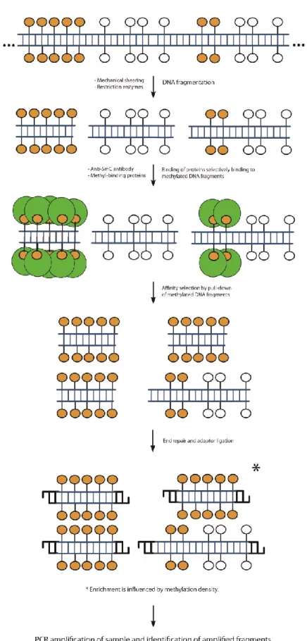
1.4.1.3. Affinity-based ... 104
1.5. Downstream methods for identifying selected sequences ... 108
1.5.1. Array-based identification ... 113
1.5.2. Sequencing based identification ... 114
1.5.3. Other options for the study of DNA methylation and new technologies in development ... 116
1.6. Successful application of the methodologies to the study of DNA methylation in early embryos ... 117
1.7. The challenges associated with the study of the epigenome in early embryos ... 122
1.8. Functional analysis of DNA methylation ... 126
1.9. Methods to analysis novel cytosine modifications ... 127
1.10. Conclusions and perspectives... 129
2. Hypothesis, Rationales, and Objectives ... 132
2.1. Hypothesis ... 132
2.2. The first rationale ... 132
2.3. The first specific hypothesis ... 133
2.4. The first objective ... 133
2.5. The second rationale ... 134
xii
2.7. The second objective ... 134
2.8. The third rationale ... 135
2.9. The third specific hypothesis ... 135
2.10. The third objective ... 135
3. An integrated platform for bovine DNA methylome analysis suitable for small samples ... 136 3.1. Résumé ... 138 3.2. Abstract ... 140 3.3. Background ... 142 3.4. Results ... 145 3.4.1. Platform Design ... 145
3.4.2. The Analysis Pipeline ... 148
3.4.3. DNA methylation profiles of bovine sperm and early embryos ... 150
3.4.3.1. Genome-wide overview of DNA methylation profiles ... 150
3.4.3.2. Characteristics of differentially methylated regions (DMRs). ... 150
3.4.3.3. DMR validation ... 151
3.4.4. Discussion ... 151
3.4.4.1. Development of EDMA ... 151
3.4.4.2. The importance of genome-scale parallel analysis of the DNA methylome and transcriptome. ... 157
3.4.4.3. Genome-wide profiles of bovine sperm and blastocyst DNA methylomes ... 158
3.5. Conclusion ... 160
3.6. Methods ... 161
3.6.1. Ethics statement ... 161
3.6.2. Microarray design ... 161
3.6.3. Sample production and genomic DNA extraction ... 162
3.6.4. Sample treatment ... 163
3.6.4.1. Fragmented genome DNA adapter ligation ... 164
3.6.4.2. HpaII tiny fragment enrichment by ligation-mediated PCR (HELP) cocktail cleavage of ligated genomic DNA ... 165
xiii
3.6.4.3. Verification of cleavage: qPCR of spiked-in templates ... 165
3.6.4.4. Fragment selection by ligation-mediated PCR ... 166
3.6.4.5. Adaptor removal ... 167
3.6.4.6. Sample labeling and hybridization ... 168
3.6.5. Bioinformatics ... 169
3.6.5.1. Data analysis pipeline ... 169
3.6.5.2. Parameters definitions ... 170
3.6.6. Data validation ... 172
3.6.7. Availability of supporting data ... 173
3.6.8. Competing interests ... 173 3.6.9. Acknowledgements ... 173 3.6.10. Author details ... 174 3.7. References ... 175 3.8. Figures ... 188 3.9. Supplementary Figures ... 202 3.10. Supplementary Tables ... 205
4. Genome-wide analysis of sperm DNA methylation from monozygotic twin bulls 210 4.1. Résumé ... 212
4.2. Abstract ... 214
4.3. Background ... 215
4.4. Materials and Methods ... 216
4.4.1. Sample collection and sperm characteristics ... 216
4.4.2. Genome-wide DNA methylation analysis ... 217
4.5. Results and discussion ... 219
4.5.1. Semen end points ... 219
4.5.2. Sperm DNA methylation profiles ... 219
4.6. Conclusions ... 223
4.7. Competing interests ... 224
4.8. Acknowledgements ... 224
xiv
4.10. Figures ... 230
4.11. Supplementary figures ... 234
5. Responses of bovine early embryos to S-adenosyl methionine supplementation in culture ... 235
5.1. Résumé ... 236
5.2. Abstract ... 238
5.3. Introduction ... 239
5.4. Materials and Methods: ... 244
5.4.1. Ethics statement ... 244
5.4.2. In vitro embryo production ... 244
5.4.3. S-Adenosyl Methionine treatment of the bovine early embryos ... 246
5.4.4. Sex ratio determination ... 247
5.4.5. Differential staining ... 248
5.4.6. Evaluation of mitochondrial activity in expanded blastocysts ... 249
5.4.7. Genome-wide parallel DNA methylome and transcriptome analysis ... 250
5.4.7.1. Simultaneous DNA and total RNA extraction from expanded blastocysts ... 250
5.4.7.2. Genome-wide DNA methylation analysis ... 250
5.4.7.3. Transcriptome analysis ... 251
5.4.8. Analysis of microarray and bioinformatics data... 252
5.4.9. Data validation ... 253 5.4.9.1. DNA methylome ... 253 5.4.9.2. Transcriptome ... 254 5.4.10. Statistical Analyses ... 255 5.5. Results ... 256 5.5.1. Phenotypic observations ... 256
5.5.1.1. Developmental speed, cell numbers and mitochondrial activities .... 256
5.5.1.2. Sex ratio ... 257
5.5.2. Genome-wide DNA methylome profile ... 259
xv
5.5.2.2. Functional annotation clustering of genes with differentially
methylated regions ... 260
5.5.3. Parallel analysis of the DNA methylome and transcriptome of SAM-treated expanded blastocysts ... 261 5.5.4. Transcriptome profile ... 262 5.6. Discussion ... 263 5.7. Conclusion ... 271 5.8. Competing interests ... 272 5.9. Authors’ contributions ... 272 5.10. Acknowledgements ... 272 5.11. Executive Summary ... 273 5.12. References ... 275 5.13. Figures ... 303 5.14. Tables ... 314 5.15. Supplementary Figures ... 316 5.16. Supplementary Tables ... 321 6. General Conclusion ... 332
6.1. Achievements and original contributions ... 334
6.2. Research perspectives ... 338
6.2.1. The first objective ... 338
6.2.2. The second objective ... 341
6.2.3. The third objective ... 345
6.3. Final statement ... 349
xvi
List of tables
Chapter 1
Table 1-1 Summaries of current possible methods of sperm quality assessments.
... 20
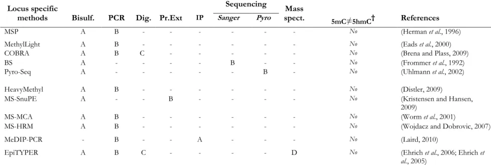
Table 1-2 Survey of different methodological platforms developed to study DNAmethylation of candidate loci. ...109 Table 1-3 Survey of different methodological platforms developed to study
genome-wide DNA methylation. ...110
Chapter 3
Table 3-1 (Supplementary) The designed Primers for the MSRE digestion quality
control step. ...205 Table 3-2 (Supplementary) Genomic and CpG coverage by MseI fragments
targeted by EDMA probes as a function of the MSRE sites present within those fragments ...206 Table 3-3 (Supplementary) Gene and CpG Island coverage by EDMA probes. ...207 Table 3-4 (Supplementary) Breakdown of the location of EDMA probes in relation
to annotated features of the bovine genome ...208 Table 3-5 ( Supplementary ) The properties of the selected hypermethylated
xvii
Chapter 5
Table 5-1 Information relating to the production of bovine embryos in vitro. Adding S-adenosyl methionine to the culture medium did not have any
significant impact on the percent reaching the blastocyst stage. ...314 Table 5-2 (Supplementary) The GO terms of genes analyzed by DAVID for all
levels of biological process ontology (GOTERM_BP_ALL)
...321
Table 5-3 (Supplementary) The list of identified differentially expressedtranscripts in the bovine embryo genome and their characteristics. ...322 Table 5-4 (Supplementary) Properties of the selected differentially methylated
regions of the bovine embryo genome and the primers designed for their
pyro-sequencing...329 Table 5-5 (Supplementary) Properties of the selected differentially methylated
regions of the bovine embryo genome and the primers designed for their
pyro-sequencing (continued).
...330
Table 5-6 (Supplementary) Properties of the selected genes and the primersxviii
List of figures
Chapter 1
Figure 1-1 Different spermatic characteristics that can be evaluated at the
molecular level to either assess sperm quality or gender selection. ...13 Figure 1-2 Ovarian follicular and corpus luteum development correlated with
endocrine changes during the bovine oestrous cycle. ...30 Figure 1-3 Reproductive management alternatives to improve reproductive
performance of lactating dairy cows with the use of presynchronization. Ovsynch for timed artificial insemination (TAI), post-insemination endocrine
treatments and resynchronization for TAI. ...35 Figure 1-4 Typical programs designed to synchronize ovulation of the dominant
follicle and facilitate success with a timed AI (TAI) protocol.. ...38 Figure 1-5 Biopsy of bovine embryo for sex diagnosis. ...46 Figure 1-6 Timing and extent of pregnancy losses in the high producing lactating
dairy cow. ...50 Figure 1-7 Terms and concepts in the pre-attachment period in a bovine embryo. ...52 Figure 1-8 Diagram of the one-carbon metabolism pathway. ...76 Figure 1-9 Pathways for removal of DNA methylation.. ...82 Figure 1-10 The general outline of the most relevant current sample treatment
options and downstream applications and identification platforms for the
xix
Figure 1-11 General flow of events of platforms for the study of DNA methylation
based on methylation-sensitive restriction enzymes. ... 103
Figure 1-12 General flow of events of platforms for the study of DNA methylation based on affinity enrichment of methylated sequences. ... 106
Figure 1-13 General outline of the reduced representation bisulfite sequencing (RRBS) platform dedicated to the study of DNA methylation. ... 120
Chapter 3
Figure 3-1 Sequence of steps involved in the generation of methylome and transcriptome data from the same sample using EDMA platform. ... 188Figure 3-2 The characteristics of EDMA array. ... 190
Figure 3-3 Snapshots from the UCSC genomic web-browser describing the genomic locations of two bovine imprinted genes (NNATand PEG10), positioning of the probes (methylome and transcriptome) and other associated bovine genomic features. ... 192
Figure 3-4 The feature of EDMA spiked-in controls and the quality control process of MSREs digestion assessment. ... 193
Figure 3-5 The two types of quality control (QC) plots generating after EDMA microarray hybridization and data analysis. ... 194
Figure 3-6 The EDMA DNA methylation analysis pipeline. ... 196
Figure 3-7 List of the graphs generated by EDMA analytical pipeline. ... 197
xx
Figure 3-9 The Circos plot showing the genome-wide DNA methylation profile of
bovine sperm DNA and the blastocyst genome, by chromosome.. ... 199
Figure 3-10 The comparative analysis of differentially methylated regions (DMRs) enrichments between bovine sperm and blastocysts. ... 200
Figure 3-11 Validation of the selected DMRs by pyrosequencing. ... 201
Figure 3-12 (Supplementary) ... 202
Figure 3-13 (Supplementary) ... 203
Figure 3-14 (Supplementary) ... 204
Chapter 4
Figure 4-1 Sperm samples phenotypic characteristics. Analysis of sperm morphology and viability of MZ twin bulls. ... 230Figure 4-2 Genome-wide perspective of sperm DNA methylation profiles of MZ twin bull pairs.. ... 232
Figure 4-3 Distribution of differentially methylated probes in the semen of each pair of twins. ... 233
Figure 4-4 (supplementary) ... 234
Chapter 5
Figure 5-1 Hatching of cultured bovine blastocyst embryos. . ... 303Figure 5-2 Cell number, TE/ICM allocation and mitochondrial activity of cultured bovine blastocysts. ... 304
xxi
Figure 5-3 Impact of S-adenosyl methionine on the sex ratio of cultured bovine
embryos. ... 306
Figure 5-4 Distribution of differentially methylated regions of the genome of cultured bovine embryos. ... 308
Figure 5-5 Hypomethylation of genes involved in active DNA demethylation and hypermethylation of de novo DNA methylation genes in bovine embryos cultured in a medium supplemented with S-adenosyl methionine.. ... 310
Figure 5-6 Embryonic stem cell pluripotency (ESC) genes (in color) found differentially methylated in SAM-treated day 7 expanded blastocysts.. ... 311
Figure 5-7 The relative expression differentially methylated genes of interest. ... 312
Figure 5-8 Volcano plot of transcripts expressed differentially in non-treated and SAM-treated day 7 expanded bovine blastocysts in culture. ... 313
Figure 5-9 (Supplementary) ... 317
Figure 5-10 (Supplementary) ... 319
xxii
List of abbreviations
% Percent
°C Celsius degree
2D-PAGE Two-dimensional polyacrylamide gel electrophoresis
5caC 5-carboxylcytosine 5fC 5-formylcytosine 5hmC 5-hydroxymethylcytosine 5mC 5-methylcytosine 5-methyl-THF 5-methyl-tetrahydrofolate AI Artificial insemination
AID Activation-induced cytidine deaminase
AOT Acridine orange test
APEX APEX nuclease (multifunctional DNA repair enzyme)
APOBEC Apolipoprotein B mRNA editing enzyme, catalytic
polypeptide-like,
AR reaction Acrosome reaction
aRNA Anti-sense RNA
ART Assisted reproductive technologies
ART2A ADP-ribosyltransferase 2a
ASMA Automated sperm head morphology analysis
ATF Artificial transcription factors
BER Base excision repair pathway
Bisulf Sodium bisulfite treatment
Blst Blastocysts
BMP Bone morphogenetic proteins
BODIPY 4-bora-3a,4a-diaza-s-indacene
BovA2 Short interspersed nuclear element (SINE) sequences of the
xxiii
BOvB Bov-B consensus sequence
BovtA Short interspersed nuclear element (SINE) sequences of the
Bovidae
bp Base pair
BRD9 Bromodomain Containing 9
BS Bisulfite sequencing
BSA Bovine Serum Albumin
BSE Breeding soundness evaluation
bST Bovine somatotropin
BWS Beckwith Wiedemann syndrome
C Non-methylated cytosine
CAM Calcein acetomethyl ester
CARS Cysteinyl- tRNA Synthetase
CASA Computer Assisted Sperm Analysis
CChIP Carrier Chromatin Immunoprecipitation
cDNA Complementary DNA
CE Capillary electrophoresis
CFDA 6-carboxyfluorescein diacetate
CG Cytosine phosphate Guanine
CGIs CpG islands
CHARM Comprehensive high-throughput arrays for relative
methylation
CHG Cytosin H Guanine (where H corresponds to A, T, or C)
ChIP-chip Chromatin immunoprecipitation (ChIP) followed by
microarray hybridization
CHOTHF 10-Formyltetrahydrofolate
CIDR Controlled intravaginal drug release
CMA3 Chromomycin A3
xxiv
Cnt.-EB Non-treated expanded blastocysts
CNV Copy number variants
CO2 Carbon dioxide
COBRA Combined bisulfite restriction analysis
COCs Cumulus oocyte complexes
CpG Cytosine phosphate guanine
Ct Threshold cycle
∆CT Delta cycle threshold
∆∆CT Delta Delta cycle threshold
CTC Chlortetracycline
d Day
DAVID Database for Annotation, Visualization and Integrated
Discovery
DE Differentially expressed
DEG Differentially expressed gene
DIC Differential interference contrast
Dig Restriction endonuclaese digestion
DMR Differentially methylated region
DNA Deoxyribonucleic acid
DNMTs DNA methyltransferase enzymes
dNTPs Deoxynucleotides
DOHaD Developmental Origin of Health and Diseases
DTT Dithiothreitol
dUMP Deoxyuridylate
E2 Oestradiol
EB Expanded blastocysts
EBVs Estimated breeding values
ECP Oestradiol cypionate
xxv
EDTA Ethylenediaminetetraacetic acid,
EGA embryonic genome activation
ELMA EmbryoGENE LIMS and microarray analysis
EmbryoGENE A Network was created to address important issues
concerning embryo development in livestock, mainly cattle and swine.
EMBV3 EmbryoGENE bovine transcriptome version 3
EpiTYPER A bisulfite-treatment-based method for
detection and quantitation of DNA methylation
ERCC External RNA Controls Consortium
ERCR Estimated relative conception rate
ERVs Endogenous retroviruses
ESCs Embryonic stem cells
ET Embryo transfer
EtBr Ethidium bromide
FA Folic acid
FACS Fluorescence-activated cell sorting
FAD Folate acid deficiency
FC Flowcytometry
FC Fold change
FDR False discovery rate
FITC Fluorescein isothiocyanate
FQRNT Fonds Québécois de la Recherche sur la Nature et les
Technologies
FR Folate receptor
FSH Follicle-stimulating hormone
FTAI Fixed-timed artificial insemination
FTET Fixed-timed embryo transfer
xxvi
GC-MS Gas chromatography-MS
gDNA Genomic DNA
GEO Gene Expression Omnibus
GnRH Gonadotrophin-releasing hormone
GO Gene ontology
GV Germinal vesicle
GVBD Germinal vesicle breakdown
GWAS Genome wide association studies
h, hr Hour
H2AK119 Histone 2A Lys4 119
H2DCFDA 2',7'-dichlorodihydrofluorescein diacetate
H3K27me3 Histone 3 lys27 trimethylation
H3K4 Histone 3 lys4
H3K4me3 Histone 3 lys4 trimethylation
H3K9me3 Histone 3 lys9 trimethylation
hCG Human chorionic gonadotropin
HCP High CpG density promoters
HDL High-density lipoproteins
HE Hydroethidine
HELP HpaII tiny fragment enrichment by ligation-mediated PCR
HEPES (4-(2-hydroxyethyl)-1-piperazineethanesulfonic acid
HOST Hypo-osmotic swelling test
HPCE-ESI/MS High-performance capillary electrophoresis–electron spray Ionization/mass spectrometry
hpf Hour post-fertilization
HPLC High Performance Liquid Chromatography
IAPs Intracisternal A particle genes
ICM Inner cell mass
xxvii
ICRs Imprinting control regions
IGF2 Insulin-like growth factor 2
IGFBP-4 and -5 Insulin-like growth factor binding proteins 4 and 5
IP Immunoprecipitation
IPA Ingenuity Pathway Analysis
iPSs Induced pluripotent stem cells
ITC Fluorescein isothiocyanate
IVC In vitro cultured embryo
IVF In vitro fertilization
IVM In vitro maturation
IVP In vitro embryo production
JC-1
5,5',6,6'-tetrachloro-1,1',3,3'-tetraethylbenzimidazolylcarbocyanine iodide
K Kilo
Kb Kilo base
Kbp Kilo base pair
LAMP Loop-mediated isothermal amplification
LC Liquid chromatography
LC-MS LC-mass spectrometry
LCP low density CpG promoters
LH Luteinizing hormone
LIG Ligase I, DNA, ATP-Dependent
LIMMA Linear Models for Microarray and RNA-Seq Data
LINE Long Interspersed Nuclear Elements
LM-PCR Ligation-mediated PCR
lncRNAs Long noncoding RNA
LOS Large offspring syndrome
LTR Long-terminal-repeat retrotransposons
xxviii
M Use for millions, and molar
M540 Merocyanine 540
MACS Magnet-activated (or magnetic-bead activated) cell sorting
MALDI-TOF Matrix-assisted laser desorption ionization–time of flight
Mass spec. Mass spectrophotometry
MAT Methionine adenosyltransferase
Mb Mega base
MBD2 Methyl-CpG binding domain protein 2
MBD4 Methyl-CpG-binding domain protein 4
MCA Methylated CpG island amplification
MeCP2 Methyl CpG binding protein 2
MeDIP Methylated DNA immunoprecipitation
MET Maternal embryonic transition
mg Milligrams
Mg2+ Magnesium
MgCl2 Magnesium chloride
MI Metaphase I
MII Metaphase II
miRNA Micro RNAs
mL Milliliter
mm Millimeter
mM Millimolre
MOET Multi-Ovulations Embryo Transfers
mRRBS Multiplexed RRBS
MS Mass spectrometry
MS-HRM Methylation-sensitive high-resolution melting
MS-MCA Methylation-sensitive melting curve analysis
MSP Methylation specific PCR
xxix
MS-SnuPE Methylation-sensitive single nucleotide primer extension
MTHF Methyltetrahydrofolate
MW Molecular-weight
MZ Monozygotic
Nacl Sodium chloride
natRNA Natural antisense
ncRNA Non-coding RNA
ng Nanogram
NGS Next generation sequencing
nmol Nanomolar
NNAT Neuronatin gene
NRC National Research Council
NRR Non-return rate
NSERC Natural Sciences and Engineering Research Council of Canada
nt Nucleotide
NTDs Neural tube defects
Oligo dT A short sequence of deoxy-thymine nucleotides
OPU Ovum pick-up
OvF Ovulatory follicle
oxBS-Seq Oxidative bisulfite sequencing
p and P Stands for probability
P4 Progesterone
PARP1 Poly (ADP-Ribose) Polymerase 1
PBS Phosphate-buffered saline
PCA Principal component analysis
PCFT Proton-coupled folate transporter
PCNA Proliferating cell nuclear antigen
PCR Polymerase chain reaction
xxx
PEG10 Paternally expressed 10 gene
pg Picogram
PGCs Primordial germ cell
PGF2α Prostaglandin F2α
PGF Placental Growth Factor
phi29 DNAP Wild-type phi29 DNA polymerase
PI Propidium iodide
piRNA Piwi-interacting RNA
PNA Peanut agglutinin
PNKP Polynucleotide Kinase 3'-Phosphatase
PR Progesterone receptor
PRC2 Polycomb Repressive Complex 2
Pr.Ext Primer extension
PSA Pisum sativum agglutinin
pY Phosphotyrosine
Pyro Pyrosequencing
QC Quality control
q-RT-PCR Quantitative real-time reverse-transcriptase polymerase chain reaction
QTL Quantitative trait loci
R123 Rodamine 123
rasiRNAs Repeat-associated siRNA
RdDM RNA-directed DNA methylation
RdDM RNA-directed DNA methylation
RE Restriction endonuclease
REDIH CIHR Training Program in Early Development, and the Impact
on Health
RFC Reduced folate carrier
xxxi
RLGS Restricted landmark genome scanning
RNA Ribonucleic acid
RNAseq RNA sequencing
ROS ROS reactive oxygen species
RPM Revolutions per minute
RQR Réseau Québécois en Reproduction
RRBS Reduced-representation bisulfite sequencing
rRNA Ribosomal RNA
RTE A non-long-terminal-repeat (non-LTR) retrotransposable
element
s second
SAH S-adenosylhomocysteine
SAM S-adenosyl methionine
SAM-EB SAM-treated expanded blastocysts
SCNT Somatic cell nuclear transfer cloning
SCR Sire conception rate
SCSA Sperm chromatin structure assay
SD Standard Deviation
shRNA Short hairpin RNA
SINEs Short Interspersed Nuclear Elements
siRNA Small interfering RNA
SMRT Single-molecule real-time
SMUG1 Single strand-selective monofunctional uracil DNA
glycosylase 1
SNARF-1 Seminaphtorhodafluor-1
SNP Single nucleotide polymorphism
snRNA Small nuclear RNA
SOF Synthetic oviduct fluid
xxxii
SSRs Simple sequence repeats
SYBR14 Membrane-permeant fluorescent nucleic acid stain
T Thymine
TAI Timed artificial insemination
TALE Transcription-activator-like effector
TALEM Transcription-activator-like effector methyltransferase
TBARS Thiobarbituric acid reactive substances
TDG Thymine DNA glycosylase
TE Trophectoderm
TE Transposable elements
TEP Trophectodermal projections
TEs Transposable elements
TETs Ten eleven translocase enzymes including
TFO triplex-forming oligomers
THF Tetrahydrofolate
TLC Time-lapse cinematography
TLH HEPES-buffered Tyrode’s medium
Tsix X-Inactivation-Specific Transcript-Antisense
TSS Transcription start sites
TUNEL Terminal deoxynucleotidyl transferase dUTP nick end labeling.
TZP Transzonal projections
U Unit
UCSC University of California, Santa Cruz
UHRF1 Ubiquitin-Like With PHD And Ring Finger Domains 1
ULI-NChIP ultra-low-input micrococcal nuclease-based native ChIP
ULS Universal Linkage System
USDA United States Department of Agriculture
V Volt
xxxiii
XCI X chromosome Inactivation
xg Times gravity
Xi Inactive X chromosome
Xic X inactivation center
Xist X-inactive specific transcript
Xm Maternal X
Xp Paternal X
XRCC X-Ray repair complementing defective repair in chinese
hamster cells
YOPRO1 A commercial green-fluorescent stain
ZFP Zinc finger proteins
ZGA Zygotic genome activation
μg Microgram
μL Microliter
μm Micrometer
xxxiv
Dedication
I dedicate this thesis to my wife, Somayeh Golzari Movafagh, who has been beside me in every moment of my life since our marriage to support, encourage and help me. It was not possible to accomplish my PhD and achieved all of my successes during the life since I have been with her without her patients, care and love.
I also dedicate this thesis to my mother, Maryam Abdollahi and my father Hamid Shojaei for being such a great parents supporting me with their infinite love. Nothing can explain their dedication and self-devotion for their children especially me to succeed and be happy in my life. Thank you mom and dad!
xxxv
Acknowledgments
I would like to express my most deeply appreciation to my supervisor Dr. Claude Robert, who has provided encouragement, motivation, enthusiasm, help and great support. His supports helped me in all the time of PhD career and had key roles for all of my success during PhD.
This is a great opportunity to appreciate Prof. Marc-André Sirard, my co-supervisor for his enormous supports, encouragements and valuable advice during both academic and non-academic discussions. I gratefully thank Prof. Frans van der Hoorns, Department of Biochemistry & Molecular Biology, University of Calgary, AB for all of his comments and advices, and precious times to generously support me during my PhD.
I am also very grateful to my internal examiner Prof. Marie-Claude Vohl, École de nutrition Centre de Recherche du CHUQ, INAF, Prof. Janice Bailey and my external examiner Dr. Mellissa Mann, Departments of Obstetrics and Gynecology and Biochemistry, Schulich School of Medicine & Dentistry, Western University, ON who accepted to be a part of my thesis’ evaluation committee and have put their valuable time into reading and evaluating my thesis.
I gratefully acknowledge Prof. John P. Kastelic, Department of Production Animal Health (PAH), University of Calgary, AB for his enourmous kind helps, corrections and supports. Special thanks go to my previous supervisors Dr. Jacob Thundathil (MSc.
xxxvi
University of Calgary, AB. Canada) and Prof. Rajabali Sadrkhanlou (DVM. Urmia University, Iran) to help, train and encourage me to pursue my interests.
I would like to take the opportunity to acknowledge Natural Sciences and Engineering Research Council of Canada (NSERC), Fonds de recherche du Québec - Nature et technologies (FRQNT), Réseau Québécois en reproduction (RQR), Canadian Institutes of Health Research (CIHR) training program for Reproduction, Early Development, and the Impact on Health (REDIH), Centre de recherche en Reproduction, Développement et Santé Intergénérationnelle (CRDSI) for their scholarships and bourses. Semex Alliance, L'Alliance Boviteq (LAB) inc. for their generous donation of the MZ twin semen samples and sexing kits for this project. I also thank Institut sur la nutrition et les aliments fonctionnels and Laval University for their travel awards.
I would also acknowledge Mr. Eric Fournier, Dominic Gagné, Dr. Isabelle Gilbert, Dr. Isabelle Dufort, Isabelle Laflamme for their kind helps and technical supports during my PhD. Thanks to Dr. Roberts’ students who provided a very friendly lab environment and supported me in any respect during the completion of the Project; Dr. Angus Macaulay, Dr. Luis Manuel Baldoceda Baldeon, Dr. Sara-Myriam Scantland-Marchand, and Dr. Julieta Caballero.
Here is the great opportunity to thank Dr. Alan M. O'Doherty (University College Dublin, Ireland) for his time and kind helps in this project and Dr. Dessie
Salilew-xxxvii
Wondim (University of Bonn, Germany) for the opportunity for collaboration and scientific discussions.
Special thanks and gratitude go to Dr. Amin Rezvanfar (University of Tehran, Iran) for being my best and very supportive friend throughout my work and life, helping me with his advices, comments and inspirations.
Many thanks go in particular to Dr. Esmail Behboodi, for his comments and insight and Prof. Mohammad Abdollahi (University of Tehran, Iran) for his generous supports and the opportunities to help me to contribute more in science.
Last but not the least; I would like to thank Mahboubeh, Mona & Hamed for being supportive and caring siblings.
xxxviii
Publications
The chapter 1(introduction) comprises a published book chapter and a review article in addition to some updated information which incorporated appropriately in the text. Chapter 3 & 4 are published articles and chapter 5 will be submitted shortly.
Chapter 1
Book chapter: Shojaei, H. and Robert, C. (2014). Modern reproductive technologies
and breeds improvement. The Genetics of Cattle. 2nd Edition. Chapter 13. CABI. UK.
Authors’ contributions
HASS and CR wrote the manuscript; designed the figure and tables. All authors read and approved the final manuscript.
Review article : Shojaei, H*., McGraw*, S., Robert, C. Meeting the methodological
challenges in the molecular mapping of the embryonic epigenome. Molecular Human Reproduction. 2013; 12:809-827. * These authors contributed equallly to this work.
Authors’ contributions
HASS and SM provided equal contribution to the conception and writing of the manuscript; C.R. wrote parts of the manuscript; S.M. and C.R. revised the manuscript. All authors approved the final version.
xxxix
Chapter 3
Shojaei, H., O'Doherty, A., Gagné, D., Fournier, E., Grant, J. R., Sirard, MA., Robert, C.
(2014) An integrative platform for efficient survey of the bovine DNA methylome suitable for low amount of starting material. BMC Genomic. 15:451
Authors’ contributions
HASS worked on platform development, performed bovine sperm and blastocyst experiments, and designed the analysis pipeline. AOD performed pyrosequencing for candidates. DG leaded platform development. EF performed bioinformatic analyses, array design and programmed the plot generation pipeline. JG performed the in silico survey of CpG islands. MAS and CR conceived the general strategy for the platform’s development. CR designed and supervised the work. HASS and CR drafted the final manuscript. All authors read and approved the final manuscript.
Chapter 4
Shojaei, H., Fournier, E., Vigneault, C., Bailey, J., Robert, C. (2016) Genome-wide
analysis of sperm DNA methylation from monozygotic twin bulls. Reproduction Fertility
and Development Journal. Published online: 12 January 2016;
http://dx.doi.org/10.1071/RD15384
Authors’ contributions
HASS worked on bovine sperm intact gDNA extraction development, performed semen and sperm analysis, MZ twin sperm DNA methylation experiments. EF performed
xl
bioinformatic analyses. CV and PB provided the MZ information and samples. JB provided the CASA analysis system and reviewed the manuscript. CR designed and supervised the work. HASS and CR drafted the final manuscript. All authors read and approved the final manuscript.
Chapter 5
Shojaei, H., Fournier, E., Gagné, D., Baldoceda, L.M., Sirard, MA., Robert, C. Responses
of bovine early embryos to S-adenosyl methionine supplementation in culture (Accepted in Epigenomics journal to publish in July 2016).
Authors’ contributions
HASS, MAS and CR designed the study. HASS performed the IVF, phenotypic, transcriptome (RT-qPCR) and epigenetics (DNA methylation and pyrosequencing) experiments, acquired and contributed to dara analysis. DG performed the transcriptome microarray. EF performed bioinformatics analyses. LMBB and HASS performed the mitochondria staining. CR supervised the work. HASS and CR drafted the final manuscript. All authors read and approved the final manuscript.
1
1. Introduction
1.1. Reproductive systems
Reproductive systems are different for male and female and consist of a series of glands and tubes that produce and nurture gametes and transport them to the site of fertilization. Therefore, the major function of the reproductive system is to ensure survival of the species. The processes of male (sperm) and female (oocyte) haploid gamete production are known as spermatogenesis and oogenesis, respectively. Following puberty, spermatogenesis can persist throughout lifetime in males. In contrast, oogenesis initiates during fetal development, arrests at prophase I of meiosis before birth, and does not resume until prior to ovulation in adulthood.
1.1.1. Male reproductive system
The male reproductive system of mammals consists of two testes (testicles) in the scrotum, accessory organs including ducts and glands, and the penis. The organs and glands of the male reproductive tract produce the sperm and deliver it to the female reproductive tract during intercourse.
1.1.1.1. Testis
The testicles have two functions; i) production of: spermatozoa (also called sperm) and ii) synthesis of the male sex hormones, most importantly testosterone. The testicles are located outside of the body cavity in the scrotum, which is important for thermoregulation of the testicles and provides a favorable environment for the production and maturation of spermatozoa (Jung and Schuppe 2007; Kim et al., 2013). This is a favorable environment as successful sperm development occurs at a
2
temperature several degrees below normal body temperature. Each testis consists of a mass of coiled seminiferous tubules where spermatogenesis takes place. In bovine, it has been estimated that the approximately 2 × 10 9 sperm are produced in a single
day (Rowen D. Frandson, 2009). Histologically, within the seminiferous tubules there are sertoli cells that surround and support developing spermatozoa and their precursors (Kaur et al., 2014). Sertoli cells are involved with nourishing the developing sperm and mediate the effects of follicle-stimulating hormone (FSH) and testosterone on the germ cells (Kaur et al., 2014). The connective tissue between the seminiferous tubules contains the interstitial cells (leydig cells) which secrete the main male hormone (testosterone) when stimulated by the pituitary gonadotropin luteinizing hormone (LH) (Smith and Walker, 2014).
1.1.1.2. Other male reproductive system structures
These structures include the epididymis and ductus deferens, accessory sex glands (ampullary glands, vesicular glands, prostate, and bulbourethral glands), the urethra, and the penis. Following spermatogenesis, testicular spermatozoa must undergo further maturation to enable fertilization of oocytes. Maturation occurs during the transit of immature spermatozoa through the male reproductive tract via interactions with epididymal epithelium (usually 10–15 days) and accessory gland fluid (Caballero
et al., 2011). Male accessory sex glands include ampulla of the ductus deferens,
vesicular gland, prostate gland, and bulbourethral gland which produce semen to provide favorable conditions for nutrition of sperm and acts as a buffer against the natural acidity of the female genital tract (Rowen D. Frandson, 2009).
3
1.1.1.3. Hormones of Male Reproduction and functions
The secretion of gonadotrophin-releasing hormone (GnRH), stimulates the release of both follicle-stimulating hormone (FSH) and luteinizing hormone (LH) from the anterior pituitary (adenohypophysis) (Schlatt and Ehmcke, 2014). Both FSH and LH are protein hormones which are the primary endocrine regulators of testicular function, therefore both are considered to be gonadotrophins. FSH promotes spermatogenesis by its actions on germ cells and sertoli cells in the seminiferous tubules to support the development of the spermatozoa (Schlatt and Ehmcke, 2014). However, LH acts on leydig cells to promote the secretion of androgens, primarily testosterone. Testosterone produced by the leydig cells is necessary for the completion of spermatogenesis (Smith and Walker, 2014).Therefore, both FSH and LH are required for successful spermatogenesis.
1.1.2. Female reproductive system
In mammals, the female reproductive system generally consists of two ovaries, uterine tubes (also called oviducts), uterus and vagina. Oocytes grow and mature in the ovaries, and following ovulation transit into the oviduct, the site of fertilization and early embryo development. Subsequently, embryos transit to and implant in the uterus resulting in pregnancy.
1.1.2.1. Ovary
Mature female gametes (oocyte) and some of the hormones necessary for female reproduction are produce in ovaries. Unlike the testis, ovaries contain a limited number of gametes following birth, (Van den Hurk and Zhao, 2005). They contain a variant
4
population of follicles including primordial, primary, secondary and tertiary follicles which the latter can be classified as small and large antral follicles (Aerts and Bols, 2010). In primates and most domesticated animal species, the stock of primordial follicles develops during fetal life. According to dogma in the field, in females, the peak number of ovarian follicles occurs in the fetus at mid-gestation and is approximately 2.7 million and seven million follicles in cow and women, respectively. This number then declines to 135,000 and 700,000 follicles in cow and women, respectively at birth meaning up to 95% loss at birth due to atresia (Van den Hurk and Zhao, 2005). Furthermore, in ovaries, folliculogenesis takes place which is the process of ovarian follicle development causing the primordial follicle to undergo several steps of maturation in order to develop into a preovulatory follicle. In fact, these changes occur within the three major cell populations: oocytes, granulosa cells, and theca cells under control of several hormones and growth factors. In order to reach the ovulatory stage, an ovarian follicle will pass through the following stages: primordial (resting), primary, secondary (pre-antral), tertiary (antral), and, finally, the pre-ovulatory (Graafian) follicle stage. Folliculogenesis can be divided into two phases: i) preantral growth which is gonadotropin-independent phase and initiates from recruitment of a primordial follicle to the end of the secondary stage and ii) antral growth which is gonadotropin-dependent phase when a fluid-filled cavity or antrum develops within the follicle which is tertiary (Graafian follicle) and ultimately fully developed into a preovulatory follicle (Conti and Chang, 2016).
5
1.1.2.2. Other female reproductive system structures
Other female reproductive system structures consist the Fallopian tubes (also called oviducts), uterus and vagina. The oviducts comprise infundibulum, ampulla, isthmus, uterotubal junction and are the site of fertilization and function in the transfer of the oocyte to the uterus. Furthermore, oviducts support sperm survival and have functions to prevent infection (Sheldon et al., 2014). The uterus of the domestic mammal consists of a body, a cervix (neck), and two horns and is the site for embryo implantation and fetus development during pregnancy.
1.1.2.3. Hormones of Female Reproduction and functions
Similar to male, in female the hypothalamic secretion of GnRH promotes the release of both FSH and LH from the adenohypophysis (Christensen et al., 2012). However, ovarian steroids (estradiol and progesterone) and peptide hormones (inhibin) can be modulated by the release of GnRH. Cellular receptors for FSH and LH are expressed in the granulosa and theca of secondary follicles, respectively and they become responsive to these hormones (Xu et al., 1995; Aerts and Bols 2010a). From this point, the coordinated effects of FSH and LH are both required for normal follicular development. During the gonadotrophin-dependent stage of follicular development, FSH- and LH-signaling pathways play essential role in follicular differentiation, selection and survival (Palermo, 2007; Gervásio et al., 2014). Under the effect of LH, theca cells produce androgen which diffuses into the mural granulosa cell (MGC) layer (those granulosa cells surround the antrum), where it is the substrate for FSH-induced aromatase for follicular estradiol synthesis. This is the landmark of ‘two cells-two
6
gonadotrophins’ concept for follicular estradiol biosynthesis and the physiology of ovarian function in mammals (Palermo, 2007; Gervásio et al., 2014; Conti and Chang, 2016). Estrogen produced by the granulosa cells acts as a paracrine agent with positive feedback that promotes the development of the follicle, however systematic circulation of estrogen has a negative feedback effect on FSH secretion from the adenohypophysis (Aerts and Bols 2010a; Forde et al., 2011). In fact, follicles are involved in the positive and negative feedback mechanisms of the hypothalamic– pituitary–gonadal (HPG) axis which also has a governing role in the regulation of the oestrous cycle of cattle (Forde et al., 2011). Together these feedback mechanisms can select which follicle ultimately produces the ovum and ovulates (Aerts and Bols, 2010b). In addition, estrogens from developing follicles are also essential for modulating of the hypothalamic–adenohypophyseal axis for ovulation and increasing the LH receptors in thecal cells to response to short-term large LH release (LH surge) necessary for ovulation (Aerts and Bols, 2010b).
7
1.2. Introduction to Assisted Reproductive Technologies (ARTs)
Assisted reproductive technologies (ARTs) encompass a wide range of techniques
designed primarily to aid couples unable to conceive without medical assistance (E. Oluwole Akande, 2008). One in six couples worldwide experience some form of
infertility problem at least once during their reproductive lifetime. It is now estimated that more than 5 million babies have been born worldwide since the first IVF baby was born in 1978 (ESHRE, July 2014).
The largest part of the agriculture economy in developing countries relies on cattle and small ruminants (Rodriguez-Martinez, 2012). In many countries of the southern hemisphere, such as Australia, New Zealand and South American countries, cattle and small ruminants represent the major economical asset in terms of milk, meat and wool production (Rodriguez-Martinez, 2012). In Canada, there are more than 11,600 dairy farms, with approximately 1,000,000 dairy cow and 500,000 dairy heifers accounting for an income over six billion dollars in 2014 (http://www.dairyinfo.gc.ca/) (Canadian Dairy Information Center, 2015).
In cattle, ARTs are routinely carried out to shorten generational intervals and to propagate genetic material among breeding animal populations (Rodriguez-Martinez 2013). ARTs are important tools for improving the production of dairy and beef cattle. Hence, ARTs have been developed over the years for manipulation of both male and female gametes and embryos. In a way that today ARTs face a strong wave of increasing commercialization and profits for the cattle industry through a positive economical impact on beef and milk production and genetics exports
(Rodriguez-8
Martinez, 2012). For instance, in Canada up to 80 % of the Canadian Dairy Genetics Exports in 2014 were accounted for by embryo and semen exportation, whereas export of dairy cattle provided only 20 % of the Canadian Dairy Genetics Exports money values (Canadian Dairy Information Centre, 2015). Among different ARTs in cattle, artificial insemination (AI) is by far the most important and routinely performed of the ARTs (see page 17). Worldwide, it has been estimated that over 130 million cattle are inseminated artificially each year (Vishwanath, 2003). In addition, in vitro embryo production is one of the most frequently employed ARTs in the cattle industry. However, in vitro production of bovine embryos is not without consequence, as they have been shown to differ greatly from their in vivo produced counterparts in many facets, including developmental competence; which has been suggested to be related to disruption of the epigenetic profile of the gametes and/or embryos (Urrego et al., 2014). Similar observations have been reported for other species such as human and mouse (van Montfoort et al., 2012; El Hajj and Haaf, 2013; de Waal et al., 2015; Song
et al., 2015).
ARTs represent an exciting prospect for conservation biologists for the preservation of both the ecologically valuable, wild, rare, and indigenous species and the genetic variability within such species (Long, 2008). Considering the intensive agriculture, extinction of natural habitats, environmental pollutions and several other factors, biodiversity has been sharply threatened through extinction of several mammalian species. Hence, due to the cultural, historical and genetics importance attempts are being made to conserve genomes by employing ART and biotechnology for the
9
endanger species (Cseh and Solti, 2000; Long, 2008). However, in practice there are few examples of success, of course not as the result of a failure on the part of the technologies per se, but rather is due to lack of knowledge about the fundamental biology of the species in question (Pukazhenthi et al., 2005; Long, 2008).
1.2.1. ART applications in cattle
Well-defined objectives are essential for success in livestock breeding and will usually evolve over time in relation to societal concerns as well as technological and economic developments. In case of cattle, higher output per unit input have been the main breeding objective but other issues including well-being, longevity and reproduction have been raised (Flint and Woolliams, 2008). Dairy cattle breeding is undergoing a paradigm shift to genomic selection of sires and dams (Amann and DeJarnette, 2012). Genetic improvement relies on two different physiological applications namely genetic selection and selective mating. The first requires estimation of genetic potential, the second pertains to the selection of mating pairs and reproductive technologies used to disseminate high merit genetics and is the focus of this chapter. Reproductive technologies have a crucial role in improvement of livestock reproduction to meet the rising challenges to the food supply of the 21st century (Murphy, 2012 ). Despite major advances associated with reproduction in dairy cattle (Moore and Thatcher, 2006), reproductive decline in elite dairy cattle is a serious concern of farmers and the dairy industry worldwide (Lucy, 2007; Dobson et al., 2007; Lucy, 2001; Pryce et al., 2004; Walsh et al., 2011). Fertility has emerged as a growing and biggest genetic and
10
management challenge in high-producing dairy herds (Funk, 2006). Fertility requires both paternal and maternal contributions to produce an embryo which eventually leads to live birth. The following aims at portraying the current and emerging reproductive technologies that are destined to increase the rate of genetic improvement with a focus on dairy herds.
1.2.1.1. Gametes 1.2.1.1.1. Sperm
Male fertility is an important factor in cattle breeding as bulls in service are used to breed numerous cows and defective semen quality can have a profound contribution on reproductive failure (Kathiravan et al., 2011; DeJarnette et al., 2004). It was estimated that sperm from majority of sires are not able to fertilize 100% of the oocytes (>90% oocytes in heifers or non-lactating cows) and male-associated deficiencies account for approximately 5 to 20% of embryos dying by Day 8 of development (Amann and DeJarnette, 2012). In cattle, primarily, post-thaw semen evaluation and analysis of several sperm characteristics are routinely performed in breeding centers to assess potential bull fertility.
Sperm is the ultimate example of a structural and functionally differentiated cell in which its genetic component located in the highly condensed nucleus results in gene expression silencing (Eddy, 2006; Johnson et al., 2011). This high level of compaction is achieved through chromatin remodeling during spermatogenesis by removing and replacing most somatic histone cores with transition proteins and then protamines (arginine-rich, nuclear proteins which have higher DNA affinity), leading to extreme
11
chromatin compaction in the late haploid phase of spermatogenesis (Ward, 2010). The extent of histone replacement differs between species and varies between 85-98% in mammals (Balhorn, 2007; Ward, 2010). Interestingly, recent studies imply that retained histones in mature sperm genome are believed to be gene-specific and non-randomly distributed and have unique histone modifications, indicating their potential epigenetic regulatory mechanisms in early embryo development (Hammoud et al., 2009; Arpanahi et al., 2009; Miller et al., 2010). In the sperm nucleus, interaction of the protamines and DNA make a unique structure of sperm DNA coiling called toroidal subunits, or doughnut loops, that contain roughly 50 kb of DNA (D'Occhio et al., 2007).
Mammalian spermatozoa are endowed with a unique cytoskeleton (Fouquet and Kann, 1994) that varies in morphology, biochemical and physiological characteristics among the species (Pesch and Bergmann, 2006). Substantial differences in sperm head shape and size were found within and between breeds in livestock (Phetudomsinsuk et al., 2008). Bull sperm has a very flat and highly condensed sperm head due to the extent and/or efficiency of disulfide bonding in its nucleus (Perreault et al., 1988). Sperm DNA is enclosed in a vestigial nuclear envelope that is protected by several outer membranes formed when the typical elongated spermatozoa take shape during spermiogenesis (Hess and Franca, 2009). These membranes are known to delineate subcellular compartments and different regions that are essential for the complex physiological transformation which sperm must undergo to achieve successful fertilization (Brewis and Gadella, 2010). Acrosome is a unique membranous secretory organelle located in the sperm head (Mayorga et al., 2007; Berruti and Paiardi, 2011;
12
Yanagimachi, 1994) which contains enzymes to digest and interact with zona pellucida of the oocyte (Buffone et al., 2008). During the early stages of sperm-egg interaction acrosome undergoes an exocytotic process known as the acrosome reaction (AR) which is an irreversible step and functions as a behavioral switch, converting sperm into a state in which they are competent to interact with oocytes (Florman et al., 2008). However, the dogma of the zona pellucida-induced acrosome reaction was challenged in 2011. Prior to the AR in the female reproductive tract, mammalian sperm must undergo capacitation which is a complex series of biochemical and physiological modifications followed by changes in sperm motility pattern (hyperactivation) which altogether are crucial changes in sperm for successful fertilization (Ickowicz et al., 2012). Depending on the species, about 100 mitochondria are localized in the sperm mid-piece, having several key roles in sperm pathology and physiology such as providing energy (ATP) for sperm metabolism, membrane function and motility (Pena
et al., 2009).
Some of these structural, physiological and biochemical characteristics of sperm have been routinely examined through techniques to evaluate and determine the quality of the semen and characterizing the most elite bulls in terms of fertility potential. These sperm attributes can be evaluated in vitro at molecular and cellular levels. Semen evaluation using light microscopy provides useful information at cellular level (morphology and motility), but is subjective which limits its prognostic value for the reproductive performance of males or the outcome of assisted fertilization. However, at molecular level sperm characteristics (mainly abnormalities) can be detected by an
13
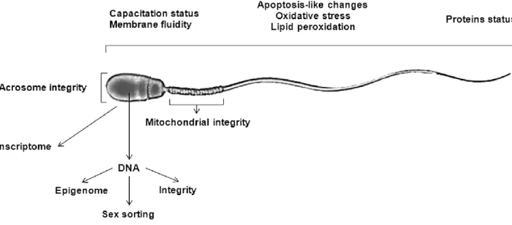
array of biomarkers including fluorescent markers for sperm plasmalema integrity, permeability and stability, acrosomal status, sperm mitochondrial integrity and activity, capacitation status and membrane fluidity of sperm, altered sperm chromatin or DNA integrity, apoptotic, oxidative stress and lipid peroxidation events in sperm, and antibodies detecting proteins that are either up- or down-regulated in defective spermatozoa (Figure 1-1) (Sutovsky and Lovercamp, 2010). Using flow cytometry (FC), the majority of these biomarkers can be efficiently evaluated (Martínez-Pastor et al., 2010). Flow cytometry enables objective measurement of these biomarkers (individually or simultaneously) in semen through automated, high throughput and rapid methods (Gillan et al., 2005; Hossain et al., 2011).
Figure 1-1 Different spermatic characteristics that can be evaluated at the molecular level to either assess sperm quality or gender selection.
14
1.2.1.1.1.1. Current applied ARTs for cattle sperm
1.2.1.1.1.1.1. Breeding soundness evaluation (BSE) and estimation of sire fertility Recently, bull fertility has received increasing attention as the results of artificial insemination (AI) are declining in highly selected dairy cattle populations (Karoui et al., 2011). One of the most important challenge in AI industry is to identify bulls producing large numbers of fertile sperm and accurately predicting fertility of dairy bulls with apparently normal semen (Foote, 2003). Breeding soundness which refers to a bull’s ability to get cows pregnant can be classically evaluated to identify bulls with substantial deficits in fertility, but does not consistently identify sub-fertile bulls (Kastelic and Thundathil, 2008). There is urgent need for accurate biomarkers of fertility to complement traditional breeding soundness evaluation, identifying and eliminating bulls with inferior fertility/semen quality (Shojaei et al., 2012; Sutovsky and Kennedy, 2013). Currently, relative sire fertility can be retrospectively estimated based on AI records for the calculation of the very general parameter of non-return rate (NRR) which is based on whether or not the female had a second insemination (indicative of a failed gestation causing a return to estrus) within a certain period (either 28, 56 or 128 days post-insemination). Usually 56 days after the first insemination (day of AI = Day 0) is currently the most commonly used time point for recording fertility in Canada and Europe (Rodríguez-Martínez, 2003). While, NRR provides reliable results over longer periods of time, it is not completely accurate and is highly prone to bias by several factors including unexplained variation. (Rodríguez-Martínez, 2003; Foote, 2003; Amann and DeJarnette, 2012). The NRR value represents the combined