HAL Id: dumas-00631137
https://dumas.ccsd.cnrs.fr/dumas-00631137
Submitted on 11 Oct 2011HAL is a multi-disciplinary open access archive for the deposit and dissemination of sci-entific research documents, whether they are pub-lished or not. The documents may come from teaching and research institutions in France or abroad, or from public or private research centers.
L’archive ouverte pluridisciplinaire HAL, est destinée au dépôt et à la diffusion de documents scientifiques de niveau recherche, publiés ou non, émanant des établissements d’enseignement et de recherche français ou étrangers, des laboratoires publics ou privés.
Transplantation pulmonaire et dysfonction chronique du
greffon : caractérisation de biomarqueurs diagnostiques
par analyse protéomique SELDI-TOF du liquide
bronchiolo-alvéolaire
Sébastien Quetant
To cite this version:
Sébastien Quetant. Transplantation pulmonaire et dysfonction chronique du greffon : caractérisation de biomarqueurs diagnostiques par analyse protéomique SELDI-TOF du liquide bronchiolo-alvéolaire. Médecine humaine et pathologie. 2009. �dumas-00631137�
UNIVERSITE JOSEPH FOURIER
FACULTE DE MEDECINE DE GRENOBLE
Année 2009
N°
Transplantation pulmonaire et dysfonction chronique du greffon :
Caractérisation de biomarqueurs diagnostiques par analyse
protéomique SELDI-TOF du liquide bronchiolo-alvéolaire
THESE PRESENTEE POUR L’OBTENTION DU DOCTORAT DE MEDECINE
DIPLÔME D’ETAT
QUETANT Sébastien
Né le 31 mai 1979 à Annecy
SOUTENUE PUBLIQUEMENT A LA FACULTE DE MEDECINE DE GRENOBLE
Le 14 octobre 2009
DEVANT LE JURY COMPOSE DE :
Monsieur le Professeur Christophe PISON
Président du jury
Madame le Professeur Françoise MOREL
Directeur de thèse
Monsieur le Professeur Christian BRAMBILLA Monsieur le Professeur François BERGER Madame le Docteur Candice TROCME
- 2 -
REMERCIEMENTS
Je tiens tout d’abord à remercier Monsieur le Professeur Christophe PISON pour avoir accepté de présider ce jury. Je vous remercie également pour toutes les connaissances que vous m’avez apportées durant mon internat et pour votre enthousiasme à toute épreuve. Soyez assuré de mon profond respect.
Je témoigne toute ma gratitude à Madame le Professeur Françoise MOREL pour m’avoir accueilli au sein du laboratoire GREPI CNRS UMR 5525. Je vous remercie également d’avoir accepté de diriger cette thèse. Soyez assuré de ma reconnaissance et de l’honneur que j’ai eu de travailler à vos côtés.
Je remercie Monsieur le Professeur Christian BRAMBILLA pour avoir accepté de juger ce travail. Merci pour votre enseignement, votre énergie débordante et votre franchise. Veuillez trouver ici le témoignage de toute ma considération.
Je remercie Monsieur le Professeur François BERGER pour avoir accepté de participer à ce jury et pour m’avoir permis de m’initier à l’utilisation de la technologie protéomique SELDI-TOF en recherche clinique. Soyez assuré de ma plus sincère reconnaissance.
Un immense remerciement au Docteur Candice TROCME pour m’avoir initié à la recherche et guidé tout au long de ce travail avec beaucoup de patience, de disponibilité et d’humour. Avec toute mon amitié.
Merci à Anne Claire et Clémence, mes co-internes de choc, pour votre amitié et votre soutien sans faille durant ces années d’internat. Merci également à Régine, Xinthia et Natacha pour leur dévouement, et leur gentillesse.
Merci aux Pr Denis Moro-Sibilot et Pr Jean-Louis Pépin pour votre disponibilité et vos précieux conseils. Merci à Christel, Bérengère, Marie, Thomas, Samia, Claire, Patricia, Olivier, Agnès, Rebecca, Carole, Renaud, Bernard et Sandrine pour tout le savoir que vous m’avez transmis, chacun à votre façon, avec patience et gentillesse. Amitiés à tous.
- 3 -
Merci à tous les autres pneumologues qui m’ont appris les ficelles du métier (François, Gille, Dominique, Emilie…) et tout particulièrement à Natalie Chouri qui m’a soutenu et encadré à mes débuts d’interne.
Merci à tout le personnel de pneumologie et de réanimation avec qui j’ai partagé tant de bons moments et aussi un certain nombre de nuits blanches. Merci à Bernadette, Manu, Zahia et Audrey pour m’avoir appris tous les secrets de la ventilation non invasive et à Céline pour nos crises de fourires au 4ème D. Merci à aux « drôles de dames » (Christelle, Danielle et Fabienne) pour avoir supporté mes coups de gueule.
Merci aux équipes du GREPI, du CIC et de Cytologie pour leur accueil et pour avoir pris le temps de répondre à mes questions de néophyte en science.
Merci,
A ma compagne, Aurélie, pour son soutien sans faille, son amour, ses bons petits plats qui remontent le moral et les discussions gastro-pneumo à n’en plus finir.
A mes parents, pour leur amour, leur dévouement et leur soutien tout au long de ces années. A ma sœur, Béatrice et sa petite famille, Jack et petit Paul, pour toute la joie que vous me procurez.
A ma belle famille, Elisabeth, Bernard, Carole et Vivien pour tous ces bons moments passés en Ardèche.
A Caro et Eric (et Nael), Audrey et Sven (et Sarah), Caro Caro, Renaud et Estelle (et Arwen), Max, Christophe et Marilyn pour votre amitié.
A Rim, Jérôme (et Emeline) pour les apéros et dîners improvisés.
Et pour finir aux vieux copains de lycée Nico, Christophe, Yves, Gaël, QuéQué et Cyril, avec qui j’ai passé tant de bons moments et qui ont égaillé mes années de lycée et de médecine.
- 4 -
TABLE DES MATIERES
Résumé ... 6
Mots clés et Abréviations ... 8
Article ... 10
ABSTRACT ... 12
INTRODUCTION ... 13
PATIENTS AND METHODS ... 15
Study population ... 15 Sample processing ... 16 SELDITOF MS analysis ... 17 Statistical analysis ... 17 RESULTS ... 18 Patient characteristics ... 18
BALF proteome profiling ... 18
BOS versus stable groups ... 19
BOS versus AR groups ... 20
AR versus stable groups ... 20
-- 5 --
Characterization and correlation of the detected biomarkers ... 21
DISCUSSION ... 22 REFERENCES ... 26 TABLES ... 33 LEGENDS ... 36 FIGURES ... 38 Conclusion ... 44
-- 6 --
- 7 -
RESUME
Objectifs : Malgré les progrès récents réalisés en transplantation pulmonaire, le pronostic à
long terme est compromis par la survenue d’une dysfonction chronique du greffon dont le diagnostic est basé sur le déclin de la fonction respiratoire. Nous avons analysé les profils protéiques du liquide de lavage bronchiolo-alvéolaire (LBA) de patients transplantés pulmonaires par une technologie protéomique innovante afin de caractériser des biomarqueurs diagnostiques de dysfonction chronique du greffon.
Matériels et méthodes : Nous avons comparé de façon transversale le protéome des LBA de
13 patients avec un syndrome de bronchiolite oblitérante (groupe BOS), 21 patients avec une fonction pulmonaire stable (groupe ST) et 8 patients avec rejet aigu (groupe AR) par analyse protéomique SELDI-TOF MS (Surface Enhanced Laser Desorption/Ionization Time-Of-Flight Mass Spectrometry).
Résultats : 45 pics d’intensité statistiquement différente ont été caractérisés entre le groupe
BOS et ST, et 42 entre les groupes BOS et AR. Parmi les pics les plus discriminants, des biomarqueurs correspondant aux protéines S100 (10812 Da, 10412 Da et 13335 Da, p<0,002) et aux alpha-défensines 1-3 (3354 Da, 3424 Da et 3470 Da, p<0,01) étaient surexprimés dans le groupe BOS. La protéine S100A8 (10812 Da) permet de différencier les patients BOS des patients stables avec une sensibilité de 92% et une spécificité de 72% (AUC=0.87) et les alpha-défensines présentent également une performance diagnostique importante.
Conclusion : L’analyse protéomique du LBA par technique SELDI-TOF MS a permis de
caractériser plusieurs biomarqueurs d’intérêt pour le diagnostic de dysfonction chronique du greffon et potentiellement impliqués dans sa physiopathologie : les protéines S100A8, S100A9, S100A12 et les alpha-défensines.
- 8 -
Mots clés et
Abréviations
- 9 -
KEYWORDS
Lung transplantation, chronic allograft dysfunction, biomarkers, proteomic, SELDI-TOF MS (Surface Enhanced Laser Desorption / Ionization Time-Of-Flight Mass Spectrometry), bronchoalveolar lavage fluid, S100 proteins, alpha-defensins.
ABBREVIATIONS
AR Acute Rejection
AUC Area Under the Curve
BALF BronchoAlveolar Lavage Fluid
BCA Bicinchoninic Acid
BOS Bronchiolitis Obliterans Syndrome
CF Cystic Fibrosis
CsA Cyclosporin A
Da Dalton
FEV1 Forced Expiratory Volume in one second
ISHLT International Society of Heart and Lung Transplantation
MALDI-TOF MS Matrix Assisted Laser Desorption / Ionization - Time-Of-Flight Mass Spectrometry
MMF Mycophenolate Mofetil PBS Phosphate Buffer Saline PMN PolyMorphonuclear Neutrophil
RAGE Receptors for Advanced Glycation End products ROC Receiver Operator Characteristic
RT Room Temperature
SDS Sodium Dodecyl Sulphate
SELDI-TOF MS Surface Enhanced Laser Desorption / Ionization -Time-Of-Flight Mass Spectrometry
SP-A Surfactant Protein A
ST Stable
- 10 -
- 11 -
Characterization of chronic lung allograft dysfunction biomarkers in
bronchoalveolar lavage fluid by SELDI-TOF proteomic analysis
Sébastien Quétant*1,2, Candice Trocme*1,3, (PhD), Marie Arlotto4, Athan Baillet1, Chrystel Saint-Raymond2 (MD), François Berger4 (MD, PhD), Françoise Morel1,3 (MD, PhD), Christophe Pison2 (MD, PhD).
*Both authors contributed equally to this work
1. GREPI TIMC-Imag CNRS UMR 5525, Université J. Fourier, Grenoble/France 2. Clinic of Respiratory Medicine, CHU Hôpital Michallon, Grenoble/France 3. Laboratory of Enzymology / DBPC, CHU Hôpital Michallon, Grenoble/France 4. Grenoble Institute of Neurosciences, INSERM U836, Grenoble/France
Source of support: SQ was supported by a grant from the Société de Pneumologie de Langue
Française (SPLF)
Corresponding author :
Sébastien Quétant
Clinique de Pneumologie, Pôle de Médecine Aiguë Communautaire CHU de Grenoble
B.P. 217 - 38043 Grenoble Cedex 09/France Tel : +33 (0)4 76 76 84 56
Fax : +33 (0)4 76 76 52 21 Email: squetant@chu-grenoble.fr
Key words (MeSH): Lung transplantation, Proteomics, Chronic allograft dysfunction, S100
proteins, Bronchiolo-alveolar fluid, Biological Markers.
Running head: proteomic in chronic lung allograft dysfunction.
- 12 -
ABSTRACT
Objective: Despite progresses in lung transplantation, long-term prognosis is compromised by
chronic allograft rejection. Its diagnosis is based on a delayed functional criterion and its physiopathology is still unclear. We investigated proteomic profiles of bronchoalveolar lavage fluid (BALF) of lung transplanted patients by an innovative proteomic technology in order to characterize diagnostic biomarkers of chronic allograft rejection.
Methods: In a cross sectional pilot study, protein profiles of 13 lung transplant recipients with
bronchiolitis obliterans syndrome (BOS), 21 patients with lung function stability (ST) and 8 patients with acute rejection (AR) were studied by SELDI-TOF MS (Surface Enhanced Laser Desorption Ionization Time-Of-Flight Mass Spectrometry) analysis.
Results: Forty five differentially expressed proteins were detected in BOS group vs. ST
population and forty two in BOS group vs. AR set. Among the most discriminating peaks, biomarkers corresponding to S100 proteins (10,812 Da, 10,412 Da and 13,335 Da, p<0.002) and alpha-defensins (3,354 Da, 3,424 Da and 3,470 Da, p<0.01) were up regulated in BOS patients compared to ST or AR patients. S100A8 protein (10,812 Da) discriminate BOS patients from ST patients with a sensitivity of 92% and a specificity of 72% (AUC=0.87), while alpha-defensins offer a slightly weaker diagnostic accuracy.
Conclusion: BALF SELDI-TOF analysis allows detection of relevant biomarkers linked to
BOS diagnosis and potentially involved in its physiopathology: S100 proteins and alpha-defensins.
- 13 -
INTRODUCTION
Lung transplantation is currently a valuable therapeutic option for a spectrum of end-stage pulmonary diseases with no other possible effective medical treatment. Outcomes have improved over time in term of survival and quality of life (1, 2), but long-term survival remains disappointing and chronic allograft rejection has emerged as the primary obstacle to long-term outcome (3, 4). Indeed, according to the latest International Society for Heart and Lung Transplantation (ISHLT) Registry report (5), chronic allograft dysfunction, as clinically manifested by bronchiolitis obliterans syndrome (BOS), affects up to 50-60% of lung transplant recipients who survive 5 years after transplantation and accounts for the leading cause of death beyond the first year (6).
Obliterative bronchiolitis is the histologic hallmark of chronic rejection, and is characterized by submucosal fibrosis of the membranous and respiratory bronchioles, which results in luminal obliteration. Histologic confirmation of obliterative bronchiolitis is difficult because of the paucity of the bronchioles on transbronchial lung biopsies and the non-homogeneous nature of the disorder. Bronchiolitis obliterans syndrome (BOS) is the clinical feature for obliterative bronchiolitis and refers to a physiological criteria: a sustained decline of pulmonary function (decrease of 20% or more in forced expiratory volume in one second (FEV1)) in absence of other potential causes (anastomotic complications, infection or acute rejection (AR)) (7). Unfortunately this internationally accepted criterion reflects an advanced stage of the disease.
The exact pathogenesis of BOS as well as early markers of the disease are still unknown. Obliterative bronchiolitis is considered to be a multifactorial process involving both immune-mediated pathways (acute rejection, human leukocyte antigen mismatches or antibody-mediated rejection) and alloimmune-independent pathways (infection, ischemia-reperfusion injury, gastro esophageal reflux…) which leads to an aberrant tissue remodeling (8, 9).
- 14 -
Complex mechanisms of obliterative bronchiolitis remain unclear; however increasing body of evidence suggests that both cells of innate and adaptive immunity, as well as structural cells are involved (10-14). A number of studies have highlighted the critical role of polymorphonuclear neutrophil (PMN) and have shown that neutrophilia in bronchoalveolar lavage fluid (BALF) correlates with chronic allograft rejection (15-18). Previous studies have shown that various biomarkers such as cytokines (19, 20), chemokines (21, 22), growth factors (23, 24) or other proteins (25, 26) that are under or overexpressed in BALF were linked to BOS pathogenesis but their diagnostic value is not currently established.
Traditional research approaches that target specific compounds on the basis of previous data can not completely catch the process complexity. In contrast system-based methodologies, such as proteomics, analyze global biological changes and provide an opportunity to identify new biomarkers of diseases. Proteomic tests have been developed in the pulmonary field for several years (27-30). First 2D gel electrophoresis (31) and more recently surface enhanced laser desorption / ionization -time-of-flight mass spectrometry (SELDI-TOF MS) technology were used to analyze the proteome of BALF (32-36), blood (37, 38), induced sputum (39-41) or exhaled breath condensate (42). SELDI-TOF MS is an innovative high throughput proteomic method specifically dedicated to clinical applications (43, 44) whose relevance was emphasized in a wide range of disease such as cancer (45-47) or inflammatory diseases (48-50). In lung transplantation, only two studies have focused on BALF proteome analysis: Nelsestuen and colleagues analyzed proteome profiles of BALF samples from lung transplant recipients (LTRs) using a matrix assisted laser desorption / ionization - time-of-flight mass spectrometry (MALDI-TOF MS) technology, and characterized some biomarkers of BOS, in particular alpha-defensins (51). More recently, using gel electrophoresis and protein identification by MALDI-TOF MS (2DE-MS), Meloni and colleagues suggested that surfactant protein A (SP-A) levels in BALF may predict BOS onset in LTRs. (52).
- 15 -
In the present study we performed BALF proteome profiling using SELDI-TOF MS technology to characterize BOS-specific biomarkers.
PATIENTS AND METHODS
Study populationThis cross-sectional study consisted of the analysis of 42 BALF samples prospectively acquired between July 2007 and September 2008 from 42 LTRs at the Grenoble University Hospital. The protocol was approved by the local ethics committee and all patients gave their informed consent to the study.
LTRs underwent regular clinical visits including functional and radiological evaluation. Spirometry was obtained routinely every month during the first postoperative year, and every 3 months after. They systematically underwent bronchoscopy with collection of BALF samples and transbronchial biopsies at 1, 3, 6 and 12 months after lung transplantation or in case of suspected infection or AR based on clinical, radiological or pulmonary function criteria. Microbiological examination was systematically performed in BALF. Infection was defined as a positive bacterial or fungal or viral culture, combined with medical findings (fever, need of antibiotics, antifungal or viral treatment), high level of C-reactive protein (CRP), and radiological examination (new radiological infiltrates). Colonization was defined as a positive culture of the investigated BALF samples without clinical findings, no increased CRP and no new radiological infiltrates.
The following groups were defined. (1) The BOS group comprised patients fulfilling the criteria of the ISHLT based on the decline of FEV1 (7). (2) The stable group comprised clinically stable patients who displayed normal and stable respiratory function (FEV1 ≥ 80% of baseline) and no clinical symptom of infection or AR. (3) The AR group included LTRs with a diagnosis of AR defined and graded according to the histological analysis of
- 16 -
transbronchial biopsies (53). If LTRs underwent repeated BALF sampling during the study period only the first BALF sample was considered. LTRs with infection criteria were excluded but not those with colonization.
Patients received the same immunosuppressive regimen throughout the study. Anti-IL-2 monoclonal antibodies (basiliximab, Novartis, France) were given for the induction therapy on day 1 and 5. The standard maintenance treatment consisted of tacrolimus (whole blood target levels between 10 and 15 ng/ml according to the postoperative delay), mycophenolate mofetil and prednisone. Episodes of AR ≥ grade A2 were treated with intravenous bolus of methylprednisolone (15 mg/kg/day on 3 consecutive days). Azithromycin (250 mg/day) was added to the immunosuppressive drug regimen as soon as the diagnosis of BOS was made.
Sample processing
BALF was performed during a flexible bronchoscopy by instilling a total volume of 120 ml of sterile isotonic saline solution (3 fractions of 40 ml) into a sub-segmental bronchus of either the lingual or the right middle lobe (54). The first recovered BALF aliquot was removed because of bronchial contamination. The next BALF samples were immediately sent to the laboratory and processed within 60 min. Cytology (total and differential cell count by cytospin and May-Grünwald Giemsa staining) and microbiology contents were evaluated on one aliquot. The last sample was processed for the proteomic analysis. After a first centrifugation (2000g, 10 min, room temperature (RT)) to remove cells, supernatant was centrifuged a second time (14000g, 10 min, +4°C) to discard any bacteria. Then, supernatant was treated by a protease inhibitor cocktail (Roche Diagnostics, Meylan, France), aliquoted and stored at -80°C until further analysis.
- 17 -
SELDI-TOF MS analysis
BALF protein profiles were obtained by the SELDI-TOF-MS method (Ciphergen Biosystems, Fremont, CA), using a cation exchange array (CM10) and an anion exchange array (Q10). Protein concentration of BALF was measured using the bicinchoninic acid (BCA) method (Pierce, Rockford, IL, USA): 1 μg was loaded on each array. Samples were processed robotically on a Biomek® 2000 Automation Workstation (Beckman Coulter, Villepinte, France). First, arrays were equilibrated with 200 μl of the corresponding loading/washing buffer (20 mM Tris, 0.1% Triton X100 (v/v) pH 8 for Q10; 20 mM sodium acetate, 0.1% Triton X100 pH 4 for CM10) during 5 minutes at RT. Respective amounts of BALF diluted in 50 μl of loading/washing buffer were incubated on each chip for 60 min at RT. Spots were washed twice with 200 μl of washing buffer, twice with 200 μl of the same buffer without Triton X100 and finally with 200 μl of 5 mM Tris pH 8 (Q10) or 5 mM sodium acetate pH 4 (CM10). After drying, spots were loaded twice with 0.8 μl of saturated sinapinic acid in 0.5% trifluoroacetic acid, 50% acetonitrile and chips were analyzed on the PCS-4000 ProteinChip® reader. Two laser intensities (1,800 nJ and 3,000 nJ) were used to analyze protein profiles and mass data processing was performed with the Ciphergen Express™ software, following the classical steps of spectra calibration, normalization, peak detection and aligning. The analysis was restricted to peptide peaks above 2,500 Da.
Statistical analysis
Data were analyzed using the SPSS/PC software package (SPSS 17.0, SPSS Inc, Chicago, IL). Continuous variables were represented as median [range] and compared using the Student’s t-test, the Kruskall-Wallis test or the ANOVA test. Categorical variables are described by frequencies and percentages with 95% confidence interval (CI) and compared by using the exact Fischer test. In the case of comparisons involving more than two groups, p-values were
- 18 -
adjusted using the Bonferroni method. A p-value below 0.05 was considered statistically significant. Mann-Whitney U tests were used to compare SELDI-TOF MS peak intensities. ROC analyses were performed to measure the diagnostic accuracy of each biomarker. Correlations between continuous variables were determined using the Spearman’s rank test. Multivariate comparisons between two populations were performed by supervised hierarchical clustering analyses of the characterized biomarkers and results were visualized by heat maps which are a graphical representation of overexpressed (red), decreased (green) or unchanged (black) proteins previously identified as differentially expressed in the two studied populations. Heat maps and hierarchical clustering were performed automatically by the Ciphergen Express™ software.
RESULTS
Patient characteristics
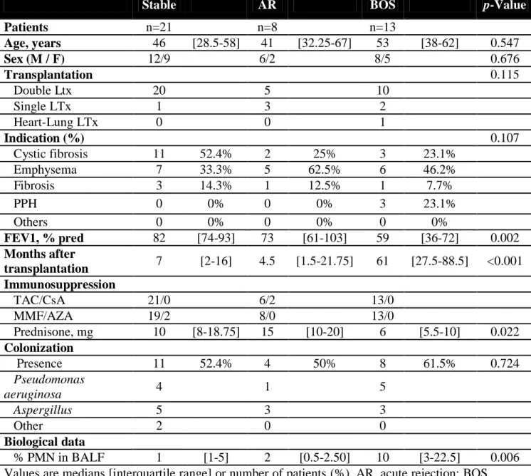
We included a total of 42 BALF samples obtained from 13 LTRs who developed BOS (6 BOS grade 1, 5 BOS grade 2 and 2 BOS grade 3), 21 LTRs who still had stable graft function and 8 LTRs with an AR (3 grade A1 and 5 grade A2). Demographic parameters are summarized in Table 1.
BALF proteome profiling
An example of generated data is shown in Figure 1. Distinct protein profiles of BALF were observed on CM10 and Q10 arrays. SELDI-TOF MS spectra generated from BALF samples typically displayed proteins ranging between 2,500 and 20,000 Da. Comparison of those spectra revealed a large number of protein peaks that are differentially expressed between groups.
- 19 -
BOS versus stable groups
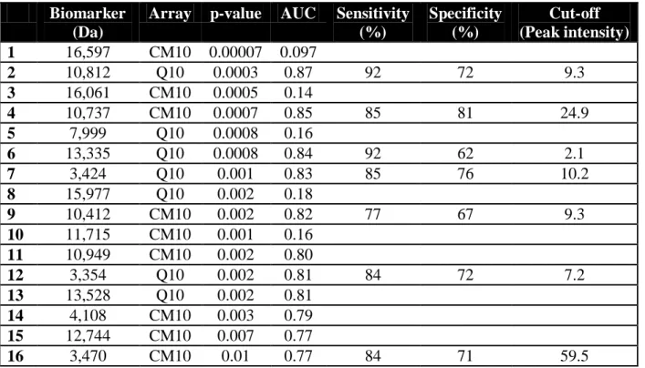
Analyses of protein profiles revealed 177 protein peaks. Statistical comparisons of all peak intensities between BOS and stable groups indicated that 45 proteins were differentially expressed in both populations (Table 2). We focused on two particularly abundant clusters of proteins that were located at approximately 3,400 Da and 10-13,000 Da. The most differentially expressed biomarkers were four proteins isolated on both arrays at 10,812 9.4 Da, 10,737 11 Da, 10,412 8.3 Da and 13,335 17.5 Da which were all overexpressed in the BOS group (Fig 2A, 2C, 2D). Their mean intensities were two to five times higher in the BOS population with significant statistical differences between p=0.0003 and p=0.002. ROC curve analyses showed that these biomarkers discriminated BOS patients from stable patients with high sensitivities and specificities (Table 3). The most discriminating biomarker was the protein of 10,812 Da that provided an accurate diagnosis of BOS with a sensitivity of 92% and a specificity of 72% (AUC=0.87; 95%IC 0.76-0.98) (Fig 2B and Table 3).
Another cluster of three peptides of approximately 3,400 Da was differentially expressed in BALF of BOS group compared to the stable population (Table 3). These biomarkers of 3,354
3.5 Da, 3,424 3.6 Da and 3,470 5.1 Da, also characterized on both arrays, were statistically enhanced in the BOS group (Fig 2E). They were a little less accurate than the previous biomarkers for the discrimination of the BOS population from the stable group (Table 3).
The 45 biomarkers whose expression was significantly different in BOS and stable groups were visualized in a heat map (Fig 3). Most of them were increased in BOS samples and decreased in stable BALF. Classification of samples according to those 45 proteins did not improve the identification of BOS patients compared to the discriminating potential of individual markers: this hierarchical clustering map displayed a sensibility of 85% and a
- 20 -
specificity of 67% in differentiating BOS from stable samples. These results were not affected by the presence of bacterial or fungal colonization. Comparison between colonized patients and no colonized patients in the whole population showed that the expression of the previous discriminating biomarkers was not related to the colonization status (Data not shown).
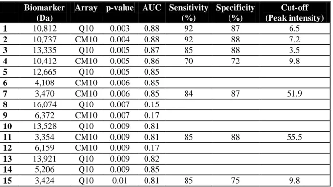
BOS versus AR groups
Among 177 proteins detected in BOS and AR BALF samples, 42 were differentially expressed in both populations (Table 2). Moreover, the previously characterized 10,812 Da, 10,737 Da, 13,335 Da and 10,412 Da proteins were the most relevant to differentiate the both populations (Fig 4A, 4C, 4D). For example the average intensity of 10,812 Da biomarker was 13.7 2.2 in the BOS group versus 5.4 2.8 in the AR set (p=0.003) (Fig 4A), providing an accurate diagnosis of BOS with a sensitivity of 92% and a specificity of 87% (AUC=0.88; 95% IC 0.71-0.97) (Fig 4B).
The three proteins of approximately 3,400 Da were also characterized in the comparison of BOS and AR protein profiles with peak intensities significantly higher in the BOS population (Fig 4E). These biomarkers displayed a weaker accuracy than the above-mentioned biomarkers in discriminating BOS from AR patients (Table 4). Concerning the hierarchical clustering classification, the sensitivity of this analysis in classifying BOS patients in this group was 77%, with 3 misclassified samples. Only one AR sample was classified in the BOS pattern group, corresponding to a specificity of 87.5% (Fig 5).
AR versus stable groups
We also compared protein profiles of stable patients with those of the AR group. Among 177 peaks detected, only 4 were differentially expressed between both groups (Table 2). We did
- 21 -
not detect any statistical difference in the intensity of the previously described biomarkers.
Differential cell count of BALF
The differential cell count of BALF showed a significantly elevated percentage of PMN in BOS patients compared to stable patients (p=0.003) and AR patients (p=0.017). No significant difference was found between AR and stable population (p=0.667). The percentage of PMN in BALF discriminated BOS patients from other groups with a sensitivity of 66% and a specificity of 84% (AUC=0.75; 95%CI 0.66-0.96), which was less accurate than the biomarkers characterized in the proteomic analysis.
Characterization and correlation of the detected biomarkers
According to published data concerning the proteome of BALF we could hypothesize on the identity of the detected biomarkers. The cluster of the 3,400 Da proteins was previously identified in several SELDI-TOF MS studies as alpha-defensin-1, -2 and -3 also called human neutrophil peptides 1-3 (HNP 1-3) (35). Moreover molecular weights of the 3 biomarkers between 10 and 13 kDa were in agreement with published molecular masses of several S100 proteins (30, 32, 35, 39). The biomarkers of 10,412 Da, 10,812 Da and 13,335 Da respectively corresponded to S100A12 (calgranulin C), S100A8 (also called MRP-8 or calgranulin A) and S100A9 proteins (MRP-14 or calgranulin B). The only 10,737 Da protein was yet unidentified in BALF proteome databases. A strong correlation was found between peak intensities of this biomarker and S100A8 (rho=0.641, p<0.001) (Fig 6E).
S100 proteins and alpha-defensins are mainly produced by PMN in inflammatory diseases. Relative peak intensities of S100A8, S100A9 and S100A12 were correlated with the percentage of PMN in BALF samples (respectively rho=0.613, p<0.001; rho=0.531, p < 0.001 and rho=0.492, p=0.001) (Fig 6A, 6B, 6C). Similarly we demonstrated a statistical correlation
- 22 -
between peak intensities of alpha-defensins and percentage of PMN (respectively rho=0.531, p<0.001; rho=0.485, p=0.002; rho=0.366, p=0.02 for biomarkers of 3,354 Da, 3,424 Da and 3,470 Da) (Fig 6D).
DISCUSSION
Chronic allograft dysfunction after lung transplantation remains a challenge. Its diagnosis is often delayed because of the poor sensitivity of functional and biological tests. The discovery of new biomarkers could improve disease diagnosis and knowledge of its pathophysiology, which may lead to the identification of new therapeutic targets. Proteomic technologies are particularly powerful tools to address these issues. Applying the SELDI-TOF MS technology to the analyses of BALF samples of LTRs, we were able to determinate a large panel of biomarkers that differentiate, with a high statistical significance, LTRs with chronic allograft dysfunction from stable patients or LTRs with acute rejection.
SELDI-TOF MS is a high throughput technic which analyze the proteome, i.e. the whole content of a biological fluid. It associates a first phase of protein fractionation on chips with distinct chromatographic surfaces (cationic, anionic…) to a second detection step of protein molecular weight by mass spectrometry. This technology has been applied to a large range of body fluids such as serum, urine and synovial fluid (55) to characterize disease specific biomarkers, but only few data are available concerning its application to BALF samples. Although the BALF procedure has been used for more than 30 years for diagnostic purposes in lung diseases, information about the protein content of BALF is still limited at least in part because of methodologic problems in sample processing (56, 57). Dilution of proteins by the lavage procedure, high salt content, and/or low concentrations of some proteins are restricting factors for many analyses and the desalting and concentration processes lead to significant loss of BALF proteins (32, 56). However these sample characteristics do not interfere with
SELDI-- 23 SELDI--
TOF MS analysis. Indeed, concentration and desalting of BALF samples are not required because of the femtomole sensitivity of the technic and the dilution of the sample in the specific array-type buffer. To avoid any concentration-related biases in our study, supernatants were adjusted to a total protein concentration of 1g/50l before applying on CM10 and Q10 arrays.
This analysis of BALF protein expression patterns revealed respectively 45 and 42 protein peaks that are associated with BOS phenotype in comparison with stable patients and LTRs with acute rejection. Among them, the most relevant biomarkers were three proteins of 10,412 Da, 10,812 Da and 13,335 Da significantly increased in BOS patients. Previous published data suggested that these proteins respectively corresponded to S100A12, S100A8 and S100A9 proteins (30, 32, 35, 39). These S100 proteins are calcium-binding proteins mainly produced by PMN (58) and are involved in pro-inflammatory processes associated with various inflammatory conditions (59). The S100A8 and S100A9 proteins are often complexed as a S100A8/S100A9 heterodimer called calprotectin. Calprotectin is chemotactic for PMN both in
vitro and in vivo (60) and is involved in the activation of NADPH oxidase, leading to an
increased release of reactive oxygen species (61). S100A12 is implicated in a novel inflammatory axis, involving Receptors for Advanced Glycation End products (RAGE) as a receptor transducing proinflammatory signals in endothelium (62), and its chemotactic properties contribute to leucocytes extravasation at sites of inflammation (63). A study of human alveolar epithelial cells showed that S100A8/S100A9 heterodimer stimulates IL-8 production in pulmonary epithelium and amplifies local neutrophilic inflammation (64). Previous clinical studies reported an up-regulation of S1008 and/or S1009 proteins in inflammatory lung diseases (65), particularly in cystic fibrosis (CF) (66, 67). A high level of calprotectin in serum was associated to an inflammation profile in CF (68) and more recently, using a SELDI-TOF MS approach, McMorran and colleagues demonstrated that S100A8, S100A9 and S100A12 proteins isolated in BALF were strongly associated with CF airway
- 24 -
inflammation (35). Interestingly, we reported here for the first time the potential involvement of these S100 proteins as diagnostic biomarkers of BOS. This result was consistent with the pivotal role of pro-inflammatory proteins in BOS pathogenesis. Indeed these S100 proteins could induce lung damages and aberrant tissue repairs in small airways by promoting neutrophil oxidative burst and leukocytes recruitment.
Another BOS biomarker was also characterized in the same molecular weight range. However the identity of this 10,737 Da protein is still unknown. The narrow difference of its molecular weight with the S100A8 mass and the strong correlation of their peak intensities suggested that this protein may be an isoform of S100A8. A truncated form of S100A8 has already been reported in BALF (35), but it is slightly shorter (10,580 Da) than this biomarker.
We also characterized a cluster of three proteins of approximately 3,400 Da overexpressed in BALF of BOS patients and that strongly discriminates the BOS population from stable or AR groups. According to the predicted molecular weight mentioned in BALF proteome databases, we speculated that these proteins corresponded to alpha-defensins (35, 51). These proteins stored in the azurophilic granules and released from neutrophils during degranulation are important antimicrobial peptides involved in the innate immune response (69-71). They also play a key role in the orchestration of subsequent adaptive immune response and repair processes (72, 73). Few publications reported the role of alpha-defensins in chronic lung allograft dysfunction. In a retrospective study, Nelsestuen and colleagues showed that alpha-defensins may serve as a biomarker predicting BOS onset (51). Moreover in our study, the overexpression of alpha-defensins in recipients with BOS was not affected by the presence of airway pathogens. Our findings are in agreement with the results of Anderson and colleagues (74) who found a higher level of alpha-defensins in BOS LTRs than in stable LTRs, whatever their colonization status. These data suggest that the increased level of alpha-defensins in BALF of BOS patients contributes to a proinflammatory microenvironment and may promote an exaggerated repair response, independently of colonization by airway pathogens.
- 25 -
A drawback of this study is the absence of formal identification of the biomarkers. The molecular weights of the biomarkers are closely related to their predicted molecular weight established in publications and/or protein databases. However, we cannot exclude that these protein peaks represent other proteins. Moreover the use of SELDI-TOF MS has allowed us to characterize other relevant biomarkers including proteins down-regulated in BOS patients compared to stable patients. For example a protein of 16,597 Da is highly under-expressed in BOS BALF compared to stable samples, but its identity is unknown.
Innovative bioinformatic tools presently provide an opportunity to fully apprehend the complexity of proteomic data. It would be interesting to apply bioinformatic algorithms to this data set in a decision tree that combines several biomarkers and then improves the diagnostic accuracy.
In conclusion, we underlined the usefulness of SELDI-TOF MS platform to characterize in BALF a large panel of new biomarkers that differentiate, with high statistical significances, BOS patients from other populations. To improve the strength of our results we are currently validating detected biomarkers in an independent cohort of LTRs. In addition to their diagnostic value, these biomarkers provide additional molecular details regarding BOS pathogenesis. Additional research is necessary in this area to validate prospectively these biomarkers in a larger cohort of patients and to clarify the possible link of increased levels of S100 proteins and alpha-defensins with chronic allograft dysfunction in lung transplantation. Their predictive value will be evaluated in a longitudinal proteomic analysis of LTRs followed over 5 years. Moreover, these proteins interacting with key modulators of proinflammatory pathways might be appropriate candidates for targeted intervention in BOS.
CONFLICT OF INTEREST
- 26 -
REFERENCES
1. Yusen RD. Technology and outcomes assessment in lung transplantation. Proc Am Thorac Soc 2009;6(1):128-136.
2. Costache V, Chavanon O, St Raymond C, Sessa C, Durand M, Duret J et al. Dramatic improvement in survival after lung transplantation over time: a single center experience. Transplant Proc 2009;41(2):687-691.
3. Boehler A, Estenne M. Post-transplant bronchiolitis obliterans. Eur Respir J 2003;22(6):1007-1018.
4. Dumonceaux M, Knoop C, Rondelet B, Estenne M. [Complications of lung transplantation: peri-operative complications, acute and chronic rejection.]. Rev Mal Respir 2009;26(6):639-653.
5. Christie JD, Edwards LB, Aurora P, Dobbels F, Kirk R, Rahmel AO et al. The registry of the international society for heart and lung transplantation: twenty-sixth official adult lung and heart-lung transplantation Report-2009. J Heart Lung Transplant 2009;28(10):1031-1049.
6. Hachem RR. Lung allograft rejection: diagnosis and management. Curr Opin Organ Transplant 2009.
7. Estenne M, Maurer JR, Boehler A, Egan JJ, Frost A, Hertz M et al. Bronchiolitis obliterans syndrome 2001: an update of the diagnostic criteria. J Heart Lung Transplant 2002;21(3):297-310.
8. Reynaud-Gaubert M. [Pathophysiology of obliterative bronchiolitis in lung transplants]. Rev Mal Respir 2003;20(2 Pt 1):224-232.
9. Sharples LD, McNeil K, Stewart S, Wallwork J. Risk factors for bronchiolitis obliterans: a systematic review of recent publications. J Heart Lung Transplant 2002;21(2):271-281.
10. Belperio JA, Weigt SS, Fishbein MC, Lynch JP, 3rd. Chronic lung allograft rejection: mechanisms and therapy. Proc Am Thorac Soc 2009;6(1):108-121.
11. Grossman EJ, Shilling RA. Bronchiolitis obliterans in lung transplantation: the good, the bad, and the future. Transl Res 2009;153(4):153-165.
12. Shilling RA, Wilkes DS. Immunobiology of chronic lung allograft dysfunction: new insights from the bench and beyond. Am J Transplant 2009;9(8):1714-1718.
13. Robertson AG, Griffin SM, Murphy DM, Pearson JP, Forrest IA, Dark JH et al. Targeting allograft injury and inflammation in the management of post-lung transplant
- 27 -
bronchiolitis obliterans syndrome. Am J Transplant 2009;9(6):1272-1278.
14. Verleden GM, Vos R, De Vleeschauwer SI, Willems-Widyastuti A, Verleden SE, Dupont LJ et al. Obliterative bronchiolitis following lung transplantation: from old to new concepts? Transpl Int 2009;22(8):771-779.
15. Devouassoux G, Drouet C, Pin I, Brambilla C, Brambilla E, Colle PE et al. Alveolar neutrophilia is a predictor for the bronchiolitis obliterans syndrome, and increases with degree of severity. Transpl Immunol 2002;10(4):303-310.
16. DiGiovine B, Lynch JP, 3rd, Martinez FJ, Flint A, Whyte RI, Iannettoni MD et al. Bronchoalveolar lavage neutrophilia is associated with obliterative bronchiolitis after lung transplantation: role of IL-8. J Immunol 1996;157(9):4194-4202.
17. Riise GC, Andersson BA, Kjellstrom C, Martensson G, Nilsson FN, Ryd W et al. Persistent high BAL fluid granulocyte activation marker levels as early indicators of bronchiolitis obliterans after lung transplant. Eur Respir J 1999;14(5):1123-1130. 18. Zheng L, Whitford HM, Orsida B, Levvey BJ, Bailey M, Walters EH et al. The
dynamics and associations of airway neutrophilia post lung transplantation. Am J Transplant 2006;6(3):599-608.
19. Meloni F, Vitulo P, Cascina A, Oggionni T, Bulgheroni A, Paschetto E et al. Bronchoalveolar lavage cytokine profile in a cohort of lung transplant recipients: a predictive role of interleukin-12 with respect to onset of bronchiolitis obliterans syndrome. J Heart Lung Transplant 2004;23(9):1053-1060.
20. Keane MP, Gomperts BN, Weigt S, Xue YY, Burdick MD, Nakamura H et al. IL-13 is pivotal in the fibro-obliterative process of bronchiolitis obliterans syndrome. J Immunol 2007;178(1):511-519.
21. Reynaud-Gaubert M, Marin V, Thirion X, Farnarier C, Thomas P, Badier M et al. Upregulation of chemokines in bronchoalveolar lavage fluid as a predictive marker of post-transplant airway obliteration. J Heart Lung Transplant 2002;21(7):721-730. 22. Belperio JA, Keane MP, Burdick MD, Lynch JP, 3rd, Xue YY, Berlin A et al. Critical
role for the chemokine MCP-1/CCR2 in the pathogenesis of bronchiolitis obliterans syndrome. J Clin Invest 2001;108(4):547-556.
23. Elssner A, Jaumann F, Dobmann S, Behr J, Schwaiblmair M, Reichenspurner H et al. Elevated levels of interleukin-8 and transforming growth factor-beta in bronchoalveolar lavage fluid from patients with bronchiolitis obliterans syndrome: proinflammatory role of bronchial epithelial cells. Munich Lung Transplant Group. Transplantation 2000;70(2):362-367.
- 28 -
24. Magnan A, Mege JL, Escallier JC, Brisse J, Capo C, Reynaud M et al. Balance between alveolar macrophage IL-6 and TGF-beta in lung-transplant recipients. Marseille and Montreal Lung Transplantation Group. Am J Respir Crit Care Med 1996;153(4 Pt 1):1431-1436.
25. Smith GN, Jr., Mickler EA, Payne KK, Lee J, Duncan M, Reynolds J et al. Lung transplant metalloproteinase levels are elevated prior to bronchiolitis obliterans syndrome. Am J Transplant 2007;7(7):1856-1861.
26. Hubner RH, Meffert S, Mundt U, Bottcher H, Freitag S, El Mokhtari NE et al. Matrix metalloproteinase-9 in bronchiolitis obliterans syndrome after lung transplantation. Eur Respir J 2005;25(3):494-501.
27. Magi B, Bargagli E, Bini L, Rottoli P. Proteome analysis of bronchoalveolar lavage in lung diseases. Proteomics 2006;6(23):6354-6369.
28. Bowler RP, Ellison MC, Reisdorph N. Proteomics in pulmonary medicine. Chest 2006;130(2):567-574.
29. Wattiez R, Falmagne P. Proteomics of bronchoalveolar lavage fluid. J Chromatogr B Analyt Technol Biomed Life Sci 2005;815(1-2):169-178.
30. Noel-Georis I, Bernard A, Falmagne P, Wattiez R. Database of bronchoalveolar lavage fluid proteins. J Chromatogr B Analyt Technol Biomed Life Sci 2002;771:221-236. 31. Wattiez R, Hermans C, Cruyt C, Bernard A, Falmagne P. Human bronchoalveolar
lavage fluid protein two-dimensional database: study of interstitial lung diseases. Electrophoresis 2000;21(13):2703-2712.
32. de Torre C, Ying SX, Munson PJ, Meduri GU, Suffredini AF. Proteomic analysis of inflammatory biomarkers in bronchoalveolar lavage. Proteomics 2006;6(13):3949-3957.
33. Kriegova E, Melle C, Kolek V, Hutyrova B, Mrazek F, Bleul A et al. Protein profiles of bronchoalveolar lavage fluid from patients with pulmonary sarcoidosis. Am J Respir Crit Care Med 2006;173(10):1145-1154.
34. Merkel D, Rist W, Seither P, Weith A, Lenter MC. Proteomic study of human bronchoalveolar lavage fluids from smokers with chronic obstructive pulmonary disease by combining surface-enhanced laser desorption/ionization-mass spectrometry profiling with mass spectrometric protein identification. Proteomics 2005;5(11):2972-2980.
35. McMorran BJ, Patat SA, Carlin JB, Grimwood K, Jones A, Armstrong DS et al. Novel neutrophil-derived proteins in bronchoalveolar lavage fluid indicate an exaggerated
- 29 -
inflammatory response in pediatric cystic fibrosis patients. Clin Chem 2007;53(10):1782-1791.
36. Macgregor G, Gray RD, Hilliard TN, Imrie M, Boyd AC, Alton EW et al. Biomarkers for cystic fibrosis lung disease: Application of SELDI-TOF mass spectrometry to BAL fluid. J Cyst Fibros 2008.
37. Bowler RP, Canham ME, Ellison MC. Surface enhanced laser desorption/ionization (SELDI) time-of-flight mass spectrometry to identify patients with chronic obstructive pulmonary disease. Copd 2006;3(1):41-50.
38. Bozinovski S, Hutchinson A, Thompson M, Macgregor L, Black J, Giannakis E et al. Serum amyloid a is a biomarker of acute exacerbations of chronic obstructive pulmonary disease. Am J Respir Crit Care Med 2008;177(3):269-278.
39. Gray RD, MacGregor G, Noble D, Imrie M, Dewar M, Boyd AC et al. Sputum proteomics in inflammatory and suppurative respiratory diseases. Am J Respir Crit Care Med 2008;178(5):444-452.
40. Sloane AJ, Lindner RA, Prasad SS, Sebastian LT, Pedersen SK, Robinson M et al. Proteomic analysis of sputum from adults and children with cystic fibrosis and from control subjects. Am J Respir Crit Care Med 2005;172(11):1416-1426.
41. Nicholas B, Skipp P, Mould R, Rennard S, Davies DE, O'Connor CD et al. Shotgun proteomic analysis of human-induced sputum. Proteomics 2006;6(15):4390-4401. 42. Fumagalli M, Dolcini L, Sala A, Stolk J, Fregonese L, Ferrari F et al. Proteomic
analysis of exhaled breath condensate from single patients with pulmonary emphysema associated to alpha1-antitrypsin deficiency. J Proteomics 2008;71(2):211-221.
43. Poon TC. Opportunities and limitations of SELDI-TOF-MS in biomedical research: practical advices. Expert Rev Proteomics 2007;4(1):51-65.
44. Kiehntopf M, Siegmund R, Deufel T. Use of SELDI-TOF mass spectrometry for identification of new biomarkers: potential and limitations. Clin Chem Lab Med 2007;45(11):1435-1449.
45. Zhang Z, Bast RC, Jr., Yu Y, Li J, Sokoll LJ, Rai AJ et al. Three biomarkers identified from serum proteomic analysis for the detection of early stage ovarian cancer. Cancer Res 2004;64(16):5882-5890.
46. De Petris L, Orre LM, Kanter L, Pernemalm M, Koyi H, Lewensohn R et al. Tumor expression of S100A6 correlates with survival of patients with stage I non-small-cell lung cancer. Lung Cancer 2009;63(3):410-417.
- 30 -
lung cancer: insights into biology and potential clinical applications. Eur Respir J 2009;34(2):489-506.
48. Trocme C, Marotte H, Baillet A, Pallot-Prades B, Garin J, Grange L et al. Apolipoprotein A-I and platelet factor 4 are biomarkers for infliximab response in rheumatoid arthritis. Ann Rheum Dis 2009;68(8):1328-1333.
49. de Seny D, Fillet M, Meuwis MA, Geurts P, Lutteri L, Ribbens C et al. Discovery of new rheumatoid arthritis biomarkers using the surface-enhanced laser desorption/ionization time-of-flight mass spectrometry ProteinChip approach. Arthritis Rheum 2005;52(12):3801-3812.
50. de Seny D, Fillet M, Ribbens C, Maree R, Meuwis MA, Lutteri L et al. Monomeric calgranulins measured by SELDI-TOF mass spectrometry and calprotectin measured by ELISA as biomarkers in arthritis. Clin Chem 2008;54(6):1066-1075.
51. Nelsestuen GL, Martinez MB, Hertz MI, Savik K, Wendt CH. Proteomic identification of human neutrophil alpha-defensins in chronic lung allograft rejection. Proteomics 2005;5(6):1705-1713.
52. Meloni F, Salvini R, Bardoni AM, Passadore I, Solari N, Vitulo P et al. Bronchoalveolar lavage fluid proteome in bronchiolitis obliterans syndrome: possible role for surfactant protein A in disease onset. J Heart Lung Transplant 2007;26(11):1135-1143.
53. Stewart S, Fishbein MC, Snell GI, Berry GJ, Boehler A, Burke MM et al. Revision of the 1996 working formulation for the standardization of nomenclature in the diagnosis of lung rejection. J Heart Lung Transplant 2007;26(12):1229-1242.
54. Haslam PL, Baughman RP. Report of ERS Task Force: guidelines for measurement of acellular components and standardization of BAL. Eur Respir J 1999;14(2):245-248. 55. Hu S, Loo JA, Wong DT. Human body fluid proteome analysis. Proteomics
2006;6(23):6326-6353.
56. Plymoth A, Lofdahl CG, Ekberg-Jansson A, Dahlback M, Lindberg H, Fehniger TE et al. Human bronchoalveolar lavage: biofluid analysis with special emphasis on sample preparation. Proteomics 2003;3(6):962-972.
57. Lenz AG, Meyer B, Weber H, Maier K. Two-dimensional electrophoresis of dog bronchoalveolar lavage fluid proteins. Electrophoresis 1990;11(6):510-513.
58. Roth J, Vogl T, Sorg C, Sunderkotter C. Phagocyte-specific S100 proteins: a novel group of proinflammatory molecules. Trends Immunol 2003;24(4):155-158.
- 31 -
as clinical laboratory markers of inflammation. Clin Chim Acta 2004;344(1-2):37-51. 60. Ryckman C, Vandal K, Rouleau P, Talbot M, Tessier PA. Proinflammatory activities
of S100: proteins S100A8, S100A9, and S100A8/A9 induce neutrophil chemotaxis and adhesion. J Immunol 2003;170(6):3233-3242.
61. Berthier S, Paclet MH, Lerouge S, Roux F, Vergnaud S, Coleman AW et al. Changing the conformation state of cytochrome b558 initiates NADPH oxidase activation: MRP8/MRP14 regulation. J Biol Chem 2003;278(28):25499-25508.
62. Foell D, Wittkowski H, Vogl T, Roth J. S100 proteins expressed in phagocytes: a novel group of damage-associated molecular pattern molecules. J Leukoc Biol 2007;81(1):28-37.
63. Collison KS, Parhar RS, Saleh SS, Meyer BF, Kwaasi AA, Hammami MM et al. RAGE-mediated neutrophil dysfunction is evoked by advanced glycation end products (AGEs). J Leukoc Biol 2002;71(3):433-444.
64. Ahmad A, Bayley DL, He S, Stockley RA. Myeloid related protein-8/14 stimulates interleukin-8 production in airway epithelial cells. Am J Respir Cell Mol Biol 2003;29(4):523-530.
65. Lorenz E, Muhlebach MS, Tessier PA, Alexis NE, Duncan Hite R, Seeds MC et al. Different expression ratio of S100A8/A9 and S100A12 in acute and chronic lung diseases. Respir Med 2008;102(4):567-573.
66. Tirkos S, Newbigging S, Nguyen V, Keet M, Ackerley C, Kent G et al. Expression of S100A8 correlates with inflammatory lung disease in congenic mice deficient of the cystic fibrosis transmembrane conductance regulator. Respir Res 2006;7:51.
67. Foell D, Seeliger S, Vogl T, Koch HG, Maschek H, Harms E et al. Expression of S100A12 (EN-RAGE) in cystic fibrosis. Thorax 2003;58(7):613-617.
68. Golden B, Clohessy P, Russell G, Fagerhol M. Calprotectin as a marker of inflammation in cystic fibrosis. Arch Dis Child 1996;74(2):136-139.
69. Bals R. Epithelial antimicrobial peptides in host defense against infection. Respir Res 2000;1(3):141-150.
70. Bals R, Hiemstra PS. Innate immunity in the lung: how epithelial cells fight against respiratory pathogens. Eur Respir J 2004;23(2):327-333.
71. Ganz T. Defensins: antimicrobial peptides of innate immunity. Nat Rev Immunol 2003;3(9):710-720.
72. Lillard JJ, Boyaka P, Chertov O, Oppenheim J, McGhee J. Mechanisms for induction of acquired host immunity by neutrophil peptide defensins. Proc Natl Acad Sci USA
- 32 - 1999;96:651-656.
73. van Wetering S, Tjabringa GS, Hiemstra PS. Interactions between neutrophil-derived antimicrobial peptides and airway epithelial cells. J Leukoc Biol 2005;77(4):444-450. 74. Anderson RL, Hiemstra PS, Ward C, Forrest IA, Murphy D, Proud D et al. Pulmonary
Antimicrobial Peptides in Lung Transplant Recipients with Bronchiolitis Obliterans Syndrome. Eur Respir J 2008.
- 33 -
TABLES
Table 1. Patient characteristics
Values are medians [interquartile range] or number of patients (%). AR, acute rejection; BOS, bronchiolitis obliterans syndrome; LTx, lung transplantation; PPH, primary pulmonary hypertension; FEV1, forced expiratory volume in 1 s; TAC, tacrolimus; CsA, cyclosporin A; MMF, mycophenolate mofetil; AZA, Azathioprine; PMN, Polymorphonuclear neutrophil; BALF, Bronchoalveolar lavage fluid.
Stable AR BOS p-Value
Patients n=21 n=8 n=13 Age, years 46 [28.5-58] 41 [32.25-67] 53 [38-62] 0.547 Sex (M / F) 12/9 6/2 8/5 0.676 Transplantation 0.115 Double Ltx 20 5 10 Single LTx 1 3 2 Heart-Lung LTx 0 0 1 Indication (%) 0.107 Cystic fibrosis 11 52.4% 2 25% 3 23.1% Emphysema 7 33.3% 5 62.5% 6 46.2% Fibrosis 3 14.3% 1 12.5% 1 7.7% PPH 0 0% 0 0% 3 23.1% Others 0 0% 0 0% 0 0% FEV1, % pred 82 [74-93] 73 [61-103] 59 [36-72] 0.002 Months after transplantation 7 [2-16] 4.5 [1.5-21.75] 61 [27.5-88.5] <0.001 Immunosuppression TAC/CsA 21/0 6/2 13/0 MMF/AZA 19/2 8/0 13/0 Prednisone, mg 10 [8-18.75] 15 [10-20] 6 [5.5-10] 0.022 Colonization Presence 11 52.4% 4 50% 8 61.5% 0.724 Pseudomonas aeruginosa 4 1 5 Aspergillus 5 3 3 Other 2 0 0 Biological data % PMN in BALF 1 [1-5] 2 [0.5-2.50] 10 [3-22.5] 0.006
- 34 -
Table 2. Number of biomarkers detected in BALF samples.
Number of protein peaks detected in
BALF samples
p<0.05
p<0.01
Total
BOS vs. ST
45
25
177
BOS vs. RA
42
15
RA vs. ST
4
1
BALF, Bronchoalveolar lavage fluid; BOS, bronchiolitis obliterans syndrome; AR, acute rejection; ST, stable.
Table 3. Characteristics of the biomarkers differentially expressed in BOS vs. stable groups.
Biomarker (Da)
Array p-value AUC Sensitivity
(%) Specificity (%) Cut-off (Peak intensity) 1 16,597 CM10 0.00007 0.097 2 10,812 Q10 0.0003 0.87 92 72 9.3 3 16,061 CM10 0.0005 0.14 4 10,737 CM10 0.0007 0.85 85 81 24.9 5 7,999 Q10 0.0008 0.16 6 13,335 Q10 0.0008 0.84 92 62 2.1 7 3,424 Q10 0.001 0.83 85 76 10.2 8 15,977 Q10 0.002 0.18 9 10,412 CM10 0.002 0.82 77 67 9.3 10 11,715 CM10 0.001 0.16 11 10,949 CM10 0.002 0.80 12 3,354 Q10 0.002 0.81 84 72 7.2 13 13,528 Q10 0.002 0.81 14 4,108 CM10 0.003 0.79 15 12,744 CM10 0.007 0.77 16 3,470 CM10 0.01 0.77 84 71 59.5 AUC, Area Under Curve.
- 35 -
Table 4. Characteristics of the biomarkers differentially expressed in BOS vs. AR groups.
Biomarker (Da)
Array p-value AUC Sensitivity (%) Specificity (%) Cut-off (Peak intensity) 1 10,812 Q10 0.003 0.88 92 87 6.5 2 10,737 CM10 0.004 0.88 92 88 7.2 3 13,335 Q10 0.005 0.87 85 88 3.5 4 10,412 CM10 0.005 0.86 70 72 9.8 5 12,665 Q10 0.005 0.85 6 4,108 CM10 0.006 0.85 7 3,470 CM10 0.006 0.85 84 87 51.9 8 16,074 Q10 0.007 0.15 9 6,372 CM10 0.007 0.17 10 13,528 Q10 0.009 0.81 11 3,354 CM10 0.009 0.81 85 88 55.5 12 6,159 CM10 0.009 0.17 13 13,921 Q10 0.009 0.82 14 5,206 Q10 0.009 0.85 15 3,424 Q10 0.01 0.81 85 75 9.8 AUC, Area Under Curve.
- 36 -
LEGENDS
Figure 1. Representative example of proteomic profiles of bronchiolitis obliterans
syndrome (BOS) and stable (ST) patients on Q10 array (3 to7 kDa) (A) and CM10 array
(7 to 15 kDa) (B). In the range of 3 to 7 kDa a cluster of 3 peaks of about 3,400 Da was
up-regulated in the BOS group (solid line) as well as a protein of 10,812 Da in the range of 7 to 15 kDa (dotted line).
Figure 2. Characterization and diagnostic value of biomarkers discriminating
bronchiolitis obliterans syndrome (BOS) patients from stable (ST) patients. Relative peak
intensity (A) and Receiver Operator Characteristic (ROC) curve with area under curve (AUC) (B) of the most discriminating biomarker of 10,812 Da. Relative peak intensities of 10,412 Da and 13,335 Da proteins (C); 10,737 Da (D); and of 3,354 Da, 3,424 Da and 3,470 Da biomarkers (E). Boxes represent the interquartile range, the line across the box is the median and the whiskers represent the 5th and 95th percentile.
‡
p<0.0001, p<0.005, § p<0.05.
Figure 3. Heat map visualization of the 45 biomarkers significantly differentially
expressed in bronchiolitis obliterans syndrome (BOS) vs. stable (ST) bronchoalveolar
lavage fluid (BALF). Enhancement of the 10,412 Da (°), 10,812 Da (°°), 13,335 Da (°°°),
3,354 Da (*), 3,424 Da (**) and 3,470 Da (***) biomarkers in patients suffering from BOS (red numbers) compared to ST patients (blue numbers).
- 37 -
Figure 4 Characterization and diagnostic value of biomarkers discriminating
bronchiolitis obliterans syndrome (BOS) patients from acute rejection (AR) patients.
Relative peak intensity (A) and Receiver Operator Characteristic (ROC) curve with area under curve (AUC) (B) of the most discriminating biomarker of 10,812 Da (S100A8). Relative peak intensities of 10,412 Da and 13,335 Da proteins (C); 10,737 Da (D); and of 3,354 Da, 3,424 Da and 3,470 Da biomarkers (E). Boxes represent the interquartile range, the line across the box is the median and the whiskers represent the 5th and 95th percentile. ‡ p<0.0001, p<0.005, § p<0.05.
Figure 5. Heat map visualization of the 42 biomarkers significantly differentially
expressed in bronchiolitis obliterans syndrome (BOS) vs. acute rejection (AR)
bronchoalveolar lavage fluid (BALF). Enhancement of the 10,412 Da (°), 10,812 Da (°°),
13,335 Da (°°°), 3,354 Da (*), 3,424 Da (**) and 3,470 Da (***) biomarkers in patients suffering from BOS (red numbers) compared to AR patients (blue numbers).
Figure 6. Origin of the biomarkers. Correlation between percentage of polymorphonuclear
neutrophil (PMN) in bronchoalveolar lavage fluid (BALF) and peak intensities of S100A8 (A), S100A9 (B), S100A12 (C) and alpha-defensin-1 protein (D) .Correlation between peak intensities of the biomarker of 10,737 Da and S100A8 (E).
- 38 -
- 44 -
- 45 - Thèse soutenue par QUETANT Sébastien
TITRE:
Transplantation pulmonaire et dysfonction chronique du greffon : Caractérisation de biomarqueurs diagnostiques par analyse protéomique SELDI-TOF du liquide bronchiolo-alvéolaire.
CONCLUSION ET PERSPECTIVES :
La dysfonction chronique du greffon constitue un challenge pour la transplantation pulmonaire. Cette complication tardive de la greffe pulmonaire apparait de façon insidieuse et il n’existe pas à l’heure actuelle de marqueurs non invasifs permettant son diagnostic précoce avant l’apparition d’une baisse irréversible de la fonction respiratoire. L’analyse protéomique SELDI-TOF est une technologie innovante fondée sur une approche originale : elle permet un screening haut débit de l’ensemble des protéines d’un milieu biologique avec une grande sensibilité et la recherche de marqueurs d’une pathologie n’est pas basée sur l’analyse de protéines prédéterminées a priori.
Nous avons donc réalisé une analyse protéomique SELDI-TOF transversale de 42 lavages bronchiolo-alvéolaires (LBA) prélevés entre juillet 2007 et septembre 2008 chez des patients transplantés pulmonaires. Trois groupes ont été déterminés selon le statut clinique des patients : un groupe de 13 patients ayant une dysfonction chronique du greffon ou bronchiolitis obliterans syndrome (BOS), un groupe de 8 patients présentant un rejet aigu (RA) et un groupe de 21 patients stables.
La comparaison des profils protéomiques des 3 groupes de patients a permis de caractériser de nombreux biomarqueurs différenciant les transplantés pulmonaires ayant une dysfonction chronique du greffon des autres patients. Parmi les biomarqueurs les plus discriminants pour le diagnostic de BOS, 3 pics à 10812, 13335 et 10412 Da correspondant probablement aux protéines S100A8, S100A9 et S100A12 permettent de différencier les patients BOS des patients stables ou RA avec une spécificité et une sensibilité élevée. Ces protéines S100 sont principalement exprimées dans les cellules de la lignée myéloïde et ont un rôle crucial dans la modulation de la réponse inflammatoire. D’autre part, nous avons mis en évidence un cluster de protéines à 3354, 3424 et 3470 Da exprimées différentiellement entre le groupe BOS et les deux autres groupes. Ces 3 biomarqueurs surexprimés chez les sujets BOS correspondraient aux alpha-défensines, qui sont des peptides antimicrobiens largement impliqués dans l’immunité anti-infectieuse innée et dans la régulation de la réponse immune adaptative. En plus de leur intérêt diagnostique, ces 6 biomarqueurs semblent intéressants pour la compréhension des mécanismes physiopathologiques du rejet chronique et la recherche de
- 46 -
nouvelles cibles thérapeutiques. Ces résultats nous incitent à poursuivre les investigations sur le rôle des protéines S100 et des alpha-défensines dans la physiopathologie du rejet chronique. Les autres pics protéiques discriminants sont pour l’instant inconnus et nécessitent d’être identifiés. Par ailleurs, nous allons réaliser une analyse longitudinale des profils protéomiques d’une cohorte prospective de patients transplantés pulmonaires au sein d’un projet national multicentrique (COhort in Lung Transplantation – COLT) qui a pour ambition principale la recherche de biomarqueurs prédictifs de dysfonction chronique du greffon par diverses approches fondamentales (protéomique, transcriptomique, immunologique…).