Université de Sherbrooke
The Nod-like Receptor, Nlrp12, plays an anti-inflammatory role in Experimental Autoimmune Encephalomyelitis
Par
Tara M. Mahvelati Programme d’Immunologie
Mémoire présentée à la Faculté de médecine et des sciences de la santé en vue de l’obtention du grade de maitre ès sciences (M. Sc.)
en Immunologie
Sherbrooke, Québec, Canada Décembre, 2017
Membres du jury d’évaluation Dr. Denis Gris, programme d’Immunologie Dr. Jana Stankova, programme d’Immunologie Dr. Diane Rottembourg, programme d’Immunologie
Dr. Alfredo Menendez, département de microbiologie et d’infectionlogie © Tara M. Mahvelati, 2017
Le Récepteur Nod-like, Nlrp12, joue un rôle anti-inflammatoire dans l’encéphalopathie expérimentale autoimmune
Par
Tara M. Mahvelati Programmes d’Immunologie
Mémoire présentée à la Faculté de médecine et des sciences de la santé en vue de l’obtention du diplôme de maitre ès sciences (M.Sc.) en Immunologie, Faculté de
médecine et des sciences de la santé, Université de Sherbrooke, Sherbrooke, Québec, Canada, J1H 5N4
La sclérose en plaques est une maladie auto-immune déclenchée par une réaction inflammatoire anormale et caractérisée par la dégradation de myéline au niveau du système nerveux central. Durant la sclérose en plaques, la microglie promeut l’expression de molécules pro-inflammatoires et joue le rôle de cellules présentatrices d’antigènes pour forcer les cellules T à adopter un phénotype pro-inflammatoire. Outre les réponses associées à la microglie, les astrocytes sont aussi impliqués dans le développement des lésions. Jusqu’à présent, plusieurs voies moléculaires ont été identifiées comme cibles pour des interventions thérapeutiques tel que les voies de NF-kB et Nlrs. Les récepteurs Nlrs sont des protéines régulatrices du système immunitaire inné et adaptatif. Nlrp12 joue un rôle important dans les réponses inflammatoires immunes en régulant négativement la voie NF-kB et la migration de cellules dendritiques. L’objectif de cette étude est d’étudier l’hypothèse dans laquelle Nlrp12 joue un rôle anti-inflammatoire dans l’encéphalopathie expérimentale autoimmune (EAE), un modèle murin de la sclérose en plaques. Durant 9 semaines, des souris n’exprimant pas Nlrp12 ont démontré un état sévère de la maladie comparativement aux souris de type sauvage (WT). Dans les deux types de génotypes, la maladie était observée à son maximum autour de la 3ème semaine après immunisation. Une
augmentation significative de l’expression d’ARNm de Nlrp12 était observée dans les souris contrôles malades comparativement aux souris saines. Une augmentation significative de l’expression de Ccr5, COX-2 ainsi qu’IL-1β était détectée dans les souris Nlrp12 KO par rapport aux souris WT. De plus, aucune différence dans le pourcentage de gliose était observée dans les deux génotypes à 3 semaines post-injection. Par contre, le pourcentage de gliose activée augmentait dans les souris Nlrp12 KO après 9 semaines de maladie. Nous avons remarqué une activation prononcée de l’inflammation dépendante de NF-kB dans des cultures cellulaires primaires de microglie provenant de souris Nlrp12 KO soumise à une stimulation au LPS. Finalement, la quantification des niveaux de nitrates et des cytokines TNFα et IL-6 traduisait une signature pro-inflammatoire de la microglie des souris Nlrp12 KO comparativement aux souris WT. Ces résultats suggèrent un rôle anti-inflammatoire de Nlrp12 durant le développement de la sclérose en plaques considérant la réponse inflammatoire accrue en absence de Nlrp12.
S
UMMARYThe Nod-like Receptor, Nlrp12, plays an anti-inflammatory role in Experimental Autoimmune Encephalomyelitis
By
Tara M. Mahvelati Immunology Program
Thesis presented to Faculty of Medicine and Health Sciences for the obtention of a Master’s degree (M.Sc.) in Immunology, Faculty of medicine and health sciences,
Université de Sherbrooke, Sherbrooke, Québec, Canada, J1H 5N4
Multiple Sclerosis (MS) is an organ-specific autoimmune disease characterized by the presence of demyelinating plaques throughout the central nervous system (CNS) as a result of an abnormal inflammatory response. During MS, activated microglia can play the role of antigen presenting cells and can, therefore, skew T cell responses towards a inflammatory phenotype. Once activated, microglia upregulate the expression of pro-inflammatory molecules. In addition to microglial responses during MS, astrocytes are also implicated in the development of MS lesions. Upon injury and nearby neuronal death, astrocytes undergo astrogliosis. To date, several molecular pathways were identified as targets for therapeutic interventions for MS such as NF-kB & Nlrs. Nlrs are regulatory proteins of the immune system capable of regulating both innate and adaptive responses. Nlrp12 is a pyrin-containing intracellular protein, largely expressed in cells of myeloid origin. Nlrp12 plays an important role in immune inflammatory responses by negatively regulating the NF-κB pathway and modulatory roles, such as dendritic cell migration. The focus of this study was to evaluate the hypothesis where Nlrp12 plays an anti-inflammatory role in Experimental Autoimmune Encephalomyelitis (EAE), a well-characterized mouse model to study MS. Over a course of 9 weeks, Nlrp12 KO mice demonstrated increased severity in disease levels compared to WT mice. In both genotypes, the disease was observed to peak around the 3rd week post immunization A significant increase in Nlrp12
mRNA was observed in diseased WT compared to healthy WT mice. A significant increase in the expression of CCR5, COX-2, and IL-1 β in Nlrp12 KO mice relative to WT mice, was observed. Interestingly, no differences in the percentage of gliosis was seen at 3 weeks post injection in both genotypes, however after 9 weeks of diseases, in Nlrp12 KO mice we observed a significant increase in the percentage of reactive gliosis compared to WT mice. A significant activation of NF-kB-dependent inflammation was seen in primary microglial cell cultures from Nlrp12 KO relative to WT following LPS stimulation. Moreover, supernatants of analysis for the level of nitrates with Griess reagent and with ELISA for TNFα and IL-6, demonstrated an increase in the pro-inflammatory phenotype from microglia from Nlrp12 KO mice compared to WT mice. These results suggest a critical role of Nlrp12 in suppressing inflammation during the development of the disease given that in its absence, we observed an increase in the inflammatory response.
Résumé ... iii
Summar y... iv
Tab le of contents ... v
List of figures ... vii
List of tab les ... viii
List of ab breviations ... ix
1. Intr oduction ... 1
1.1Multiple Sclerosis ... 1
1.1.1 The risk factors of Multiple Sclerosis ... 3
1.1.2 The different types and symptoms of Multiple Sclerosis... 4
1.1.3 The Pathophysiology of Multiple Sclerosis ... 6
1.1.4 The Treatments of Multiple Sclerosis ... 10
1.1.5 Experimental Autoimmune Encephalomyelitis ... 12
1.2 The Central Nervous System ... 14
1.2.1 The Cells of the Central Nervous System ... 14
1.2.2 The Blood-Brain Barrier ... 22
1.2.3 The Immunology and Lymphatics of the Central Nervous System ... 24
1.2.4 Inflammation in the Central Nervous System ... 25
1.3 Innate Immunity ... 27
1.3.1 Pathogen Recognition Receptors ... 27
1.3.1.1 Toll-Like Receptors ... 29
1.3.1.2 RIG-I-Like Receptors ... 30
1.3.2 NLRs ... 31
1.3.2.1 NLRs in the Central Nervous System ... 34
1.3.2.2 NLRs and their Implications in Neuroinflammation ... 34
1.3.3 The Inflammatory response... 35
1.3.3.1 The NF-ĸB Pathway ... 36
1.4 NLRP12 ... 39
1.4.1 Discovery and Expression of NLRP12 gene ... 39
1.4.2 NLRP12 and its identified roles ... 39
2. Hypothesis and Objectives ... 43
2.1 Hypothesis ... 43
2.2 Objective 1 ... 43
2.3 Objective 2 ... 43
3. Article ... 44
4. Discussion ... 77
4.1 Innate Immunity in the pat hophysiology of MS ... 77
4.2 NLRP12 and its role in MS ... 80
4.2.1 Nlrp12 at the level of CNS ... 82
4.2.2 Nlrp12 at the level of the periphery ... 84
Acknowledgments ... 88 List of r eferences ... 89
THESIS
Figure 1 Geographical risk of MS ... 2
Figure 2 Pathophysiology of MS ... 9
Figure 3 Current FDA-approved MS therapies ... 10
Figure 4 Effector phases of the EAE model ... 13
Figure 5 Resident CNS cells ... 15
Figure 6 Structure of a neuron ... 16
Figure 7 Types of neurons ... 17
Figure 8 Activated microglia cell ... 22
Figure 9 Neurovascular unit forming the BBB ... 23
Figure 10 Activation of microglia in neuroinflammation ... 26
Figure 11 Pathogen Recognition Receptors ... 28
Figure 12 NLR classification based on inflammasome formation ... 33
Figure 13 Canonical and alternative NF-B pathways ... 38
Figure 14 Anti-inflammatory role of Nlrp12 ... 41
Figure 15 Nlrp12's role in CNS and periphery ... 83
Figure 16 Mouse model of EAE ... 83
Figure 17 Primary microglia cell culture model ... 84
Figure 18 Nlrp12 in MS pathology ... 88
ARTICLE Figure 1 Nlrp12 mRNA expression reaches a peak at third week post injection ... 57
Figure 2 Nlrp12-/- mice exhibit exacerbated form of disease compared to WT mice ... 58
Figure 3 Photomicrograph pictures of Spinal Cords stained with GFAP ... 60
Figure 4 Photomicrograph pictures of Spinal Cords stained with Iba1 ... 61
Figure 5 Percent level of Astrogliosis following EAE ... 62
Figure 6 Precent level of Microgliosis following EAE... 63
Figure 7 The proliferation of activated T cells from WT and Nlrp12-/- mice in vitro ... 64
Figure 8 IL-4 production by activated T cells from WT and Nlrp12-/- mice in vitro and in vivo ... 65
Figure 9 Nlrp12 deficiency augments expression of pro-inflammatory molecules in the CNS after EAE... 66
Figure 10 Expression of iNOS in primary microglia cells ... 67
Figure 11 TNF- and IL-6 concentrations following treatment with LPS in primary microglia cell ... 68
A
AIM ALS AOMS ASC ATPB
BAFF BBB BDNF BIR BLIMP1C
CAMs CAPS CARD CATERPILLAR CCL5 CCL20 CCL21 CCR5 CD4 CD8 CD11b CD14 CD17a CD20 CD200 CD200R CD40 CD47 CD80 CD86 CFA CIITA CIS CLRsA
Absent in MelanomaAmyotrophic Lateral Sclerosis Adult Onset Multiple Sclerosis
Apoptosis-associated Speck-like protein containing CARD Adenosine Triphosphate
A
B cell Activating Factor Blood Brain Barrier
Brain-Derived Neurotrophic Factor Baculoviral Inhibitory Repeat
B Lymphocyte-Induced Maturation Protein-1
B
Cytokine-induced Adhesion Molecules Cryopyrin-Associated Periodic Syndromes Caspase-Recruitment Domain
CARD Transcription Enhancer, R (Purine)-binding, Pyrin, Lots of Leucine Repeats
Chemokine (C-C motif) Ligand 5 Chemokine (C-C motif) Ligand 20 Chemokine (C-C motif) Ligand 21 Chemokine (C-C motif) Receptor 5 Cluster of Differentiation 4
Cluster of Differentiation 8 Cluster of Differentiation 11b Cluster of Differentiation 14 Cluster of Differentiation 17a Cluster of Differentiation 20 Cluster of Differentiation 200
Cluster of Differentiation 200 Receptor Cluster of Differentiation 40
Cluster of Differentiation 47 Cluster of Differentiation 80 Cluster of Differentiation 86 Complete Freund’s Adjuvant Class II Transactivator Clinically Isolated Syndrome C-type Lectin Receptors
CNS COX2 CSF CSF1R CXCL1
D
DAMPs DCs DD DMA DNA ds-RNAE
EAE ERKG
GDNF GFAP GWASH
HET-E HL60 HLA HLA-DR HSP HSP60I
IBD ICAM-1 IFIH1 IFN IFN-1 IFN-1 IFN-1a IFN-1b IFN- IGF-1 IB IKKCentral Nervous System Cyclooxygenase 2 Cerebrospinal Fluid
Colony-Stimulating Factor 1 Receptor Chemokine (C-X-C motif) Ligand 1
B
Danger Associated Molecular Patterns Dendritic Cells
Death Domain
Disease Modifying Agents Deoxyribonucleic Acid
Double-Stranded Ribonucleic Acid
B
Experimental Autoimmune Encephalomyelitis Extracellular Signal-Regulated Kinase
B
Glial-Derived Neurotrophic Factor Glial Fibrillary Acidic Protein Genome-Wide Association Scan
B
Heterokaryon incompatibility gene E Human Leukemia cell line 60
Human Leukocyte Antigen Human Leukocyte Antigen Heat Shock Proteins Heat Shock Proteins 60
B
Inflammatory Bowel Disease Intracellular Molecule-1
Interferon Induced with Helicase C domain 1 Interferon Interferon-1 Interferon-1 Interferon--1a Interferon--1b Interferon-
Insulin-like 1 Growth Factors Inhibitor of B
IKK IL-1 IL-1 IL-1R IL-4 IL-6 IL-10 IL-12 IL-13 IL-17 IL-17A IL-17F IL-18 IL-22 IL-23 iNOS IPS-1 IRAK IRAK1 IRAK4
L
LAMs LGP2 LOMS LPS LRRs LTM
MAPK MBP MCP-2 MDA5 MHC MHC-II MIP1 MIP1 MMP-9 MOG MS MRI MyD88 Inhibitor of B kinase Interleukin-1 Interleukin-1 Interleukin-1 Receptor Interleukin-4 Interleukin-6 Interleukin-10 Interleukin-12 Interleukin-13 Interleukin-17 Interleukin-17A Interleukin-17F Interleukin-18 Interleukin-22 Interleukin-23Inducible Nitric Oxide Synthase Interferon Promoter Stimulator 1 IL-1R Associated Kinase
IL-1R Associated Kinase 1 IL-1R Associated Kinase 4
B
Leukocyte Adhesion Molecules
Laboratory of Genetics and Physiology 2 Late Onset Multiple Sclerosis
Lipopolysaccharide Leucine-rich repeats Lymphotoxin β
B
Mitogen-Activated Protein Kinase
Myelin Binding Protein/Myelin Basic Protein Monocyte Chemoattractant Protein 2
Melanoma Differentiation Associated Factor 5 Major Histocompatibility Complex
Major Histocompatibility Complex class II Macrophage Inflammatory Protein 1 Macrophage Inflammatory Protein 1 Matrix Metalloproteinase 9
Myelin Oligodendrocyte Glycoprotein Multiple Sclerosis
Magnetic Resonance Imaging
Myeloid Differentiation primary-response gene 88
N
NACHT NAIP NAIP1 NAIP4 NBD NBS NF-B NIK NK NKT cells NLRs NLRC4 NLRP1 NLRP3 NLRP10 NLRP12 NLRX1 NLS NMSS NO NOD NOD1 NOD2O
OCD OPCsP
PAMPs PLP PML PMN PNS POMS PPMS PRMS PRRs PYPAF7N
NAIP, CIITA, HET-E and TP1 domain NLP family Apoptosis Inhibitor Protein NLP family Apoptosis Inhibitor Protein 1 NLP family Apoptosis Inhibitor Protein 4 Nucleotide-Binding Domain
Nucleotide-Binding Site Nuclear Factor-Kappa B NF-κB inducing kinase Natural Killer
Natural Killer T cells NOD-Like Receptors
NLR family CARD domain containing 4 NLR family Pyrin domain containing 1 NLR family Pyrin domain containing 3 NLR family Pyrin domain containing 10 NLR family Pyrin domain containing 12 NLR family X1
Nuclear Localization Signal
National Multiple Sclerosis Society Nitric Oxide
Nucleotide-binding Oligomerization Domain Nucleotide-binding Oligomerization Domain 1 Nucleotide-binding Oligomerization Domain 2
B
Obsessive-Compulsive Disorder Oligodendrocyte Precursor Cells
B
Pathogen Associated Molecular Patterns Proteolipid Protein
Progressive Multifocal Leukoencephalopathy Polymorphonuclear
Peripheral Nervous System Pediatric Onset Multiple Sclerosis Primary-Progressive Multiple Sclerosis Progressive-Relapsing Multiple Sclerosis Pathogen Recognition Receptors
R
RA RANKL RANTES RIG1 RKs RLR RNA RNO ROS RRMSS
Shh SPMS ss-RNAT
TGF- TH1 TH17 TIR TLRs TLR1 TLR2 TLR3 TLR4 TLR5 TLR7 TLR9 TNF- TNF- TP1 TREM2 TREM2LV
VCAMW
WTB
Rheumatoid ArthritisReceptor Activator of NF-κB Ligand
Regulated upon Activation, Normal T cell Expressed and Secreted Retinoic Acid Inducible Gene I
Receptor Kinases RIG-Like Receptor Ribonucleic Acid
Regulated by Nitric Oxide Reactive Oxygen Species
Relapsing-Remitting Multiple Sclerosis
B
Sonic Hedgehog
Secondary-Progressive Multiple Sclerosis Single-Stranded Ribonucleic Acid
B
Transforming Growth Factor T helper 1 T helper 17 Toll/Interleukin-1 Receptor Toll-Like Receptors Toll-Like Receptors 1 Toll-Like Receptors 2 Toll-Like Receptors 3 Toll-Like Receptors 4 Toll-Like Receptors 5 Toll-Like Receptors 7 Toll-Like Receptors 9 Tumor Necrosis Factor- Tumor Necrosis Factor-
Telomerase-associated Protein 1
Triggering Receptor Expressed on Myeloid Cells 2
Triggering Receptor Expressed on Myeloid Cells 2 Ligand
B
Vascular Cell Adhesion Molecule
B
1.1 Multiple Sclerosis
Multiple Sclerosis (MS) is among one of the most common inflammatory demyelinating and neurodegenerative diseases affecting approximately 400 000 individuals in the United States and an estimated 2.5 million individuals worldwide, with Canada having one of the highest rates (Beck, Metz, Svenson, & Patten, 2005; Files, Jausurawong, Katrajian, & Danoff, 2015; Goldmann & Prinz, 2013; MSAA, 2015; NMSS, 2015). The occurrence of this chronic disease greatly varies across the globe with increased prevalence particularly in the populations of northern latitudes of Europe and North America and southern areas of Australia and New Zealand (Files et al., 2015; Rosati, 2001). Moreover, the prevalence of the disease is however, reduced in African, Mexican, Puerto Rican, Japanese, and Chinese populations, as well as in the Philippines (Ramagopalan & Sadovnick, 2011). Thus, the prevalence rates of the disease increase in countries that are farthest from the equator and countries found closer to the equator have lower rates (Figure 1). These differences are not only observed between geographical locations but as well as in different ethnicities, for example 1 in 400 Canadians are affected by MS whereas the disease affects only 1 in 5000 Brazilians (Beck et al., 2005; Callegaro et al., 2001). Interestingly, it appears that in individuals who migrate during their childhood to regions of greater MS prevalence rates will adopt the risk of their new country, whereas those who relocate in later years will actually preserve the risk of their country of origin (Gale & Martyn, 1995).
Figure 1. Geographical risk of MS. The prevalence rates of MS increase in countries that are farthest from the equator particularly in northern latitudes of Europe and North America and southern areas of Australia and New Zealand. Countries closer to the equator have lower rates of developing the disease. Figure inspired based on the
information provided by (Files et al., 2015), (Rosati, 2001), and (Files et al., 2015; Rosati, 2001) (Ramagopalan & Sadovnick, 2011).
This devastating disease is a lifelong condition. MS patients’ life expectancy is about 6 to 14 years less than the general healthy population (Kingwell et al., 2012; Scalfari et al., 2013). MS primarily develops in young adulthood between the ages of 20 and 40 years old (J. H. et al. Noseworthy, 2000). However, between 2% to 5% of MS cases occur before the age of 18 and this form of MS, Pediatric Onset Multiple Sclerosis (POMS), follows a course of disease with a more inflammatory phenotype (Ghezzi, 2005; Ness et al., 2007). Late Onset Multiple Sclerosis (LOMS) typically occurs after the age of 50 and includes between 3% to 12% of all MS cases and follows a more frequent and rapidly progressive course of the disease with fewer inflammatory incidents compared to Adult Onset Multiple Sclerosis (AOMS) (Hooge & Redekop, 1992; Kis, Rumberg, & Berlit, 2008; J. Noseworthy, Paty, Wonnacott, Feasby, & Ebers, 1983).
1.1.1 The risk factors of Multiple Sclerosis
Despite its first description in 1868, the exact etiology of MS still remains unknown today. However, over the past two decades tremendous advancements have been made in understanding the disease, and these suggest that development of MS is a contribution of both genetic and environmental factors (Rae-Grant, MD, Fox, & Bethoux, 2013; Ramagopalan & Sadovnick, 2011). Aside of race and geographical factors, prominent MS risk factors include genetics, sex factors, environmental factors such as vitamin D, lifestyle factors such cigarette smoking, and infections such as Epstein-Barr virus (Ascherio, Munger, & Lünemann, 2012).
Genetic Susceptibility
Unlike sickle cell anemia and other diseases where a mutation in a single gene is responsible for the pathogenesis of the disorder, MS is rather a genetically complex case (Rizvi & Coyle, 2011). The genetic susceptibility of the disease is apparent with the greater risk in first-degree relatives (Ramagopalan & Sadovnick, 2011). The general population has a MS prevalence rate of 1 in 1000, whereas full siblings have a risk of 35 in 1000, and monozygotic twins have a risk of 270 in 1000 (Ebers, 2008). Interestingly, it appears that there is a considerable maternal effect, where the risk for maternal half siblings is 24 in 1000 compared to paternal half siblings with a risk of 13 in 1000 (Ebers, 2008; Files et al., 2015). Moreover, the genome-wide association scan (GWAS), performed by the International Multiple Sclerosis Genetic Consortium in 2007 and the 2009, GWAS resulted in the identification of several loci related to MS susceptibility, which was a ground-breaking discovery since the only chromosomal locus previously known was the Major Histocompatibility Complex (MHC) (Baranzini et al., 2009; Hafler et al., 2007; Rizvi & Coyle, 2011). Another breakthrough in 2013 revealed more than 100 different genes related to MS susceptibility, where 48 genes were immune-related (Beecham et al., 2013). The strongest and replicated MS-associated gene in MS genetics is the Human Leukocyte Antigen (HLA) genes, which are located on chromosome 6p21 (Ramagopalan & Sadovnick, 2011; Rizvi & Coyle, 2011; Sachs, 1977). HLA genes are involved in the process of antigen presentation to T cells and consequently the triggering of an immune response (Files et al., 2015).
Gender factors
Ever since the first report conducted by the National Multiple Sclerosis Society (NMSS) in the 1940s, it has become increasingly evident that women are at higher risk of developing the disease. Indeed, the first report described a sex ratio of 1:1 however, an increasing female to male ratio continues to grow, reaching 2:1 in the 1980s and exceeding 3:1 today (Alonso & Hernán, 2008; Ascherio et al., 2012; Bove & Chitnis, 2013; Confavreux, Aimard, & Devic, 1980; Grytten et al., 2006; Sadovnick, 2009; Wallin et al., 2012). Though the difference in the sex ratio is typically reported as 1.5 to 2.5 times more in women (Ascherio & Munger, 2008; Compston & Coles, 2008; Rizvi & Coyle, 2011).
The predisposition to many autoimmune conditions is increased amongst women (Tiniakou, Costenbader, & Kriegel, 2013). Numerous theories and explanations have been proposed to account for the gender skewing of autoimmune diseases such as genetic, environmental, epigenetic and nutritional factors (Harbo, Gold, & Tintoré, 2013; Tiniakou et al., 2013). Genetic factors involve both the direct effects of encoded genes on sex chromosomes as well as the indirect effects mediated by genes encoding sex hormones (Smith-Bouvier et al., 2008). Interestingly, the study conducted by Smith-Bouvier et al. demonstrated that the presence of two female X chromosomes influences autoimmunity independently of hormone productions (Smith-Bouvier et al., 2008; Tiniakou et al., 2013).
1.1.2 The different types and symptoms of Multiple Sclerosis
MS is a disease characterized by multifocal demyelinating plaques, inflammation, and neuronal loss throughout the brain and the spinal cord. Consequently, the manifestations of the disease as well as its symptoms varies greatly, which can range from a benign to a rapidly evolving form (Files et al., 2015). Prior to 2013, there were four disease courses defined for MS; Relapsing-Remitting (RRMS), Primary-Progressive (PPMS), Secondary-Progressive (SPMS), and Secondary-Progressive-Relapsing (PRMS). In 2013, the NMSS revised the descriptions of the different types of MS, removing PRMS and adding the new course, Clinically Isolated Syndrome (CIS) (National Multiple Sclerosis Society, 2014).
CIS represents the first incident of inflammatory demyelination within the central nervous system which could progress to MS if any other additional inflammatory event occurs. RRMS is the most common form of MS found in around 85% of MS patients (Goldmann & Prinz, 2013). This form is characterized by episodes of acute worsening of existing symptoms or new symptoms followed by a period of total or partial recovery, with no progression in disease (Conor Mc Guire, Prinz, Beyaert, & van Loo, 2013). SPMS is characterized by a steadily progressive form following an initial relapsing-remitting course, with or without relapses. Approximately half of the patients with RRMS will progress to SPMS (Feinstein, Freeman, & Lo, 2015; Goldmann & Prinz, 2013; National Multiple Sclerosis Society, 2014). Lastly, PPMS is a progressively worsening form of the disease, where neurological functions are affected from the onset of symptoms (C. Mc Guire, Prinz, Beyaert, & van Loo, 2013; National Multiple Sclerosis Society, 2014). Moreover, all three forms RRMS, SPMS, and PPMS can be further characterized as either being an active or not active and stable or worsening form of MS, over a specified time period. Active MS is defined by any evidence of new relapses, new enhancing lesions, and/or new enlarging lesions seen on MRI. Worsening form of MS is defined as an increase in disability over a specified time following a period of relapse (National Multiple Sclerosis Society, 2014; Thorpe et al., 2015).
The diagnosis of MS is based on neurological signs and symptoms as well as evidence of dissemination of lesions within the CNS in space and time (Brownlee, Hardy, Fazekas, & Miller, 2017). MS symptoms vary depending on the course of the disease and the location of the lesions. The most common and typical presentations of RRMS include acute unilateral optic neuritis, diplopia due sixth nerve palsy or to an internuclear ophthalmoplegia, nystagmus, cerebellar ataxia, partial myelopathy, asymmetric limb weakness, urge incontinence, facial sensory loss or trigeminal neuralgia, and sensory symptoms in a CNS pattern (Brownlee et al., 2017; Feinstein et al., 2015). Thus, amongst MS symptoms the most common deficits include visual disturbances, locomotive problems, fatigue, weakness, bladder dysfunction, and cognitive dysfunctions (Brownlee et al., 2017; Feinstein et al., 2015; Conor Mc Guire et al., 2013).
1.1.3 The Pathophysiology of Multiple Sclerosis
The description of MS entails a chronic organ-specific autoimmune disease characterized by recurrent episodes of demyelination and axonal damage, which can eventually lead to neuronal cell death (Hernández-Pedro, Espinosa-Ramirez, de la Cruz, Pineda, & Sotelo, 2013). However, what remains uncertain is what components of the immune and inflammatory response are contributory and/or a result of the disease process. A distinguishing feature of MS pathology from other inflammatory diseases are the MS plaques or lesions, which are widely spread throughout the CNS particularly in the periventricular white matter, optic nerve, brain stem, and spinal cord areas (Lucchinetti et al., 2000). Pathological features of these plaques include oligodendrocyte cell damage, myelin destruction, axonal damage, glial scar formation, disruption and leakage of the blood brain barrier (BBB), and the presence of inflammatory infiltrates composed of autoreactive T cells, macrophages, astrocytes, B lymphocytes, ependymal cells (Bar-Or, 2008; Frohman, Racke, & Raine, 2006; Morales, Parisi, & Lucchinetti, 2006). The four different types of demyelinating plaques described, demonstrate heterogeneity in the process of demyelination however, activated macrophages, microglial, and astrocytes have been described in the demyelinating activity of lesions (Brück et al., 1995; Lucchinetti et al., 2000; Ulvestad et al., 1994). Type I and II are primarily T-cell mediated or T-cell and antibody-mediated with defined perivenular demyelination loss and sparing of oligodendrocytes. Type II is further distinguished amongst the other types by a marked immunoglobulin and complement reactivity. Type III and IV are defined by oligodendrocyte death, the demyelinating lesions possess indistinct borders, are devoid of any compliment and immunoglobulin reactivity, and are not centered around veins (Lucchinetti et al., 2000).
The innate immune response plays a pivotal role in both the initiation and the progression of MS pathogenesis as well as in the activation of effector functions of T and B cells (Sospedra & Martin, 2005; Weiner, 2008). Under normal homeostatic conditions, antigen-activated cells are able to perform basal surveillance throughout the CNS, with restricted entry primarily through the expression of endothelial cytokine-induced adhesion molecule (CAMs) (Carman, 2009). In response to leukocyte-derived cytokines such as Tumor
Necrosis Factor- (TNF-), interleukin-17 (IL-17), interleukin-22 (IL-22), interleukin-1 (IL-1), and interferon- (IFN-), an induction of the upregulation of CAM expression results in further recruitment of leukocytes and thus, leads to an increase in the inflammatory cascade (Hernández-Pedro et al., 2013). Both Toll-Like Receptors (TLRs) and NOD-Like Receptors (NLRs) have causative roles in autoimmune diseases such as EAE and MS (Inoue & Shinohara, 2013; Reynolds, Martinez, Chung, & Dong, 2012). TLRs have diverse role in axonal path finding and cell fate determination. TLR2 ligands are able to block differentiation of mesenchymal stem cells and TLR4 has been shown to differentially regulate hippocampal neurogenesis (Kostjuk et al., 2012; Rolls et al., 2007). Moreover, TLR3 signaling has been shown to play a role in the suppression of relapsing demyelination in EAE and is involved in triggering a neuroprotective role in astrocytes (Wolf, Amouzegar, & Swanborg, 2007). In addition to TLRs, NLRs are not only involved in the first line of defence, but also sense a diverse range of signals such as reactive oxygen species (ROS) (Virginie Pétrilli, Dostert, Muruve, & Tschopp, 2007). NLRs play a role in mediating the activation of caspase-1, which once active cleaves pro-IL-1 and pro-IL-18. Both IL-1 and IL-18 are rapidly secreted under trauma, stress, and infection and are involved in inducing changes within the BBB permeabilization (Dinarello, 2006). Inflammasome-derived cytokines have been shown to play a crucial role in MS pathogenesis (De Jong et al., 2002). A key event in the pathogenesis of MS lesions is the disruption of the BBB, where lesions occur when leukocytes enter the CNS and result in an inflammatory cascade (Høglund, 2014). Initially, leukocytes migrate into the perivascular space and then further migrate into the brain parenchyma (Bechmann, Galea, & Perry, 2007).
The interaction between the immune system and all of the CNS elements determine the pathogenesis of MS (Constantinescu, Farooqi, O’Brien, & Gran, 2011). The role of T lymphocytes in MS pathogenesis has been well established. Following BBB crossing, activated autoreactive T cells secrete inflammatory cytokines resulting in the activation of macrophages and microglial cells, which in turn secrete chemokines contributing to the recruitment of other T cells, dendritic cells, macrophages and consequently further amplify the ensuing inflammatory cascade. Furthermore, recruited T cells are then activated by
local antigen presenting cells (Mallucci, Peruzzotti-Jametti, Bernstock, & Pluchino, 2015). Numerous T cell types and subsets are involved in MS however, CD4+ T helper 1 (TH1)
and T helper 17 (TH17) are key components in the inflammatory response in MS (Høglund,
2014). TH1 differentiation is favored in the presence of IL-12 and once activated
subsequently release IFN as well as TNF-, TNF-, and IL-2 (Mallucci et al., 2015; Sospedra & Martin, 2005). TH17 differentiation and development occurs in the presence of
IL-23, as well as IL-6, and TGF- and the activation of this subtype of CD4+ T helper cell results in the secretion of IL-17A, IL-17F, IL-9, IL-21, IL-22, and TNF- (Mallucci et al., 2015; McFarland & Martin, 2007; Sospedra & Martin, 2005). Furthermore, CD8+ T cells are also implicated in MS and are found primarily surrounding lesions as well as in the perivascular area in MS (McFarland & Martin, 2007). These cells are the principal constituents in the inflammatory plaques, they play a crucial role in the axonal and oligodendrocyte damage and in BBB disruption, primarily through cytokine, granzyme and perforin production (Mallucci et al., 2015; Sospedra & Martin, 2005). Interestingly, CD4+ T cells seem to play a role in the initiation of MS lesions, whereas CD8+ T cells seem to be further involved in the amplification of the response and in the ensuing damages (Bhat & Steinman, 2009; Høglund, 2014; Mallucci et al., 2015; McFarland & Martin, 2007).
B lymphocytes are also implicated in the regulation of MS pathology and inflammation. B lymphocyte differentiation into plasma cells results in the secretion of immunoglobulins, which can bind and activate complement or induce antibody-dependent cytotoxicity (Constantinescu et al., 2011). Plasma cells can also play a role in the activation of T cells, enhancing macrophage phagocytosis, they can serve as antigen presenting cells to autoreactive T cells, and can secrete pro-inflammatory cytokines such as IL-6, IL-12, and TNF- and anti-inflammatory cytokines such as IL-10 (Høglund, 2014; Mallucci et al., 2015). Moreover, both immunoglobulins and complements have been found in MS lesions (Storch et al., 1998).
The process of axonal demyelination and neuronal degeneration in MS pathology implicates the disruption of the BBB, activation of macrophages and microglial cells, infiltration and activation of B and T cells, and the resulting inflammatory cascade
(Goldmann & Prinz, 2013). Furthermore, an increased influx of calcium, mitochondrial damage, and increased glutamate-associated excitatory responses are also contributory to axonal damage (McFarland & Martin, 2007). Microglia cells are important players in the neural-immune interactions both during physiological and pathological conditions (Jack, Ruffini, Bar-Or, & Antel, 2005). Microglia cells’ implication in MS involve their ability to play a crucial role in the initiation of the inflammatory response, in phagocytosis and clearance of damaged tissues, in antigen presentation to T cells, and in irreversible tissue injury (Jack et al., 2005).
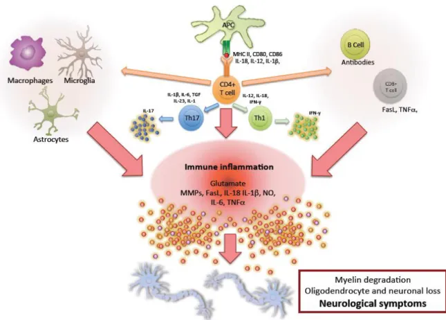
Figure 2. Pathophysiology of MS. Immune mediators involved in the pathophysiology of MS include macrophages, microglia, and astrocytes within the CNS and antigen presenting cells, CD4+ T cells, CD8+ T cells, and B cells. Upon activation, these cells secrete inflammatory molecules resulting in an inflammatory cascade of events. Consequently, this state of chronic inflammation will lead to myelin destruction, oligodendrocyte and neuronal cell loss and result in the neurological symptoms observed in MS patients. Figure inspired by (Mallucci et al., 2015), (McFarland &
Martin, 2007), (Jack et al., 2005), (Saijo & Glass, 2011), (Amor et al., 2009), (Hernández-Pedro et al., 2013), (Goverman, 2009), (Constantinescu et al., 2011), and (Krumbholz et al., 2012).
1.1.4 The Treatments of Multiple Sclerosis
To this day there are no cures for MS and current treatment therapies primarily target the inflammatory phase of the disease which is aimed at reducing symptoms (Thorpe et al., 2015). Current disease-modifying agents (DMA) are only minimally effective in reducing relapses in MS (Rizvi & Coyle, 2011). Moreover, these treatments need to be to ongoing and potentially lifelong, in order to avoid adverse effects such as worsening of symptoms, appearance of new lesions or an increase in the frequency and severity of relapses (Multiple Sclerosis Coalition, 2017). Currently approved agents for MS treatment include injectable, oral, and infused therapies listed in Figure 3 (Costello, Halper, Kalb, Skutnik, & Rapp, 2017; Mallucci et al., 2015; Rizvi & Coyle, 2011).
Figure 3. Current FDA-approved MS therapies. Current MS treatments include injectable medications, oral medications, and infused medications. Figure inspired by
(Costello et al., 2017).
The objectives of current therapies are to reduce the onset of relapses and to reduce the accumulation of new lesions detected by MRI, in order to delay MS progression (Mallucci et al., 2015). The use of therapies can be divided into two main categories, a short-term and long-term treatments. Corticosteroids are primarily used for short-term treatment of acute relapses. Their mode of action is to decrease the inflammatory response by reducing the
Injectable Medications •Avonex (IFN-1) •Rebif (IFN-1) •Betaseron (IFN-1) •Extavia (IFN-1) •Plegridy (pegylated
IFN-1) •Copaxone (glatiramer acetate) •Glatopa (glatiramer acetate) •Zinbryta (daclizumab) Oral Medications •Aubagio (teriflunomide) •Gilenya (fingolimod) •Tecfidera (dimethyl fumarate) Infused Medications •Lemtrada (alemtuzumab) •Novantrone (mitoxantrone) •Ocrevus (ocrelizumab) •Tysabri (natalizumab)
release of pro-inflammatory cytokines, inhibiting antigen presentation and lymphocyte proliferation, and stabilizing the BBB in order to elicit in rapid recovery from relapses (Mallucci et al., 2015; Rizvi & Coyle, 2011). Long term use of corticosteroids can result in adverse reactions such as cataracts, limb weakness, and osteoporosis. Long-term therapies are more specific in their mode of action, such as inhibiting immune cell recruitment, inhibiting cellular replication, and immunomodulatory effects (Mallucci et al., 2015).
Interferon (IFNs) mode of action is primarily through cell receptors and involves a wide range of immunological effects, such as inhibition of T cell activation and migration, anti-inflammatory cytokine production, and a decrease in BBB permeability (Boiko et al., 2002). IFNs are generally well tolerated, however, adverse reactions are more common with higher dosages and frequency of administration. These side effects include flu-like symptoms, depression, injection site reactions, and lymphopenia (Dale, 2000; Mallucci et al., 2015). Interferon beta-1b (IFN-1b) was the first DMA for the treatment of RRMS and is administered subcutaneously (Betaseron) every other day. Interferon beta-1a(IFN-1a) is administered intramuscularly (Avonex) once weekly or subcutaneously (Rebif) three times weekly.
Glatiramer acetate is a synthetic molecule whose its exact mechanism of actions are unknown, however, there is evidence showing that it might play a competitive role with Myelin Binding Protein (MBP) for antigen binding and have effects on TH1 to TH2
phenotypic cell shifts (Mikaeloff et al., 2004). Adverse reactions include anxiety, injection site reactions, and chest pain (Mallucci et al., 2015).
Fingolimod is an immunomodulatory agent that was the first approved oral treatment for MS. It is a structural analog of sphingosine which modulates the immune system by binding to sphingosine-1-phosphate receptors on lymphocytes. This binding induces receptor internalization and results in T cells being retained in lymphoid tissues and prevents their migration towards inflammatory sites (Wingerchuk & Lucchinetti, 2007). Potential serious side effects of the drug include macular oedema, reactivation of latent
infections such as shingles, delayed reconstitution of peripheral lymphocytes, and bradycardia (Hahn, Shroff, Blaser, & Banwell, 2004; Rizvi & Coyle, 2011).
Natalizumab was the first humanized monoclonal antibody to be approved for MS treatment. It is primarily recommended to patients who have inadequate response to the first line DMA. Its mechanism of action is to inhibit leukocyte adhesion to vascular cell adhesion molecule (VCAM) receptors by binding to alpha-4-integrin, resulting in the prevention of leukocyte migration across the CNS and thus, in a significant decrease of CD4+ and CD8+ T cells within the CNS (Alper & Wang, 2009; Clerico et al., 2017). The primary concerns around this drugs resides in its potential to reactivate latent JC virus found in the majority of the population, which can result in progressive multifocal leukoencephalopathy (PML) (Banwell & Anderson, 2005; Clerico et al., 2017). Other adverse reactions include headaches, liver toxicity, fatigue, hypersensitivity reactions, and infections (Rizvi & Coyle, 2011).
Ocrelizumab is a human monoclonal antibody that was recently approved in March 2017 for the treatment of RRMS and for the first time ever, for PPMS treatment. It targets mature B cells by binding to CD20 and result in a reduction of circulating B cells. Potential side effects include injection site reactions, headaches, flushed face, and irritation in the throat (Baker, Marta, Pryce, Giovannoni, & Schmierer, 2017).
1.1.5 Experimental Autoimmune Encephalomyelitis
Much of the understanding of MS pathogenesis and current drug therapies today are derived from the experimental autoimmune encephalomyelitis (EAE) model. EAE is the best characterized animal model for human autoimmune diseases (Constantinescu et al., 2011). Major resemblance features in EAE and MS are the destruction of myelin, the presence of multiple lesions within the CNS distributed in space and time particularly pronounced in the brain and the spinal cord, a predominant perivascular lesion location, the relative sparing of other elements of the nervous tissue, the presence of immunoglobulins within the CNS, inflammation, gliosis, and partial remyelination (Adams, Kubik, & Bezer, 1952; Constantinescu et al., 2011). On the other hand, a major difference between EAE and
MS is the requirement of an external immunization step in EAE (Gran, O’Brien, Fitzgerald, & Rostami, 2007).
EAE is a CD4+ T cell-mediated autoimmune disease consisting of perivascular T cells, CD4+ TH1- and TH17-mediated tissue damage, mononuclear cell infiltration and
demyelination within the CNS (Miller, Karpus, & Davidson, 2010). EAE consists of two phases; the induction and the effector phase. The induction phase involves the priming of CD4+ T cells following immunization with myelin proteins or peptides such as proteolipid protein (PLP), myelin basic protein (MBP) or epitopes of MBP such as Myelin Oligodendrocyte Glycoprotein (MOG), MOG35-55, in complete Freund’s adjuvant (CFA)
(Miller et al., 2010). The effector phase involves multiple steps depicted in Figure 4.
Figure 4. Effector phases of EAE model. Four stages define the effector phase of EAE. Stage 1 involves the migration of activated myelin-specific T cells and extravasation of T cells. In stage 2, myelin-specific T cells secrete cytokines and chemokines and result in an influx of peripheral mononuclear phagocytic cells within CNS parenchyma. Following these events, stage 3 occurs when peripheral monocytes, macrophages, and microglia cells are activated. Lastly, stage 4 results in demyelination of CNS axonal tracts. Figure inspired based on the description of stages given by (Miller, Karpus, &
Davidson, 2007).
Stage 4
CNS axonal tracts demyelination Stage 3
Activation of peripheral monocytes and macrophages and resident CNS microglia Stage 2
Cytokine and chemokine secretion by
myelin-specific T cells Influx of peripheral mononuclear phagocytic cells into CNS parenchyma Stage 1
1.2 The Central Nervous System
The human nervous system is divided into two categories, the Central Nervous System (CNS) and the Peripheral Nervous System (PNS). In addition to this division, another division of note within the CNS is the distinction between the grey and white matter. The grey matter is the region that contains high numbers of neuronal cell bodies, whereas the white matter is the region that contain predominantly myelinated axons (Jacobson & Marcus, 2008). The PNS originates from either the brain or the spinal cord and is responsible for the signals from and to the CNS through efferent and afferent nerves, respectively. The CNS consists of the brain, enclosed within the skull, and the spinal cord is found in the vertebral column. In addition to the bony protection, the brain and the spinal cord are enclosed by membranous coverings; the pia, arachnoid and dura membranes. The dura mater is the outermost layer and underneath the dura is the arachnoid layer, which lacks blood vessels and is closely associated to the pia mater. The pia consists of the innermost layer and contains blood vessels. Together, the arachnoid and pia mater form the subarachnoid space, filled with cerebrospinal fluid (CSF) (Hagan, Bolon, & Keene, 2012).
The CNS is conceivably the most exquisite system in the human body, it is responsible for setting apart humans from other species, and for the wonders that humans have achieved and continue to do so. Moreover, the brain is arguably the most complex and fascinating structure. This single organ controls all of the body’s functions and possesses extraordinary computational capabilities, ranging from heart rate, movement control, emotions, memory, learning, thought, attention, language, visual and auditory perceptions to shaping an individual’s hopes, dreams, and personality.
1.2.1 The Cells of the Central Nervous System
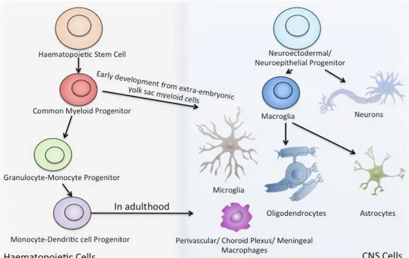
There are two main categories of cells in the CNS, the nerve cells, generally referred to as neurons, and the glial cells, which groups macroglia (astrocytes and oligodendrocytes) and microglia cells. The human nervous system contains approximately 100 billion neurons and 10 times more glial cells (Nolte, 2002). Cells of the CNS lineage originate from a neuroectodermal/neuroepithelial progenitor that gives rise to neurons and macroglia cells (Ransohoff & Cardona, 2010a). Interestingly, microglia cells derive from a different source
of progenitor cells (Figure 5). Indeed, they derive from a common myeloid progenitor early on during development from the extra-embryonic yolk sac, they then migrate towards the CNS, and only once they’ve entered the CNS do they become resident microglia cells (Ransohoff & Cardona, 2010a).
Figure 5. Resident CNS Cells. CNS cells originate from a neuroectodermal/ neuroepithelial progenitor that gives rise to neurons and macroglia cells. Macroglia cells can further differentiate into oligodendrocytes or astrocytes. Microglia cells derive from haematopoietic stem cell and myeloid progenitor early on during development from the extra-embryonic yolk sac. Figure inspired by (Ransohoff &
Cardona, 2010).
Neurons
Within the CNS, neurons are the basic functional conducting units. In a typical neuron, three distinct regions can be defined: (1) the cell body or soma, or perikaryon, (2) the dendrites, and (3) the axons (Figure 6). The cell body is the center of the neuron and contains the nucleus and the organelles that are necessary to maintain proper neuronal structure and function (Jacobson & Marcus, 2008). Dendrites are found at variable numbers depending on the type of neuron. They function to receive input signals from pre-synaptic
neurons and to transmit signals to post-synaptic neurons at the axon terminal region (Nolte, 2002). A synapse constitutes the location where the dendrites of different neurons form what is called a synaptic contact to transmit signals typically, in the form of neurotransmitters. Signals received at dendrites can be either excitatory or inhibitory and an action potential is generated at the site of axon origin, also called the axon hillock, if the accumulating sum of stimuli reaches a threshold value. Axons are an extension of the cell body, which can measure from a few microns to a meter long (Jacobson & Marcus, 2008). Axons are enveloped by a protective myelin sheath, which allows proper functioning to propagate and speed up signal transmission through what is called salutatory conduction.
Figure 6. Structure of a neuron. The cell body (soma) is the center of the neuron and contains the nucleus and the organelles necessary to maintain proper neuronal structure and function. Dendrites function to receive input signals from pre-synaptic neurons and to transmit signals to post-synaptic neurons at the axon terminal. A synapse constitutes the location where the dendrites of different neurons form connections. Axons are an extension of the cell body and are enveloped by a protective myelin sheath. Nodes of Ranvier are unmyelinated areas involved in signal propagation. Figure inspired by (Son et al., 2012).
Neurons can be classified based on several different features such as anatomical, molecular, and based on their electrophysiological properties (Sharpee, 2014). Perhaps, the most simplified classification of neurons is based on their shape: unipolar, bipolar, and multipolar. Unipolar neurons have only one dendrite and constitute the most common type of sensory neuron and function to transmit sensory information to the CNS. Unipolar neurons are particularly found in invertebrates (Poulain & Sobel, 2010). Furthermore, bipolar neurons possess two processes and are also sensory neurons involved in the transmission of signals of sensory pathways such as olfactory, visual, and auditory. Interneurons, which are abundantly found within the nervous system, are a form of bipolar neurons. Lastly, multipolar neurons constitute the vast majority of neurons, which are involved in motor and sensory processing. They possess numerous dendritic extensions from the soma and an axon. Typically, the arrangement of dendritic projections is a characteristic of a particular type of neuron for example the multipolar neurons in the cerebral cortex have different shape than those found in the cerebellar cortex as illustrated in Figure 7 (Jacobson & Marcus, 2008; Nolte, 2002).
Figure 7. Types of neurons. A) Unipolar neurons have only one dendrite and function to transmit sensory information to CNS. B) Bipolar neurons possess two processes and are sensory neurons involved in the transmission of signals from sensory pathways. C, D. Multipolar neurons are involved in motor and sensory processing. The arrangement of their dendritic projections is characteristic of the specific type of neuron. Figure inspired by (Jacobson & Marcus, 2008).
Oligodendrocytes
Oligodendrocytes, derived from oligodendrocyte precursor cells/progenitors (OPCs), are the cells responsible for the formation and maintenance of the myelin sheath tightly wrapped around neuronal axons (Jacobson & Marcus, 2008). This lipid covering enhances neuronal communication and transmission of signals to their effector organ extremely rapidly. Additionally, axonal insulation by myelin covering also serves as a protective layer against axonal degeneration and is also involved in the regulation of axonal transport (Fitzner et al., 2006). Interestingly, myelin is actually a single sheet of oligodendrocyte plasma membrane and a single oligodendrocyte is capable of forming and maintaining numerous myelin sheaths. Each axonal fragment that is myelinated is termed an internode due to the bare segment of axon between each internode, which is called node of Ranvier (Byrne, Byrne, & Roberts, 2009). This type of insulation allows action potentials to jump from node to node, which eliminates the necessity of the signal to be continuously regenerated and allows the efficient and fast communication between neurons. Additionally, oligodendrocytes provide neuronal support by releasing neurotrophic factors such as glial-derived neurotrophic factor (GDNF), brain-derived (BDNF), and insulin-like 1 growth factors (IGF-1) (Bradl & Lassmann, 2010).
In the process of OPC differentiation to mature oligodendrocytes, the capacity for myelination occurs during a very brief period of time and very early on, and once differentiated into mature oligodendrocytes, they are relatively unable to myelinate (Bradl & Lassmann, 2010). Accordingly, this process is highly regulated and coordinated. Neuronal electrical activity has been suggested to control myelin assembly (Bradl & Lassmann, 2010). Typically, oligodendrocytes must synthesize and assemble many proteins to be transported to the myelin membrane. A major myelin protein, myelin basic protein, has been suggested to bring different layers of myelin closer together and it appears that a neuronal- and an MBP-dependent mechanism is responsible this (Fitzner et al., 2006).
Astrocytes
Astrocytes are treasured cells of the CNS and are the most abundant, making up to 90% of the brain cells (He & Sun, 2007). There are two major types of astrocytes in the CNS: fibrous, which are commonly found in the white matter, and protoplasmic astrocytes which are found in the grey matter (Jacobson & Marcus, 2008). Besides differences in anatomical localizations, protoplasmic astrocytes processes were shown to cover synapses, whereas the processes of fibrous astrocytes were shown to form a connection with nodes of Ranvier (Sofroniew & Vinters, 2010). A third type, radial glia, are present during development and serve to guide growing axons (Nolte, 2002). Radial glial cells retract their processes and eventually serve as progenitor cells of astrocytes in the mature CNS (Byrne et al., 2009). Astrocytes connect to one another by gap junctions and are identified by their star shape, their end feet on capillaries, and their expression of large intermediate filaments that are composed of glial fibrillary acidic protein (GFAP) (Byrne et al., 2009).
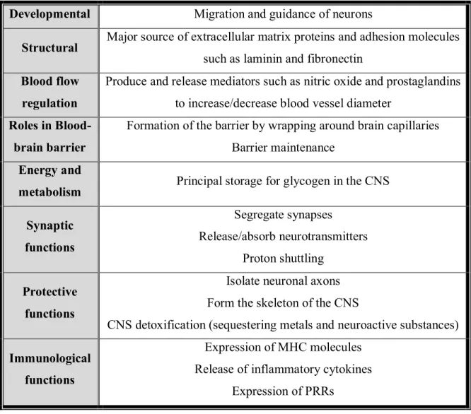
These star-shaped cells possess numerous functions and roles in maintaining CNS homeostasis. Astrocytes execute important roles for proper CNS development such as guidance for developing axons, and the formation and function of developing synapses (Christopherson et al., 2005; Ullian, Sapperstein, Christopherson, & Barres, 2001). Moreover, given the extensive contacts astrocytes make with blood vessels, it is not surprising that they also regulate CNS blood flow (Sofroniew & Vinters, 2010). Table 1 lists some of the various functions of astrocytes beyond their roles in neuronal and glial survival (Byrne et al., 2009; Jacobson & Marcus, 2008; Koehler, Roman, & Harder, 2009; Perea, Navarrete, & Araque, 2009; Sofroniew & Vinters, 2010).
Developmental Migration and guidance of neurons
Structural Major source of extracellular matrix proteins and adhesion molecules such as laminin and fibronectin
Blood flow regulation
Produce and release mediators such as nitric oxide and prostaglandins to increase/decrease blood vessel diameter
Roles in Blood-brain barrier
Formation of the barrier by wrapping around brain capillaries Barrier maintenance
Energy and
metabolism Principal storage for glycogen in the CNS Synaptic functions Segregate synapses Release/absorb neurotransmitters Proton shuttling Protective functions
Isolate neuronal axons Form the skeleton of the CNS
CNS detoxification (sequestering metals and neuroactive substances) Immunological
functions
Expression of MHC molecules Release of inflammatory cytokines
Expression of PRRs
Table 1. Roles of astrocytes in the CNS. Astrocytes possess numerous functions and roles in maintaining CNS homeostasis. They play key role in the proper development of the CNS. They provide structural, regulatory, metabolic, immunologic, and protective barrier and functions. Table inspired by (Sovrea & Bosca, 2013),
(Christopherson et al., 2005), (Byrne et al., 2009), and (Sofroniew & Vinters, 2010).
Microglia
Microglia cells are the resident innate immune cells of the CNS and compromise about 10% to 15% of the glial cell population (Nayak, Roth, & McGavern, 2014). They are primarily localized within the brain, spinal cord, retina, and optic nerve (Goldmann & Prinz, 2013). These specialized macrophages of the CNS differ from astrocytes and oligodendrocytes by their origin, morphology, gene expression and numerous functions
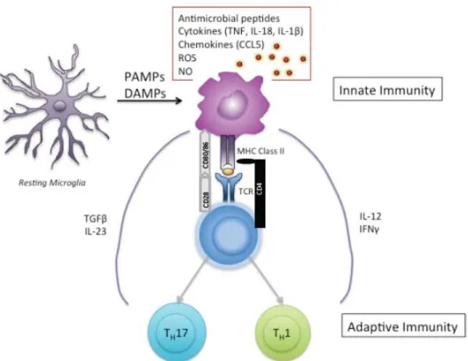
(Kettenmann, Hanisch, Noda, & Verkhratsky, 2011). Unlike other cells of the CNS, microglia cells derive from cells of haematopoietic origin and the myeloid lineage, which migrate into the CNS during embryogenesis (Chan, Kohsaka, & Rezaie, 2007; Graeber & Streit, 2010; Saijo & Glass, 2011). They express a variety of macrophage-associated markers such as CD11b, CD14, CSF1R, CD80/86, and MHC II (Graeber & Streit, 2010; Kettenmann et al., 2011; Saijo & Glass, 2011). In their resting phase, they are highly ramified, which allows their distinction from other macrophages and dendritic cells. They play a role in a variety of functions such as CNS development and homeostasis (Nayak et al., 2014). Indeed, microglia cells are highly dynamic cells and are in constant surveillance of their environment, through the extension and retraction of their ramified branches and clearance of debris, and through their phagocytotic activities (Nayak et al., 2014; Shastri, Bonifati, & Kishore, 2013). Microglia cells also play an important role in neurogenesis, neuroprotection, and synapse formation and pruning (Shastri et al., 2013). Their important role within the normal CNS development and homeostasis is elucidated by microglial dysfunction in many neurological disorders such as Obsessive-Compulsive Disorder (OCD), and Rett Syndrome as well as neurodegenerative diseases such as Alzheimer’s Disease, Parkinson’s Disease, and Multiple Sclerosis (Nayak et al., 2014). In response to CNS insults or injuries, microglia cells rapidly change morphology from a ramified to an amoeboid shape and can adopt two states of activation, either M1 or M2 (Tang & Le, 2015). The M1 phenotype, also known as classical activation, occurs in an inflammatory setting where microglia cells upregulate their migratory, proliferative, and phagocytic ability as well as increased production of multiple pro-inflammatory molecules including but not restricted to TNF-α, IL-1β, IL-6, IL-12, MIP1-α, iNOS, CCL5, ROS and NO (Perry & Holmes, 2014) (Figure 8). In this state, they can play a role of antigen presentation to T cells and skew T cells towards TH1 and TH17 phenotypes (Goldmann &
Prinz, 2013; Kettenmann et al., 2011; Kim & De Vellis, 2005). The alternative activation, also referred to as the M2 phenotype, is involved in tissue remodeling, repair, and healing (Tang & Le, 2015). Typically, a M2 phenotype occurs in the presence of IL-4, IL-10, and IL-13, which results in the M2 cells producing IL-10 and TGF-β (Goldmann & Prinz, 2013; Tang & Le, 2015).
Figure 8. Activated micrgolia cell. In an inflammatory setting, microglia cells upregulate their migratory, proliferative, phagocytic ability, and secrete multiple pro-inflammatory molecules. They can play a role of antigen presentation to T cells and skew T cells towards TH1 and TH17 phenotypes. Figure inspired by (Saijo & Glass, 2011).
1.2.2 The Blood-Brain Barrier
Proper brain functioning requires an adequate blood supply and metabolic energy in a tightly controlled environment, free of toxins and harmful bodies (Andreone, Lacoste, & Gu, 2014; Attwell & Laughlin, 2001). The Blood-Brain Barrier (BBB) delivers this environment through a physiological barriers required for protecting the CNS and controlling the influx and efflux of substances (Andreone et al., 2014). This control is selective and limits the entry of large hydrophilic proteins however, allows the passage of small lipophilic and small gaseous molecules (Rizvi & Coyle, 2011). The BBB is formed by what is called the neurovascular unit, which is composed of endothelial cells, pericytes, and astrocytes (Siegenthaler, Sohet, & Daneman, 2013) (Figure 9).
Figure 9. Neurovascular unit forming the BBB. The neurovascular unit is composed of endothelial cells, pericytes, and astrocytes. Figure inspired by (Abbott et al., 2010).
CNS endothelial cells are the chief cells responsible for forming the BBB. They possess a mixture of structural, physiological, and cellular properties which permit them to serve their function to tightly control the trafficking of molecules, ions, and cell types between the brain and the blood. There are four particular features of CNS endothelial cells that are responsible for their contribution to BBB integrity conferring tightness and preventing leakiness (Andreone et al., 2014; Siegenthaler et al., 2013). First, they possess remarkably tight junctions, which limits the movement of most polar molecules across the blood plasma to the brain extracellular fluid (Abbott, Patabendige, Dolman, Yusof, & Begley, 2010). Second, transcellular transport which is the transport between the luminal and abluminal cell membranes, is very slow. Furthermore, CNS endothelial cells express numerous transporters, unidirectional or bidirectional, which import nutrients such as amino acids, glucose, and ions. Additionally, these transporters also function to remove neurotoxic substances. Lastly, the low expression of leukocyte adhesion molecules (LAMs) on CNS endothelial cells serves to limit the movement of immune cells into the brain (Abbott et al., 2010; Andreone et al., 2014; Siegenthaler et al., 2013).
Pericytes are microvascular cells, which include capillaries, venules, and arterioles, and consist of the contractile cells of the neurovascular unit that provide structural support and vasodynamic capacity (Andreone et al., 2014; Ballabh, Braun, & Nedergaard, 2004). Their contribution to BBB structure is observed in mice with significantly reduced pericytes, where in these mice, there is an increase in vascular permeability thus, BBB leakiness was observed to correlate to the extent of pericyte loss. In addition, defects in transcytosis rates and in association of astrocytic end-feet were also observed (Armulik et al., 2010).
Beyond serving a multitude of functions within the CNS, astrocytes also function to maintain an optimal BBB environment. Astrocytes regulate BBB functioning in various ways notably in the maintenance of BBB functionality (Andreone et al., 2014). Astrocytes secrete several factors that regulate BBB properties such as sonic hedgehog (Shh), angiotensin, and retinoic acid, to name a few (Andreone et al., 2014; Ballabh et al., 2004; Siegenthaler et al., 2013). Shh secreted by astrocytes increases tight junction component expression components and decreases the expression of cell adhesion molecules on CNS endothelial cells (Alvarez et al., 2011). Angiotensin II, which is cleaved from angiotensinogen, is also implicated in CNS endothelial tight junctions properties, particularly by promoting their efficient organization (Wosik et al., 2007). Moreover, Mizee et al. demonstrated that astrocyte-derived retinoic acid plays a role in promoting barrier function and development (Mizee et al., 2013).
1.2.3 The Immunology and Lymphatics of the Central Nervous System
Sir Peter Medawar was the first to describe the CNS as an immune-privileged site, that this lead to the notion that immune responses within the CNS are tightly regulated. The idea of immune privilege designates that antigens within these areas do no provoke an immune response (Amor et al., 2014). However, it is well established that immune surveillance occurs within the CNS and that adaptive immune responses can occur against CNS antigens (Ransohoff & Engelhardt, 2012). In their 2013 study, Kamimura et al. selectively stimulated sensory neurons of different muscles and observed in spinal cord vessels, the expression of local chemokines (Kamimura et al., 2013). Interestingly, they found an increase in the expression of CCL20 in the L3 cord dorsal vessels following stimulation of
thigh muscles. Their studies lead to the suggestion that blood vessels act as gates for the infiltration of immune cells and this is dependent upon regional neural stimulation, which they termed “the gateway theory” (Kamimura et al., 2013). It is well recognised that adaptive immune responses against CNS antigens occur in the periphery and are subsequently disseminated to the CNS by circulating memory T cells, which are successively re-stimulated within the CNS (Ransohoff & Engelhardt, 2012). The healthy CNS is composed of various immune cells such as parenchymal microglia, choroid plexus macrophages and dendritic cells, meningeal macrophages, and perivascular macrophages. In addition, the CSF contains primarily T cells and also B cells, monocytes, and dendritic cells (Ransohoff & Cardona, 2010b; Ransohoff & Engelhardt, 2012). In 2015, Louveau A, et al. published a very interesting study where they sought to identify routes for recirculation of meningeal immune cells (Louveau et al., 2015). By using whole-mount preparation of dissected mouse brain meninges, they were able to identify a high concentration of T cells, MHC II-expressing cells, and endothelial cells in proximity to the dural sinuses. Additionally, they found CCL21 expression in meningeal lymphatic vessels (Louveau et al., 2015). Meningeal lymphatics, although similar to peripheral lymphatics, possesses unique organization and distributional features. Accordingly, they cover less tissues and form a network of lower complexity, with narrower vessels. However, vessels are larger in the transverse sinuses (Louveau et al., 2015). Moreover, it appears that meningeal lymphatics drain into the deep cervical lymph nodes first and then, into superficial cervical lymph nodes and are responsible for the drainage of CSF soluble and cellular components (Louveau et al., 2015).
1.2.4 Inflammation in the Central Nervous System
The immune system and the nervous system are two biological entities that are indistinguishably interlinked (Amor et al., 2014). Many common mediators exist between the two systems and both innate and adaptive immunity occurs in CNS, which are essential in debris clearance, infection, tumor elimination, and tissue repair (Amor, Puentes, Baker, & van der Valk, 2010). Several measures are put in place in order to avoid damage to the CNS such as limitation of peripheral immune cell influx. However, the hallmark of the majority of neurodegenerative conditions is the underlying chronic inflammatory state as a