Université de Montréal
Regulation of the proinflammatory properties of angiopoietins
par
Ricardo J.M. Maliba Département de pharmacologie
Faculté de Médecine
Thèse présentée à la Faculté des Études Supérieures en vue de l’obtention du grade de Phitosophiae Doctor (Ph.D.) en pharmacologie
L
N (N
Université
ll
de Montréal
Direction des bibliothèques
AVIS
L’auteur a autorisé l’Université de Montréal à reproduite et diffuser, en totalité ou en partie, par quelque moyen que ce soit et sur quelque support que ce soit, et exclusivement à des fins non lucratives d’enseignement et de
recherche, des copies de ce mémoire ou de cette thèse.
L’auteur et les coauteurs le cas échéant conservent la propriété du droit
d’auteur et des droits moraux qui protègent ce document. Ni la thèse ou le mémoire, ni des extraits substantiels de ce document, ne doivent être
imprimés ou autrement reproduits sans l’autorisation de l’auteur.
Afin de se conformer à la Loi canadienne sur la protection des renseignements personnels, quelques formulaires secondaires, coordonnées
ou signatures intégrées au texte ont pu être enlevés de ce document. Bien
que cela ait pu affecter Ta pagination, il n’y a aucun contenu manquant.
NOTICE
The author of this thesis or dissertation has granted a nonexclusive license
allowing Université de Montréal to reproduce and publish the document, in
part or in whole, and in any format, solely for noncommercial educational and research purposes.
The author and co-authots if applicable retain copyright ownership and moral
rights in this document. Neither the whole thesis or dissertation, nor
substantial extracts from it, may be printed or otherwise reproduced without the author’s permission.
In compliance with the Canadian Privacy Act some supporting forms, contact
information or signatures may have been removed from the document. While this may affect the document page count, t does not tepresent any loss of
II
Université de Montréal Faculté des Études Supérieures
Cette thèse intitulée:
Regulation of the proinflammatory properties of angiopoietins
présentée par: Ricardo J.M. Maliba
A été évaluée par un jury composé des personnes suivantes:
Docteur Jean-François Gauchat président rapporteur
Docteur Martin G. Sirois directeur de recherche
Docteur Lucie Germain membre du jury
membre du jury /: -r2
----//)
111
Résumé
La découverte récente des angiopoïétines (Angi et Ang2) a permis de démontrer qu’elles peuvent, via leur liaison au récepteur Tie2, moduler la perméabilité et la stabilité vasculaire, et ainsi contribuer à diverses étapes du processus angiogénique. Nous avons démontré que les angiopoïétines sont capables de moduler des processus proinflammatoires dans les cellules endothéliales (CE), notamment la synthèse du facteur d’activation plaquettaire (PAF) et la transiocation de la P-sélectine.
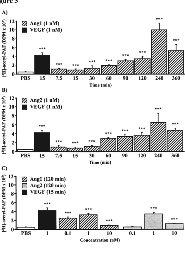
Le vascutar endothetiat growth factor (VEGF) est le seul facteur angiogénique capable d’induire une réponse inflammatoire et cet effet inflammatoire est médié par la synthèse endothéliale du PAF. De plus, la synthèse de PAF endothéliale induite par le VEGF-A165 implique l’activation des voies de signalisation des p38 et p42/44 mitogen-activated protein kinases (MAPK). Sachant que l’inflammation joue un rôle important dans l’angiogénèse et que les angiopoïétines agissent de concert avec le VEGF dans la modulation de la plasticité vasculaire, nous avons donc étudié la capacité des angiopoïétines d’induire la synthèse du PAF et les mécanismes intracellulaires par lesquels cette synthèse se produit dans les CE. Nous avons observé qu’Angl et Ang2 induisent la phosphorylation rapide et transitoire de Tie2, des voies p42144 et p38 MAPK ainsi que PI3KIAkt et finalement la synthèse de PAF de façon dose- et temps-dépendante dans les cellules d’aortes bovines (BAEC). L’effet maximal d’Angl et Ang2 sur la synthèse de PAF est obtenu après 240 minutes de stimulation (1695% et 851%
iv
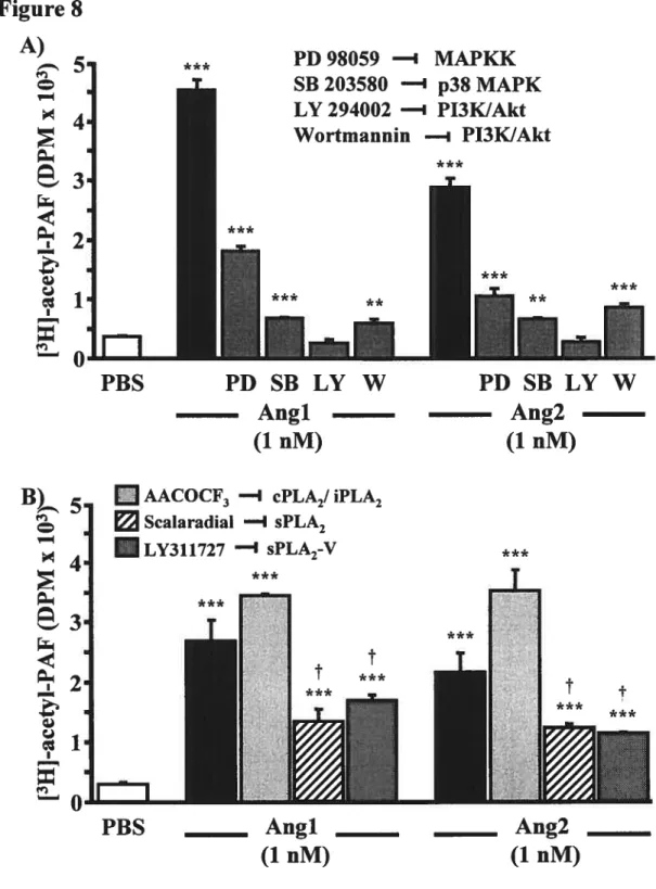
nécessite l’activation des voies de signalisation intracellulaires des p38 MAPK, p42!44 MAPK et PI3K ainsi que d’une phospholipase A2 sécrétée de type V (sPLA2-V). De plus, nos résultats démontrent que la synthèse de PAF endothéliale induite par les angiopoïétines est en partie médiée par une relocalisation de VEGF endogène vers la membrane cellulaire.
Nous avons récemment démontré que le PAF est impliqué dans la translocation de la P-sélectine et l’adhésion de neutrophiles sur les CE induite par le VEGF-A165 et que Angi et Ang2 étaient capables de promouvoir la translocation de la P-sélectine endothéliale et l’adhésion de neutrophiles sur des CE. Par conséquent, nous avons voulu déterminer les mécanismes impliqués et le rôle du PAF endogène dans la translocation de la P-sélectine endothéliale par les angiopoïétines. Nous avons observé que Angi et Ang2 (11Y9 M) pouvaient induire une activation rapide du récepteur Tie2 et une translocation rapide et transitoire (maximale à 7,5 minutes et correspondant à des augmentations de 125% et 100% sur les valeurs témoins, respectivement) de la P-sélectine. De plus, nous avons décrit pour la première fois que la translocation de la P-sélectine endothéliale induite par les angiopoïétines dépend du calcium en plus d’être régulée par l’activation de la phospholipase C-y (PLC-y).
En conclusion, nos résultats démontrent que les angiopoïétines possèdent la capacité de moduler des effets proinflammatoires tels la synthèse de PAF et la translocation de la P-sélectine au niveau de l’endothélium.
Mots clés: Angiopoïétines, inflammation, angiogénèse, facteur d’activation plaquettaire (PAF), P-sélectine, Tie2.
V
Summary
Recently discovered, Angi and Ang2, upon binding to the Tie2 tyrosine kinase receptor, modulate vascular permeabïlity and integrity, contributing to angiogenesis. Herein, we demonstrated that angiopoietins are capable of modulating proinflammatory processes in endothelial ceils (EC), namely the synthesis of platelet activating factor (PAF) and P-selectin transiocation. In addition, we also identified the intracellular mechanisms responsible for these biological effects.
Vascular endothelial growth factor (VEGF) is the only angiogenic growth factor capable of inducing an inflammatory response and we have demonstrated that this effect is mediated by the endothelial synthesis of PAF. Furthermore, VEGF-A165-mediated endothelial PAF synthesis requires the activation of both p38 and p42144 mitogen-activated protein kinases (MAPK). Since inflammation exists in a mutually dependent association with angiogenesis and angiopoietins act in concert with VEGF to modulate vascular plasticity during postnatal neovascularization, we sought to determine whether Angi andlor Ang2 were capable of inducing endothelial PAF synthesis and if so, what mechanisms were implicated. Treatment of bovine aortic endothelial ceils (BAEC) with Angi or Ang2 (iOE9 M) induced a rapid Tie2 phosphorylation and a rapid, progressive and sustained endothelial PAF synthesis maximal wïthin 240 minutes (1695% and 851% increase, respectively). Angiopoietin mediated endothelial PAF synthesis requires the activation of the p38 and p42144 MAPKs, PI3K intracellular signalling pathways, and a secreted phospholipase A2 (sPLA2-V). Furthermore, angiopoietin-mediated PAF synthesis is partly driven by a
vi
We have recently reported that VEGF-A165 inflammatory effects are mediated through the synthesis of PAF by endothelial ceils which contributes to the induction of endothelial P-selectin transiocation and neutrophil adhesion onto activated EC. Furthermore, our laboratory demonstrated that Angi and Ang2 are also both capable of promoting endothelial P-selectin transiocation and neutrophil adhesion onto EC. We therefore sought to investigate the cellular mechanisms implicated in
angiopoietin-mediated P-selectin transiocation in BAEC and assess the role of PAF
in this process. We observed that Angi and Ang2 (11Y9 M) are both capable of
mediating a rapid Tie2 phosphorylation as well as a rapid and transient endothelial P selectin transiocation maximal within 7.5 minutes (125% and 100% increase, respectively over control values). In addition, we demonstrate for the first time that angiopoietin-mediated endothelial P-selectin transiocation is calcium-dependent and regulated through phospholipase C-y (PLC-7) activation.
In conclusion, our data demonstrate that angiopoietins are capable of modulating proinflammatory events, namely endothelial PAF synthesis and P selectin transiocation in BAEC and shed light on the intracellular pathways through which angiopoietins regulate these events.
Keywords: Angiopoietins, inflammation, angiogenesis, platelet-activating factor (PAF), P-selectin, Tie2.
vii
Table of Contents
Résumé iii
Summary y
Table of Contents vii
List of Tables xi
Listoffigures xii
List of Abbreviations xiv
Acknowledgements xvi
1.0 Introduction 1
1.1 Biology of the vascular endothelium
1.1.1 Role of the endothelium in vascular homeostasis 1
1.1.2 Role of the endothelium in the pathogenesis of vascular disorders 2
1.2 Angiogenesis 3
1.2.1 Overview 3
1.2.2 Physiological angiogenesis 6
1.2.3 Pathological angiogenesis 7
1.3 Regulation of angiogenesis 8
1.3.1 Vascular endothelial growth factor (VEGF-A) 8
1.3.1.1 Regulatïon of VEGF-A165 expression 13
1.3.1.2 VEGF receptors (VEGFR) 14
viii
1.3.2.2 Tie Receptors .22
1.3.2.3 Role of angiopoietins and Tie2 receptor in angiogenesis 24
1.3.2.4 Regulation of the expression ofangiopoietins 30
1.3.2.5 Signalling pathways activated by angiopoietins 31
1.3.2.6 Angiopoietins in pathological angiogenesis 33
1.3.2.6.1 Angiopoietins in tumour vascularization 33
1.3.2.6.2 Angiopoietins in inflammation 35
1.4 The inflammatory response and angiogenesis 37
1.4.1 PAF, an inflammatory mediator 3$
1.4.1.1 PAF synthesis 39
1.4.1.2 Role of PAF in angiogenesis 41
1.4.2 The phospholipase A2 family 42
1.4.3 Adhesion of leukocytes to the endothelium 44
1.4.3.1 The selectins 46 1.4.3.2 The integrins 49 1.4.3.3 The 1g superfamily 50 1.5 Rationale 51 1.6Purpose 52 2.0 Article #1 53 2.1SUMMARY 58 2.2 INTRODUCTION 59
ix 2.4 RESULT$ .66 2.5 DISCUSSION 72 2.6 ACKNOWLEDGEMENTS $1 2.7 REFERENCES 82 2.8 FIGURE LEGENDS 86 2.9 FIGURES 90 3.0 Article #2 99 3.1 ABSTRACT 103 3.2 INTRODUCTION 104
3.3 MATERIALS & METHOD$ 107
3.4RESULTS 112 3.5 DISCUSSION 117 3.6 ACKNOWLEDGEMENTS 124 3.7 REFERENCES 125 3.8 FIGURE LEGENDS 130 3.9 FIGURES 134 4.0 Discussion 140
4.1 Angi and Ang2 behave as Tie2 agonists in BAEC 142
4.2 Angi and Ang2 induce PAF synthesis in BAEC 144
X
4.5 Perspectives .158
5.0 Conclusion 161
xi
List of Tables
Table
1:
Summary of the experiments conducted in transgenic or knockout mice to determine the respective roles of Tie2, Angi, and Ang2 in angiogenesis.xii
List of Figures
Figure 1: The stepwise progression of budding (classical) angiogenesis. Figure 2: VEGF-A isoforms.
Figure 3: Members of the VEGf family and their respective receptors. Figure 4: Intracellular signalling pathways activated by VEGF-A165. Figure 5: The angiopoietin family.
Figure 6: The Tie receptors and their respective ligands.
Figure 7: Interaction between VEGF and angiopoietins during angiogenesis. Figure 8: Intracellular signalling pathways activated by the angiopoietins. Figure 9: Summary of the remodelling pathway of endothelial PAF synthesis. Figure 10: Leukocyte rolling and adhesion.
Figure 11: Structural organization of selectins.
Article #1
Figure 1: Activation and expression of Tie2 in BAEC.
Figure 2: Angi and Ang2 activate p38 MAPK, p42144 MAPK, and PI3K pathways. Figure 3: Angi and Ang2 are both capable of inducing endothelial PAF synthesis in
a time and concentration-dependent manner.
Figure 4: Pretreatment with VEGF receptor (VEGFR) inhibitors attenuates angiopoietin-mediated endothelial PAF synthesis at 4 hours.
Figure 5: Endothelial distribution of VEGF upon stimulation with angiopoietins. Figure 6: Treatment of BAEC with angiopoietins induces a relocalization of
xiii Figure 7: Treatment of BAEC with Angi or Ang2 promotes VEGFR-2
phosphorylation.
Figure 8: Angiopoietin-mediated endothelial PAF synthesis requires the activation of p38 MAPK, p42144 MAPK, and PI3K signalling pathways and sPLA2-V. Figure 9: Proposed mechanism by which angiopoietins mediate endothelial PAF
synthesis.
Article #2
Figure 1: Angi and Ang2 activate Tie2 in a time-dependent manner.
Figure 2: Angi and Ang2 induce endothelial P-selectin transiocation in a time- and concentration-dependent manner.
Figure 3: Angi and Ang2 mediate endothelial P-selectin translocation.
Figure 4: Endothelial P-selectin transiocation mediated by Angi or Ang2 requires PLC-y and PKC signal transduction.
Figure 5: p38 MAPK, p42/44, and PI3K signalling is flot required for angiopoietin mediated endothelial P-selectin transiocation.
xiv
List of Abbreviations
AA: arachidonic acid
Angi, Ang2: angiopoietin-1 and -2 BAEC: bovine aortic endothelial ceils cPLA2: cytosolic phospholïpase A2 DAG: diacylglycerol
EC: endothelial celis
eNOS: endothelial nitric oxide synthase ESL-1: E-selectin ligand-1
FGF: fibroblast growth factor HIE hypoxia-inducible factor
HUVEC: human umbilical vein endothelial ceils ICAM- 1: intercellular adhesion molecule- 1 IL: interleukin
1P3: inositol 1 ,4,5-triphosphate
iPLA2: calcium-independent phospholipase A2 LPS: Iipopolysaccharide
Lyso-PAF AT: lyso-PAF acetyl transferase MAPK: mitogen-activated protein kinase NO: nitric oxide
NRP- 1: neuropilin- 1
xv
PAFR: platelet-activating factor receptor
PECAM- 1: platelet-endothelial cellular adhesion molecule- 1 PGI2: prostaglandin 12 (prostacyclin)
PI3K: phosphatidyl inositol-3 kinase PLC-y: phospholipase C-a’
PKC: protein kinase C
PJGF: placental growth factor PMA: phorbol myristate acetate PMN: polymorphonuclear celis
PSGL- 1: P-selectin glycosylated ligand- 1 RTK: receptor tyrosine kinase
sPLA2: secreted phospholipase A2
Tic 1, Tie2: yrosine kinase with jmmunoglobulin and piderma1 growth factor homology domains receptor-1 and -2
VCAM-1: vascular ccli adhesion molecule- 1 VE-cadherin: vascular-endothelial cadherin VEGF: vascular endothelial growth factor
VEGFR: vascular endothelial growth factor receptor WPB: Weibel-Palade bodies
xvi
Acknowledgements
Although only my name appears on the cover of this manuscript, several individuals contributed either directly or indirectly in bringing this project to fruition. I would like to thank the following individuals:
1. First and foremost, Dr. Martin G. Sirois for giving me this opportunity and providing an amazing environment in his laboratory. Your support and guidance throughout this process are most appreciated. At every occasion, your insightful comments (both positive and negative) and judgement helped me tremendously.
2. Secondly, my spouse, Annie for her unwavering support (both moral and
financial) and apparently limitless love. I also wish to thank my son, Arthur for just being himself. Thank-you, both of you, for letting me embark on this long and crazy trip. That’s it, I’m done with school, I promise.
3. My parents for teaching me at a young age to pursue my dreams.
4. My colleagues, past and present. I’m fortunate to have had the opportunity to encounter some extraordinary people who have made this experience memorable. Several of you, regardless of the fact that our paths will someday diverge, have become cherished friends. My most heartfelt thanks to Stéphanie Lapointe, Catherine Marchand, Simon Rollin, François Tremblay, Pascal Bematchez, Sonia Grandi, Angelino Calderone, Bruce Allen, and especially Paul-Eduard Neagoe and Alexandre Brkovic for ail of the laughs. Merci.
1.0 Introduction
1.1 Biology of the vascutar endothelium
1.1.1 Role of the endothehum in vascular homeostasis
The vascular system has fascinated mankind for thousands of years. Egyptian physicians recognized that ‘there were vessels in him for every part of the body’ , Aristotie considered that ‘bfood vessels are like watercourses in gardens: they start from one spring and branch off into numerous channels so as to supply every part of the garden’ ‘, and Leonardo da Vinci (1452-1519) speculated that the vasculature developed like a tree from a seed (the heart) by sprouting roots (capillaiy network) and atrunk with major branches (the aorta and arteries) 2 Indeed, the complexity of the vascular system prompted the formulation of several hypotheses in an attempt to understand its functioning and regulation. However, the defence of these postulates was rendered difficuit by the absence of anatomic proof. William Harvey (157$-1657) was the first physiologist to describe the circulation of blood in large vessels yet he could flot explain how blood passed from the arteries to the veins. Thirty years later, Malpighi (162$-1694) observed, through the newly developed microscope, the interconnecting capillary network. Pioneering work conducted by Ruysch, (163$-1731) enabled the development of techniques permitting the identification of the micro- and macroscopic constituents of blood vessels enabling anatomic research to progress.
The ceilular monolayer carpeting the luminal aspect of blood vessels attracted the attention of various researchers. The term endothelium was coined by His in 1865 who believed that this layer served solely as a physicai barrier to prevent blood
from flowing into the surrounding tissues .
However, others beiieved that the endothelium played a crucial foie in homeostasis, namely preventing the formation of
thrombi and the invasion of smooth muscle celis (SMC) into the vascular lumen .
Due to their direct contact with biood, endothelial cells perform a critical role in ail
aspects of tissue homeostasis. Endothelial cells regulate vascular tone through
interaction with components of the peripheral nervous system ami are involved in
thrombolysis and blood coagulation. Endothelial celis are aiso implicated in
inflammatory and immunological processes. The endothelial cells surface consists of a layer of surface glycoprotein (glycocalyx) that provides flot only a local charged barrier to the transendotheliai migration of blood celis and plasma proteins under
normai physiological conditions but is also physioiogicaily active 6
CeIls within the vessel wall communicate with surrounding celis, can sense changes in pressure and
flow, and can secrete molecules that perform various functions. The beiief that
endothelial celis (EC) were merely barriers between blood and the interstitium was revolved.
1.1.2 Role of the endothelium in the pathogenesis ofvascular disorders
Despite their role in the maintenance of vascular homeostasis, endothelial ceils may aiso play a deleterious role under certain situations such as inflammation. British
surgeon John Hunter (172$-1793) reported that the characteristic redness associated with inflammation is due to increased blood flow through dilated vessels. Early work placed an emphasis on the fact that inflammation is characterized by an endothelium dependent alteration in the vasculature. Furthermore, the endothelium was directly
associated with the accumulation and transmigration of leukocytes into underlying
tissues
Recent studies have demonstrated the crucial role played by the endothelium in the development of various types of cancer. In fact, early studies reported that cancerous tumours contain a high density network of blood vessels that supply the rapidly growing cancerous celis with the necessary oxygen and nutrients and that the disruption or inhibition of this vascular network may be a key in the treatment of
cancer .
It is now commonly accepted that the unorganized growth of new blood vessels is present in other pathologies such as atherosclerosis, retinopathies, psoriasis,
and rheumatoid arthritis 9-11 Thus, the endothelium possesses a dual nature. On one
hand, the endothelium plays a predominant role in the maintenance of homeostasis yet it can regulate the progression ofvarious pathologies.
1.2 Angiogenesis 1.2.1 Overview
Angiogenesis is defined as the formation of blood vessets from pre-existing ones. Tissue and ceils require blood to supply the nutrients and oxygen required for
angiogenesis in the placenta ofpregnant monkeys. Classic angiogenesis, or sprouting angiogenesis, lias been extensively studied and its stepwise progression was initially detailed by Dr. Folkman in 1971 ° Angiogenesis initiates with vasodilation and an increase in vascular permeability in response to local hypoxia. Hypoxia is an important stimulus for expansion of the vascular bed once tissues are no longer oxygenated by simple diffusion. Hypoxia triggers angiogenesis tbrough the hypoxia indicible factor (HIF-la). HIF is an c43 heterodimer initially identified as a DNA binding factor implicated in erythropoietin hypoxia-inducible activity 12 HIf-la subunits are inducible by hypoxia and are able to interact with hypoxia-responsive element to induce transcriptional activity. Under normoxia, HIF-la is rapidly inactivated by proteolysis but in a liypoxic environment, HIF-la mediates the increase in expression of various angiogenic factors such as vascular endothelial growth factor (VEGf) which increases vascular permeability. The ensuing protein extravasation of plasma proteins lays down a provisional scaffold for migrating endothelial cells 13 The increase in permeability is mediated by the formation of fenestrations and a redistribution of platelet endothelial celi adhesion molecule (PECAM) and vascular endothelial (VE)-cadherin. The intercellular junctions are Ioosened and the endothelial ceils may disengage from supporting structures (pericytes or smooth muscle ceils) in response to the degradation of the basement membrane and the extracellular matrix. Pericytes, or perivascular ceils surround and provide endothelial cells, which lack their own blood supply, with a balanced cellular microenvironment. Liberated, the proliferating celis can then migrate to the site of neovascularization and form tubular structures that mature through vessel
bm
)
EC
-. . — s e e e * .(2)
+(4)
• — * • e • u • • • • • • • • • e ê • • • u • 1: The stepwise progression of budding (classical) angiogenesis. The destabilization of the basement membrane (bm) (1) for the migration (2) and proliferation of endothelial celis (EC) (3) which can then differentiate and form tubular structures (4). from Savona et aï. Rev Prat. 1997; 47: 2239-43)remodelling. Once assembled into vessels, endothelial ceils become quiescent and survive for years. A summary ofclassic angiogenesis appears in Figure 1.
1.2.2 Physiological angiogenesis
In the embryo, endothelial progenitors originating from the differentiation of angioblasts, assemble in a primitive capillary plexus in a process termed vasculogenesis. Afier the primary capillary plexus is formed, more endothelial celis are generated and may form new capillaries through angiogenesis. Endothelial ceils can differentiate into either arterial or venous cells in embryonic development indicating remarkable phenotypic plasticity 14 Angiogenesis is a necessary physiological process in embryonic development whereas neovascularization in the adult occurs under specific conditions.
Angiogenesis is commonly observed during wound healing where sprouting induces the formation of new capillaries from pre-existing ones. In aduits, endothelial ceils differentiate from endothelial progenitor celis (EPC), mesoangioblasts, multipotent aduit progenitor ceils, or side-population ceils in bone marrow During the formation of new connective tissue, hypervascularization is observed and once the injury repaired, most ofthe neovessels regress and normal vascularization is restored. During prolonged physical efforts, localized hypoxia may occur in skeletal muscle in response to the increased oxygen demand and this situation may be compensated for, in the long-term, through the formation of new vessels 16 Lastly, the fernale reproductive system consists of the only tissues whose vasculature undergoes regular
growth and regression. Upon ovulation and follicular rupture, the remaining ceils form the corpus luteum and secrete estradiol and progesterone. The ensuing growth and maturation of the corpus luteum resuits in an intense vascular development and at the end of the menstrual cycle, this vascular network regresses. In unison, the uterine lining also undergoes angiogenesis in preparation for the potential implantation of the embryo. In absence of fertilization, the endometrial vasculature regresses.
1.23 Pathological angiogenesis
Dysregulated vessel growth may have a significant impact on health and contribute to the pathogenesis of many disorders. Indeed, numerous disorders are characterized or caused by excessive angiogenesis including cancer, psoriasis, rheumatoid arthritis, retinopathies, and atherosclerosis
t’
for review). Interestingly, a high fat diet has been demonstrated to promote angiogenesis in adipose tissue and angiogenic factors stimulate adipogenesis 17,18 Other disorders may be attributable to abnormal vessel regression and maturation such as purpura, preeclampsia, nephropathy, and impaired reendothelialization afier arterial injury j.If blood supply is impaired, tissue ischemia may resuit leading to patient death or disability. Conversely, excessive vessel growth could lead to increased tumour growth or inflammation. An in-depth understanding of the molecular mechanisms of angiogenesis is of paramount importance for the development of therapeutic strategies to combat inflammatory, malignant, and ischaemic disorders. Natural inhibitors of angiogenesis, such as angiostatin and endostatin 19,20
have been identified and soluble receptors of angiogenic factors have been shown to efficiently block tumour angiogenesis and
growth21,22 However, efforts to therapeutically generate new blood vessels have flot been as successful as those to inhibit angiogenesis and strategies must be explored.
1.3 Regulation of angiogenesis
The aforementioned angiogenic processes are subject to a tight and rigorous control at the molecular level. The consensus is that the regulated induction, stabilization, and regression of the vasculature are the resuit of a balance between angiogenic and
angiostatic elements. Once the equilibrium is disrupted and the balance shifis,
angiogenesis is either induced or repressed. Until recently, vascular endothelial
growth factor (VEGf) was the only growth factor proven to be specific to and critical
for blood vessel formation 11,23 Other growth factors, such as fibroblast growth
factor (fGf) had effects in endothelial ccli assays 2
but also acted on many other celi
types. New growth factors, namely the angiopoietins, acting on the vascular
endothelium in a tightly regulated complementary and coordinated manner have
recently been identifled24,25
1.3.1 Vascular endothelial growth factor (VEGF-A)
Initially identified as vascular permeability factor (VPf) in the 1 980s because tumour growth is associated with increased microvascular permeabiÏity, VEGf is the most
important modulator of vascular formation. The gene coding for VEGf-A is
composed of 8 exons separated by 7 introns (figure 2). Altemate spiicing gives rise
D
D
S’LYI’R A’[G TC Snn,iI ]l)iinciisation VlGF—R2 ‘ sjULflL inding Ilicparin hmdingI
l
I IN-IycosybIion jcuriiptii n inding slcI kavagc inding ic VEGF19 tEGF, E( ;i•’ — VEGF11 JxI 1N2 F3 I,4 Ex5 Fx6 1x7 figure 2: VEGf-A isoforms. The gene coding for VEGF-A produces several isoforms tbrough alternative spiicing. The presence of exons 6 and 7 confers upon VEGF-A,06 complete sequestration within the extracellular matrix. The ability of VEGF-A165 to bind to the neuropilin-1 (NRP-1) coreceptor stems from it having exon 7, whereas it is absent in the sequence coding for VEGf-A121. (from Bates et al., Vascular Pharmacology, 2002; 39: 225-37)to 6 different isoforms ofVEGF-A respectively comprising 206, 189, 183, 165, 145,
or 121 amino acids 26-2$ These isoforms differ from one another by the presence or
absence of sequences encoded by exons 6 and 7, sequences responsible for binding to
heparin and the extracellular matrix. VEGf-A165, the most abundant and potent
isoform, does flot possess exon 6 and displays a moderate affinity for heparin which explains why 50 to 70% of secreted VEGF-A165 remains bound to ceils or the
extracellular matrix 29 On the other hand, VEGf-A121 has neither exon 6 or 7 and
does flot bind heparin and is thus ftee to diffuse in the circulation°.
In addition, the lack of exon 7 also prevents VEGf-A121 from binding to the neuropilin-1 (NRP-1) coreceptor and thus it is less powerful than VEGf-A165 at inducing biological effects. In contrast, VEGF-A189 and —A206 possess exons 6 and 7 and thus are completely
sequestered with the extracellular matrix31•
Other members of the VEGF family were identified based on their homology to VEGF-A: placental growth factor (P1GF), VEGF-B, VEGF-C, VEGF-D, and VEGF E 32-35 Placenta! growth factor and VEGF-B both play a predominant role in
embryonic development 32 P1Gf knockout mice do flot have an apparent phenotype.
However, these mice recover poorly from experimental myocardial infarction and
exhibit impaired collateral formation in response to hind limb ischemia 36
Overexpression of P1Gf in the skin of transgenic mice resuits in a hypervascular
phenotype with increased inflammatory and permeabitity responses while local
administration of P1Gf with recombinant adenoviruses or as a recombinant protein induces the formation of mature, leakage-resistant vessels in a macrophage
dependent manner .
The precise role of VEGf-B in vivo is flot known. Since VEGF-B is highly expressed in striated muscle, myocardium and brown fat 40,41,
its function may be linked to high cellular energy metabolism.
On the other hand, VEGF-C and —D are synthesized as precursors that may be metabolized into by-products with various properties 42,43•
VEGf-C is expressed predominantly in regions where lympliatic vessels develop The expression and mice lacking both VEGF-C alleles fail to develop lymphatic vessels and succumb to tissue edema at E15.5—E17.5 VEGF-D is present in most human tissues, most abundantly in the lung and skin during embryogenesis 46•
In experimental tumours VEGF-D increases lymphatic vesse! growth and lymphatic metastasis “.
VEGF-D is expressed by melanoma ceils and has been proposed to have a ro!e in tumour angiogenesis and !ymphangiogenesis in this tumour 48•
VEGF-E, identified in the genome of the Orf virus (0V), possesses similar mitotic and vascular permeability effects as VEGF-A165 a!though it shows about 25% homology with mammalian VEGF
VEGF-A165 possesses several biological fiinctions and it is produced by many different celi types, including endothelial celis and vascular smooth muscle celis, upon activation by various stimuli including hypoxia, oxidative stress, hormones and other growth factors In angiogenesis, it is the key cytokine and p!ays a role throughout the process. VEGf-A165 mediates vasodi!ation through nitric oxide (NO), prostacyc!in (PGI2), and PAF synthesis which also modulates vascular permeability 55.56
laboratory has recently demonstrated that the activation of endothelial NOS (eNOS) by VEGf-A165 may occur through two pathways: a rapid, calcium-dependent one or a delayed, Akt-dependent one The resulting vasodilation permits the induction of morphological changes required to enhance the protiferative and mitogenic effects of VEGf-A165 The breakdown of the basal membrane by the localized release of MMPs from endothelial ceils is also mediated by VEGF-A165 as is the subsequent celi proliferation, migration, and capillary-like tubule formation 60-62
VEGF-A165 is also an inflammatory mediator 63,64 through its translocation and expression of various ce!! adhesion molecules such as P-se!ectin, E-selectin, interce!!ular adhesion molecu!e-1 (ICAM-l) and vascu!ar celi adhesion molecu!e-l (VCAM-l) which favour the binding of leukocytes such as neutrophils and monocytes to the endothe!ium 65,66 The mitogenic effects of VEGF-A165 are mediated by the activation of the p42144 MAPK intracellular signalling pathway through a Ras independent (favouring PLC and PKC) or a Ras-dependent pathway requiring scaffolding proteins such as Sos, Grb2, and Shc leading to the transcription 67
The ability of VEGF-A165 to mediate the migration of endothelia! celis is due to a rapid interaction with VEGf receptor-2 (VEGf R-2) and integrins and the activation of the p38 MAPK pathway. VEGF-A165 is also a celi survival factor through the expression of anti-apoptotic mo!ecules such as Bd-2, Ai, survivin, and XIAP68,69
VEGf -A165-mediated ceil survival depends on the activation ofthe PI3K!Akt pathway which may !ead to NO synthesis or inhibition of p38 MAPK-mediated apoptotic mechanisms 69-71
1.3.1.1 Regulation of VEGf-A165 expression
The expression of VEGF-A165 is primarily induced under hypoxic conditions via hypoxia inducible factor (HIF) regulated elements of the VEGF gene 72•
The HIF-1
complex is composed of two subunits, HIF-la and HIF-1f3 Under normal
conditions (ie. when oxygen is present), HIF-1f3 is readily present within the ceil whereas HIF- 1 ct is absent since it is rapidly degraded by the ubiquitin-proteosome
system by direct proline hydroxylation ‘7.
This hydroxylation permits the recruitment of the Von Hippel Lindau tumour suppressor gene product (pVHL)
which targets HIF-1Πfor proteosomal degradation 76,77
Constitutive degradation of hypoxia inducible factor (HIF)- 1 a is blocked in hypoxia because of the oxygen requirement of HIF-specific proline hydroxylases, promoting the stabilization of HIF-1Πand its heterodimerization with HIF-1f3, also called the aryl hydrocarbon nuclear transiocator (ARNT). These complexes then bind hypoxia-responsive elements (HREs) in the promoters of hypoxia inducible genes and initiate transcription of approximately seventy genes, including genes involved in glucose
transport, glycolysis, and angiogenesis 72,78
Regulation of the expression of VEGF A165 through oxygen levels may also occur in collaboration with other growth factors such as epidermal growth factor (EGF), transforming growth factor (TGF-Πand -3),
FGF, and platelet-derived growth factor and locally expressed inflammatory
1.3.1.2 VEGf receptors (VEGFR)
Three VEGf receptors, ail members of the receptor tyrosine kinase (RTK) family. have been identified: VEGfR- 1, -2, and -3 with molecular weights ranging from 180 to 220 kDa. The expression of the three receptors at the ceil surface membrane is in
the monomeric form. However, in the presence of the ligand, dimerization of the
monomers occurs tbrough disulfide bonds forming homo- (R1/R1, R2/R2, or R3/R3)
or heterodimers (R1/R2 or R2/R3) VEGfR-1 and -2 were initially identified as
characteristic to endothelial celis $2,83
but studies have recently observed VEGFR-1 to be present in monocytes, macrophages, and trophoblasts whereas VEGFR-2 is also
expressed on hematopoietic stem ceils, mesangial celis, and p1atelets 84•
VEGFR-3 is
expressed in most embryonic endothelial ceils and primarily in the lymphatic
system in aduits ‘.
Neuropilin-1 (NRP-1), a 140 kDa membrane protein originally
identified as a participant in neuronal growth 86,
is also extensively expressed in endothelial ceils and can interact with VEGFR-2 and potentiate the effects of VEGF
A165 65,80,87,88 A summary of the VEGF receptors and their respective Iigands
appears in Figure 3.
VEGf-A165 can bind to VEGFR-1 and R-2 but flot R-3. VEGFR-1 (fms-like tyrosine kinase or Fit-1) is composed of seven extracellular immunoglobulin (1g) homology domains, a single transmembrane region and an intracellular tyrosine kinase (TK)
domain that is interrupted by a kinase-insert domain VEGFR-1 was the flrst
receptor identified and its VEGf-A165 binding site is located within the second 1g
domain90 A soluble form of VEGFR- I (sFlt- 1) exists and has neither the seventh
PIGF VEGF-A 14 165e VEGF-A12,
-A%¾
N’
VEGF-C VEGFDJÏ’
VEGFR-1
VEGFR-2
VEGFR-3
figure 3: Members of the VEGF famlly and their respective receptors. PIGf and VEGF-B bind to VEGFR-J whereas VEGf-C and VEGf-D bind to VEGFR-3. VEGF-A165, the most potent member ofthe family can bind to VEGFR-J, VEGFR-2, and the VEGfR-2INRP-1 complex, whereas VEGf-A121, which lacks exon 7, can bind to VEGf R-1 and VEGFR-2 but flot NRP-1.1g domain, transmembrane domain, nor the kinase domain. Since VEGf-A165 binds
to this soluble form of VEGfR-1 with great affinity, sflt-1 sequesters VEGf-A165 and inhibits its various effects. Hypoxia increases VEGFR-1 expression, just like
VEGf-A165 through HIF-lu 91 In addition to binding VEGF-A165, VEGFR-1 may
also bind P1GF and VEGF-B 92,93•
Binding of VEGf-A165 elicits only a weak autophosphorylation and it was long believed that VEGFR- 1 was a decoy receptor
The role of VEGFR-1 in response to VEGF-A165 binding remains unclear 96-9$
The inactivation of the VEGFR-1 kinase domain (VEGFR-1 TKj in mice embryos
produced viable animais with no apparent vascular defects These observations
supported the suggestion that VEGFR- 1 was in fact a clearance receptor capable of negatively regulating the mitogenic effects of VEGF-A165 by preventing its binding
to VEGFR-2 92 In mice deficient for the gene coding for VEGFR-1 (VEGFR-1j,
endothelial celis migrate and proliferate normally but the three-dimensional arrangement ofthe vasculature is impaired and death occurs between embryonic days
$.5 and 9.5 (E$.5 and E9.5) In other instances, the kinase activity ofVEGFR-1 is
required, namely for the migration of monocytes and the release of plasminogen (tPA and uPA) and matrix metalloprotease-9 (MMP-9) following treatment with VEGF
A165 We have demonstrated in vitro that a selective stimulation of VEGFR- 1 by
P1Gf only marginaiiy induced endothelial PAF synthesis and an intermediate
endothelial P-selectin transiocation 65
Furthermore, we reported that in vivo,
blocking the expression of VEGFR-1 reduced VEGF-A165-mediated angiogenesis by
85% 102 We have also recently reported that VEGF-A165-induced prostacyclin
(PGI2) synthesis requires the activation of VEGfR- 1 and -2 heterodimer Indeed,
pretreatment with antisense oligomers targeting VEGfR- 1 or R-2 mRNA reduced
PGI2 release mediated by VEGf-A165 and —A121 by up to 79% °.
Based on the aforementioned evidence, it would therefore appear that VEGfR- 1 is involved in selective biological activities mediated by VEGf-A165.
Although the affinity of VEGF-A165 for VEGFR-1 is 10 times greater for than for VEGfR-2, the latter is the primary mediator of VEGf-A165-induced angiogenic
activity. The overali structure of VEGfR-2 (kinase-insert domain receptor,
KDRJfetal liver kinase or f 1k-1) is similar to that of VEGfR-1 103,104
Genetic inactivation of VEGfR-2 (VEGfR-2j is lethal between embryonic days 8.5 (E8.5)
and 9.5 (E9.5) 105 In presence of its ligand, dimerization ofVEGFR-2 occurs which
leads to transphosphorylation of the receptor and the autophosphorylation of tyrosine
residues in the kinase domain 106 The affinity of the Src homology -2 (SH2) domain
for certain tyrosine residues favors the interaction of the receptor with phospholipase
C-y (PLC-’y) 107 In addition to a robust phosphorylation of VEGfR-2, VEGF-A165
activates several intracellular signalling pathways (figure 4). The chemotactic
properties of VEGf-A165 towards endothelial cells depend on the activation of the p38 MAPK, an intracellular molecule responsible for the rearrangement of the
cytoskeleton 108 Cellular proliferation is under the control of the p42144 MAPK
pathway through the activation of nuclear transcription factors 108,109
Activation of PLC-y and the subsequent hydrolysis of phosphatidylinositol 4,5 diphosphate (PIP2) into diacylglycerol (DAG) and inositol
D
$
38MAPKc1
4-MAPKI—
— — — ‘—————i.D
VEGF-A165
4
FR2 NRP-1‘k\IÇit’Çj
çs1t
s
— -—» pMigration
Ir
—I.’.--,c
Celi
survival
Relaxation
Transcription
factors
Prohferation
Inhibition
of
apoptosis
Figure 4: Intracellular signalling pathways activated by VEGF-A165. Stimulation of VEGFR-2 with VEGf-A165 activates the p38 MAPK pathway which is responsible for VEGF-A165 chemotactic effects. Activation of the p42144 MAPK pathway regulates VEGF-A165-mediated mitogenic effects whereas PI3KJAkt activation is responsible for cellular survival.1,4,5 triphosphate (1P3), an important regulator of intracellular calcium, also resultsin
the activation of VEGFR-2 by VEGf-A165 67,10$
The anti-apoptotic effects of
VEGF-A165 are mediated by the activation of the PI3K!Akt pathway 110
This pathway is also capable of activating NO synthase (NOS) which is responsible for the
VEGf-A1 65-mediated NO synthesis 70,hhl•
The interaction between NRP-l and VEGFR-2 markedly increases
VEGF-A165-mediated phosphorylation of VEGFR-2 and ceil migration Initially, NRP-1 was
shown to participate in axonal growth through its ability to bind semaphorins and
collapsins $6 Subsequently, NRP-1 was described as capable of binding VEGf-A165
but flot VEGf-A121, which lacks exon 7 and thus cannot bind NRP-1 112
Strongly expressed in endothelial ceils, NRP-1, when coexpressed with VEGfR-2, potentiates
the binding of VEGF-A165 and the ensuing biological effects .
Genetic inactivation (NRP-F’) resuits in severe defects in vascular development and overexpression caused excess formation of dilated capillaries and blood vessels resulting in
embryonic death between E1O.5 and E12.5 $6,113
Together, these data confirm the important role of neuropilin-1 in supporting VEGF-A165-mediated angiogenesis. We have recently reported that NRP-1 increases the migration and proliferation of
endothelial ceils, platelet-activating factor (PAF) synthesis 88,
endothelial P-selectin translocation and adhesion of neutrophils onto endothelial cells 65
and prostacyclin
(PGI2) synthesis 80
1.3.2 Angiopoietins and the Tie2 receptor
In light of its critical role in angiogenesis, VEGf must nonetheless work in concert
with other factors to fine-tune vessel formation and regression. Indeed, early
therapeutic efforts based on the delivery of a single growth factor to favour
angiogenesis resulted in the formation of leaky and dysfunctional vessels 114
Recently discovered, the angiopoietins, namely Angi and Ang2, appear to be important partners for VEGf-A165. Angiopoietins were discovered as ligands for the lyrosine kinase with jmmunoglobulin and pidermal growth factor homology
domains (lie) receptor family 24,25 selectively expressed within the vascular
endothelium (and in other types such as haematopoietic celis) 11511$
1.3.2.1 The angiopoietins
The angiopoietin family of ligands is comprised of four members, numbered 1 to 4, with similar molecular structures containing a fibrinogen-like domain which binds to Tie2, a coiled-coil domain necessary for dimerization of angiopoietin monomers, and a N-terminal that favors the formation of oligomeric structures essential to the
activation of Tie2 (figure 5) 119-122 Angi was isolated by screening conditioned
media from ceil unes for specific binding affinity for Tie2 24 and Ang2 was isolated
by screening a cDNA library using Angi cDNA as a probe 25 Angi and Ang2 bind
to the Tie2 receptor with similar specificity and affinity24,25 Mgi
D
197AA
fflW?
q1nnmnwn1m
Angi:
498
AA
196AA
Ang2:
496
AA
206AA
I
rrirnrrmïrrrunïnrrrn
Ang3:
509
AA
200ÀA
I
__fl7fl7flflTm7fl7ïmmfflïr\__
Ang4:
503
AA
I
I
I
I
Coiled-coil
Fibrinogen-like
Domain
Domain
Figure 5: The angiopoietin family. These structurally similar Tie2 receptor ligands possess a coiled-coil domain and a fibrinogen tike domain. (Adapted from Koh et al. Exp Mol Med., 2002; 34: 1-1 1.)has been characterized as a lie2 agonist, having the capacity to stabilize and promote
the maturation of unstable vessels in the presence of VEGf-A165 123
On the other hand, Ang2 was initially described as a natural endogenous Tie2 antagonist, thereby
destabilizing existing vessels prior to VEGf-A165-induced angiogenic sprouting 25
However, recent findings have shown that Ang2 may, under certain circumstances (concentration, autocrine or paracrine stimulation, duration of treatment, and endothelial ceil type) induce Tie2 phosphorylation and biological activities such as
EC migration, and in vitro tubule capillary-like formation 124 12$ Angiopoietin-3
(mouse) and -4 (human) are interspecies orthologs identified through low stringency
hybridization screening using Angl and Ang2 cDNA 121
Ang4 is reported to be a lie2 agonist whereas Ang3 inhibits Tie2 activation by Angi. However, as with
Ang2, agonist activities for Ang3 have recently been reported 129
although marginal effects have been observed in human endothelial celis.
1.3.2.2 lie receptors
Identified in the early 1 990s, the lie receptors are a family of receptor tyrosine kinases (RTK) containing two members, Tiel and Tie2. These two receptors share a unique extracellular amino-terminal domain consisting of three epidermal growth factor (EGf)-like domains, two Ig-like domains flanking the EGF-like repeats, and three fibronectin-type III repeats located just above the transmembrane region (f igure 6). The intracellular domain of the lie receptors inctudes two conserved
tyrosine kinase domains 116,130,131 Although ligands for lie2 are well
D
figure 6: The Tie receptors and their respective Jigands. The extracellular region ofthe Tie receptors consists of immunoglobulin (1g)-like dornains, epidermal growth factor (EGf)-like cysteine repeats, and fibronectin type III (FN) homology domains. The intracellular region ofTie receptors contains the tyrosine kinase (TyrK) domains. Tiel remains an orphan receptor whereas Tie2 can bind Angi, Ang2, Ang3, or Ang4. (Adapted from Fiedier et al. J Biol Chem, 2003; 3; 172 1-27.)?
1g
1g
EGF
FN
1g
1g
EGf
FN
TyrK
TyrK
Tie
1
Tie2
documented, ligands for Tiel have yet to be identified and thus, Tiel is considered an
orphan receptor. However, evidence is growing that Tie receptors can
heterotypicaliy associate through their intracellular domains, suggesting that Tiel
may modulate Tie2 signaliing 132 furthermore, Tiel has been shown to undergo
proteolytic cleavage by VEGF-A165, releasing a soluble extracellular domain capable of forming a complex with Tie2 132-134 A recent study has reported that a chimeric form of Tiel signais in the absence of Tie2 and activates the PI3K!Akt signalling
pathway 135
1.3.2.3 Role of angiopoietins and the Tie receptors in angiogenesis
Important observations on the biological significance of angiopoietins and the Tie receptors have been made through the use of transgenic and knockout mice. Table 1 presents a summary of the various studies dissecting the role of the Tie receptors and the angiopoietins. Embryos lacking Tie2 or Angi die in utero by embryonic days
10.5 (E10.5) and 12.5 (E12.5), respectively and display similar phenotypes 136,137
Both types of embryos display retarded cardiac growth 136,137
and the primary capiiiary plexus fails to differentiate resulting in a vasculature of iow complexity. Furthermore, the existing vessels appear dilated and dispiay dismpted connection of
endothelial ceils to the extracellular matrix 136,137
These resuits suggest that Ang recruits and sustains periendothelial support ceils and is needed for the maturation of blood vessels during embryonic development. In comparison, the deletion of a single allele ofVEGF-A165 is sufficient to induce lethality at E 11.5 and these embryos show
severe defects in their vascuiar networks 138,139
D
Gene Lethality Cause of Lethality Reference Tie 1 E13.5 -Poor vessel integrity; edema and hemorrhage Puri et al . 1995 KiO Pi Defective vessel remodeling, organïzation, and Dumont et aï . 1994 Tic 2 ElO sprouting; heart trabcculation dcfects; lack of Sato et al . 1995 K/O pericyte recruitment; simplification ofvessel Patan et al . 1998 branching; heart defects Puri et al . 2001 Tic 2 Epidermal hyperproliferation; inflammatory ccli Voskas et al . 2001 . nia accumulation; altered dermal angiogenesis; Transgemc psoriatic-hke skin phenotype Angi Ei2 Defective vessel remodeling, organization, and Davis et al . 1996 1(/0 sprouting; heart trabeculation defects Suri et al . 1996 Angi (K14) Larger, more branched vessels; resistant to Suri et al . 1998 Transgenic a VEGf-induced leakage Thurston et al . 1999 Ang2 P14 Lymphatic drainage problems; poor vessel integrity; Gale (personal K/0 edema; hemorrhage communication) Ang2 19.5 -Endocardial-myocardial separation; absence of Maisonpierre et al . 1997 Transgenic E 10.5 vessels Table 1: Summary of the experiments conducted in transgenic or knockout mice te determine the respective roles of Tie2, Angi, and Ang2 in angiogenesis.Interestingly, one can gain insight into the chronology of vessel formation by
comparing the phenotype of Tie 2 (Tie2
j
and VEGFR-2 (VEGfR-2j
kuockouts.Deletion of the gene coding for VEGFR-2 resuits in lethality between E$.5 and E9.5 and these embryos are characterized by an absence of blood islands and primary capiliary plexi which illustrates the critical role of VEGfR-2 signalfing in vascular
deveiopment 105rn
Embryos lacking Tiel die between E13.5 and birth, with prominent edema in various internai organs, resulting in haemorrhages due to impaired endothelial integrity in the
microcirculation 136 Although flot lethal during embryonic development, Ang2
knockout mice rarely exceed 14 days of postnatal life due in part to impaired
lymphatic organization and angiogenic defects 126
In these mice, the lymphatic defects may be resolved upon gene repiacement with cDNA encoding for Angi,
suggesting that Ang2 behaves as a Tie2 agonist in lympliatic tissue 126
Transgenic mice overexpressing Angi under the control of the human keratinocyte promoter (Ki 4-Ang 1) present a hypervascularization characterized by an increased number, branching, number of endothelial ceils, and diameter of dermal blood
vessels 123,137 As with K14-Angl mice, K14-VEGf-A164 (VEGF-A164 in mice is the
equivaient of VEGf-A165 in humans) mice appear normal and show an increased
redness of the skin due to the hypervascularization 123,137
However, analysis of the phenotype of these animais shows an increased vascular penneabiiity, leukocyte
infiltration, and the dermal biood vessels resemble capiiiaries 123,137
Interestingly,
mice overexpressing Angi and VEGF-A164 display a markedly increased dermai vascularization characterized by greater redness, vesseis are more numerous and possess characteristics of both phenotypes although no leukocyte infiltration was
observed 123,137 In addition, mice overexpressing Ang2 display a phenotype similar
to what was described for AngF’ animais. Indeed, overexpression of Ang2 induces iethality between E9 and ElO due to a discontinuous vascular network devoid of
capiilary branching as welI as cardiac malformations 25
Taken together, these observations suggest that Angi inhibits certain inflammatory actions of VEGf-A165 and stabilizes the vasculature whereas Ang2 behaves like a Tie2 antagonist during embryonic development (Figure 7).
Although Mgi does flot stimulate endothelial cell proliferation 24
it can nonetheless
induce endothelial ceii migration 140
tubule formation 141 and sprouting 142,143 and
survival from a variety of apoptotic insuits 144-146
in vitro. Taken together, these
observations and the resuits of the above animal studies demonstrate that Angi is a potent proangiogenic factor. Angi can aiso interact directly with integrins at the surface of endotheiial celis to favour their adhesion and, in the event of angiogenesis,
their migration through its chemotactic effects 140,147•
In addition, prolonged
treatment with Mgi reduces VEGF-A165-mediated neutrophil adhesion and the
Ang1, Tie2 Maturation and stabilization of the vascular wall Ang2
w
Capillary sprouting In absence ofVEGF, Ang2 leads to vesse! regression and apoptosis Figure 7: Interaction bebveen VEGF and angiopoietins during angiogenesis. (Adapted from Fam et al. Circulation. 2003; 10$: 2613-2618.)D
D
VEGFU VEGF Ang2 VEGFRso
Migration and proliferation of endothelial celis Vessels destabilization Tube Formation Allows access to angiogenic inducer Formation of new blood vessels 00The role ofAng2 in blood vessel regulation, on the other hand, is quite complex. The expression of Ang2 in endothelial ceils suggests that Ang2 may act in an autocrine manner to control endothelial ceil quiescence and responsiveness. As mentioned above, transgenic overexpression of Ang2 is lethal, suggesting that it behaves as a
Tie2 antagonist 25 Initially, in vitro experiments confirmed this by demonstrating
that Ang2 prevented Angl-mediated Tie2 phosphorylation 25
and migration I4O
However, Ang2 was shown to activate ecotopically-expressed Tie2 on fibroblasts 25
Furthermore, recent studies have demonstrated that under certain circumstances, Ang2 may induce, following Tie2 activation, biological activities such as endothelial
cel! (EC) migration, neutrophil activation, vascular permeability, and in vitro tubule
capillary-like formation 124,125,127,149,150 Ang2 also promotes endothelial ce!! survival
through activation of the PI3KIAkt signalling pathway 127
In addition, HUVEC and
fibrob!asts plated on various surfaces coated with Ang2 were shown to adhere and
spread. In the presence of VEUF, Ang2 induces the pro!iferation and migration of
endothelial celis and promotes the sprouting of new vessels in vivo whereas vessel
regression occurs when the activity of endogenous VEGF is inhibited 151•
Ang2 has a!so recently been shown to be stored in endothelial celis within organelles ca!!ed
Weibel-Palade bodies (WPB) from which it can be rapidly secreted, suggesting that
1.3.2.4 Regulation ofthe expression of angiopoietins
Angi mRNA is primarily expressed in periendothelial ceils (pericytes and vascular
smooth muscle celis) 146whereas Ang2 mRNA is selectively expressed in endothelial
ceils in aduit tissues that undergo vascular remodelling such as the ovaries, uterus,
and placenta25
In the aduit, Angi is expressed in many tissues such as the reproductive system, skeletal muscle, and small quantities in the heart and liver. A recent study reports that hypoxia modulates the expression levels of Angi in pericytes but flot in
endothelial celis 153 The presence of a hypoxia-responsive element (HRE) on the
Angi promoter has yet to be determined. This upregulation may also be mediated in collaboration with other growth factors, namely VEGF-A165 which can double Angi
mRNA expression in pericytes 153
However, TNF-a has been shown, depending on the study and ceil type used to either increase or decrease Angi mRNA expression
levels 154,155
Hypoxia also increases Ang2 expression in macro- and microvascular endothelial
ceils 156,157 furthermore, VEGF-A165, bfGf, and TNf-a increase Ang2 mRNA
expression in endothelial celis 156-15$
In a rat ovary model of angiogenesis, Ang2 and VEGF mRNA were coexpressed at the front of invading sprouts during active angiogenesis whereas Ang2, but flot VEGF, was upregulated during vessel regressiofi 25
Studies show that tyrosine kinase, MAPK, and PKC-dependent pathways play a
crucial role in VEGF-induced Ang2 expression 157
TNf-cx increases Ang2
expression in part through Nf-icB activation in human umbilical vein endothelial ceils (HUVEC) It has also been demonstrated that angiotensin II induces Ang2 expression but flot Mgi in cardiac and microvascular celis 159,160
High levels of Ang2 are expressed in a wide variety of highly vascular tumours (161
for review) suggesting that Ang2 plays an important role in the initiation oftumour angiogenesis.
1.3.2.5 Signalling pathways activated by angiopoietins
Previous studies reported that Angi is capable of activating p38 and p42144 MAPKs 162
as well as the PI3KIAkt signal transduction pathways 146
Prior to our study, liffle was known with regards to potential intracellular events following the activation of Tie2 by Ang2. A summary of the signalling pathways activated upon angiopoietin binding to Tie2 appears in figure 8. In addition, other studies have reported that Tie2 activates signal transducers and activators of transcription-3 and -5 (STATs) 163 known to play a role in celi proliferation, differentiation, migration, and survival.
Phosphorylated Tie2 interacts cytoplasmically with the Grb family of scaffolding proteins and the p85 subunit of PI3K via their SH2 domains 164
The interaction of p85 and Tie2 resuits in the activation of PI3K and the subsequent activation of Akt
165
Angl is implicated in ceil survival 144,145 and this activitiy is mediated by the PI3KJAkt pathway 146,166 furthermore, the activation of p42/44 MAPK signalling by Mgi regulates apoptosis in endothelial celis through the phosphorylation of transcription factors favouring the inhibitory action of caspases
-D
-I..1I\jIIç1I\1r1IJ
Ii
L.’Migration
Transcription
factors
Inhibition
of
apoptosis
Figure 8: Intracellular signalling pathways activated by the angiopoietîns. Stimulation of Tie2 with Angi activates the p38 MAPK, p42/44 MAPK, and PI3K/Akt pathways. Ang2 has been shown to activate the PI3KJAkt pathway which mediates celi survival. Signalling by the Tiel receptor remains to be described.D
Angi
Ang2
Apoptosis
4,
€4MAPK IL ______Ceil
Aktsurvival
Relaxation
3, -7, and 162 The promotion of ce!! survival contributes to the quiescence and stability of endotheliai ceils. Another function of Angi is the recruitment of pericytes. This activity requires the activation of the p38 MAPK pathway 167 Ang2 has also been shown to be a ceil survival factor through the activation of the PI3KIAkt signalling pathway 127 However, littie is known with respect to the signalling pathways activated by Ang2 in endothelial celis.
1.3.2.6 Angiopoietins in pathologicai angiogenesis
Angiogenesis plays an important foie in severai beneficial biologicai events such as embryonic development, wound healing and, regulation of the femaie reproductive cycle. However, angiogenesis is aiso a component of various pathologies. Recent insight into the angiogenic process has determined that the complex sequence of events leading to the formation of neovessels requires the collaboration of various angiogenic mediators, namely VEGF and the angiopoietins. In addition, recent studies have begun defining roies for the angiopoietins in inflammation. Therefore, it is important to present the contribution of angiopoietins to pathoiogical angiogenesis.
1.3.2.6.1 Angiopoietins in tumour vascuiarization
In addition to a genetic component, tumour progression and propagation requires the induction of tumour vascularization, termed the ‘angiogenic switch’ (168 for review). Induction of the angiogenic switch depends on the balance between anti-angiogenic and pro-angiogenic factors and can be induced at various stages of tumour development. Tumours have lost the appropriate balance between positive and
negative regulatory mechanisms that characterize normal, physiological
angiogenesis. The switch begins with perivascular detachment and vessel dilation
folÏowed by angiogenic sprouting, new vessel formation and maturation to provide
the hypoxic regions of the tumour supplied with oxygen and nutrients 168
Tumour blood vessels are irregularly shaped, dilated, torturous, may have dead ends, and are
ofien leaky and haemorrhagic due to an overproduction of VEGF. It has been
suggested that cancer ceils initially encroach upon existing microvessels (co-option) followed by destabilization and regression of the vessels in the center of the tumour,
and initiation of new capillary growth at the periphery of the tumour 15$.
In certain cases, tumours may recruit endothelial precursor celis from bone marrow which become incorporated into the walls of growing vessels through the action of VEGf
A165, PIGF, and Angi 169,170 However, the number of circulating endothelial precursors is low and although some tumours have been shown to progress in this
manner 171 most tumour neovascularization occurs through angiogenesis.
The role of Angi in tumour angiogenesis is not clear. Overexpression of Angi has
been observed in malignant glioblastoma 172,173,
neuroblastoma 174 non-small cell
lung cancer 175
and variably in other tumours as well 161•
However, certain studies
using xenograft models demonstrate ectopic expression of Angi in breast 176
and
colon 177 cancer ceils resuits in decreased tumour proliferation and angiogenesis. In
addition, a similar tumour inhibitory role for Angi has been observed in squamous
ceil carcinoma (SCC) models overexpressing Angi’ The explanation for this
apparent paradox may lie in the ability of Angl to recruit mural celis to stabilize
vessels since vessels in the aforementioned tumours ail displayed increased pericyte
recniitment, suggesting that mature blood vessels functionally inhibit tumour
angiogenesis through enhanced celi-celi junctions, rendering the vessel iess prone to
angiogenesis 161 In reviewing the literature, what becomes apparent is that although
absolute levels of either Angi or Ang2 increase or decrease depending on the type of tumoural mode!, the ratio of Angi to Ang2 within tumours shifis in favour of Ang2
179
High !evels ofAng2 mRNA are observed in highly vascularized tumours such as g!ioblastoma, Kaposi’s sarcoma. cutaneous angiosarcoma, hemangioma, thyroid
tumours, as weil as gastric and colorectal cancer 172,180-184
The overexpression of
Ang2 in tumours is ofien accompanied by that of VEGf 161
suggesting that the destabilization of vesse!s by Ang2 permits VEGf-mediated angiogenesis to proceed. It would therefore appear that Ang2 plays an important role in the initiation of tumour angiogenesis through its ability to antagonize Angl-induced vesse! stabi!ization. The abi!ity of tumours to shift the angiogenic balance towards a pro angiogenic state by altering the balance between angiopoietins may impiicate Ang2 as a candidate for the angiogenic switch
1.3.2.6.2 Angiopoietins in inflammation
The ro!e of Angi in inflammation is current!y under investigation and in light of the anima! studies demonstrating the abi!ity of Angi to inhibit certain inflammatory actions of VEGF-A165, recent reports indicate that Angi possesses anti-inflammatory
abi!ities. Indeed, Angi was shown to reduce VEGf-A165-stimu!ated !eukocyte
namely intercellular adhesion molecule-l (ICAM-1), vascular ccli adhesion molecule-l (VCAM-1), and E-selectin 148
In this study, Angi by itselfhad no effect on leukocyte adhesion or the expression of adhesion molecules within 4 hours but it counteracted VEGF-induced inflammation. The antiinflammatory effects of Angi were further demonstrated when endotheliai permeability, neutrophil adhesion to endothelial ceils, and interleukin-8 (IL-8) production by endothelial ceils were
inhibited following at least 1 hour of treatment with Angi 185
In addition, polymorphonuclear neutrophil (PMN) migration across endothelial monolayers stimulated by TNF-a was atso reduced in response to treatment with angiopoietin-1 186
Moreover, the localization of proteins such as platelet endothelial ce!! adhesion molecu!e-1 (PECAM-1) into junctions between endothelia! ceils was enhanced as evidenced by a decrease in basal permeability and inhibition of permeabiiity
responses to thrombin and vascular endothelial growth factor 186
VEGf-mediated permeabi!ity is associated with the disruption of endothelial celi junctionai complexes, dissociation of f3-catenin from vascular endothelial (VE)-cadherin, and
accumulation of f3-catenin in the cytosol 187,188
and this pathway requires the
activation of protein kinase C-f3 (PKCf3) isoforms which is b!ocked by Angi‘.
Taken together, the above in vitro studies appear to confirm that Angi inhibits the proinflammatory effects ofVEGF-A165.
Until recently, !ittle was known about the potential role of Ang2 in inflammatory processes. However, Ang2 has been shown in vivo to induce edema formation in the mouse paw in a dose-dependent marmer which is blocked by administration of
soluble Tie2 receptor or Angi 150
In addition to edema, a weak stimulatory effect on leukocyte migration was observed in the mouse paw following treatment with Ang2 Similarly, Ang2 injected into the mouse air pouch produced a modest effect on leukocyte migration ‘°
The recent identification of Ang2 as a stored, rapidly secreted molecule in endothelial celis strongly suggests that Ang2 may play a role in rapid vascular homeostatic reactions such as inflammation 152
Stimulation of endothelial ceils with thrombin, phorbol myristate acetate (PMA), or histamine induced a rapid release of Ang2 (within 5 minutes) from Weibel-Palade bodies whereas treatment with VEGF-A165, Angi, bFGF, or transforming growth factor
f3
(TGF-f3) did flot induce Ang2 release 152 The released Ang2 could, in mm, modulate the destabilization of the vasculature in the presence of VEGf-A165 or act in an independent manner and promote proinflammatory events.1.4 The inflammatory response and angiogenesis
It is now well established that the inflammatory response and angiogenesis are mutually inclusive based on observations that tumours demonstrate increased vascular permeability and the presence of inflammatory ceils such as neutrophils and macrophages at angiogenic sites 8,23,190
During inflammation, new vessels provide essential oxygen and nutrients to inflamed tissues. furthermore, the vasculature enables inflammatory ceils, namely neutrophils and monocytes/macrophages, to reach the affected site and secrete cytokines, growth factors (ie. VEGf-A165) 191
and proteases (ie. MMP-2 and MMP-9) 192,193
Inflammation can be divided into two major categories, chronic or acute. Chronic inflammatory diseases such as rheumatoid arthritis, atherosclerosis, and inflammatory bowel disease are characterized by a prolonged duration (weeks to years) and infiltration of monocytes and fibrosis in which active inflammation, tissue destruction, and attempts at tissue repair are occurring simultaneously 194
Acute inflammation is of relatively short duration (minutes to days) and is characterized by vasodilation which facilitates the local delivery of soluble mediators and inflammatory celis, the exudation of protein-rich plasma, and the migration of neutrophils to the site of injury ‘. The vasodilatory response is mediated by nitric oxide (NO) throught the action of nitric oxide synthase (NOS) and prostaglandins mostly PGI2 produced from arachidonic acid through the action of cyclooxygenases
196
The exudation of plasma into the surrounding tissues is termed edema and is representative of an increase in vascular permeability resulting from the actions of inflammatory mediators such as VEGF-A165, histamine, bradykinin, leukotrienes, and PAF which act on intercellular junctions between endothelial ceils. The inflamed region swells and displays a characteristic redness and warmth.
1.4.1 PAF, an inflammatory mediator
Initially identified in rabbit leukocytes, platelet-activating factor (PAF; l-O-alkyl-2-acetyl-sn-glycero-3-phosphocholine) has now been shown to possess a variety of biological activities 197 PAF has been shown to participate in embryogenesis and angiogenesis through its ability to induce endothelial ceil migration 19$
Studies have also reported that PAF can act as both an inter- and intracellular messenger 199,
can