HAL Id: hal-02073889
https://hal.archives-ouvertes.fr/hal-02073889
Submitted on 20 Mar 2019
HAL is a multi-disciplinary open access
archive for the deposit and dissemination of
sci-entific research documents, whether they are
pub-lished or not. The documents may come from
teaching and research institutions in France or
abroad, or from public or private research centers.
L’archive ouverte pluridisciplinaire HAL, est
destinée au dépôt et à la diffusion de documents
scientifiques de niveau recherche, publiés ou non,
émanant des établissements d’enseignement et de
recherche français ou étrangers, des laboratoires
publics ou privés.
Metabolisation of eutypine by plant tissues: an HPLC
determination
Maha Afifi, Marie-Carmen Monje, Valérie Legrand, Jean-Paul Roustan,
Françoise Nepveu
To cite this version:
Maha Afifi, Marie-Carmen Monje, Valérie Legrand, Jean-Paul Roustan, Françoise Nepveu.
Metaboli-sation of eutypine by plant tissues: an HPLC determination. Analytica Chimica Acta, Elsevier
Mas-son, 2004, 513 (1), pp.21-27. �10.1016/j.aca.2003.10.018�. �hal-02073889�
OATAO is an open access repository that collects the work of Toulouse
researchers and makes it freely available over the web where possible
Any correspondence concerning this service should be sent
to the repository administrator:
[email protected]
This is an author’s version published in: http://oatao.univ-toulouse.fr/23180
To cite this version:
Afifi, Maha and Monje, Marie-Carmen
and Legrand, Valérie and Roustan, Jean-Paul and
Nepveu, Françoise Metabolisation of eutypine by plant tissues: an HPLC
determination. (2004) Analytica Chimica Acta, 513 (1). 21-27. ISSN 0003-2670
Metabolisation of eutypine by plant tissues:
an HPLC determination
Maha Afifi
a, Marie-Carmen Monje
a, Valérie Legrand
b,
Jean-Paul Roustan
b, Françoise Nepveu
a,∗aLaboratoire Pharmacochimie des Substances Naturelles et Pharmacophores Redox, Faculté des Sciences Pharmaceutiques,
IRD-UPS 152, Université Paul Sabatier, 35 Chemin des Maraˆıchers, F-31062 Toulouse Cedex 4, France
bInstitut National Polytechnique, Ecole Nationale Supérieure Agronomique de Toulouse, UMR INRA/INP-ENSAT 990,
BP 107, F-31326 Castanet-Tolosan Cedex, France
Abstract
Eutypine, 4-hydroxy-3-(3-methyl-3-butene-1-ynyl) benzaldehyde, is a toxin produced by Eutypa lata, the causal agent of eutypa dieback
of grapevine. The tolerance of some grapevine cultivars to the disease has been ascribed to the potential reduction of eutypine into its
corresponding non-toxic alcohol, eutypinol. In the present study, eutypine biotransformation in different tissues of grapevine was investigated
by HPLC and LC–MS. Grape callus tissues were able to biotransform eutypine into eutypinol within the first 3 h of culture. The grape plantlets
cultured in vitro can also transform eutypine into eutypinol. Grape plantlet leaves do not have any effect on the uptake of eutypine, which
goes through the tissues following a concentration gradient. Results revealed that the toxicity of eutypine in grape tissues is an active process
showing that eutypinol is rapidly metabolised into other compounds. The use of micro-cuttings and in vitro plants showed that eutypine strongly accumulates in the bottom part of the diseased plant stems.
Keywords Eutypine; Eutypinol; Eutypa dieback; Eutypa lata; Grapevine; HPLC
1. Introduction
The ascomycete fungus Eutypa lata, responsible for eu-typa dieback, is having important economic consequences
on grapevines throughout the world [1,2]. The disease
is characterised by dwarfed or withered new growth, marginal necrosis of the leaves, dryness of the inflores-cences, sartorial necrosis of the stems, burnishing dryness
and death of one or more branches [3]. The fungus
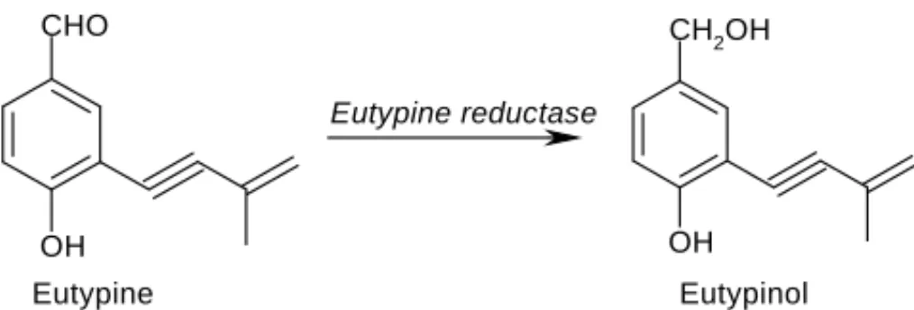
syn-thesises a toxin, named eutypine, structurally characterised as 4-hydroxy-3-(3-methyl-3-butene-1-ynyl) benzylaldehyde
[4,5] (Fig. 1). The toxin is transported by the sap to the herbaceous parts of the vine and plays a prominent role in
the expression of eutypa dieback symptoms [3]. Previous
works brought some information on the mode of action of eutypine at the cellular level and it has been shown that
∗Corresponding author. Tel.:+33-5-6225-6869;
fax:+33-5-6225-6888.
E-mail address [email protected] (F. Nepveu).
eutypine exhibits a protonophoric activity [6,7]. Eutypine
levels in diseased plant extracts might explain differences in expression of symptoms according to different culture or climatic parameters. The internal toxic concentration of eutypine, which can cause toxic symptoms in plant tissues, is not known yet.
It was established that in the grapevine cells, eutypine is metabolised into another compound identified as 4-hydroxy-3-(3-methyl-3-butene-1-ynyl) benzyl alcohol, or eutypinol (Fig. 1). This compound does not exhibit toxic effects towards the grapevine and has no protonophoric activity. Reduction of the carbonyl group of eutypine into an alcohol function is catalysed by eutypine reductase. This enzyme
was partially purified from grapevine cell suspensions[8].
Our aim was to characterise the biotransformation of eu-typine into eutypinol in grape plant tissues. The time-course of eutypine biotransformation into eutypinol was stud-ied using different grape plant materials (callus cells, micro-cuttings, plantlets) and culture media. Sample analy-ses were carried out with HPLC and LC–MS.
OH CH2OH CHO OH Eutypine Eutypinol Eutypine reductase
Fig. 1. The biotransformation of eutypine, 4-hydroxy-3-(3-methyl-3-butene-1-ynyl) benzaldehyde, into its corresponding alcohol metabolite eutypinol, 4-hydroxy-3-(3-methyl-3-butene-1-ynyl) benzyl alcohol.
2. Experimental
2.1. Chemicals
Eutypine, 4-hydroxy-3-(3-methyl-3-butene-1-ynyl)
ben-zylaldehyde, was synthesised by Raffaele Tabacchi
(Neuchˆatel, Switzerland), as previously described[9].
2.2. In vitro culture incubation conditions
In all the experiments, in vitro cultures were incubated at
25◦C, under a 16 h light/8 h dark cycle with a photon flux of
100M m−2s−1(Osram L36 W 36 Nature tubes) and 70%
humidity.
2.3. Plant material preparation 2.3.1. Grape callus cell cultures
Callus cell cultures of Vitis vinifera L. cv. Gamay, orig-inating from grape berry thin skin, was grown in solidified
medium (0.7% agar) as previously described[10]. After 21
days (exponential growth phase) of culture, calli were trans-ferred to fresh medium containing various concentrations of eutypine.
2.3.2. Grape micro-cuttings
Vitro plants of V. vinifera cv. Ugni-blanc were cultured as
previously described[11]. After 5 weeks of culture, leaves
and roots of each plantlet were removed under sterile condi-tions. Each main stem was divided into micro-cuttings each of which carried one bud. The micro-cuttings were cultured
in the presence of 500M eutypine in petridishes
contain-ing solidified medium (0.7% agar) as previously described
[10].
2.3.3. Grape plantlets
Vitro plants of V. vinifera cv. Ugni-blanc were collected after 5 weeks of culture. Roots of each plantlet were re-moved under sterilised conditions to give a plantlet with three vegetal levels. Plantlets without roots were cultured in Magenta vessels containing 40 ml of Murashige and Skoog’s
medium culture as previously described[12]in the presence
of eutypine at 500M. After 2, 5 and 10 days of culture,
plantlets and culture media were collected. Each plantlet
contained three vegetal levels and in each level two leaves. Plantlets were divided into three parts starting with the basal level.
2.4. Eutypine extraction from plant materials
Plant materials in each experiment were ground using liquid nitrogen and were extracted with acetone (20 ml for 3.5 g fresh weight (FW) tissues) for 3 h under agitation. Af-ter filtrating, the residues were treated with dioxane (2.5 ml for 18 ml acetone) for 1 h under stirring. Pigments were eliminated by extraction with water (4 ml for 20 ml extract).
Samples were placed at −18◦C overnight and were then
filtered using ash-free filter paper. Residual filtrate was evaporated to form a syrup-like residue which was diluted in 200 ml water and the pH adjusted to 5 with 0.1 M NaOH. Eutypine extraction was carried out with three portions of 100 ml ether. The raw etheric extracts were combined, dried by adding magnesium sulphate, filtered and then dried under
vacuum at 35◦C using a rotary evaporator. The residue was
suspended in 2 ml absolute ethanol for further analysis[13].
2.5. Eutypine extraction from culture media
The culture media were ultra-centrifuged at 100 000 g for
8 h at 10◦C. The liquid phase was harvested, adjusted to pH
5 using 0.1 M NaOH and extracted four times with 50 ml of ether. Organic phases were collected, dried by adding magnesium sulphate and filtered. The resulting filtrate was finally evaporated to dryness in a rotary evaporator under
vacuum at 35◦C. After evaporating the solvent from the
extract, the residue was dissolved in 2 ml absolute ethanol for further analysis.
2.6. HPLC analysis of the extract samples
Eutypine and its metabolites contained in the extracts were assayed by HPLC. HPLC conditions are quite close to those
described previously[5]with some modifications. A Waters
HPLC system was used consisting of a Waters 600 solvent delivery system, a Waters 717 plus autosampler and a
Wa-ters 2487 Dual absorbance detector. Data were acquired
and processed in a Waters Millenium32workstation, which
gave retention times and measured peak areas. A Lichrosorb
RP-C18column (5M, 150 mm ×4.6 mm) was used to
sep-arate the eutypine and its metabolites. The column
temper-ature was kept at 35◦C. The mobile phase was a mixture
of solvents A and B: water and acetonitrile, respectively. A
linear elution gradient was used as shown in Table 1. The
flow rate was 1.5 ml min−1during the run time. Before the
injection, samples were filtered through a 0.22M nylon
filter (Millipore). The 15l injection volume of each
sam-ple was taken automatically by the autosamsam-pler. The needle was washed with water/methanol (50/50). The analysis was monitored at 260 nm.
Agar concentration (g l-1 medium) 7 9 12 Concentration ( µ mol l -1 ) 0 20 40 60 80 100 120 140 eutypine T0 eutypine T12 eutypinol T12
Fig. 3. Effect of agar concentration on eutypine absorption into the cells and eutypinol diffusion back to the culture media (extracts from media) (T0: at the beginning time, T12: after 12 h of culture).
tissue into the culture media (Fig. 3). These results also
revealed a highly significant difference in eutypine uptake between the three agar concentrations studied. The lowest
agar concentration (7 g l−1) led to the highest eutypine
ab-sorption rate and of course the highest eutypinol diffusion from the callus to the media. The highest agar
concentra-tion (12 g l−1) led to the lowest eutypine absorption by the
callus, and hence the lowest eutypinol production. In the
presence of 9 g l−1agar, 75% of the eutypine in the culture
media had been absorbed by the grapevine cells after 12 h
of culture. This agar concentration (9 g l−1) was selected to
carry out the following experiments.
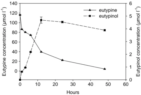
3.3. Eutypine biotransformation by grape callus tissues To study the time-course of eutypine metabolisation, pre-liminary experiments using eutypine at a concentration of
200M were designed. Samples of callus tissues and
cul-ture media were collected after each hour of culcul-ture during 12-, 24- and 48-h cultures. The chromatogram of the callus extracts showed that Gamay callus tissues contained neither eutypine nor eutypinol during the experiment time. How-ever, both were detected in the culture media as shown in
Fig. 4. These data indicate that eutypine was absorbed and transformed rapidly by the callus. It was also observed that the eutypine concentration in the culture media decreased rapidly between 0 and 48 h of culture. The largest amount of eutypine was absorbed within the first period of culture between 0 and 12 h and the highest eutypinol concentration was detected after 48 h.
Because of the impossibility to detect either the eutypine or the eutypinol within the callus using a culture medium
with 200M eutypine, two other experiments were carried
out using a higher eutypine concentration (500M). Callus
Hours 0 10 20 30 40 50 60 E u typine c o ncent rat ion ( µ m o l l -1 ) 0 20 40 60 80 100 120 140 E u ty pinol c o nc ent rat ion ( µ mo l l -1 ) 0 1 2 3 4 5 6 eutypine eutypinol
Fig. 4. Eutypine and eutypinol concentrations in Gamay culture media during the short experiment of Gamay callus culture in the presence of 200M eutypine.
tissues and culture media were collected in a first experi-ment after 1, 3 and 6 h of culture, extracted and analysed. HPLC analysis revealed the presence of both eutypine and eutypinol both in callus tissues and in the culture media 1 h after the beginning of the culture. The eutypine detection in the callus extract samples after 1 h of culture, indicated rapid absorption of eutypine by the callus tissues. Three hours
later eutypinol increased to its highest level of 0.05mol g−1
fresh weight and then decreased after 6 h to 0.03mol g−1
FW within the callus tissues as shown inFig. 5A. A small
in-crease of eutypine concentration was observed in the callus
tissues after 6 h of culture (0.013mol g−1FW). Moreover,
results also show that within the callus tissues during the experiment period, the eutypinol concentration was higher than the eutypine concentration. In the same experiment, the eutypine concentration in culture media decreased rapidly between 0 and 1 h of culture. After that eutypine concen-tration remained stable for 6 h. The eutypinol concenconcen-tration
increased from 2.43 to 8.94mol l−1after 1 and 6 h from
the beginning of culture (Fig. 5B).
Eutypine and eutypinol in the different extracts were iden-tified by LC–MS analysis. LC–MS scans displayed a
molec-ular ion peak atm/z = 185 corresponding to eutypine and
a molecular ion peak atm/z = 187 corresponding to
eu-typinol.
As shown inFig. 4andFig. 5A and B, grapevine callus
tissues are able to absorb eutypine and metabolise it into eu-typinol very rapidly—within the first 3 h of culture. In this context, plant defence is known to be dependent on the ef-ficiency of the rapid initiation and development of defence
responses [14–16]. The effectiveness of defence response
induction often depends more on its rapid initiation, devel-opment and accumulation than on the plant’s ability to
syn-thesise defence metabolites and proteins[14].
In order to study the stability and the behaviour of eutyp-ine during long periods, a second experiment was carried out. Samples of callus and culture media were collected af-ter 2, 5 and 10 days of culture. HPLC analysis revealed the
penetrated grapevine cells quickly and was then metabolised very rapidly into eutypinol. Eutypinol is excreted into the culture medium and also metabolised into other compounds. The use of micro-cuttings and in vitro plants showed that eutypine circulates in the plant and accumulates strongly in the bottom part of the stem. Moreover, leaves and stems acted similarly in the accumulation and metabolisation of eutypine.
Acknowledgements
We thank Raffaele Tabacchi for providing eutypine, Hélène Mondies for skilful technical assistance and the Midi-Pyrénées Région and its Health-Food Network for financial support.
References
[1] G.P. Munkvold, J.A. Duthie, J.J. Marois, Phytopathology 84 (1994) 186.
[2] R. Tabacchi, Pure Appl. Chem. 66 (1994) 2299.
[3] J. Fallot, C. Deswarte, S. Dalmayrac, C. Colrat, J.P. Roustan, CR Acad. Sci. Paris 320 (1997) 149.
[4] J.M. Renaud, G. Tsoupras, R. Tabacchi, Helv. Chim. Acta 72 (1989) 929.
[5] P. Tey-Rulh, I. Philippe, J.M. Renaud, G. Tsoupras, P. de Angelis, J. Fallot, R. Tabacchi, Phytochemistry 30 (1991) 471.
[6] C. Deswarte, J. Eychenne, J. Davy de Virville, J.P. Roustan, F. Moreau, J. Fallot, Arch. Biochem. Biophys. 334 (1996) 200. [7] B.E. Amborabé, P. Fleurat-Lessard, J. Bonmort, J.P. Roustan, G.
Roblin, Plant Physiol. Biochem. 39 (2001) 51.
[8] S. Colrat, C. Deswarte, A. Latché, A. Klaébé, M. Bouzayen, J. Fallot, J.P. Roustan, Planta 207 (1999) 544.
[9] E. Defrancq, R. Tabacchi, J. Labelled Comp. Radiopharm. 31 (1992) 1057.
[10] C. Ambid, M. Moissef, J. Fallot, Physiol. Vég. 21 (1983) 82. [11] O. Soulié, J.P. Roustan, J. Fallot, Vitis 32 (1993) 243. [12] T. Murashige, F. Skoog, Physiol. Plant 15 (1962) 473.
[13] I. Philippe, Ph.D. Thesis, INP Toulouse, Ecole Nationale Supérieure d’Agronomie, 1991.
[14] F. Smit, I.A. Dubery, Phytochemistry 44 (1997) 811. [15] I.A. Dubery, V. Slater, Phytochemistry 44 (1997) 1429.
[16] I.A. Dubery, L.G. Teodorczuk, A.E. Louw, Mol. Cell Biol. Res. Commun. 3 (2000) 105.
[17] C. Deswarte, H. Canut, A. Klaebe, J.P. Roustan, J. Fallot, J. Plant Physiol. 149 (1996) 336.