HAL Id: hal-03025390
https://hal.archives-ouvertes.fr/hal-03025390
Submitted on 26 Nov 2020HAL is a multi-disciplinary open access
archive for the deposit and dissemination of sci-entific research documents, whether they are pub-lished or not. The documents may come from teaching and research institutions in France or abroad, or from public or private research centers.
L’archive ouverte pluridisciplinaire HAL, est destinée au dépôt et à la diffusion de documents scientifiques de niveau recherche, publiés ou non, émanant des établissements d’enseignement et de recherche français ou étrangers, des laboratoires publics ou privés.
Effect of coordination dissymmetry on the catalytic
activity of manganese catalase mimics
Ripul Mehrotra, Micaela Richezzi, Claudia Palopoli, Christelle Hureau,
Sandra Signorella
To cite this version:
Ripul Mehrotra, Micaela Richezzi, Claudia Palopoli, Christelle Hureau, Sandra Signorella. Effect of coordination dissymmetry on the catalytic activity of manganese catalase mimics. Journal of Inorganic Biochemistry, Elsevier, 2020, 213, pp.111264. �10.1016/j.jinorgbio.2020.111264�. �hal-03025390�
1
Effect of Coordination Dissymmetry on the Catalytic Activity of Manganese
Catalase Mimics
Ripul Mehrotra,a,1 Micaela Richezzi,a Claudia Palopoli,a Christelle Hureau,b and Sandra R.
Signorellaa,*
aIQUIR (Instituto de Quimica Rosario), Consejo Nacional de Investigaciones Cientificas y
Tecnicas (CONICET), Facultad de Ciencias Bioquimicas y Farmaceuticas, Universidad Nacional de Rosario, Suipacha 531, S2002LRK Rosario, Argentina
bCNRS, LCC (Laboratoire de Chimie de Coordination) and UPS, INPT, LCC, Université de Toulouse, 205 route de Narbonne, F-31077 Toulouse, France
*Corresponding author: signorella@iquir-conicet.gov.ar
1Present address: Department of chemistry, Govt. College Chhapara, District-Seoni, Madhya Pradesh, India.
2
Abstract
Two mixed-valence Mn(II)Mn(III) complexes, [Mn2L1(OAc)2(H2O)]BPh4·2.5H2O and
[Mn2L2(OAc)2]·4H2O, obtained with unsymmetrical N4O2-hexadentate L1(2-) (H2L1 =
2-(N,N-
bis(2-(pyridylmethyl)aminomethyl)-6-(N-(2-hydroxybenzyl)benzylaminomethyl)-4-methylphenol) and N4O3-heptadentate L2(3-) (NaH2L2 =
2-(N,N-bis(2-
(pyridylmethyl)aminomethyl)-6-(N’-(2-hydroxybenzyl)(carboxymethyl)aminomethyl)-4-methylphenol sodium salt) ligands, have been prepared and characterized. Both complexes share a µ-phenolate-bis(µ-acetate)Mn(II)Mn(III) core and N3O3-coordination sphere around the
Mn(II) ion, but differ in the donor groups surrounding Mn(III) (NO4(solvent) and NO5). In
non-protic solvents, these two complexes are able to disproportionate at least 3600 equiv. of H2O2
without significant decomposition, with first-order dependence on catalyst and saturation kinetics on [H2O2]. Spectroscopic monitoring of the reaction mixtures revealed the two
complexes disproportionate H2O2 employing a different redox cycle, with retention of
dinuclearity. The higher catalytic efficiency of [Mn2L2(OAc)2] was rationalized in terms of the
larger labilizing effect of the heptadentate ligand that favors the acetate-shift and the replacement of the non-coordinating benzyl arm of L1 by a carboxylate arm in L2 which
facilitates the formation of the catalyst-H2O2 adduct, placing [Mn2L2(OAc)2] as the most efficient
among the phenolate-bridged diMn catalysts based on the kcat/KM criterion.
Keywords
3
1. Introduction
H2O2 is a by-product of aerobic metabolism in respiratory and photosynthetic electron-transport
chains and a product of enzymatic activity [1]. Excess of H2O2 and hydroxyl radical, its
decomposition product formed via a Fenton-type reaction, are harmful for almost all cell components. Thus, the rapid and efficient removal of H2O2 is essential to all aerobically living
prokaryotic and eukaryotic cells. Catalase (CAT) enzymes are present in most aerobic forms of life and are responsible for the decomposition of H2O2 into molecular oxygen and water [2].
In many pathological conditions and diseases, including cancer, neurological disorders, atherosclerosis, hypertension, ischemia/perfusion, diabetes, etc., the endogenous antioxidant systems can be overwhelmed. Administration of exogenous enzymes for the treatment of oxidative stress is limited by their short half-life, antigenicity, high cost, and large sizes that disable the enzymes to cross the cell membranes [3,4]. Therefore, low molecular weight enzyme mimics constitute an alternative to overcome these limitations [5-7], some of which have proven to protect cells from oxidative damage in animal models [8].
Manganese catalases (MnCAT) contain two Mn ions at their active site and catalyze the disproportionation of H2O2 through a Mn(II)2 and Mn(III)2 redox cycle [9-11]. The two metal ions
of the diMn active site are triply bridged through a µ1,3-carboxylate of a glutamate residue and
two solvent-derived bridges (either aqua, hydroxo, or oxo ligands) and terminally bound to His and Glu [12,13]. Both Mn subsites are six-coordinate but only one of them is bound to a labile water molecule where initial substrate binding is presumed to occur. This coordination dissymmetry around the two metal ions could be essential for catalysis. However, most diMn complexes tested as MnCAT mimics have been prepared with symmetrical dinucleating ligands with a central alcoholate or phenolate group that afford synthetic models with identical environment around the two metal centers [14-18], while only a few ones were made from unsymmetrical ligands [19-21].
With the aim of reproducing the singularities of the biosite, we report here the synthesis, properties and CAT activity of two diMn complexes obtained with unsymmetrical ligands bearing a central phenolate group:
2-(N,N-bis(2-(pyridylmethyl)aminomethyl)-6-(N-(2-4
hydroxybenzyl)benzylaminomethyl)-4-methylphenol (H2L1) and
2-(N,N-bis(2-
(pyridylmethyl)aminomethyl)-6-(N’-(2-hydroxybenzyl)(carboxymethyl)aminomethyl)-4-methylphenol sodium salt (NaH2L2), and compare the kinetic results to other diMn mimics.
These ligands offer two different coordination compartments to build bimetallic sites with different environment around each metal center and will be used for assessing the influence of the ligand dissymmetry on the CAT activity of the diMn complexes.
2. Experimental part
2.1. Synthesis of the ligands
The secondary amines, bis(2-pyridylmethyl)amine, (2-hydroxybenzyl)benzylamine and N-(2-hydroxybenzyl)glycine chlorohydrate were prepared as reported previously [22-24]. Intermediates (6-Methyl-2-phenyl-2,4-dihydro-1,3-benzodioxin-8-yl)methanol (1),
8-(bromomethyl)-6-methyl-2-phenyl-2,4-dihydro-1,3-benzodioxine (2), 8-(bis(2-pyridylmethyl)aminomethyl)-6-methyl-2-phenyl-2,4-dihydro-1,3-benzodioxine (3),
2-(N,N-bis(2-(pyridylmethyl)aminomethyl)-6-(hydroxymethyl)-4-methylphenol (4) and
2-(N,N-bis(2-(pyridylmethyl)aminomethyl)-6-(chloromethyl)-4-methylphenol (5), were synthesized
following procedures described in references [23,25-27]. Synthetic details for the obtention of these compounds are described in the supplementary information.
2.1.1. 2-(N,N-Bis(2-(pyridylmethyl)aminomethyl)-6-(N-(2-hydroxybenzyl)benzylaminomethyl)-4-methylphenol (H2L1). To a solution of 5 (533.6 mg, 1.45 mmoL) and N-(2-hydroxybenzyl)benzylamine (309 mg, 1.45 mmoL) dissolved in THF (15 mL), 200 µL of Et3N
were added and left with stirring at room temperature for 6 days. The solid formed was removed by filtration. After evaporation of THF, a red yellow oil was obtained. This oil was further dissolved in 5 mL CH2Cl2 and washed with phosphate buffer (pH 7.0). The organic
layers were collected, dried over Na2SO4 and filtered. After solvent evaporation the solid
product was obtained. Yield: 380.2 mg, 48.1 %. 1H NMR spectrum (δ, CDCl
3, ppm): 8.56 (d,
2H, Ar-Hpy); 7.60-7.54 (td, 2H, Ar-H); 7.45 (d, 1H, Ar-H); 7.36-7.31 (m, 5H, Ar-H); 7.28-7.24
5
(s, 1H, Ar-H); 6.80-6.70 (t, 2H, Ar-H); 6.74-6.71 (dd, 1H, Ar-H); 3.86 (s, 4H, Arpy-CH2-N); 3.79
(s, 2H, -CH2); 3.74 (s, 2H, -CH2); 3.72 (s, 2H, -CH2); 3.68 (s, 2H, -CH2); 2.22 (s, 3H, CH3). 13C
NMR spectrum (δ, CDCl3, ppm): 158.23, 157.7, 154.15, 148.92, 136.73, 131.42, 130.44,
129.71, 128.90, 128.41, 128.30, 127.46, 127.24, 126.55, 123.27, 122.67, 122.18, 118.82, 115.98, 59.16, 57.93, 57.13, 56.70, 20.43. Significant IR bands (ATR, νcm-1): 3356, 2914,
1589, 1454, 1433, 1357, 746, 696.
2.1.2. 2-(N,N-Bis(2-(pyridylmethyl)aminomethyl)-6-(N’-(2-hydroxybenzyl)(carboxymethyl) aminomethyl)-4-methylphenol sodium salt (NaH2L2). A solution of 5 (1.36 mmol, 500 mg) in 15 mL THF was added to N-(2-hydroxybenzyl)glycine chlorohydrate (296 mg, 1.36 mmol) in 5 mL of 0.55 M NaOH aqueous solution. The mixture was left with stirring for 48 h. THF was removed under vacuum and 20 mL CH2Cl2 added to the aqueous residue. The organic layer was
separated and washed with phosphate buffer of pH 7.0 (3 x 10 mL), dried over Na2SO4 and
filtered. After concentration of the filtrate, hexane was added and a yellow solid formed when cooled at 4ºC. Recrystallization from 2:8 CHCl3:hexane mixture afforded a bright yellow solid.
Yield: 139 mg (19.2%). 1H NMR spectrum (δ, MeOD, ppm): 8.49 (d, 1H, Ar-HPy), 8.38 (d, 1H,
Ar-HPy), 7.69 (dt, 1H, Ar-H), 7.61 (dt, 1H, Ar-H), 7.39 (m, 2H, Ar-H), 7.23 (m, 2H, Ar-H), 7.02
(m, 2H, Ar-H), 6.85 (m, 2H, Ar-H), 6.78 (s, 2H, Ar-H), 3.84 (s, 4H, Arpy-CH2N), 3.74 (s, 2H,
-CH2-), 3.72 (s, 2H, CH2), 3.68 (s, 2H, CH2), 3.56 (s, 2H, -CH2-CO2), 2.23 (s, 3H, CH3). 13C NMR
spectrum (δ, MeOD, ppm): 168.93, 157.96, 157.92, 156.45, 154.03, 148.22, 148.01, 137.44,
137.22, 132.73, 131.75, 131.17, 128.29, 128.06, 127.57, 123.68, 123.51, 123.45, 123.18, 122.44, 122.40, 119.62, 115.12, 59.34, 58.64, 58.28, 58.12, 56.40, 55.90, 54.85, 54.32, 18.94. Significant IR bands (ATR, νcm-1): 3420, 2075, 3010, 2917, 2847, 1636, 1591, 1435, 1381,
862, 760.
2.2. Synthesis of complexes
2.2.1. Complex [Mn2L1(OAc)2(H2O)]BPh4·2.5H2O (6·2.5H2O). A solution of H2L1 (113.2 mg,
0.208 mmoL) in CH3OH (2 mL) was added to a methanolic solution of manganese(II) acetate
tetrahydrate (102 mg, 0.416 mmoL) and left with stirring at room temperature for 2 h. The addition of a methanolic solution (2 mL) of NaBPh4 (71.1 mg) caused the formation of a brown
6
precipitate, which was collected by filtration, washed with methanol and hexane and dried under vacuum. Yield: 0.1920 g (0.167 mmol), 80.0 %. Thin layer chromatography (TLC) of 6
using different mobile phases (in different ratios) and three different stationary phases (silica, alumina and cellulose) showed only one spot. Anal. calcd. for BC63H62Mn2N4O7·2.5H2O: C
65.6, H 5.9, Mn 9.5, N 4.9, %; Found: C 65.5, H 5.6, Mn 9.2, N 5.3 %. Molar conductivity (CH3OH) = 129 Ω−1 cm2 mol−1. UV–vis, λmax nm (ε M−1 cm−1) in N,N-dimethylformamide (DMF):
275 (20800), 365 (4168), 450 (sh), 608 (264). Significant IR bands (KBr, νcm-1): 3445 (broad),
3055, 3030, 2920, 2853, 1625, 1595, 1560, 1431, 864, 734, 705, 644, 611. The content of 2.5 molecules of non-coordinated water per complex molecule was confirmed by thermogravimetric analysis of the complex which showed 3.9 % mass loss below 120 °C. 2.2.2. Complex [Mn2L2(OAc)2]·4H2O (7·4H2O). A solution of NaH2L2 (100 mg, 0.187 mmol) in
8 mL CH3OH was mixed with a solution of Mn(OAc)2·4H2O (92 mg, 0.378 mmol) in 2 mL
CH3OH, and the red brown mixture was left with stirring for 3 h. Then the volume was reduced
to 2 mL under vacuum, 10 mL ethyl ether were added, and the mixture cooled to 4 ºC. After 3 h, the formed brown precipitate was filtered off, washed with cool CH3OH and ether and dried
under vacuum. Yield: 108 mg, 78.3%. TLC of 7 using different mobile phases (in different
ratios) and three different stationary phases (silica, alumina and cellulose) showed only one spot. Anal. calcd. for C34H35Mn2N4O8·4H2O: C 50.4, H 5.4, Mn 13.6, N 6.9, %; Found: C 50.1,
H 5.1, Mn 13.7, N 6.7 %. UV–vis, λmax nm (ε M−1 cm−1) in DMF: 279 (21350), 345 (4105), 462
(1665), 694 (293). Significant IR bands (KBr, νcm-1): 3435 (broad), 3065, 3032, 3004, 2921,
2859, 1624, 1590, 1574, 1422, 806, 761, 654, 630. The content of 4 molecules of non-coordinated water per complex molecule was confirmed by thermogravimetric analysis of the complex which showed 8.9 % mass loss below 120 °C.
2.3. Analytical and physical measurements
Infrared spectra were recorded on a Perkin-Elmer Spectrum One FTIR spectrophotometer in the 4000–400 cm−1 range. UV–visible spectra were recorded on a Jasco V-550
spectrophotometer, with thermostated cell compartments. Metal content was determined with an Inductively coupled plasma mass spectrometer (ICP-MS) Perkin Elmer NexION 350X. EPR
7
spectra were obtained at 115 K on an Elexsys E 500 Bruker spectrometer, operating at a microwave frequency of approximately 9.5 GHz. TGA measurements were conducted on a Perkin-Elmer Diamond TG/DTA Instrument. The compound was heated at the rate of 10 °C/min between RT and 800°C. Conductivity measurements were performed using a Horiba F-54 BW conductivity meter, on 1.0 mM solutions of the complexes in methanol. Electrospray ionization (ESI) mass spectra were obtained with a Thermo Scientific LCQ Fleet. The solutions for electrospray were prepared from solutions of complex or reaction mixtures diluted with acetonitrile to a final ~10−5 M concentration. 1H NMR spectra were recorded on a Bruker AC
300 NMR spectrometer at ambient probe temperature (ca. 25 °C). Chemical shifts (in ppm) are referenced to tetramethylsilane and paramagnetic NMR spectra were acquired employing superWEFT sequence, with acquisition time of 50 ms. The electrochemical experiments were performed with a computer-controlled Princeton Applied Research potentiostat, VERSASTAT II model, with the 270/250 Research Electrochemistry Software. Studies were carried out under Ar, in DMF solution using 0.1 M Bu4NPF6 as a supporting electrolyte and ≈10−3 M of the
complexes. The working electrode was a glassy carbon disk, and the reference electrode was a calomel electrode isolated in a fritted bridged with a Pt wire as the auxiliary electrode. All potentials are referred to the SCE electrode. Under these conditions, E(ferrocene/ferrocenium) = 474 mV, in DMF.
2.4. Evaluation of CAT activity
Reaction rates were determined by volumetric measurement of the O2 evolved after addition
of excess of H2O2 to a DMF solution of the complex. A round-bottom flask with a rubber septum,
containing the degassed solution of the complex, was thermostated at 25°C and connected to a gas-measuring burette (precision of 0.1 mL). Previously thermostated H2O2 was injected
through the septum to the stirred complex solution, and the evolved O2 was measured with the
burette. The initial reaction rates were obtained by fitting the [O2] versus time data to a
polynomial expression and calculating the slope of the tangent at time zero. Each rate constant reported here represents the mean value of multiple determinations that fall within ± 5%. All
8
experiments were carried out at 25°C. Blanc experiments performed with 100 mM H2O2 in DMF
without the catalyst showed no decomposition after 1 h.
3. Results and discussion
3.1. Synthesis and characterization of complexes 6 and 7
The synthesis of the unsymmetrical ligands H2L1 and NaH2L2 is shown in Scheme 1.
OH OH OH OH O O Br O O N N N N N O O N N N OH OH N N N OH Cl N N OH N N OH N N HO NHCl HO OH O N N OH N N ONa HO O i ii iii iv v vi vii H2L1 NaH2L2 1 2 3 4 5
Scheme 1. Synthesis of unsymmetrical ligands. (i) PhCH(OMe)2, p-MeC6H4SO3H, DMF, 50ºC; (ii) CBr4,
9
The key step in the synthesis for introduction of dissymmetry is the formation of the cyclic acetal 1 to protect one of the terminal hydroxymethyl groups of the starting
2,6-bis(hydroxymethyl)-4-methylphenol [25]. Next, bromination and alkylation of bis-2-picolylamine afforded the N3-branch. Acid hydrolysis of the acetal, followed by chlorination and subsequent
alkylation of a second amine bearing terminal benzyl/phenol or carboxylate/phenol groups afforded the N4O2-hexadentate (H2L1) and N4O3-heptadentate (H2NaL2) ligand, respectively.
Dimanganese complexes 6 and 7 were prepared from 1:2 mixtures of the corresponding ligand
and Mn(OAc)2·4H2O in methanol. Acetate facilitates the deprotonation of the ligand for
coordination to the metal [28] and complexation occurs immediately after mixing, as evidenced by the change of color of the reaction mixture from pale yellow to brown. Complex 6 was
precipitated as a brown solid from the reaction mixture upon addition of the bulky BPh4−, while
complex 7 separated from the solution as a brown solid after addition of ether at 4 ºC.
The IR spectra of powdered 6 and 7 (Figure S1) exhibit strong phenolate and pyridyl ring
absorptions between 1625 and 1575 cm-1 as well as absorption bands at 1560-1574 cm−1 and
1431-1422 cm−1 attributable to the antisymmetrical and symmetrical stretching vibrations of
carboxylate groups. For 6, the three bands at 611, 705 and 734 cm−1 together with a strong
and sharp stretching band at ~3055 cm−1 can be assigned to non-coordinated BPh4− counter
anion, in agreement with its molecular structure. In line with the loss of H2O evidenced by the
thermogravimetric analysis, the two compounds display a broad band at ≈ 3440 cm−1 assigned
to water molecules.
ESI-mass spectra of complexes 6 and 7 in CH3CN (Figure 1(a)) confirmed their chemical
composition as well as retention of their nuclearity in solution. For complex 6, the parent peak
is observed at m/z = 811.2 in the positive mode ESI-mass spectra and originates from the [Mn2L1(OAc)2(CH3CN)]+ monocation. Other peaks generated during the electrospray
correspond to the exchange of AcO− by HCO
2− ([Mn2L1(OAc)(HCO2)]+, m/z = 757.2) and the
loss of Mn(OAc)2 ([MnL1H]+, m/z = 598.2). Complex 7 affords a mass spectrum with the major
10
[Mn2L2(OAc)]+ at m/z = 678.1 and [MnL2Na]+ at m/z = 587.1. The isotopic patterns of these
peaks match very well their simulated spectra. In addition, negative ion ESI-mass spectrum of complex 6 shows only one peak at m/z = 319 corresponding to BPh4−anion, in agreement with
the molar conductivity of 129 Ω−1 cm2 mol−1 for 6 in methanol, a value expected for a complex
that behaves as a 1:1 electrolyte in solution.
Figure 1(b) illustrates the X-band EPR spectra of the two powdered complexes at 120 K. These spectra exhibit two features, one around g = 2 which dominates the spectra and a low-field component at g = 5 for 6 and 6.8 for 7, which can be associated with spin transitions in S > ½
spin states of the weakly coupled phenolate-bridged Mn(II)Mn(III) center, as already observed for other Mn(II)Mn(III) complexes [29-30].
The electronic spectra of complexes 6 and 7 (Figure 1(c)) recorded in DMF exhibit an intense
absorption at 275 and 279 nm, respectively, assigned to π- π* transitions within the ligands, and two moderately intense bands in the region 330–550 nm attributed to ligand-to-metal charge transfer (LMCT) transitions from pπ orbital of phenolate (either bridging or terminal phenolate) to partially filled dσ* and dπ* orbitals of Mn(III) [17,19,31,32] with an additional contribution of carboxylate-to-Mn(III) CT on the lower energy band [33]. The low intensity broad band observed around 608 nm (ε = 265 M-1 cm-1) for 6 and at 698 nm (ε = 294 M-1 cm-1) for 7,
corresponds to d-d transitions of the Mn(III) ion in agreement with reported values for related Mn(II)Mn(III) complexes [31,34]. The red shift of the d-d band can be interpreted in terms of the effect of the axial or equatorial position of the terminal phenolate group in the two complexes (Figure 2). The more distant binding of phenolate to the Mn(III) ion on the elongation Jean-Teller axis weakens the metal-phOax bond reducing the d-d energy gap and this can
account for red shift of this band in the spectrum of 7 relative to 6 [35]. The contribution of other
electronic transitions disable direct comparison of the energy of the phenolate-to-Mn(III) CT bands. Electronic spectra of the two complexes registered at different times after preparing the DMF solutions (up to 24 h), showed identical molar absorption coefficients, indicating that both are stable in this solvent.
11
Figure 1. (a) ESI-mass spectra of 6 and 7, in MeCN, positive mode. (b) EPR spectra of solid
complexes, ν = 9.5 GHz, T = 120 K, microwave power = 0.5 mW. (c) Electronic spectra of 6
and 7 in DMF. [complex] = 0.125 mM; l = 1 cm. (d) 1H NMR spectra of 6 in DCCl3 and 7 in
CD3OD. [complex] = 15 mM.
The 1H NMR spectrum of solutions of 6 and 7 (Figure 1(d)) show resonances outside the
diamagnetic region spanning from 70 to -30 ppm. For the two complexes the signals appear in the same spectral region according to a weak magnetic coupling between the metal ions; however, resonances are much narrower for 7 as a consequence of the different spatial
12
arrangement of the ligands in the two compounds. For 6, the spectral pattern comprises a set
of broad signals at 60, 48, 29, 20 and -13 ppm (linewidth in the range of 2500 to 3500 Hz) ascribed to phenolate/pyridine ring protons with even broader peaks of methylene protons spanning the full spectral range [36]. For 7, the spectrum is better resolved showing intense
resonances at 62, 49, 47 and 45 ppm (w1/2 = 1200 Hz), and relatively sharp resonances at 26,
19, -9, -12 and -14.5 ppm (w1/2 = 600 Hz), and -26 ppm (w1/2 = 300 Hz). These peaks can be
attributed to phenol and pyridine ring protons shifted down- and upfield as a consequence of spin-delocalization and dipolar effects, the last playing an important role in this kind of mixed-valence Mn(II)Mn(III) complexes [37].
The 1-10 ppm region of the 1H NMR spectra of the complexes (Figure S2) show peaks
belonging to the solvent, and BPh4− in the case of complex 6, without traces of the ligand,
indicating the compounds are stable towards metal dissociation. Besides, no signal attributable to terminally coordinated acetate (broad signal at around 1.9 ppm [38]) is observed, which suggests that the most plausible structure for the two complexes in solution contains two bridging acetate ligands as shown in Figure 2.
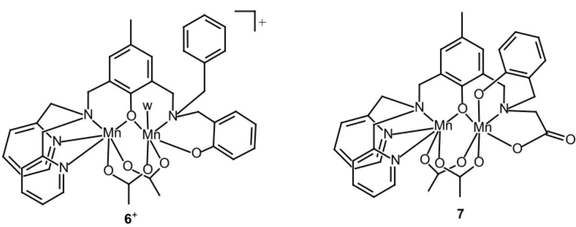
N N O N Mn Mn N O O O O O w O O O O N N O N Mn Mn N O O O + 6+ 7
Figure 2. Schematic structure formulas of the diMn complexes under study. w = water
3.2. Electrochemical studies
The electrochemical properties of complexes 6 and 7 were investigated by cyclic voltammetry
in DMF solutions containing 0.1 M Bu4NPF6 (Figure S3). The two complexes exhibit one
13
that these waves correspond to reduction processes. W1/2 values of 0.12 V for 6 and 0.13 V for 7 in the square-wave voltammetry (SWV) experiments (Figure S3(a-b)) suggest that these
reductions correspond to one-electron processes and may be attributed to the Mn(II)Mn(III)/Mn(II)Mn(II) redox couple.
On the oxidative side, 6 shows two anodic waves at Epa1 = 0.44 V (from deconvolution of the
SWV wave) and E1/2(2) = 0.54 V vs SCE (∆Ep = 0.14 V) (Figure S3(c)). These two one-electron oxidation processes might correspond to the formation of Mn(III)2 and Mn(II)Mn(III)-phenoxyl
radical. The formation of Mn(II)Mn(III)-phenoxyl radical and Mn(III)2 occurs at close potentials
and are difficult to distinguish [39]. However, the growth of the relative intensity of the second oxidation wave with increasing scan rate suggests it corresponds to the ligand-centered oxidation to generate a Mn(II)Mn(III)-phenoxyl radical [40]. In the case of complex 7, three
non-reversible anodic processes are detected at Epa1 ≈ 0.4 V, Epa2 ≈ 0.52 V and Epa3 ≈ 0.79 V vs
SCE (Figure S3(d)), which might be attributed to the formation of higher oxidation state complexes with concomitant formation of oxo-bridges. It must be observed that while reduction and first oxidation processes of 6 take place at potentials differing in 0.38 V, 7 shows
electrochemical stability over ∆E = 0.56 V. Therefore, the introduction of carboxylate instead of the non-coordinating benzyl group in the ligand framework plays a significant role in the stabilization of the mixed valence Mn(II)Mn(III) state in dry DMF. An still larger ∆E (0.965 V) was reported for another triply phenolate-bis(acetate)-bridged Mn(II)Mn(III) complex where the benzyl group is replaced by pyridine, a donor stronger than carboxylate, with redox processes taking place at Ered = -0.057V and Eox = 0.908 V vs SCE in MeCN [19].
3.3. Catalase activity studies
3.3.1. Kinetics. In the previous sections it was shown that the replacement of the non-coordinating benzyl arm by the carboxylate in the ligand influences both the ligand-field splitting and the redox potentials. Thus, it is expected that this ligand changes also affect the catalase activity of the resulting complexes.The ability of complexes 6 and 7 to catalyze H2O2
14
vigorous evolution of dioxygen was observed after addition of H2O2 to a solution of the catalyst.
Turnovers as high as 3600 without significant decomposition were measured for the two complexes in DMF, and up to 600 and 300 equivalents of H2O2 were decomposed in CH3CN
and CH3OH, respectively. Besides, in the last two solvents, the rate of O2 evolution diminished
after successive additions of H2O2. It is likely that the bridging ligands of the complexes serve
as internal bases facilitating deprotonation of the peroxide coupled to the redox reaction. Therefore, in DMF, a non protic solvent (αDMF = 0) [41], these catalysts disproportionate H2O2
with only slight loss of activity (Figure S4). However, in the protic CH3OH solvent (αMeOH = 0.93)
or CH3CN, a solvent with intermediate proton donor ability (αMeCN = 0.19), protonation of the
bridges could inactivate the catalyst resulting in moderate to low turnover numbers. Therefore, DMF was chosen as solvent for evaluation of catalase activity.
The initial rate of H2O2 disproportionation by complexes 6 and 7 in DMF was measured as a
function of the complex (Figure S5) and substrate concentrations at 25 ºC. As shown in Figure 3, for both complexes, at constant [H2O2]0 = 148 mM, the initial rate (ri) varies linearly with the
[catalyst], and the first-order rate constants 𝑘𝑘1𝟔𝟔 = 24.5(3) min−1 and 𝑘𝑘1𝟕𝟕 = 51.2(2) min−1 were
obtained from the slope of the straight line.
At constant [catalyst] = 0.25 mM, complexes 6 and 7 exhibit saturation kinetics with [H2O2]0
(Figure 3) and the ri/[catalyst] vs [H2O2]0 data could be fitted to the Michaelis-Menten equation
(eq. 1). In this equation, kcat is the catalytic rate constant (turnover number) and KM is a
measure of the complex affinity for H2O2 (the lower the KM value, the higher affinity for H2O2).
The values of catalytic turnover numbers 𝑘𝑘𝑐𝑐𝑐𝑐𝑐𝑐𝟔𝟔 = 2.3(1) s-1 and 𝑘𝑘𝑐𝑐𝑐𝑐𝑐𝑐𝟕𝟕 = 1.22(2) s-1, and Michaelis
constants 𝐾𝐾𝑀𝑀𝟔𝟔 = 6.8(4) x 10-1 M and 𝐾𝐾𝑀𝑀𝟕𝟕 = 7.3(5) x 10-2 M, were determined from the non-linear
fit of experimental data to eq. 1 (solid wine and blue lines in Figure 3).
𝑟𝑟
𝑖𝑖[catalyst] =
𝑘𝑘
𝑐𝑐𝑐𝑐𝑐𝑐[H
2O
2]
15
Kinetics results show that electron transfer between 6 and H2O2 is more effective (larger kcat)
than for 7, but 6 is 10-times less efficient at binding the substrate (𝐾𝐾𝑀𝑀𝟔𝟔 > 𝐾𝐾𝑀𝑀𝟕𝟕), so that can reach
a maximal rate (Vmax = kcat [catalyst]) at [H2O2] much higher than 7. Based on the kcat/KM
criterion, 𝑘𝑘𝑐𝑐𝑐𝑐𝑐𝑐𝟔𝟔
𝐾𝐾𝑀𝑀𝟔𝟔 = 3.38 M−1 s−1 and 𝑘𝑘𝑐𝑐𝑐𝑐𝑐𝑐 𝟕𝟕
𝐾𝐾𝑀𝑀𝟕𝟕 = 16.7 M−1 s−1, the catalytic efficiency of 7 is ≈ 5-times higher than for complex 6. The higher efficiency of 7 to disproportionate H2O2 is related to its
larger affinity for binding peroxide, probably because the presence of the terminal carboxylate facilitates H2O2 binding through hydrogen bonding favoring the formation of the catalyst-H2O2
adduct [18,42].
Figure 3. Effect of [catalyst]0 (green and orange lines; [H2O2] = 148 mM) and [H2O2]0 (brown
and blue lines; [catalyst] = 0.25 mM) on the initial rate of H2O2 disproportionation catalyzed by
(a) 6 and (b) 7 at 298 K, in DMF
3.3.2. Spectroscopic Monitoring of the Catalase Reaction. In order to get insight into the mechanism of the H2O2 disproportionation catalyzed by complexes 6 and 7 in DMF, the
reaction was monitored by using a combination of electronic and EPR spectroscopies and ESI-MS. UV-visible absorption spectra taken in DMF during the progress of the reaction of complex
6 with excess H2O2 exhibited an increase of the absorption band at 275 nm concurrently with
16
The intensity of bands at longer wavelengths changed slightly during the reaction. The same spectral changes were observed upon successive additions of 100 equiv of H2O2 to the DMF
solution of the catalyst, and the O2 production rate measured after each new addition was
essentially constant, suggesting that the catalyst is stable in these reaction conditions.
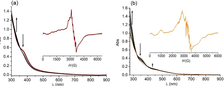
Figure 4. UV–vis spectra of (a) 6 (0.125 mM) and (b) 7 (0.1 mM) after addition of 100 equiv.
of H2O2, in DMF. T = 25ºC. Inset: X-band EPR spectra of 1 mM catalyst + 100 equiv. H2O2 a
few minutes after mixing. ν = 9.5 GHz, T = 110 K, microwave power = 0.5 mW.
After addition of 100 equiv. of H2O2 to a solution of 6 in DMF, the low-temperature X-band EPR
spectra exhibit a broad signal (Figure 4(a), inset) consistent with a weakly coupled Mn(II)Mn(III) center [17] overlapped to a six-line signal centered at g = 2 with hyperfine splitting of ≈ 93 G. This 6-line signal departs from that of the solvated Mn2+ ion and is characteristic of a Mn(II)
center bound to the ligand that could arise from a Mn(III)Mn(II) species where the phenolate-bridge disrupted through protonation [36,37]. This cleavage of the phenolate phenolate-bridge makes the diMn unit appear as the individual ions: an EPR silent Mn(III) and a mononuclear Mn(II) with a six-line signature. This was confirmed by addition of p-toluensulphonic acid to the solution of
6 in DMF giving the same EPR spectrum. This spectral pattern was kept at the end of the
reaction. Retention of the complex nuclearity during the reaction was confirmed by mass spectrometry. ESI-mass spectra obtained after addition of 100 equiv H2O2 to a DMF solution
17
of 6 showed the parent peak at m/z 811.2 and other peaks already appearing in the spectrum
of the starting solution and a new peak at m/z 773.2 corresponding to [Mn2L1(OAc)(HCO2)(OH)]+ (Figure S6(a)), thus providing a clear indication that the diMn
complex persisted during the catalytic cycle.
During the spectrophotometric monitoring of the reaction of 7 with excess H2O2 in DMF, the
intensity of the CT band at 345 nm decreased and absorbance at 279 nm grew up, with an isosbestic point at 323 nm (Figure 4(b)). Interestingly, the intensity of the band at 462 nm (PhO-to-dπ Mn(III) CT) also raised, although in a lesser proportion. These spectral changes suggest a different arrangement of phenolate groups around Mn(III) during catalysis. For this complex, the initial intensity of the LMCT bands was not restored at the end of the H2O2
disproportionation reaction. The EPR spectra taken during the 7 + H2O2 reaction course
(Figure 4(b), inset) showed broad lines expanded over the whole spectral range characteristic of diMn(II) species with weak anti-ferromagnetic exchange interactions [37,43,44] which originate from the contribution of the populated paramagnetic higher excited spin states [45]. Several sets of 11 lines are observed on the top of some of the more intense signals (i.e. signal centered at 2870 G), with an average hyperfine splitting of ≈ 43 G typical of the interaction between the electronic spin and two Mn(II) nuclear spins (IMn = 5/2). This Mn(II)2 spectrum is
superimposed to a 6-line signal at g ≈ 2 that arises from a small fraction of monomeric aqueous Mn2+ (up to 5% at the end of the reaction). The final EPR spectrum of the reaction mixture was
identical to those registered during the reaction meaning the formed Mn(II)2 species persists
in solution, in agreement with absorption spectra. The disproportionation of H2O2 catalyzed by 7 was also followed by ESI-MS. Besides the parent peak at m/z 755 and other peaks already
present in the starting complex solution, another intense peak is observed at m/z = 619.1 that can be attributed to [Mn2L2]+, thus confirming the retention of dinuclearity of the complex during
catalysis (Figure S6(b)).
3.3.3. Mechanism of the CAT-like reaction of 6 and 7. The two complexes catalyze H2O2
18
successive additions of H2O2, and O2 evolution occurs without a time-lag at the onset of the
reaction, all suggesting these complexes are responsible for the disproportionation of H2O2.
Based on spectroscopic results, both compounds retain their dinuclearity during catalysis but employ a different redox cycle for H2O2 disproportionation. From the EPR spectra, the mixed
valence Mn(II)Mn(III) complex 6 is present in solution during the catalytic cycle either in the
coupled [Mn(II)(µ-PhO)Mn(III)]+ or uncoupled [Mn(II)(PhOH)Mn(III)]2+ forms. A possible
mechanism is presented in Scheme 2, including the equilibrium of the uncoupled/coupled diMn species of 6 (observed in the EPR spectra) and species present in the ESI-mass spectra. It is
proposed that 6 is the active reduced form of the catalyst that reacts with H2O2 in the slow
reductive half-reaction (turnover-limiting step). The observation of saturation kinetics with substrate indicates that a catalyst-peroxide adduct is formed at this step, probably by substitution of the labile solvent molecule bound to Mn(III), which upon O-O bond breaking yields the µ-oxo-Mn(III)Mn(IV) and water. This oxidized form of the complex reacts with another H2O2 molecule in a fast-oxidative half-reaction to afford O2 and restores the starting complex
closing the cycle. This oxidative half-reaction must be much faster than the reductive one, so that the µ-oxo-Mn(III)Mn(IV) is not observed by EPR.
19
For H2O2 disproportionation catalyzed by complex 7, the observation of a coupled Mn(II)2
species in the EPR spectra taken during and at the end of the reaction suggests a redox cycle involving Mn(II)2 and Mn(III)2 oxidation states, as proposed in Scheme 3.
Scheme 3. Proposed catalytic cycle for complex 7
According to this scheme, initial oxidation of the starting Mn(II)Mn(III) complex by H2O2 affords
Mn(III)2(OAc)1(2)(µ-O(H)) species (EPR silent) in a fast step. In this way, the starting complex
should be a precursor of the active complex. A similar one-electron oxidation of a precursor phenolate-bridged diMn(II) complex has already been reported [36]. The H2O2 binding to the
starting complex can take place through acetate-shift to yield the µ-hydroxo-Mn(III)2 species,
where terminally bound acetate can be further protonated and partially dissociates. The higher labilizing effect of L2(3-) compared to L1(2-) may favor acetate dissociation as evidenced by the
high proportion of the Mn2L+ species in the mass spectra. The µ-oxo-Mn(III)2 species is
proposed to be the oxidized active form of the catalyst that enters the catalytic cycle of H2O2
dismutation. The absence of a lag-time at the onset of the reaction is consistent with the oxidation of the mixed valence complex being faster than the H2O2 disproportionation reaction.
20
adduct (Figure S7) may account for the higher efficiency of this complex for binding peroxide (low KM value) which renders 7 more efficient to catalyze H2O2 disproportionation than 6.
At pH 7, the electrochemical potentials for the two-electron O2/H2O2 and H2O2/H2O redox
couples are +0.04 V and +1.11 V vs SCE, respectively. If disproportionation of H2O2 occurs
through an outer-sphere mechanism, only diMn complexes with redox couples between the H2O2 reduction and oxidation potentials should be active. However, a number of complexes
that exhibit redox potentials outside the H2O2 dismutation range react efficiently [7,14],
suggesting diMn complexes dismutate H2O2 through an inner-sphere mechanism with electron
transfer taking place within the catalyst-peroxide adduct, in agreement with the saturation kinetics observed for a number of diMn mimics. In Table 1, the catalytic efficiency (kcat/KM
values) of complexes 6 and 7 is compared to other phenolate-bridged diMn complexes of
ligands with different terminal N/O donors that employ either Mn(II)2/Mn(III)2 or
Mn(II)Mn(III)/Mn(III)Mn(IV) redox cycles during catalysis. For those complexes with low affinity for the substrate (large KM value) that do not achieve saturation with [H2O2], second-order rate
constants (k2cat) are given for comparison, considering that when KM >> [H2O2], the
denominator of eq. 1 is dominated by KM, and ri = 𝑘𝑘𝐾𝐾𝑐𝑐𝑐𝑐𝑐𝑐
𝑀𝑀 [catalyst][H2O2] = 𝑘𝑘2𝑐𝑐𝑐𝑐𝑐𝑐 [catalyst][H2O2]. The catalytic efficiency of triply bridged (µ-phenolate)bis(µ1,3-carboxylate)diMn complexes falls
into two groups. Complexes bearing terminal carboxylate that react with efficiency higher than 11 M-1s-1 (entries 1-2), and those with pyridyl/phenolate arms that react with efficiency lower
than 5 M-1s-1 (entries 4-6), highlighting the role of carboxylate to facilitate the formation of the
adduct with H2O2 through hydrogen bond. Phenolate-bridged [Mn2L(OH)2(H2O)2] with no
acetate bridges but four carboxylate arms, is placed in between these two groups, with the carboxylate groups compensating the long intermetallic separation that disfavors the cooperativity between the two Mn ions for the electron transfer process (entry 3). Bis(µ -phO)diMn complexes are even less efficient than any of the (µ-phenolate)bis(µ1,3
-carboxylate)diMn complexes, one example is shown in entry 7. Enhanced activity is observed when the two Mn ions are linked by one or two alcoholate bridges (entries 8-9) where the intermetallic distance is shorter than in the phenolate-bridged complexes. As can be observed
21
in Table 1, the Mn⋅⋅⋅Mn separation in phenolate-bridged complexes is longer than 3.4 Å, while in the (µ-alcoholate)ndiMn complexes the Mn⋅⋅⋅Mn distance is closer to the value reported for
the enzyme (3.03 and 3.18 Å for the oxidized and reduced forms, respectively, entry 10), strengthening the cooperativity between the two Mn ions.
Table 1. Catalytic efficiency of phenolate-bridged diMn complexes for disproportionating H2O2
Complex kcat/KM or k (M-1s-1)
Bridging ligands Ligand terminal groups
Redox cycle Mn⋅⋅⋅Mn (Å)
Refs
1 7 16.7 (µ-phO)(µ-OAc)2 py2/phO-,CO2- Mn(II)2/Mn(III)2 - This work
2 [Mn2(bcpmp)(µ-OAc)2] 11.44 (µ-phO)(µ-OAc)2 py,CO2-/ py,CO2- Mn(II)Mn(III)/Mn(III)Mn(IV) 3.47 [17] 3 [Mn2L(OH)2(H2O)2] 5.08a (µ-phO) (CO2-)2/(CO2-)2 Mn(II)2/Mn(III)2 3.67d [18]
4 [Mn2(bphpmp)(µ-OAc)2(H2O)]+ 4.7b (µ-phO)(µ-OAc)2 py2/py,phO- NR 3.497 [19]
5 [Mn2(bpbp)(µ-OAc)2]2+ 3.76 (µ-phO)(µ-OAc)2 py2/py2 Mn(II)Mn(III)/Mn(III)Mn(IV) 3.45 [17,46]
6 6 3.38 (µ-phO)(µ-OAc)2 py2/phO- Mn(II)Mn(III)/Mn(III)Mn(IV) - This work
7 [Mn2(bphba)2(Cl)2] 1.1 (µ-phO)2 py2/py2 Mn(II)2/Mn(III)2 3.41 [47,48]
8 [Mn2(HBPClNOL)(BPClNOL)Cl]+ 80.2c (µ-phO)(µ-OR) py,RO-/py,phO- Mn(II)Mn(III)/Mn(III)Mn(IV) 3.16 [30] 9 [Mn2(µ-OMe)(OAc)(hppentO)]+ 106.3 (µ-OR)2(µ-OAc) py,phO-/py,phO- Mn(II)2/Mn(III)2 2.95 [49] 10 MnCAT (T. thermophilus) 3.1 x 10⁶ (µ-OH)(µ-OH
2)(µ-OAc) His,Glu/His,Glu,H2O Mn(II)2/Mn(III)2 3.18; 3.03 [50,51,13] NR = not reported. Bcpmp = 2,6‑bis({(carboxymethyl)[(1‑pyridyl)methyl]amino}methyl)‑4‑methylphenol]; bpbp =
2,6‑bis{[bis(2‑pyridylmethyl)amino]methyl}‑4‑tert‑butylphenol; bphba = 2‑{(N,N‑bis(2‑pyridylmethyl)amino)methyl}phenol; bphpmp = 2‑[bis(2‑pyridylmethyl)aminomethyl]‑6‑{[(2‑hydroxybenzyl)(2‑pyridylmethyl)amino]methyl}‑4-methylphenol; LH4 = 5‑methyl‑2‑hydroxo‑1,3‑xylene‑α,α‑diamine‑N,N,N’,N’‑tetraacetic acid; H2BPClNOL = N-(2-hydroxybenzyl)-N-(2-pyridylmethyl)[(3-chloro)(2-hydroxy)]propylamine; H2hppentOH = 1,5-Bis[(2-hydroxybenzyl)(2-pyridylmethyl)amino]pentan-3-ol
aSecond-order kinetics. bCalculated from reported data. cIn the presence of piperazine, pH 9.7. dCalculated from an optimized geometry.
4. Conclusions
Unsymmetrical N4O2-Hexadentate H2L1 and N4O3-heptadentate NaH2L2 ligands afford mixed
valence Mn(II)Mn(III) complexes 6 and 7 where each Mn ion is in a different coordination
environment. Both complexes share the coordination sphere of the Mn(II) ion, but differ in the donor groups surrounding Mn(III). In 6, the sixth coordination position of Mn(III) is occupied by
a water molecule, while in 7 Mn(III) is bound to the NO3-donor sites of the ligand and two
additional O-atoms from bridging acetates, leaving this metal ion coordinatively saturated. The replacement of the non-coordinating benzyl arm of L1 by the carboxylate arm in L2 modifies the
22
ligand field splitting, the reduction potential and the reactivity of the resulting complex. Therefore, complex 7 catalyzes H2O2 disproportionation more efficiently than 6, even when the
formation of the catalyst-peroxide adduct must take place through acetate-shift in 7 but by
direct replacement of a labile solvent molecule in 6. This different reactivity can be interpreted
in terms of the larger affinity of 7 for the substrate (smaller KM value), attributed to the ability of
the terminal carboxylate group of the ligand to stabilize the adduct through hydrogen bond to H2O2, and to the higher labilizing effect of the heptadentate ligand that favors the acetate-shift.
These two combined effects in 7 render KM = 0.073 M, that compares well to KM = 0.083 M of
MnCAT of T. thermophilus [50], and, based on the kcat/KM criterion, place complex 7 as the
most efficient among the phenolate-bridged diMn catalysts. Given that acetate-shift is also an initial process in the phosphohydrolase activity [52], further catalytic studies of complexes 6
and 7 as hydrolase mimics will be undertaken to test the bifunctionality of these complexes. Declaration of competing interest
The authors declare that they have no known competing financial interests or personal relationships that could have appeared to influence the work reported in this paper.
Acknowledgments
This work was supported by the National University of Rosario (PIP-BIO553) and the Consejo Nacional de Investigaciones Científicas y Técnicas (CONICET, PIP 0337 and PUE 0068), the Centre National de la Recherche Scientifique (CNRS, PICS 07121), and the Agency for Science, Technology and Innovation of Santa Fe (ASACTeI, IO 2010-164-16). R. Mehrotra thanks CONICET for a post-doctoral fellowship.
Appendix A. Supplementary data
Supplementary data to this article can be found online at
https://doi.org/10.1016/j.jinorgbio.2020.xxxxx
References
23
[2] J. E. Penner-Hahn, in Manganese Redox Enzymes, ed. V. L. Pecoraro, VCH, New York, 1992.
[3] D. Salvemini, C. Muscoli, D. P. Riley, S. Cuzzocrea, Pulm. Pharmacol. Ther. 15 (2002) 439–447.
[4] J. M. McCord, M. A. Edeas, Biomed. Pharmacother. 59 (2005) 139–142. [5] A. Mahammed, Z. Gross, Catal. Sci. Technol. 1 (2011) 535–540.
[6] C. Policar, in: J. S. Reboucas, I. Batinic-Haberle, I. Spasojevic, D. S. Warner, D. St. Clair (Eds.), Redox Active Therapeutics, Springer, Berlin, 2016, pp. 125–164. [7] S. Signorella, C. Palopoli, G. Ledesma, Coord. Chem. Rev. 365 (2018) 75–102. [8] Y. Ning, Y. Huo, H. Xue, Y. Du, Y. Yao, A. C. Sedgwick, H. Lin, C. Li, S.-D. Jiang,
B.-W. Wang, S. Gao, L. Kang, J. L. Sessler, J.-L. Zhang, J. Am. Chem. Soc. 142 (2020) 10219–10227.
[9] P. J. Riggs-Gelasco, R. Mei, J. E. Penner-Hahn, H. H. Thorp, V. L. Pecoraro (Eds.), Mechanistic Bioinorganic Chemistry, American Chemical Society, Washington, DC, 1995 (Chapter 8).
[10] W. C. Stallings, K. A. Pattridge, R. A. Strong, M. L. Ludwig, J. Biol. Chem. 260 (1985) 16424–16432.
[11] S. Signorella, C. Palopoli, V. Daier, G. Ledesma, R.H. Kretsinger, E.A. Permyakov, V.N. Uversky (Eds.), Encyclopedia of Metalloproteins, Springer, New York, 2013, pp. 1283–1292.
[12] V. V. Barynin, M.M. Whittaker, S. V. Antonyuk, V. S. Lamzin, P. M. Harrison, P. J. Artymyuk, J. M. Whittaker, Structure 9 (2001) 725–738.
[13] S. V. Antonyuk, W. R. Melik-Adamiyan, V. R. Popov, V. S. Lamzin, P. D. Hempstead, P. M. Harrison, P. J. Artymiuk, V. V. Barynin, Crystallogr. Rep. 45 (2000) 105-116.
[14] S. Signorella, C. Hureau, Coord. Chem. Rev. 256 (2012) 1229–1245.
24
[16] S. Abdolahzadeh, J. W. de Boer, W.R. Browne, Eur. J. Inorg. Chem. (2015) 3432– 3456.
[17] R. Singh, M. Haukka, C. J. McKenzie, E. Nordlander, Eur. J. Inorg. Chem. (2015) 3485–3492.
[18] V. Solís, C. Palopoli, V. Daier, E. Rivière, F. Collin, D. Moreno, C. Hureau, S. Signorella, J. Inorg. Biochem. 182 (2018) 29–36.
[19] P. Karsten, A. Neves, A.J. Bertoluzzi, J. Strahle, C. Maichle-Mossmer, Inorg. Chem. Commun. 5 (2002) 434–438.
[20] L. Dubois, R. Caspar, L. Jacquamet, P.-E. Petit, M.-F. Charlot, C. Baffert, M. -N. Collomb, A. Deronzier, J.-M. Latour, Inorg. Chem. 42 (2003) 4817–4827.
[21] G. N. Ledesma, E. Anxolabéhère-Mallart, L. Sabater, C. Hureau, S. R. Signorella, J. Inorg. Biochem. 186 (2018) 10–16.
[22] A. Neves, M. A. de Brito, V. Drago, K. Griesar, W. Haase, Inorg. Chim. Acta 237 (1995) 134-135.
[23] E. Lambert, B. Chabut, S. Chardon-Noblat, A. Deronzier, G. Chottard, A. Bousseksou, J.-P. Tuchagues, J. Laugier, M. Bardet, J.-M. Latour, J. Am. Chem. Soc. 119 (1997) 9424-9437.
[24] L. L. Koh, J. O. Ranford, W. T. Robinson, J. O. Svensson, A. L. C. Tan, D. Wu, Inorg. Chem. 35 (1996) 6466-6472.
[25] C. Belle, G. Gellon, C. Scheer, J. L. Pierre, Tetrahedron Lett. 35 (1994) 7019-7022. [26] R. N. Salvatore, C. H. Yoon, K. W. Jung, Tetrahedron 57 (2001) 7785-7811. [27] B. Capon, W. G. Overend, M. Sobell, Tetrahedron 1961 (16) 106-112
[28] C. Palopoli, G. Gómez, A. Foi, F. Doctorovich, S. Mallet-Ladeira, C. Hureau, S. Signorella, J. Inorg. Biochem. 167 (2017) 49–59.
[29] A. Gelasco, M. L. Kirk, J. W. Kampf, V. L. Pecoraro, Inorg. Chem. 36 (1997) 1829-1837.
25
[30] R. O. Costa, S. S. Ferreira, C. A. Pereira, J. R. Harmer, C. J. Noble, G. Schenk, R. W. A. Franco, J. A L. C. Resende, P. Comba, A. E. Roberts, C. Fernandes, A. Horn Jr, Front. Chem. 6 (2018) 491. doi: 10.3389/fchem.2018.00491.
[31] R. Lomoth, P. Huang, J. Zheng, L. Sun, L. Hammarström, B. Akermark, S. Styring, Eur. J. Inorg. Chem. (2002) 2965-2974.
[32] C. Hureau, L. Sabater, F. Gonnet, G. Blain, J. Sainton, E. Anxolabéhère-Mallart, Inorg. Chim. Acta 359 (2006) 339–345.
[33] M. M. Whittaker, C. A. Ekberg, R. A. Edwards, E. N. Baker, G.B. Jameson, J.W. Whittaker, J. Phys. Chem. B 102 (1998) 4668–4677.
[34] H. Diril, H.-R. Chang, M. J. Nilges, X. Zhang, J. A. Potenza, H. J. Schugar, S. S. Isied, D. N. Hendrickson, J. Am. Chem. Soc. 111 (1989) 5102-5114.
[35] M. G. Patch, K. P. Simolo, C. J. Carrano, Inorg. Chem. 21 (1982) 2972-2977. [36] L. Dubois, D.-F. Xiang, X.-S. Tan, J.-M. Latour, Eur. J. Inorg. Chem. (2005) 1565–
1571.
[37] L. Dubois, D.-F. Xiang, X.-S. Tan, J. Pécaut, P. Jones, S. Baudron, L. Le Pape, J.-M. Latour, C. Baffert, S. Chardon-Noblat, J.-M.-N. Collomb, A. Deronzier, Inorg. Chem. 42 (2003) 750-760.
[38] A. E. M. Boelrijk, G. C. Dismukes, Inorg. Chem. 39 (2000) 3020–3028.
[39] T. Kurahashi, A. Kikuchi, T. Tosha, Y. Shiro, T. Kitagawa, H. Fujii, Inorg. Chem. 47 (2008) 1674-1686.
[40] T.K. Paine, T. Weyhermüller, E. Bothe, K. Weighardt, P. Chandhuri, Dalton Trans. (2003) 3136–3144.
[41] Shorter, J. Correlation Analysis of Organic Reactivity; Research Studies Press: New York, 1983; pp 146-153.
[42] L. Dubois, J. Pecaut, M.-F. Charlot, C. Baffert, M.-N. Collomb, A. Deronzier, J.-M. Latour, Chem. Eur. J. 14 (2008) 3013-3025.
[43] C. Hureau, L. Sabater, E. Anxolabéhère-Mallart, M. Nierlich, M.-F. Charlot, F. Gonnet, E. Rivière, G. Blondin, Chem. Eur. J. 10 (2004) 1998–2010.
26
[44] C. Hureau, S. Blanchard, M. Nierlich, G. Blain, E. Rivière, J.-J. Girerd, E. Anxolabéhère-Mallart, G. Blondin, Inorg. Chem. 43 (2004) 4415–4426.
[45] S. Blanchard, G. Blain, Eric Rivière, M. Nierlich, G. Blondin, Chem. Eur. J. 9 (2003) 4260–4268.
[46] Y. Gultneh, Y. T. Tesema, T. B. Yisgedu, R. J. Butcher, G. Wang, G. T. Yee, Inorg. Chem. 45 (2006) 3023-3033.
[47] N. Reddig, D. Pursche, M. Kloskowski, C. Slinn, S. M. Baldeau, A. Rompel, Eur. J. Inorg. Chem. (2004) 879–887.
[48] S. Signorella, A. Rompel, K. Büldt-Karentzopoulos, B. Krebs, V. L. Pecoraro, J.-P. Tuchagues, Inorg. Chem. 46 (2007) 10864–10868.
[49] H. Biava, C. Palopoli, C. Duhayon, J.-P. Tuchagues, S. Signorella, Inorg. Chem. 48 (2009) 3205–3214.
[50] M. Shank, V. Barynin, G. Dismukes, Biochemistry 33 (1994) 15433-15436. [51] V. V. Barynin, P. D. Hempstead, A. A. Vagin, S. V. Antonyuk, W. R.
Melik-Adamyan, V. S. Lamzin, P. M. Harrison, P. J. Artymiuk, J. Inorg. Biochem. 67 (1997) 196.
[52] S. J. Smith, M. J. Riley, C. J. Noble, G. R. Hanson, R. Stranger, V. Jayaratne, G. Cavigliasso, G. Schenk, L. R. Gahan, Inorg. Chem. 48 (2009) 10036-10048.

![Figure 3. Effect of [catalyst] 0 (green and orange lines; [H 2 O 2 ] = 148 mM) and [H 2 O 2 ] 0 (brown and blue lines; [catalyst] = 0.25 mM) on the initial rate of H 2 O 2 disproportionation catalyzed by (a) 6 and (b) 7 at 298 K, in DMF](https://thumb-eu.123doks.com/thumbv2/123doknet/13723524.435657/16.892.78.849.435.746/figure-effect-catalyst-orange-catalyst-initial-disproportionation-catalyzed.webp)