Université de Montréal
Characterizing the Expression of Cytochrome P450s
in Breast Cancer Cells
par
Catherine Armstrong
Sciences Pharmaceutiques
Faculté de Pharmacie
Mémoire présenté à la Faculté Pharmacie
en vue de l’obtention du grade de M.Sc.
en Sciences Pharmaceutiques
option Pharmacologie
Décembre, 2011
Université de Montréal
Faculté des études supérieures et postdoctorales
Ce mémoire intitulé:
Characterizing the Expression of Cytochrome P450s in Breast Cancer Cells
Présentée par :
Catherine Armstrong
a été évaluée par un jury composé des personnes suivantes :
Marc Servant, président-rapporteur
Jacques Turgeon, directeur de recherche
i
Résumé
Une résistance aux agents anticancéreux utilisés dans le traitement du cancer du sein est souvent associée à un échec de traitement. Des variations dans le devenir des agents anticancéreux dans l’organisme, sont des facteurs pouvant expliquer des phénomènes de résistance. Notre but était d’évaluer l’impact des isoenzymes du CYP450s, dans le métabolisme local des agents anticancéreux.
Notre premier objectif était de valider un gène rapporteur pour nos analyses de PCR en temps réel. Pour ce faire, nous avons criblé l’expression de 6 gènes rapporteurs dans 23 lignées cellulaires. NUP-214 a été démontré comme étant le gène rapporteur le plus stable avec un écart-type de seulement 0.55 Ct.
Notre deuxième objectif était de déterminer le niveau d’expression des ARNm de 19 isoformes du CYP450 dans plusieurs lignées cellulaires du cancer du sein. Les ARNm des CYP450s ont démontré une très grande variabilité entre les lignées cellulaires. Les isoformes CYP1B1 et CYP2J2 démontrent l’expression la plus importante pour la majorité des lignées.
Notre troisième objectif était d’évaluer la corrélation entre l’expression des isoformes des CYP450s et leur activité métabolique en utilisant les substrats spécifiques du CYP1B1 et 2J2, 7-éthoxyrésorufine et ébastine, respectivement. Une forte corrélation (r2=0.99) fut observée entre l’activité métabolique vis-à-vis l’ébastine et l’expression du CYP2J2. De même, le métabolisme du 7-éthoxyrésorufine était fortement corrélé (r2=0.98) avec l’expression du CYP1B1.
En résumé, ces résultats suggèrent que le métabolisme local des agents anticancéreux pourrait significativement moduler le devenir des agents anticancéreux dans l’organisme, et pourrait être ainsi, une source de résistance.
ii
Mots-clés : cytochromes P450, cancer du sein, résistance aux agents anticancéreux, métabolisme, variabilité interindividuelle
iii
Abstract
Several types of cancer cells have shown an innate or accute resistance to anti-cancer agents which in turn causes a failure in treatment. This resistance has been suggested to be caused by the expression of membrane transporters in cancer cells, as well as inter-individual variability in metabolism. Our interest was to evaluate the implication of CYP450 enzymes in the local metabolism of cancer cells.
Our first objective was to screen the expression level of six housekeeping genes (HKG) using 23 different cell lines to determine which gene was the most stable. We found that NUP-214 was the most stable HKG across the panel of cell lines tested, with a standard deviation of only 0.55 Ct.
Our second objective was to determine the expression level of 19 CYP450 mRNA isoforms in various breast cancer cell lines by RT-PCR. The CYP450 mRNAs showed a large variability between the different cell lines analyzed, where CYP1B1 and 2J2 were strongly expressed in most cell lines.
Our third objective was to determine if measurable metabolic activity was present and correlates with mRNA expression in these same breast cancer cell lines using the specific substrates 7-ethoxyresorufin and ebastine for CYP1B1 and 2J2 activities, respectively. The metabolism of 7-ethoxyresorufin showed an excellent correlation of 0.98 with CYP1B1 expression while ebastine demonstrates a strong correlation (r2=0.99) with 2J2 expression.
Overall, these results suggest that local metabolism of anti-cancer agents could significantly affect drug disposition and be a source of chemoresistance.
Keywords : Cytochrome P450, breast cancer, chemotherapy resistance, drug metabolism, intersubject variability
iv
TABLE OF CONTENTS
Résume français i
Abstract in English iii
Table of Contents iv
List of Tables vii
List of Figures viii
List of Abbreviations ix
Thank you xii
Chapter 1 Introduction 1
Introduction 2
1. Breast Cancer Pathology 3
1.1. Cancer Cell Lines Characteristics/Molecular Classifications 3
1.2. Receptors present in breast cancer cells 4
1.2.1. Estrogen Receptor 4
1.2.2. Progesterone Receptor 5
1.2.3. Human Epidermal Growth Factor Receptor (HER) 6
1.3. Molecular Subtypes 7 1.3.1. Luminal A 7 1.3.2. Luminal B 8 1.3.3. Basal-Like 8 1.3.4. HER2 overexpressing 9 1.3.5. Normal breast-like 9 2. Anti-Cancer agents 10 2.1. Types of Treatments 10 2.1.1. Chemotherapy Classes 10 2.1.2. Targeted Treatments 11 2.1.2.1. Hormone Therapy 11 2.1.2.1.1. Aromatase inhibitors 12
2.1.2.1.2. Selective Estrogen Recetor Modulators (SERMs) 12
2.1.2.1.3. Seletive Estrogen Receptor Downregulators (SERDs) 12
2.1.2.2. Specific Targeted Enzymes 12
2.1.2.2.1. Human Epidermal Growth Factor Receptor (HER) Inhibitor 13
2.1.2.2.2. Angiogenesis Inhibitors 13 2.2. Metabolism 15 3. Cytochrome P450s 16 3.1. Description 16 3.1.1. Structure 17 3.1.1.1. Oxydation Cycle 17 3.2. Families 18 3.3. Roles of CYP450s 18
3.3.1. Systemic Metabolism (Exogenous molecules) 18
3.3.2. Local Metabolism (Exogenous molecules) 19
3.3.3. Synthesis of endogenous molecules 20
3.3.4. Adverse Drug Events/Drug-Drug Interactions 20
3.4. Importance in Breast Cancer 21
3.4.1. CYP1B1 21
3.4.1.1. Homology 22
v
3.4.2. CYP2J2 24
3.4.2.1. Polymorphisms in CYP2J2 24
4. Anti-Cancer Resistance 27
4.1. Drug Bioavailability-Membrane Transporters 27
4.2. Stochastic Cell Theory and Tumor-Initiating Cells 28
4.3. Cancer Stem Cell Theory 29
4.3.1. Isolation of Cancer Stem Cells 29
4.3.1.1. CD44+/CD24-/low 29
4.3.1.2. ALDH 30
4.3.2. Resistance of Cancer Stem Cells 31
4.3.2.1. Membrane Efflux Transporters 31
5. Conclusions 32
References 34
Chapter 2 Hypothesis and Objective of Study 47
Hypothesis 48
Objectives of Study 48
Chapter 3 Selection of a Stable Housekeeping Gene for RT-PCR Analysis of Cultured Cells;
NUP-214 a Preferred Choice 50
Abstract 52
Background 53
Materials and Methods 55
Results 59
Discussion 61
Conclusion 63
References 70
Chapter 4 Expression of CYP450 mRNAs in Various Breast Cancer Cell Lines 71
Abstract 73
Introduction 75
Materials and Methods 78
Results 86
Discussion 90
Conclusion 94
References 107
Chapter 5 General Discussion and Perspectives 111
General Discussion and Perspectives 112
References 118
vi
General Conclusions 123
vii
List of tables
Chapter 1: IntroductionTable 1: Breast cancer cell lines characteristics 4
Table 2: List of chemotherapy agent classes used for breast cancer and their mechanism of action 11 Table 3: List of hormonal and targeted treatment classes used for breast cancer and their mechanisms of
action 14
Table 4: Anticancer agents used in the treatment of breast cancer, and their metabolism by CYP450s 15 Table 5: Table of CYP450s of interest describing their roles and localization 19 Table 6: Known polymorphisms of CYP1B1 and their effects on metabolic activity 23 Table 7: Known polymorphisms of CYP2J2 and their effects on metabolic activity 26 Table 8: Anticancer agents used in the treatment of breast cancer, and transport by membrane efflux
transporters 28
Chapter 3: Housekeeping Gene Selection
Table 1: Panel of 6 candidate housekeeping genes 64
Table 2: Expression of housekeeping genes 65
Table 3: Cotton est database evaluation of reference gene expression 66
Chapter 4: CYP450 expression in breast cancer cell lines
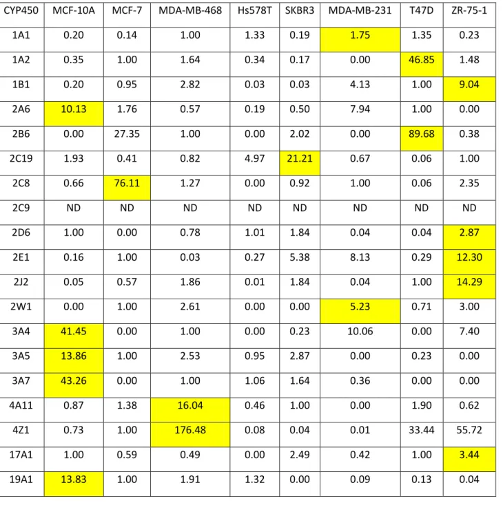
Table 1: Relative expression of CYP450 mRNAS 94
Table 2: CYP1B1 Primers 95
Table 3: Cell line CYP1B1 genotypes 96
viii
List of figures
Chapter 1: IntroductionFigure 1: Oxydation cycle of CYP450s. 18
Figure 2: Active site of CYP1A1 (green), 1A2 (blue) and 1B1 (red). 22 Figure 3: Active site of CYP2J2 (red). Ebastine is shown in the active site. 25
Chapter 3: Housekeeping Gene Selection
Figure 1: Housekeeping gene stability. 67
Figure 2: Frequency distribution of housekeeping gene expression 68
Chapter 4: CYP450 expression in breast cancer cell lines
Figure 1: Relative expression of various CYP450 Families 1, 17 and 19 mRNAs in various breast cancer
cell lines 98
Figure 2: Relative expression of various CYP450 Family 2 mRNAs in various breast cancer cell lines 99 Figure 3: Relative expression of various CYP450 Families 3 and 4 mRNAs in various breast cancer cell
lines 100
Figure 4: CYP450 profile of each cell line 101
Figure 5: Expression of total CYP450 mRNAs in each cell line 102
Figure 6: Ebastine metabolism in breast cancer cell lines 103
Figure 7: 7-Ethoxyresorfin metabolism in breast cancer cell lines 104 Figure 8: Correlation studies of mRNA expression and metabolic activity 105
ix
List of abbreviations
ADE:
Adverse Drug Event
ALDH:
Aldehyde Dehydrogenase
CD24:
Cluster of Differentiation 24
CD44:
Cluster of Differentiation 44
cDNA:
Complementary Deoxyribonucleic Acid
CSC:
Cancer Stem Cell
Ct:
Threshold Cycle
CYP450:
Cytochrome P450
DDI:
Drug-Drug Interaction
DNA:
Deoxyribonucleic Acid
dNTP:
Deoxynucleotide Triphosphate
DTT:
Dithiothreitol
H
2O:
Water
HER2:
Human Epidermal Growth Factor Receptor
EGFR:
Epidermal Growth Factor Receptor
EM:
Extensive Metabolizer
ER:
Estrogen Receptor
ERD:
Estrogen Receptor Downregulators
FACS:
Fluorescent Activated Cell Sorting
Fe:
Iron
x
FFPE:
Formalin-Fixed Paraffin-Embedded
GAPDH:
Glyceraldehyde 3 Phosphate Dehydrogenase
HKG:
Housekeeping Gene
IM:
Intermediate Metabolizer
kDa:
kiloDalton
mRNA:
Messenger Ribonucleic Acid
NADPH:
Nicotinamide Adenine Dinucleotide Phopshate
NUP214:
Nucleoporin 214kDa
O
2:
Oxygen
PBS:
Phosphate Buffered Saline
PCR:
Protocol Chain Reaction
PM:
Poor Metabolizer
PPIG:
Peptidyl-prolyl cis-trans xsomerise G
PgR:
Progesterone Receptor
PR:
Progesterone Receptor
qRT-PCR:
Quantitative Real Time Protocol Chain Reaction
RNA:
Ribonucleic Acid
SNP:
Single Nucleotide Polymorphism
SERM:
Selective Estrogen Receptor Modulators
TN:
Triple Negative
VEGF:
Vascular Endothelial Growth Factor
xi
xii
Thank you
I would first like to thank my supervisor, Dr Jacques Turgeon. Thank you for accepting me into your laboratory, having the confidence in my work and for guiding me to develop a more critical thought process. Thank you for allowing me to attend so many conferences. I will never forget them.
Thank you to my Master’s jury, Dr Marc Servant and Dr John Stagg, for revising my thesis.
I would also like to thank François Bélanger for all of his help in Real-Time PCR, and for being so nice throughout the whole process. Thank you to Fleur Gaudette for all of your time and help for the metabolism studies. This project would not have been possible without the two of you.
Thank you to all my fellow students, Jade Huguet, Jennifer Lu, Henry Leung and Liliam Gabriela Guilarte Moya for their help, support and friendship. Thank you for your contributions to the manuscripts that I am including in this thesis.
Thank you to the Canadian Institutes of Health Research (CIHR) for awarding my with the Frederick Banting and Charles Best Canada Graduate Scholarships - Master’s Award. I would also like to thank the Université de Montréal, Faculty of Pharmacy for awarding me with the Master’s Recruitment Award.
I would finally like to thank my family. Thank you to my amazing husband, Ian, who has been a great support throughout the last two years of my studies. Thank you to my parents and sister for their financial and emotional support throughout my studies.
1
CHAPTER 1
2
INTRODUCTION
Breast cancer is an important form of cancer that affects both men and women. In women, breast cancer accounts for 28% of all diagnosed cancers, and for 15% of the deaths due to cancer. In 2011, an estimated 6,200 women in Quebec (23,400 women and 190 men, in Canada) will be diagnosed with breast cancer, and 1,300 women in Quebec (5,100 women and 55 men, in Canada) will die due to breast cancer. Currently, it is estimated that the probability of developing breast cancer is 1 in 9, and the chance of death due to breast cancer is 1 in 29 (according to the Canadian Cancer Statistics 2011). Therefore, breast cancer is a disease which affects so many people, and merits the time and funding required to find a cure.
Great advancements have been made in the diagnosis and treatment of breast cancer. Large improvements have been made to mammography which can now detect smaller tumours while using less X-rays. Additionally, regular mammography screenings have been implemented leading to earlier detection, ultimately leading to better prognoses. Targeted treatments for breast cancers have also been developed leading to greater survival. While great advancements have been made in therapeutics, inter-subject variability and anti-cancer resistance remains to be a key issue in breast cancer patients.
Therefore, this work has been focused on finding a cause of anti-cancer resistance and inter-subject variability in the response to anticancer agents. Our focus was on the local expression of Cytochrome P450s, key enzymes in the metabolism of xenobiotics, in breast cancer.
3
1. BREAST CANCER PATHOLOGY
The use of molecular classifications is not only important for breast cancer patients, but also for researchers. Studies have shown that gene expression varies between subtypes, and therefore gene expression differences observed in vitro need to be related back to the molecular subtype, especially when large inter-subject variability is observed.
1.1. CANCER CELL LINES CHARACTERISTICS/MOLECULAR CLASSIFICATIONS
Treatment options for breast cancer tumors are based on many factors including morphology, histology, grade as well as gene expression profiles. The characteristics and molecular classifications of these tumors are necessary to properly determine tumor aggression and to choose the best treatment option. Based on gene-cluster analysis, five subtypes have been identified and described for breast cancers tumors: Luminal A, Luminal B, Basal-like, Human Epidermal Growth Factor Receptors (HER2) and Normal breast-like. [1, 2] However, the classification of subtypes used for breast cancer cell lines only consists of three groups: Luminal, Basal A and Basal B. [3, 4] The classification differences between cell lines and tumor classifications may be due to the absence of stromal and immune gene signatures in cell lines. While the cell line classification does not consist of a HER2 subtype, HER2 overexpression is observed in Luminal and Basal A subtypes. [3, 4]
Table 1 lists the cell lines used, along with various characteristics used in tumor diagnostics, which include gene cluster type, along with the presence or absence of the following receptors: estrogen, progesterone and HER2. As can be seen in Table 1, an equal amount of Luminal and Basal cell lines were selected in order to have each subtype present during our analyses, as well as the fact that each breast cancer cell line has a unique expression profile.
4
1.2. RECEPTORS PRESENT IN BREAST CANCER CELLS
The presence of cell surface and intracellular receptors in breast cancer is of great importance during the diagnostics of breast cancer. Hormone receptor positive tumors have available targeted treatments, which have been proven to be very effective. Hormone receptors of interest in breast cancer are Estrogen Receptor (ER), Progesterone Receptor (PR), HER1 and HER2.
Table 1: Breast cancer cell lines characteristics (Adapted from Kao, 2009, and Neve, 2006 [3, 4]) Cell Line Cell Line
Subtype
Tumor Type Source Estrogen Receptor
Progesterone Receptor
HER2 Receptor Hs578T Basal B Invasive Ductal
Carcinoma
Primary Breast
No No No
MCF7 Luminal Invasive Ductal Carcinoma
Pleural Effusion
Yes Yes No
MCF10A Basal B Non-tumorigenic Epithelial Cell Line
Primary Breast
No No No
MDA-MB-231 Basal B Adenicarcinoma Pleural Effusion
No No No
MDA-MB-468 Basal A Adenicarcinoma Pleural Effusion
No No No
SKBR3 Luminal Adenicarcinoma Pleural Effusion
No No Yes
T47D Luminal Invasive Ductal Carcinoma
Pleural Effusion
Yes Yes No
ZR-75-1 Luminal Invasive Ductal Carcinoma
Ascites Fluid
Yes No No
1.2.1. Estrogen Receptor
Many physiological processes in humans are influenced by the presence of estrogens. Estrogens mediate their effects through the binding and interactions with the estrogen receptor (ER).[5] The ER is known to be present under two forms, ERα and ERβ, which are encoded by two separate genes, ESR1 and ESR2, respectively. These two forms are tissue specific, have similar affinities for steroid ligands, and can be both localized to the nucleus, or to the plasma membrane.[6]
5 The development of breast cancer has been shown to be linked to the presence of estrogen.[5] Two hypotheses have been suggested to explain this phenomenon. First, the binding of estrogen to intracellular or membrane bound estrogen receptors cause an increase in cell division. This increase in cell proliferation leads to elevated DNA synthesis and therefore an elevated risk of DNA replication errors, ultimately leading to cancer development.[5] The second hypothesis is that estrogen metabolism produces genotoxic metabolites that can cause DNA damage.[5]
While estrogen receptors are linked to the activation of cancer, there are specific treatments that target the ER, called Selective Estrogen Receptor Modulators (SERMs) which include tamoxifen, a very effective anti-cancer agent for ER+ cancers.[5] Another class of treatments that target the ER are Selective Estrogen Receptor Modulators (SERDs). Currently only one SERDs is available for the treatment of breast cancer, Faslodex (Fulvestrant).[7, 8]
1.2.2. Progesterone Receptor
Progesterone is a steroid that is critical to normal breast development, and mediates its effects by binding to progesterone receptors (PR). Two isoforms have been identified for the progesterone receptor, PR-A and PR-B. Both isoforms are synthesized from the same gene, but have different translation start sites, creating two receptors of different masses, namely PR-A lacks the 164 N-terminal amino acids found in PR-B.[9, 10] The ratio of isoforms A to B is unknown, but appears to be a crucial element in cell homeostasis.[10]
One single nucleotide polymorphism (+331G/A) has been identified for this receptor, which affects the transcriptional activity of the gene, leading to a greater production of the PR-B receptor. There are some conflicting studies in whether or not there is a link between this SNP and the presence of breast cancer, but overall, there has been no association between this mutation and the risk of developing breast cancer.[9]
6
1.2.3. Human Epidermal Growth Factor Receptor (HER)
The Human Epidermal Growth Factor Receptor is comprised of 4 isoforms, HER1 (also known as epidermal growth factor receptor, EGFR), HER2, HER3 and HER4. These four receptors are encoded by the gene ERBB1/2/3/4, respectively.[11] All four of these receptors have been shown to influence tumor development by affecting cell proliferation, migration, angiogenesis and apoptosis protection.[12] While no defined ligands are known for the four HER receptors, it is known to play a key role in the amplification of cell signaling through a dimerization reaction. This dimerization has been shown to increase receptor-ligand affinity, enhancing its own activation. The activated receptor promotes the phosphorylation of tyrosines in its intracellular domain, which is subsequently recognized by specific cytoplasmic signal transducers, which inevitably leads to cell growth and proliferation.[13] Since these receptors cause increase cell proliferation and are capable of preventing apoptosis, their overexpression in breast cancer leads to very aggressive tumors.[11] HER2 has been studied the most due to its frequent overexpression in breast cancer, estimated to between 18-25% of human breast cancers, and is therefore a targeted receptor.[11, 14]
The attempt to explain the aggression observed in tumors overexpressing HER2 has been of interest. Ginestier at al. in 2007 demonstrated a strong correlation between ALDH1 overexpression (a marker of cancer stem cells, see section 4.3 for more details) and HER2 overexpression. It is believed that the aggression observed in HER2 breast cancers is due to a large population of cancer stem cells, where cancer stem cells have been linked to anti-cancer agent resistance. [15]
While HER2 overexpression is generally undesired due to its aggressive behavior, specific treatments targetting this receptor have been developed and have proven to be very effective. Treatments include Trastuzumab and Lapatinib, which are specific inhibitors of the HER2 receptor (see section 2.1.2.2.2 HER2 Inhibitor for more details).[1, 13, 16-18]
7
1.3. MOLECULAR SUBTYPES
Molecular subtypes used in the description of breast cancer cell lines and breast cancer tumors are slightly different. In order to better understand how these two classification systems overlap, Kao et al looked at the various gene expression patterns of all subtypes to determine how cell lines would be classified using the breast cancer tumor subtypes. It was determined that the Luminal cell lines most resembled Luminal A and Luminal B tumors, while Basal A cell lines were most similar to basal-like tumors, and Basal B cell lines were similar to basal-like or HER2 overexpressing tumors.[2, 3] While the Basal B cell lines displayed expression patterns that are similar to HER2 overexpressing tumors, Basal B cell lines do not overexpress HER2 and are referred to as as triple negative cell lines.[3]
For breast cancer cell lines, the molecular subtypes are important to take into consideration because previous studies have shown that gene expression profiles are dependent on the molecular subtype. Meaning that some genes may demonstrate an overexpression in one molecular subtype, whereas a downregulation is observed in others. Therefore gene expression variations observed may simply be due to its cell line characteristics.[3]
Since each molecular subtype is very different, and requires a different treatment plan, molecular subtypes are always determined for breast cancer patients. Much detail is available for breast cancer tumors subtypes; therefore a more comprehensive assessment of these subtypes follows.
1.3.1. Luminal A
Luminal A cancers originate from the inner cells, called luminal cells, which line the mammary duct.[1] Luminal cells are responsible for the secretion of milk, are highly differentiated and are glandular.[19] Luminal A tumors are mainly characterized by the presence of Estrogen Receptor (ER) and, or the Progesterone Receptor (PR, or PgR), but the absence of the Human Epidermal Growth Factor Receptor 2 (HER2). Luminal A cancers have been associated with a good prognosis, where the survival
8 rate is high, and the rate of reccurrence is low because they can be treated with hormone therapies, including Tamoxifen.[1, 2, 20]
1.3.2. Luminal B
Luminal B cancers also originate from the inner cells, called the luminal cells.[1] While this subtype is similar to the Luminal A subtype, where there is the presence of Estrogen Receptor (ER) and, or the Progesterone Receptor (PR, or PgR), Luminal B tumors often express the HER2 receptor. [1, 2, 21, 22] Hormone treatments are available; however because of the presence of the HER2 receptor, Luminal B tumors tend to have a higher tumor grade, and exhibit high proliferation rates and DNA amplifications, and therefore are associated with a poor prognosis.[1-3]
Treatments of Luminal B tumors include hormone therapy, Trastuzumab-based, chemotherapy and/or surgery.[1]
1.3.3. Basal-Like
Basal-like cancers originate from the outer cells, called the basal cells (myoepithelial), which line the mammary duct [1, 3]. Basal cells do not have a role in secretion, but rather in muscle contraction, and are undifferentiated.[19] These tumors are often associated with the hereditary BRCA1 breast cancer tumors and have been known to be the most aggressive subtype. [1] Most Basal-like tumors are negative for all three receptors, and are commonly referred to as the Triple Negative tumor (TN). However, this is not always the case. It is estimated that 5-45% of basal tumors are ER+ and that 14% are HER2+.[1, 17] For patients diagnosed with a tumor that is both basal-like and TN, the prognosis tends to be very poor because of the lack of targeted treatment (such that are available for hormone responsive tumors) and because the cancer is very aggressive.[1]
Stem cells, or cells with stem cell like properties are believed to be the source of basal-like cancers. There have been reports showing that the Basal phenotype are largely composed of the
9 CD44+/CD24- polulation, otherwise known as a Cancer Stem Cell population (see section 4.3 for more details).[4] The expression of markers such as cytokeratins (specifically CK5/6, 14 and 17) and the transcription factor p63 are found in basal-like breast cancers.[23]
Treatment options for Basal-Like tumors depend on the presence of receptor overexpression. If ER, PR or HER2 is overexpressed, hormone, or trastuzumab treatments are used. For TN tumors, treatment options include chemotherapy and surgery.[1]
1.3.4. HER2 overexpressing
HER2 overexpression has been linked to mammary tumorigenesis, tumor aggression and metastasis and therefore tends to have a poor prognosis.[14] HER2+ tumors are negative for both ER and PR, while they overexpress the HER2 receptor. While the tumor is known to be aggressive, targeted treatments for HER2 positive breast cancers are available. Trastuzumab (or Herceptin®), and Lapatinib are two targeted treatments for HER2 positive tumors that have proven to be very effective.[1, 13, 16-18, 24]
1.3.5. Normal breast-like
The normal breast-like cancers are cancers which do not fit within the other predefined subtypes.[1] This subtype has been shown to demonstrate similar expression patterns as a normal breast tissue. [3] Overall, these tumours are small, and usually have a good prognosis.[1]
10
2. ANTI-CANCER AGENTS
Anti-cancer agents, such as chemotherapy and targeted treatments, are used in the treatment of breast cancer. However, some patients experience resistance to these treatments, which inevitably ends in treatment failure. Research has shown that the presence of breast cancer stem cell, as well as proteins involved in drug bioavailability, such as membrane transporters and drug metabolizing enzymes, are the sources of resistance (see Section 4 for more details). Our focus is on the effects of the Phase I drug metabolizing enzymes, Cytochrome P450s on the bioavailability of anti-cancer agents. First the agents used in breast cancer will be described.
2.1. TYPES OF TREATMENTS
Two main groups of treatments are used in the treatment of breast cancer, chemotherapy agents, and targeted treatments. Different classes of each group are available; however, chemotherapy agents destroy all quickly dividing cells, whereas targeted treatments attack cells which overexpress specific proteins or processes observed in breast cancer cells.
2.1.1. Chemotherapy Classes
Chemotherapy is the process where anticancer medications are used to treat cancer. The goal of chemotherapy agents is to prevent the growth and spreading of cancer cells by interfering with normal cell processes at different points. Some chemotherapy agents mechanism of action occurs at the genetic level (DNA and RNA damage), while other interfere with normal protein function.[25] Chemotherapy agents have been categorized in different chemotherapy classes based on their mechanism of action. These classes include alkylating agents, anti-metabolites, plant alkaloids (vinca alkaloids and Taxanes), Topoisomerase inhibitors and cytotoxic antibiotics (anthracylines). See Table 2 for more details on chemotherapy agents used in breast cancer treatments.
11 Table 2: List of chemotherapy agent classes used for breast cancer and their mechanism of action
Chemotherapy Classes
Action Mechanism Examples used in breast cancer Alkylating Agents DNA damage through alkylating nucleophilic
sites of DNA bases. [26]
Cyclophosphamide and carboplatin Anti-metabolites DNA and RNA damage through masquerading
as pyrimidine and purine bases. [27]
Methotrexate, 5-fluorouracil, Gemcitabine and Capecitabine Plant Alkaloids Microtubule binding [27, 28] Vinca Alkaloids and Taxanes Vinca Alkaloids Natural product that destabilizes microtubule
by binding to the β-subunit of tubulin destroying mitotic spindles and blocks mitosis.
[27, 28]
Vinblastine and Vinorelbine
Taxanes Natural product that stabilizes microtubule by binding to the β-subunit of tubulin inhibiting depolymerisation and slowing down mitosis.
[27, 28]
Paclitaxel and Docetaxel
Topoisomerase inhibitors
Relaxes DNA supercoiling by Topoisomerases, causing DNA vulnerability to intercalating
agents.[29]
Irinotecan (TOP1) and Anthracyclines (TOP2); currently in
clinical trials. Cytotoxic antibiotics
-Anthracyclines
Intercalating agents, that inhibit topoisomerases and generated reactive
oxygen species.[30]
Doxorubicin and Epirubicin[31]
2.1.2. Targeted Treatments
Targeted treatments are often used in the treatment of breast cancer, specifically cancers that are receptor positive. Treatment groups include hormone therapies and specific targeted enzymes.
2.1.2.1.
Hormone Therapy
The majority of breast cancer tumors are hormone receptor positive and therefore can benefit from hormone therapies. Hormone receptors, namely estrogen and progesterone receptors, respond to intra- and extracellular hormone levels, which act as a signal to turn on cell growth. Therefore, hormone therapies work by blocking the hormone action potential at their specific receptors, as well as to lower the amount of hormone in the body.[7] Hormone therapies include aromatase inhibitors, Selective Estrogen Receptor Modulators and Estrogen Receptor Downregulators.
12
2.1.2.1.1.
Aromatase inhibitors
Aromatase, otherwise known as CYP19A1, is an enzyme which catalyzes the conversion of the hormone androgen to estrogen. [32, 33] Aromatase Inhibitors are molecules that inhibit aromatase activity by blocking the production of estrogen in the body (through CYP19A1 activity). These inhibitors only work in post-menopausal women, who are diagnosed with ER+ cancers, because pre-menopausal women produce significant amounts of estrogen in the ovaries without aromatase activity.[34] Aromatase inhibitors include Anastrozole, Exemestane and Letrozole.[7, 34]
2.1.2.1.2.
Selective Estrogen Recetor Modulators (SERMs)
Selective Estrogen Receptor Modulators (SERMs) work by competitively binding to the active site of Estrogen Receptors. Since estrogen is unable to bind to the estrogen receptor in the presence of SERMs, the breast cancer cell does not receive a signal to grow and divide, and therefore the proliferation of breast cancer is blocked.[35] SERMs include Tamoxifen, Raloxifene and Toremifen.[7]
2.1.2.1.3.
Seletive Estrogen Receptor Downregulators (SERDs)
Selective Estrogen Receptor Downregulators (ERDs) have a similar action mechanism as SERMs, however, they also work to reduce the amount of Estrogen receptors in the breast cell, and change the shape of the Estrogen Receptor active site, so that the receptor cannot recognize the hormone as efficiently.[8] Currently there is only one SERDs commercially available, Faslodex (Fulvestrant).[7, 8]
2.1.2.2.
Specific Targeted Enzymes
Certain enzymes or receptors are overexpressed in breast cancer. By blocking the activity of these specific enzymes, the proliferation rate of cancer cells is blocked. These enzymes include the Epidermal Growth Factor Receptor, HER2 and VEGFR (enzyme responsible for angiogenesis).
13
2.1.2.2.1.
Human Epidermal Growth Factor Receptor (HER) Inhibitor
The Human Epidermal Growth Factor Receptor (HER) is a family of receptors which play a key role in the development and proliferation of cancer as well as in the prevention of apoptosis.[12-14] The HER family is known to require tyrosine kinase activitation. In breast cancer, HER1, also known as the epidermal growth factor receptor (EGFR) and HER2 are most often studied members of the HER family, where HER2 is considered to be the most important because of its frequent overexpressed breast cancer. Several molecules have been used to inhibit this family of receptors. Gefitinib is an EGFR inhibitor that blocks the kinase activity of the EGFR receptor, however it is not approved for breast cancer.[36] Lapatinib (Tykerb®) is a small molecule which inhibits the tyrosine kinase of both the HER1 and HER2 receptors.[36] Finally, Trastuzumab (Herceptin®) is a monoclonal antibody which specifically recognizes the HER2 receptor.
2.1.2.2.2.
Angiogenesis Inhibitors
The vascular endothelial growth factor receptor (VEGFR) is responsible for the development and maintenance of tumor vasculature, or angiogenesis through a signal transduction process. [37] When the vascular endothelial growth factor (VEGF) binds to its receptor, VEGFR, signals are sent to initiate the growth of new blood vessels and to promote the maintenance of old ones. This is a vital process to all cells, and has been seen to be overexpressed in some tumors. This process has also been suggested as a possible source of cancer metastasis. Therefore, by targeting angiogenesis, cells which overexpress VEGF, such as cancer cells, would be most affected by angiogenesis inhibitors. Several treatments are available and include: monoclonal antibodies that binds specifically to VEGF, Bevacizumab (no longer approved for the treatment of breast cancer), small-molecule inhibitors of tyrosine kinase, Axitinib (not yet approved in Canada), Sunitinib, Sorafenib, as well as endogenous inhibitors of angiogenesis, Endostatin and Angiostatin. [36, 38-40] Currently no Angiogenesis inhibitors are approved for the treatment of breast cancer.
14 Table 3: List of hormonal and targeted treatment classes used for breast cancer and their mechanisms of action
Chemotherapy Classes Action Mechanism Examples used in breast cancer Aromatase Inhibitors Reduction of estrogen
production through the inhibition of aromatase activity
(through CYP19A1) [32, 33]
Anastrozole, Exemestane and letrozole [7, 34]
Selective Estrogen Receptor Modulators (SERMs)
SERMs bind to the ER preventing the growth signal of ER+ breast cancer to grow and divide. [35]
Tamoxifen, Raloxifen and Toremifen [7] Selective Estrogen Receptor
Downregulators (SERDs)
Reduction of the amount of ER present, and change in the active
site shape to reduce hormone binding efficiency. [8]
Faslodex [7, 8]
Human Epidermal Growth Factor Receptor (HER) Inhibitors
Inhibition of HER to prevent receptor activation which allow
for the apoptosis of cancer cells.[12-14]
Gefitinib (not approved for breast cancer) Trastuzuab and
Lapatinib [13, 36] Angiogenesis Inhibitors Blocking of VEGFR to reduce
blood flow to tumors by reducing the formation of new
blood vessel formation and maintenance of old vessels. [37]
Axitinib (not yet approved in Canada), Bevacizumab (no longer approved for breast cancer), Sunitinib, Sorafenib, Endostatin and Angiostatin (not
15
2.2. METABOLISM
The clearance of chemotherapeutics is an essential aspect to consider for drug administration. Many of the chemotherapy agents used in the treatment of breast cancer are known substrates and or inhibitors of the superfamily of metabolizing enzymes called Cytochrome P450 (CYP450s). Table 3 lists the various anti-cancer agents used in the treatment of breast cancer, along with the CYP450s isoforms that are responsible for their clearance. As can be seen, some agents are not metabolized by CYP450s and therefore may require other pathways for drug clearance, such as by drug transporters.
Table 4: Anticancer agents used in the treatment of breast cancer, and their metabolism by CYP450s
Anti-Cancer Agent CYP450
Cyclophosphamide CYP2B6, 2A6, 3A4/5, 2C8,2C9, 2C19 [41-44]
Carboplatin None[42-44]
Methotrexate None [43]
5-fluorouracil Inhibitor of CYP2C9 [45]
Gemcitabine None
Capecitabine None
Vinblastine CYP3A4 [44]
Vinorelbine CYP3A4 [44, 46, 47]
Paclitaxel CYP2C8, 3A4, 3A5 [41, 43, 44]
Docetaxel CYP3A4/5 [43, 44, 48]
Doxorubicin Inhibitor of CYP2D6 [45]
Epirubicin None [43]
Anastrozole CYP3A4/5 [44, 49]
Exemestane CYP3A4 [44, 50]
Letrozole CYP2A6 and 3A [44, 51]
Tamoxifen CYP1A1/2, 1B1, 2B6, 2C9/19, 2D6, 2J2 3A4/5 [44, 45, 52-55] Raloxifene Inhibitor of and metabolism by CYP3A4[56-58]
Toremifene CYP3A4 [44, 59]
Fulvestrant None
Gefitinib CYP3A4, 2D6, minor 3A5 and 1A1 [60]
Trastuzumab None
Lapatinib CYP3A4/5 and minor by 2C8/19 [12, 60]
16
3. CYTOCHROME P450S
Cytochrome P450s (CYP450) are enzymes which have been shown to be involved in the bioavailability of anti-cancer agents through phase I metabolism. Each CYP450 has a specific role in drug metabolism and tissue localization pattern. CYP450 isoforms which are locally expressed in breast cancer tissue and breast cancer cell lines are of interest because they may play an important role in the local drug metabolism of various anti-cancer agents.
3.1. DESCRIPTION
Drug metabolism, an important role in drug disposition, is executed by many enzymes in the human body, but more specifically in the liver. The most important family of enzymes responsible for the metabolism of medications is the cytochrome P450 (CYP450) superfamily. These enzymes work to detoxify the body of many xenobiotic molecules through a biotransformation reaction, rendering the molecule more hydrosoluble. This in turn facilitates the secretion of xenobiotics into urine and bile. [61] In human, this superfamily is composed of 57 known CYP450 genes, which are classified into families and subfamilies, depending on sequence homology. [62, 63] Isoforms which display greater than 40% homology are grouped within the same family, while enzymes with greater than 55% are classified within the same subfamily.[64, 65] Each CYP450 isoform has various roles in metabolism and in the synthesis of molecules, where similar functions are common within the same family.
While CYP450s localized in the liver play a major role in drug metabolism, either in drug clearance or in the activation off pro-drugs, extrahepatic CYP450s are implicated in both local drug metabolism, as well as in the synthesis and degradation of endogenous molecules, such as steroids, and fatty acids. [32, 33, 45, 63, 66-70]
17
3.1.1. Structure
All isoforms of the CYP450 superfamily are relatively small proteins with molecular weights ranging between 45 and 66 kDa.[71] CYP450s are found in every tissue at varying concentrations, and are all membrane bound protein located in the endoplasmic reticulum.[72] All CYP450s contain a ferroprotoporphyrin IX heme group, which constitutes the active site (the heme moiety). However, the specificity of each isoform is dependent upon the apoprotein and not the active site. [65]
In order for CYP450s to oxidize a molecule, these isoenzymes require two things; the presence of an energy source, which in the case of CYP450s is the reducing agent, nicotinamide adenine dinucleotide phosphate (NADPH), as well as atmospheric oxygen.[65]
3.1.1.1. Oxydation Cycle
The oxidation cycle of CYP450s has been well described, and is summarized and simplified in Figure 1. [73] Overall, the cycle is described as a dynamic process, which means that these steps do not necessarily occur in a sequential fashion. [73] In brief, a substrate binds to the active site in a reversible fashion. Using NADPH, the heme centre is reduced from the ferric state (Fe3+) to the ferrous state (Fe2+). One molecule of oxygen then binds generating a ferrous CYP-Substrate complex. The oxygen molecule is then cleaved by using a second electron (either from another molecule of NADPH or by Cytochrome b5. The molecule is then oxidized and released from the active site.[65] In the end, the iron is returned to the ferric state, and is ready for another cycle of oxidation.
18 Fe3+ Fe3+RH Fe2+RH O2 Fe2+-O2 RH RH
NADPH-P450 reductasereduced
NADPH-P450 reductaseoxidized
NADPH-P450 reductasereduced
NADPH-P450 reductaseoxidized
1 e -1 e -Fe2+-O 2 RH FeOH3+R Fe3+ROH FeO3+RH Fe2+-OOHRH H+ H2O ROH
Figure 1: Oxydation cycle of CYP450s. (Adapted from Guengerich, 2007 [73])
3.2. FAMILIES
Cytochrome P450 isoforms are grouped according to their families and subfamilies. In human, the CYP450s are grouped into 18 families, and 43 subfamilies.[74, 75] Not all families have been shown to be influenced in drug metabolism or steroid biosynthesis; therefore specific CYP450s were selected based on their role in drug metabolism, steroid metabolism, or link to breast cancer. Below is a table of all CYP450s analyzed, categorized by their families, and describing their roles and localization in human.
3.3. ROLES OF CYP450S
3.3.1. Systemic Metabolism (Exogenous molecules)
Hepatic CYP450 isoforms have been well characterized. These enzymes account for ~75% of the drug metabolizing enzymes found in the liver. The five most abundant hepatic CYP450s, accounting for 95% of hepatic metabolism are CYP3A4, 2D6, 1A2, 2C9 and 2C19, where 3A4 is the most important.[73]
19 Table 5: Table of CYP450s of interest describing their roles and localization
Family Subfamily Enzymes Roles Localization
CYP1 CYP1A CYP1A1 Hydroxylation of Steroids and Xenobiotic metabolism [63, 66-69]
Hepatic[73] CYP1A2 Hydroxylation of Steroids and Xenobiotic
metabolism [63, 66-69]
Hepatic[73] CYP1B CYP1B1 Detoxification of steroidal hormones,
therapeutic drugs and environmental toxins [76]
Adrenal glands, ovary, testis, lung, prostate[72]
CYP2 CYP2A CYP2A6 Xenobiotics Metabolism[63] Hepatic [77]
CYP2B CYP2B6 Xenobiotics Metabolism[63] Hepatic[73]
CYP2C CYP2C8 Xenobiotics Metabolism[63] Liver, kidney, adrenal gland, brain, uterus, breast, ovary and
duodenum[78]
CYP2C9 Xenobiotics Metabolism[63] Hepatic[73]
CYP2C19 Xenobiotics Metabolism[63] Hepatic[73]
CYP2D CYP2D6 Xenobiotics Metabolism[63] Hepatic[73]
CYP2E CYP2E1 Xenobiotics Metabolism (Ethanol)[45, 63] Hepatic[73]
CYP2J CYP2J2 Fatty Acid and xenobiotic Metabolism [53, 63] Heart, kidney, lung [72, 79, 80]
CYP2W CYP2W1 Procarcinogens [81] Prostate, pancreas, placenta,
lung, colon, intestine[63, 82]
CYP3 CYP3A CYP3A4 Xenobiotics Metabolism[63] Hepatic[73]
CYP3A5 Xenobiotics Metabolism[63] Hepatic, lung, small intestine, prostate[83]
CYP3A7 Xenobiotics Metabolism[63] Prenatal Tissue (liver) [65, 84]
CYP4 CYP4A CYP4A11 Fatty Acid Metabolism[63] Kidney and liver[72]
CYP4Z1 Lauric Acid Metabolism[70] Breast, breast carcinoma, kidney and liver[63, 82, 85] CYP17 CYP17A CYP17A1 Androstenedione synthesis[33, 67] Adrenal cortex[72] CYP19 CYP19A CYP19A1 Aromatase Activity[32, 33] Breast Brain, placenta
gonads[33, 72]
3.3.2. Local Metabolism (Exogenous molecules)
The local metabolism of exogenous molecules by various Cytochrome P450 isoforms has become of greater importance. The presence of many CYP450 enzymes has been identified in extra-hepatic tissues, such as the intestines, kidneys, brain, lungs and heart. [86-92] While concentrations of these enzymes may be inferior to those found in the liver, their implications in drug disposition cannot be ignored. Above all, the expression of the CYP450 isoforms appear to be tissue specific, where many isoforms identified in the extra-hepatic tissues are not found in the liver. For example, the mRNA expression of CYP2J2, an isoform not present in the liver, has been identified in abundance in cardiac
20 tissue.[79] Since many extra-hepatic CYP450s are not evaluated during the drug discovery process, the metabolism potential of these tissue specific CYP450s are unknown, and could locally metabolize exogenous molecules. As is seen in the liver, inter-subject variability is observed in the expression of CYP450s, and therefore, this same variability could be present in other tissues, such as the heart, or breast. This inter-subject variability may be another source of variation in drug effect.
3.3.3. Synthesis of endogenous molecules
The presence of steroids in breast cancer has been shown to be important, especially in hormone receptor positive breast cancers. Therefore, the expression of various CYP450s responsible for the synthesis and degradation of steroids expressed in breast cancer could be of great importance. The members of the Family 1 isoforms, namely CYP1A1, 1A2 and 1B1, have been shown to play a role in the hydroxylation of progesterone, testosterone and estrogen.[66-69] Therefore the local expression of these three isoforms may lead to the growth and proliferation of breast cancers which are stimulated by the presence of these hormones, (namely ER+ cancers being stimulated by estrogens). The expression of two other CYP450 isoforms which may be of interest in breast cancer are CYP17A1, mainly expressed in prostate tissue and 19A1, mainly expressed in breast tissue. CYP17A1 plays a role in the conversion of pregnenolone to androstenedione, wherease CYP19A1 converts androstendione to estrone through the use of aromatase activity.[33, 67, 93] Therefore the presence of these five CYP450s may be greatly implicated in the local synthesis of estrogens and other steroids.
3.3.4. Adverse Drug Events/Drug-Drug Interactions
Adverse drug events (ADEs) are events where an injury occurs from the use of a medication when the drug has been administered at a normal dose. ADE can lead to toxicity or loss of treatment efficiency, and can occur due to genetic alterations (such as polymorphisms), drug-drug interactions, or food-drug interactions. Overall, ADE results in 6.7% of hospitalizations in the United States, which has been estimated to cost 1.5 million dollars. [94, 95]
21 Drug-drug interactions (DDIs) are highly studied ADE, where interactions with the major hepatic CYP450s, such as CYP3A4 and 2D6 are commonly evaluated (substrates, inhibitors or inducers of these enzymes).[73] One example of a DDI for ER+ breast cancer patients would be the co-administration of tamoxifen, a substrate of CYP2D6, and the anti-depressant fluoxetine, a substrate of CYP2D6.[45, 52, 96] Under this situation, tamoxifen, which requires metabolism by CYP2D6 to become endoxifen, its active form, would be affected by the presence of the anti-depressant, due to competitive inhibitions of the enzyme. Therefore, a lack of active metabolite would be present, and a treatment failure would ensue.
Overall, DDI need to be avoided in all patients to ensure that no toxicity or treatment failure occurs.
3.4. IMPORTANCE IN BREAST CANCER
Two CYP450 Isoforms have been identified, through this work, to be highly expressed in the breast cancer cell lines analyzed: CYP1B1 and CYP2J2 (See Chapter 4 for more details). Both isoforms have a different role and importance in metabolic reactions, where 1B1 is involved in hormone and toxin detoxification, while 2J2 is involved in the hydroxylation of fatty acids. [63, 76]
3.4.1. CYP1B1
The expression of CYP1B1, a 58 kDa protein, has been found in many extra-hepatic tissues such as the uterus, ovaries, testis, prostate and adrenal glands.[97, 98] CYP1B1 has been shown to catalyze the hydroxylation of 17β-estradiol and testosterone.[99] However, CYP1B1s metabolic activity has also been linked to the activation of several pre-carcinogenic molecules such as benzanthracene, benzo(a)pyrine, DMBA, 1-ethynyl-pyrene, 3-methyl-cholantrene and oestradiol.[97] Since an overexpression of this isoforms has been found in tumors, such as in breast cancer, it is believed that CYP1B1 may be a source of steroid hormone-mediated cancer.[99, 100]
22 In order to understand the local metabolism potential of CYP1B1 in breast cancer cells, 7-ethoxyresorufin, a substrate which is common to CYP1A1, 1B1 and 1A2, may be used.[101] The rate of metabolism of 7-ethoxyresorufin is quite different in each of these enzymes. The maximum activity observed in CYP1A1 is greater than what is observed by CYP1B1, however, the substrate demonstrates a stronger affinity towards CYP1B1 than 1A1 (lower Km for 1B1).[101] However, since CYP1B1 is preferentially expressed in breast cancer cell lines, the metabolic activity of 7-ethoxyresorufin corresponds to CYP1B1 expression.
3.4.1.1.
Homology
CYP1B1 shares a strong homology with the two other members of the CYP1 family, CYP1A1 and 1A2. According to sequence alignment, CYP1B1 shares 39% and 37% homology with CYP1A1 and 1A2, respectively. The active site is where the most homology is seen where many of the amino acids are conserved; this leads to large substrate overlap.[102] However, there are small differences in the amino acids in the active site which leads to substrate affinity differences (see figure 2).
Figure 2: Active site of CYP1A1 (green), 1A2 (blue) and 1B1 (red). 7-Ethoxyresorufin is shown in the active site. (Adapted from Takemura, 2010.[102])
23
3.4.1.2.
Polymorphisms in CYP1B1
There are many known Single Nucleotide Polymorphisms (SNPs) for CYP1B1, both in the coding and non-coding regions. Some of these SNPS have been further characterized and have been given specific variant names (see Table 5). As can be seen, many of the CYP1B1 variants do not lead to altered metabolic activity with the exception of variant *6 and *7 which demonstrate a significant decrease in enzyme efficiency.[103] This finding was of interest because *6 and *7 variants are a combination of 3 and 4 SNPs, respectively, and when only one or two of these mutations are present, no change in enzyme activity was observed. Therefore it is hypothesized that only in the presence of all three SNPs (found in *6 and *7 variants), a change in protein folding results in a conformational change of the enzymes active site.[103] Others have reported that the *2 and *3 variants have a 2-4 increase in metabolic activity and an increased risk of developing certain types of cancers.[76] Since CYP1B1 has been linked to activation of certain pre-carcinogenic molecules, it is believed that an increased activity of CYP1B1 may lead to a greater production of carcinogens, and hence cancer development, specifically in tissues where elevated expression of CYP1B1 is found.[76]
Table 6: Known polymorphisms of CYP1B1 and their effects on metabolic activity (Adapted from Aklillu, 2001, [103])
Variant Nucleotide Change Amino Acid Substitution Protein Activity
CYP1B1*1 None None Wild Type Activity
CYP1B1*2 142C>G, 355G>T Arg48Gly, Ala119Ser Lower expression level, similar kinetic activity as wild type
CYP1B1*3 1294C>G Leu432Val Lower expression level, similar kinetic activity as wild type
CYP1B1*4 1328A>G Asn453Ser Lower expression level, similar kinetic activity as wild type
CYP1B1*5 142C>G, 1294C>G Arg48Gly, Leu432Val Unknown CYP1B1*6 142C>G, 355G>T,
1294C>G
Arg48Gly, Ala119Ser, Leu432Val
Similar expression level, kinetic activity significantly lower CYP1B1*7 142C>G, 355G>T,
1294C>G, 1328C>G
Arg48Gly, Ala119Ser, Leu432Val, Ala443Gly
Lower expression level, kinetic activity significantly lower
24
3.4.2. CYP2J2
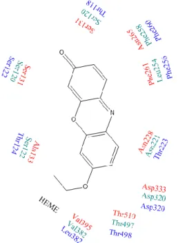
CYP2J2, a 56 kDa protein, is localized primarily in the heart, kidneys, lungs and breast. [72, 79, 80] CYP2J2 has been shown to be involved in the metabolism of endogenous molecules, such as fatty acids, arachidonic and linoleic acid, as well as xenobiotic molecules such as ebastine, terfenadine, astemizole, amiodarone, albendazole, danazol, thioridazine, tamoxifen, cyclosporin A, nabumetone and mesoridazine. [53, 63, 104-106] Substrate overlap has been observed between CYP2J2 and 3A4, because both isoforms have large active sites. However, because CYP2J2 has a slightly more cylindrical and narrow active site, substrates are more restricted and therefore can only be metabolized at a single site.[53] This restriction leads to differences in regioselectivity between CYP2J2 and 3A4.[107] Figure 3 shows the active site of CYP2J2 where ebastine has been shown in place.
In order to understand the metabolism of CYP2J2, ebastine can be used. While CYP3A4 can metabolize ebastine, a different metabolite is formed by CYP3A4 (namely N-desmethyl-ebastine) than by CYP2J2 (namely Hydroxyebastine).[106] In addition, the expression of CYP3A4 mRNAs is almost non-detectable in breast cancer cell lines, so that the metabolism of ebastine within these cells is specific to CYP2J2.
3.4.2.1.
Polymorphisms in CYP2J2
There are many known Single Nucleotide Polymorphisms (SNPs) for CYP2J2, both in the coding and non-coding regions. Some of these SNPS have been further characterized and have been given specific variant names (see Table 6). Significant loss in activity has been observed for variant *2, *3 and *6 in the metabolism of arachidonic acid and linolein acid, while *4 only showed a loss of function for arachidonic acid metabolism, and *5 showed no loss in functional activity. [108, 109] Variants *8 and *9 were analyzed using astemizole and ebastine as metabolites, where *8 showed almost a complete loss of function, while *9 showed wild type activity. [109, 110]
25 Since CYP2J2 has been shown to be capable of metabolizing xenobiotics, such as tamoxifen, an anti-cancer agent, the loss of function of this isoform, which is highly expressed in breast cancer cells, may have important implications on local drug metabolism.[53]
O N O Leu378 Thr488 Leu402 Leu83 Met116 Arg117 Val380 Ile127 Thr315 Ile376 Phe310
Figure 3: Active site of CYP2J2 (red). Ebastine is shown in the active site. (Adapted from Lafite, 2007.[111])
26 Table 7: Known polymorphisms of CYP2J2 and their effects on metabolic activity
Variant Nucleotide Change Amino Acid Substitution Protein Activity
CYP2J2*1 None None Wild Type Activity
CYP2J2*2 427A>G Thr143Ala Significant decreased in metabolism of arachidonic and linoleic acid [108, 109] CYP2J2*3 472C>T Arg158Cys Significant decrease in metabolism of
arachidonic and linoleic acid [108, 109] CYP2J2*4 575T>A Ile192Asn Significant decrease in metabolism of
arachidonic acid, but no difference of linoleic acid [108, 109]
CYP2J2*5 1024G>A Asp342Asn Wild Type activity for arachidonnic and linoleic acid [108, 109]
CYP2J2*6 1210A>T Asn404Tyr Almost complete loss of metabolism for arachidonic and linoleic acid [108, 109] CYP2J2*7 -50G>T Promotor Decreased promotor activity [109, 112] CYP2J2*8 934G>A Gly312Arg Almost complete loss of catalytic activity
for Astemizole and Ebastine [109, 110] CYP2J2*9 1052C>T Pro351Leu Wild type activity for Astemizole and
Ebastine [109, 110]
27
4. ANTI-CANCER RESISTANCE
Several types of cancer cells have shown an innate or acute resistance to anti-cancer agents which in turn causes a failure in treatment. Even though our work has been focussed on metabolic factors that could affect anti-cancer resistance, it is also important to acknowledge other sources of resistance, such as the impact of membrane transporters on drug bioavailability, as well as the presence of Tumor-Initiating cells (T-ICs) and Cancer Stem Cells (CSCs). [113, 114]
4.1. DRUG BIOAVAILABILITY-MEMBRANE TRANSPORTERS
Many anti-cancer agents are known substrates of the membrane transporters, Multidrug Resistance Protein 2 (MRP2 encoded by the gene ABCC2), Breast Cancer Resistance Protein (BCRP encoded by the gene ABCG2) and P-glycoprotein (P-gp, encoded by the gene ABCB1), which are responsible for the efflux of these medications. Table 7 lists the efflux transporters responsible for the efflux of anti-cancer agents used in the treatment of breast cancer. [43, 44, 114-119] These three transporters, MRP2, BCRP and P-gp, are highly expressed in the liver membrane, specifically the cannulicular side. Therefore the bioavailability of anti-cancer agents, which are substrates of these transporters, will be greatly affected by the presence of these transporters.
However, these transporters are also present on the basolateral membrane of other tissues, acting as efflux transporters, where an elevated expression of BCRP is observed in breast cancer. [115, 120, 121] Some studies have shown that an overexpression of membrane transporters occurs when cells are treated with anti-cancer agents. One particular study treated the breast cancer cell line, MCF-7 with Adriamycin, and demonstrated that an overexpression of P-gp was present in MCF-7-Adriamycin resistant cells. [122] Other studies demonstrated an overexpression of BCRP in mitoxantrone resistant cells while VP-16 resistant cells overexpressed MRP transporters. [121, 123] Therefore, cancer cells are either upregulating their expressions of specific membrane transporters to reduce intracellular exposure to anti-cancer agents, or some cancer cells naturally have an overexpression of the specific membrane
28 transporters which aid in their resistance, resulting in a population selection. In either case, membrane transporters are locally expressed in breast cancer, which results in a resistance to anti-cancer agents.
Table 8: Anticancer agents used in the treatment of breast cancer, and transport by membrane efflux transporters
Anti-Cancer Agent Efflux Transporters
Carboplatin MRP2 [43] Docetaxel MRP2 and P-gp [43, 44, 114, 117] Doxorubicin MRP2, BCRP and P-gp [43, 44, 115-117] Epirubicin MRP2, BCRP and P-gp [43, 115, 116] Gefitinib BCRP [115] Methotrexate MRP2 and BCRP [43, 115, 116] Paclitaxel P-gp [43, 44, 117] Raloxifene P-gp and MRP2 [58, 118] Tamoxifen P-gp and BCRP [44, 115] Toremifene BCRP [115] Vinblastine MRP2, P-gp [44, 117] Vinorelbine P-gp [119]
4.2. STOCHASTIC CELL THEORY AND TUMOR-INITIATING CELLS
Stochastic cell theory states that every cell has the potential of becoming a tumor-initiating cell (T-IC). However, this property is not present in every cell, and the chance of developing this property is very low.[113] Therefore, only a small population of cells within a tumor are able to initiate tumor growth.[113] It is believed that the cause of the tumor-initiation process is due to random mutations and subsequent clonal selection.[124] However, this process leads to the production of a homogenous tumor.[113]
The stochastic theory states that T-ICs are resistant to chemotherapy agents and will ultimately lead to the relapse of cancer. However, because it is impossible to predict which cells are T-ICs, and they cannot be separated from non-T-ICs, T-ICs cannot be targeted to prevent relapses.[113] It is also unclear as to why T-ICs have developed a resistance to chemotherapy agents.
29
4.3. CANCER STEM CELL THEORY
The cancer stem cell (CSC) theory states that all tissues are derived from organ-specific stem cells. These cells have the capacity of self-renewal and differentiation, which ensures tissue integrity. The CSC hypothesis states that cancer develops from normal stem cells that have undergone oncogenic transformation.[124] Since stem cells are believed to be long living, slow dividing cells, they have a longer period of toxin exposure than regular cells, ultimately leading to the development of cancer and cancer stem cells.[125] Since stem cells have the properties of self renewal and differentiation, the expansion of the cell population can then lead to additional genetic and epigenetic changes.[124] In the breast, it is believed that the differentiation of CSC is limited to specific cell types, and therefore leads to the development of specific breast cancer molecular subtypes.[124]
4.3.1. Isolation of Cancer Stem Cells
Several methodologies for the isolation and purification of CSC have been suggested: dye exclusion (side population), cell culture selection through tumorospheres, cell surface marker (CD44+/CD24-/low), and an enzymatic assay for ALDH+ cells (ALDEFLUOR assay).[15, 124, 126, 127] The two most commonly used methods for the isolation of CSC are the cell surface markers and the ALDEFLUOR enzymatic assay.
4.3.1.1.
CD44
+/CD24
-/lowCD44 and CD24 are cell surface markers that are often present in breast cancer cells. Cells which express CD44, but do not express CD24 have been described as being highly resistant to chemotherapy agents. Previous studies showed that placing MCF-7 cells in the presence of chemotherapy agents caused some cells to die, but others resisted. The remaining cells showed increased concentration of CD44+/CD24-/low cells. [124]
30 CD44+/CD24-/low cells have also demonstrated high tumorigenicity, where only 100-200 cells were required for tumor growth, whereas, tens of thousands of other phenotypes failed to form a tumor.[124, 126] CD44+/CD24-/low cells have also been shown to be strongly correlated to the triple negative (TN) diagnosis of breast cancer, where TN demonstrates a high level of expression of CD44+/CD24-/low cells. Specific antibodies tagged with fluorescent probes can be used to identify cells expressing CD44 and not expressing CD24.
4.3.1.2.
ALDH
Aldehyde dehydrogenase (ALDH) is a superfamily of enzymes which consists of 19 isoforms.[128] ALDH is an intracellular detoxifying enzyme which is known to metabolize aldehydes to carboxylic acids through an oxidation reaction. [128, 129] These enzymes are also known to play a role in the oxidation of retinal to retinoic acid, [128, 130] as well as in the metabolism of some chemotherapy agents, such as cyclophosphamide.[128, 131, 132]
The isolation of ALDH+ cells is possible using a kit called ALDEFLUOR which is manufactured by Stemcell Technologies.[124] Using the kit, ADLH+ cells are able to be identified and sorted by Fluorescence Activated Cell Sorting (FACS).
Using the ALDEFLUOR kit, some clinical studies have shown that an elevated expression of ALDH activity had been linked to poor clinical outcomes, and that ALDH+ cells are capable of self-renewal in vitro. [15] Studies have also been performed to determine the tumorigenicty of ALDH+ cells. It was observed that NOD/SCID mice injected with as few as 500 ALDH+ cells developed a tumor. [133] Combination of ALDH and the cell surface markers (CD44+/CD24-) further amplified the tumorigenicity where only 20 cells were required for tumor growth. [133]
31
4.3.2. Resistance of Cancer Stem Cells
CSC are believed to show similar properties to normal stem cells including relative order, resistance to drugs and toxins, active DNA repair capacity and resistance to apoptosis. Therefore, CSCs are pluripotent, chemotherapy resistant cells that are capable of reinitiating tumor growth.[124]
CSCs have also been shown to be resistant to radiotherapy. The resistance is believed to be due to DNA damage checkpoints and an increase in the DNA repair cycle, through cell cycle-regulating proteins CHEK1 and CHEK2. This same resistance was observed in the breast cancer cell line, MCF-7, where following radiotherapy an increase in the CSC population (CD44+/CD24-/low) was observed.[124]
4.3.2.1.
Membrane Efflux Transporters
The overexpression of membrane efflux transporters, namely the ABC transporter family (ATP-binding cassette transporters), have been identified as a potential source of CSC resistance. The three genes that have been most studied have been ABCB1, ABCG2 and ABCC1, which encode the proteins, P-glycoprotein, Breast Cancer Resistance Protein, and MRP1, respectively.[124]
Breast cancer resistance protein (BCRP) or ABCG2 appeared to be upregulated in breast cancer stem cells isolated from breast cancer cell lines MCF-7 and MDA-MB-231 cells by 5.8 and 3.6 fold.[115, 134] The selection of cells that overexpress BCRP through the dye exclusion assay are referred to as the side population (rather than a CSC population).[124] Therefore, the elevated expression of BCRP in breast cancer stem cells could decrease the intracellular concentrations of certain anti-cancer agents, which could lead to the resistance of anti-cancer agents by CSCs.
![Table 1: Breast cancer cell lines characteristics (Adapted from Kao, 2009, and Neve, 2006 [3, 4]) Cell Line Cell Line](https://thumb-eu.123doks.com/thumbv2/123doknet/7515283.226334/18.918.125.794.366.764/table-breast-cancer-lines-characteristics-adapted-neve-cell.webp)

![Figure 1: Oxydation cycle of CYP450s. (Adapted from Guengerich, 2007 [73]) 3.2. FAMILIES](https://thumb-eu.123doks.com/thumbv2/123doknet/7515283.226334/32.918.195.725.106.485/figure-oxydation-cycle-cyp-s-adapted-guengerich-families.webp)
![Table 6: Known polymorphisms of CYP1B1 and their effects on metabolic activity (Adapted from Aklillu, 2001, [103])](https://thumb-eu.123doks.com/thumbv2/123doknet/7515283.226334/37.918.99.817.772.1045/table-known-polymorphisms-effects-metabolic-activity-adapted-aklillu.webp)
![Figure 3: Active site of CYP2J2 (red). Ebastine is shown in the active site. (Adapted from Lafite, 2007.[111])](https://thumb-eu.123doks.com/thumbv2/123doknet/7515283.226334/39.918.306.650.295.686/figure-active-site-ebastine-shown-active-adapted-lafite.webp)