Hepatocellular carcinoma (HCC) is one of the most common cancers in the world. It is a growing public health problem and its incidence is increasing worldwide (most cases of HCC in Asia and sub-saharan Africa but recently, in western countries too (1, 2)). The presence of cirrhosis is the major risk factor, essentially due to chronic hepatitis C and hepa-titis B infection or alcohol dis-ease (1). Hepatocellular carcinoma is of poor prognosis: the five-year sur-vival rate is less than 5 percent (2). The prognosis depends on hepatic function, tumour size, and tumour extent at the time of diagnosis. Nowadays, orthotopic liver trans-plantation only remains curative, but because of the shortage of organ donors, these treatments are appli-cable only to a small part of all patients. The majority of the patients with unresectable HCC are treated by various palliative therapies included surgical resection (partial hepatectomy), or percutaneous treatment. Despite advances in imaging techniques and follow-up programs only 20% of patients are candidates for surgery at the time of diagnosis (3, 4) due to an advanced tumor stage, comorbidities, poor hepatic functional reserve or short-age of donor livers.
Case report
A 78-year-old man was referred to our hospital in September 2008 for a health deterioration associated with weight loss. His medical history was
CT-guided biopsy of the largest hepatic lesion (segment IV) con-firmed the diagnosis of well differen-tiated HCC in a cirrhotic liver. The patient was medically treated.
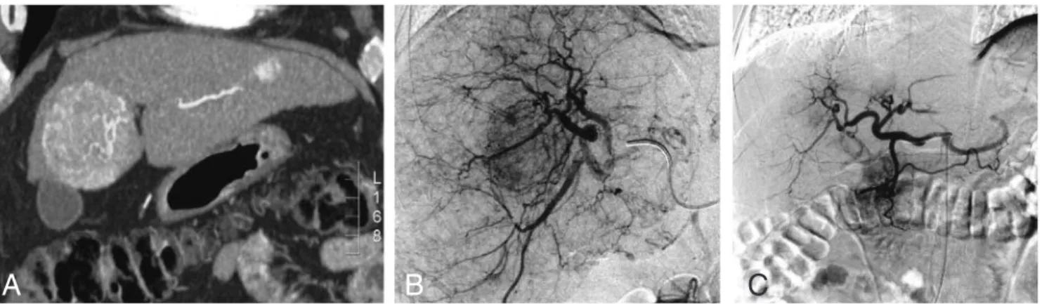
On a follow-up CT scan, a few months later, it appeared that the segment IV mass was markedly increased (6 × 5.5 × 7 cm) (Fig. 1A). The segment II and VI lesion were confirmed but stable. Additional tests did not reveal any metastasis.
After multidisciplinary consensus and given the patient’s delicate clini-cal condition, surgiclini-cal treatment was not adequate. In these conditions, the presence of multiple liver HCC nodules justified a transarterial chemoembolization (TACE). TACE was performed with no intra-opera-tive complications. Embolization of the feeding artery was performed with small gelfoam pledgets before (to reduce the flow) and after (to achieve stases) injection of 50 mg of cisplatine (Fig. 1B and C). Post -operative and one week control CT-scan (Fig. 2) didn’t show any sign of active arterial contrast extravasation and other complications.
Three weeks after transarterial chemoembolization (TACE), the patient complained about a right upper abdominal pain. This led to an abdominal CT-Scan (Fig. 3) that revealed ruptured necrotic mass with associated peritonitis and pneumo peritoneum but no active bleeding. At this time, laboratory test revealed stable anemia (haemoglo-bin level: 10.9 g/dl), hypoalbumine-mia (19 g/l), abnormal liver function tests (ALP/GGT: 359/168 IU/L) and elevated conjugated bilirubin level (5.2 mg/L).
Because of the bad general state of the patient, conservative treat-ment was decided with discharge from hospital to palliative care. Unfortunately, the patient died two months after TACE procedure. remarkable for silicosis associated
with respiratory failure, type 2 dia-betes and cerebrovascular accident. He had a previous history of smok-ing and persistent alcohol abuse was documented. On admission, his height was 165 cm and his body weight 74 kg. There was no history of hematemesis, vomiting, flushing or diarrhea.
Clinical examination revealed a
hepatomegaly without
splenomegaly. Jaundice, ascites, peripheral oedema and other signs of chronic liver disease were not observed. The patient’s blood pres-sure was 120/60 mmHg and heart rate was regular to 74 beats per minute. Laboratory test revealed marked leukocytosis (18000/mm3), hemoglobin level 11g/dl and abnor-mal liver function tests (ALP/GGT: 125/158 IU/L). Serum tumor markers were within normal range ( -fetopro-tein: 2.6 ng/ml). Other laboratory tests were also within normal range. However, glycaemia (110 mg/dl) and HbA1c (7.9%) were slightly elevated. Tests for hepatitis B and C virus markers were all negative. The patient showed a Child-Pugh score of 7 (class B).
A triphasic contrast enhanced CT-scan showed three hypervascular lesions, without clear wash out, sus-pected of malignancy. The bigger one was in segment IV (4 × 4 cm) and near to the liver capsule. The other lesions were localized in seg-ment II and in segment VI. Ultrasound was not contributive because of patient’s corpulence. A JBR–BTR, 2011, 94: 0-00.
Reference number to be mentioned by correspondence :
JBR/BRULS-RUPTURED HEPATOCELLULAR CARCINOMA FOLLOWING TRANS CATHETER
ARTERIAL CHEMOEMBOLIZATION
S. Bruls1, J. Joskin2, R. Chauveau2, J. Delwaide3, P. Meunier2
Transcatheter arterial chemoembolization (TACE) is known to be an effective palliative treatment in unresectable hepatocellular carcinoma (HCC). Although TACE can control tumour growth and palliate the patients, complications of TACE with significant morbidity are well known and adversely affect the outcome of patients. Necrotic tumor rup-ture is a serious complication of TACE and has a high mortality rate. We report a case of ruprup-tured HCC following TACE in a 78-year-old male patient who subsequently developed peritonitis and pneumoperitoneum. This case gives us the opportunity to underline the importance of such complications and demonstrates the utility of CT imaging for diag-nosis and management of patients with ruptured HCC.
Key-word: Liver neoplasms, therapy.
From: 1. Department of Surgery, 2. Department of Medical Imaging, 3. Department of
Gastroenterology and Hepatology, University Hospital of Liège, Liège, Belgium.
Address for correspondence: Dr P. Meunier, M.D., Department of Medical Imaging,
function. Tumor sizes, hypovasculari-ty on imaging and elevated INR are predictors of increased mortality after TACE therapy for HCC (13). Contraindications for TACE (19) include portal vein thrombosis, sig-nificant arteriovenous shunting and poor liver function (Child-Pugh class C).
Imaging techniques and correct anatomical evaluation is essential to select those patients who may poten-tially benefit from the procedure. CT or MR examinations should be done prior to TACE (8). Although mean survival rates of 12 months (13) have been reported, a variety of complica-tions have been described after TACE with significant morbidity. These complications are more often benign and include postembolization syn-drome (fever, abdominal pain, nau-sea, and vomiting), impaired liver function, and leukocytopenia (3, 5, 14). However major complications are described. Liver failure, as well as liver abscess, splenic infarction, treatment of choice (7). TACE
com-bines the effect of targeted chemotherapy with the effect of ischemic necrosis induced by arterial embolization (8). The basic principles of the procedure consist of a reduc-tion of hepatic arterial blood supply to the tumor as well as the delivery of cytostatic agents (Adryamycin, Cisplatin, or Doxorubicin) directly into the tumor. It is well known that HCC is hypervascularised (with a high propensity for vascular inva-sion and growth factor produce) and that this hypervascularisation is mostly dependent on the hepatic artery and its branches (9).
The survival benefit of this treat-ment is confirmed by two meta-analyses (10, 11) and two recent ran-domized controlled trials (4, 12) that allowed showing a significant higher survival rate compared to a group of best supportive care after three years. However its benefice appears only in selected patients with unre-sectable HCC and preserved liver
2 JBR–BTR, 2011, 94 (—)
Fig. 1. — A: Coronal MultiPlanar Reconstruction (MPR) of a contrast enhanced CT scan of arterial phase demonstrates a hyper
-enhancing lesion in segment IV. B: Hepatic angiography (arteriogram) shows an anarchic hypervascularised hepatic mass that suggests hepatocellular carcinoma with formation of arteriovenous shunts. C: Hepatic angiography after chemoembolization shows an almost complete regression of the anarchic vascularisation in the lesion.
A
B
A
B
C
C
Fig. 2. — Contrast enhanced CT scan
obtained one week after TACE shows the integrity of the liver capsule.
Fig. 3. — Axial (A and B) and sagittal (C) contrast enhanced CT scan, in arterial phase, obtained 3 weeks after TACE shows a
capsular rupture at the level of the necrotic mass with associated peritonitis and pneumoperitoneum.
Discussion
Transarterial chemoembolization (TACE) is an efficient palliative well accepted treatment for unresectable HCC (3, 5, 6). In the presence of mul-tiple HCC nodules, TACE remains the
upper gastrointestinal bleeding, bile duct complications, acalculous cholecystitis, pulmonary embolism, spasm or occlusion of hepatic artery and acute renal failure are associat-ed with significant morbidity and mortality.
Rupture following TACE is uncom-mon and represents less than 1% of all complications (15) but it is proba-bly the most important complication of TACE with an extremely poor prognosis (1). The mortality and morbidity rate is high because the patients usually suffer from an advanced disease.
The mechanism of rupture of HCC after TACE is unclear. It is probably to be related to tumor and capsular necrosis and increased pressure inside the friable tumor after TACE. Secondary infection, vascular injury during TACE or inflammation sec-ondary to the chemotherapeutic agents could play a role too (1). They can appear especially when there is no tumor capsule or when the lesion is located adjacent to the Glissonian liver capsule (16). Two series from Asia conclude that most patients with complications after TACE had pre-existing risk factors as large tumor size, or extracapsular exten-sion of the tumor (15, 17).
Patients with ruptured hepatocel-lular carcinoma present right upper abdominal pain and abdominal dis-tension and the diagnosis is usually confirmed by US or CT-Scan. CT imaging is the most useful modality in the imaging of HCC (18) and can detect most of complications follow-ing TACE (18). In a ruptured HCC, blood or gas (as a result of necrosis) may be seen in the lesion, around the liver or within the peritoneal cav-ity. Bleeding may also occasionally be seen.
Management of patients with rup-ture of HCC following TACE is diffi-cult and remains controversial because patients have already been diagnosed as having inoperable tumor and poor liver function. The primary objective in the manage-ment of these patients is to control potential bleeding and to maintain haemodynamic parameters stables. Hemostasis can be done by surgical laparotomy, interventional or con-servative methods. Battula et al (19) consider that a repeat TACE can be
7. Acunas B., Rozanes I.: Hepatocellular carcinoma: treatment with tran-scatheter arterial chemoembolization.
Eur J Radiol, 1999, 32:86-89.
8. Shin S.W.: The current practice of transarterial chemoembolization for the treatment of hepatocellular carci-noma. Korean J Radiol, 2009, 10: 425-434.
9. Nakashima T., Kojiro M.: Pathologic characteristics of hepatocellular carci-noma. Semin Liver Dis, 1986, 6: 259-266.
10. Llovet J.M., Bruix J.: Systematic review of randomized trials for unre-sectable hepatocellular carcinoma: Chemoembolization improves sur-vival. Hepatology, 2003, 37:429-442.
11. Geschwind J.F., Ramsey D.E.,
Choti M.A., et al.: Chemo
-embolization of hepatocellular carci-noma: results of metaanalysis. Am J
Clin Oncol, 2003, 26: 344-349.
12. Lo C.M., Ngam H., Tso W.K., et al.: Randomized controlled trial of lipidol chemoembolization for unresectable
hepatocellular carcinoma.
Hepatology, 2002, 35: 1164-1171.
13. Shen H. Agarwal D., QI R.,
Chalasani N.: Predictors of outcome in patients with unresectable hepato-cellular carcinoma receiving tran-scatheter arterial chemoembolization.
Aliment pharmacol Ther, 2007, 26:
393-400.
14. Jinglin X., Zhenggang R.,
Shenglong Y., Sharma D, Zhiying L., Yuhong G., Yi C., Zengchen M., Zhiquan W., Jia F., Lunxiu Q., Xinda Z., Zhaoyou T., Binghui Y.: Study of severe and rare complications of
transarterial chemoembolization
(TACE) for liver cancer. Euro J Rad, 2006, 59: 407-12.
15. Chung J.W., Park J.H., Han J.K., et al.: Hepatic Tumors: Predisposing Factors for Complications of transcatheter oily chemoembolization. Radiology, 1996, 198: 33-40.
16. Sokamoto I., Aso N., Nagaoki K., et al:
Complications associated with
Transcatheter arterial embolization for hepatic tumors. Radiographics, 1998, 18: 605-619.
17. Liu C.L., Ngan H., Lo C.M., et al.: Ruptured hepatocellular carcinoma as a complication of transarterial oily chemomboliation. Br J Surg, 1998, 85:612-614.
18. Kim H.C., Yang D.M., Jin W., et al.: The various manifestations of ruptured hepatocellular carcinoma: CT imag-ing findimag-ings. Abdom Imagimag-ing, 2008, 33: 633-642.
19. Battula N., Srinivasan P., Madanur M., et al.: Ruptured hepatocellular carci-noma following chemoembolization: a western experience. Hepatobiliary
Pancreat Dis Int, 2007, 6: 49-51. performed to stabilize the tumor. In
ours opinion, emergency emboliza-tion is an efficient haemostatic treat-ment in case of intra-peritoneal bleeding of a ruptured HCC. In this particular situation of our patient with a bad general health and the absence of active bleeding, TACE or embolization had to be excluded. The only left solution was conserva-tive management.
Although TACE is generally a safe procedure, tumor rupture remains a potential complication. As showed in our patient, it appears to be especial-ly important in large tumors adja-cent to the liver capsule. In case of such important masses, liver rupture is a clear but relatively rare compli-cation that may occur even in absence of TACE. This complication may appear relatively lately (several weeks) after procedure, that the rea-son why a precise follow-up is mandatory.
References
1. Gomaa A.I., Khan S.A., Toledano M.B., et al.: Hepatocellular carcinoma: epidemiology, risk factors and patho-genis. World J Gastroenterol, 2008, 14: 4300-4308.
2. El-Serag H.B., Mason A.C.: Rising incidence of hepatocellular carcino-ma in the United States. New Engl J
Med, 1999, 340: 745-750.
3. Rose M.D., Chapman W.C.,
Brokenbrough A.T., Wright J.K., Rose A.T., Meranze S., Mazer M., Blair T., Blanke C.D., Debelak J.P., Wright-Pinson C.: Transcatheter arteri-al chemoembolization as primary treatment for hepatocellular carcino-ma. Am J Surg, 1999, 177: 405-410. 4. Llovet J.M., Real M.I., Montana X., et
al.: Arterial embolisation or
chemoembolisation versus sympto-matic treatment in patients with unre-sectable hepatocellular carcinoma: a randomised controlled trial. Lancet, 2002, 359: 1734-1739.
5. A comparison of lipiodol chemoem-bolization and conservative treat-ment for unresectable hepatocellular carcinoma. Groupe d’étude et de Traitement du Carcinome Hépato -cellulaire. New Engl J Med, 1995, 332: 1256-1261.
6. Kiely J.M., Rilling W.S., Touzios J.G., Hieb R.A., Franco J., Saeian K.,
Quebbeman E.J., Pitt H.A.:
Chemoembolization in patients at high risk: results and complications.
J Vasc Inter Rad, 2006, 17: 47-53.