HAL Id: dumas-01074884
https://dumas.ccsd.cnrs.fr/dumas-01074884
Submitted on 15 Oct 2014
HAL is a multi-disciplinary open access
archive for the deposit and dissemination of
sci-entific research documents, whether they are
pub-lished or not. The documents may come from
teaching and research institutions in France or
abroad, or from public or private research centers.
L’archive ouverte pluridisciplinaire HAL, est
destinée au dépôt et à la diffusion de documents
scientifiques de niveau recherche, publiés ou non,
émanant des établissements d’enseignement et de
recherche français ou étrangers, des laboratoires
publics ou privés.
Fibromuscular dysplasia : what the radiologist should
know : a pictorial review
Lionel Varennes
To cite this version:
Lionel Varennes. Fibromuscular dysplasia : what the radiologist should know : a pictorial review.
Human health and pathology. 2014. �dumas-01074884�
AVERTISSEMENT
Ce document est le fruit d'un long travail approuvé par le
jury de soutenance et mis à disposition de l'ensemble de la
communauté universitaire élargie.
Il n’a pas été réévalué depuis la date de soutenance.
Il est soumis à la propriété intellectuelle de l'auteur. Ceci
implique une obligation de citation et de référencement
lors de l’utilisation de ce document.
D’autre part, toute contrefaçon, plagiat, reproduction illicite
encourt une poursuite pénale.
Contact au SICD1 de Grenoble :
thesebum@ujf-grenoble.fr
LIENS
LIENS
Code de la Propriété Intellectuelle. articles L 122. 4
Code de la Propriété Intellectuelle. articles L 335.2- L 335.10
http://www.cfcopies.com/V2/leg/leg_droi.php
1
UNIVERSITE JOSEPH FOURIER
FACULTE DE MEDECINE DE GRENOBLE
Année : 2014
N°
FIBROMUSCULAR DYSPLASIA : WHAT THE RADIOLOGIST
SHOULD KNOW : A PICTORIAL REVIEW
THESE
PRESENTEE POUR L’OBTENTION DU DOCTORAT EN MEDECINE
DIPLÔME D’ETAT
LIONEL VARENNES
Né le 21 janvier 1986 à Vesoul
THESE SOUTENUE PUBLIQUEMENT A LA FACULTE DE MEDECINE DE GRENOBLE
Le jeudi 9 octobre 2014
DEVANT LE JURY COMPOSE DE
Président du jury : M. le Professeur Alexandre KRAINIK.
Membres :
M. le Docteur Jean-Philippe BAGUET.
Mme le Docteur, Maître de conférence universitaire Sylvie GRAND.
Mme le Docteur Florence TAHON.
M. le Docteur Fréderic THONY.
M. le Professeur Emmanuel TOUZE.
*La Faculté de Médecine de Grenoble n’entend donner aucune approbation ni improbation aux opinions émises dans les thèses ; ces opinions sont considérées comme propres à leurs auteurs.
... ... ...
Affaire suivie par Marie-Lise GALINDO sp-medecine-pharmacie@ujf-grenoble.fr
Doyen de la Faculté : M. le Pr. Jean Paul ROMANET
Année 2013-2014
ENSEIGNANTS A L’UFR DE MEDECINE
CORPS NOM-PRENOM Discipline universitaire
PU-PH ALBALADEJO Pierre Anesthésiologie réanimation
MCU-PH APTEL Florent Ophtalmologie
PU-PH ARVIEUX-BARTHELEMY Catherine chirurgie générale
PU-PH BACONNIER Pierre Biostatiques, informatique médicale et technologies de communication
PU-PH BAGUET Jean-Philippe Cardiologie
PU-PH BALOSSO Jacques Radiothérapie
PU-PH BARRET Luc Médecine légale et droit de la santé
PU-PH BAUDAIN Philippe Radiologie et imagerie médicale
PU-PH BEANI Jean-Claude Dermato-vénéréologie
PU-PH BENHAMOU Pierre Yves Endocrinologie, diabète et maladies métaboliques
PU-PH BERGER François Biologie cellulaire
PU-PH BETTEGA Georges Chirurgie maxillo-faciale, stomatologie
MCU-PH BOISSET Sandrine Agents infectieux
PU-PH BONAZ Bruno Gastro-entérologie, hépatologie, addictologie
MCU-PH BONNETERRE Vincent Médecine et santé au travail
PU-PH BOSSON Jean-Luc Biostatiques, informatique médicale et
technologies de communication
MCU-PH BOTTARI Serge Biologie cellulaire
PU-PH BOUGEROL Thierry Psychiatrie d'adultes
PU-PH BOUILLET Laurence Médecine interne
MCU-PH BOUZAT Pierre Réanimation
PU-PH BRAMBILLA CHRISTIAN Pneumologie
PU-PH BRAMBILLA Elisabeth Anatomie et cytologie pathologiques
MCU-PH BRENIER-PINCHART Marie Pierre Parasitologie et mycologie
PU-PH BRICAULT Ivan Radiologie et imagerie médicale
PU-PH BRICHON Pierre-Yves Chirurgie thoracique et cardio- vasculaire
MCU-PH BRIOT Raphaël Thérapeutique, médecine d'urgence
PU-PH CAHN Jean-Yves Hématologie
MCU-PH CALLANAN-WILSON Mary Hématologie, transfusion
PU-PH CARPENTIER Françoise Thérapeutique, médecine d'urgence
UFR de Médecine de Grenoble
DOMAINE DE LA MERCI
38706 LA TRONCHE CEDEX – France TEL : +33 (0)4 76 63 71 44
FAX : +33 (0)4 76 63 71 70
3
PU-PH CARPENTIER Patrick Chirurgie vasculaire, médecine vasculaire
PU-PH CESBRON Jean-Yves Immunologie
PU-PH CHABARDES Stephan Neurochirurgie
PU-PH CHABRE Olivier Endocrinologie, diabète et maladies métaboliques
PU-PH CHAFFANJON Philippe Anatomie
PU-PH CHAVANON Olivier Chirurgie thoracique et cardio- vasculaire
PU-PH CHIQUET Christophe Ophtalmologie
PU-PH CHIROSSEL Jean-Paul Anatomie
PU-PH CINQUIN Philippe Biostatiques, informatique médicale et technologies de communication
PU-PH COHEN Olivier Biostatiques, informatique médicale et technologies de communication
PU-PH COUTURIER Pascal Gériatrie et biologie du vieillissement
PU-PH CRACOWSKI Jean-Luc Pharmacologie fondamentale, pharmacologie clinique
PU-PH DE GAUDEMARIS Régis Médecine et santé au travail
PU-PH DEBILLON Thierry Pédiatrie
MCU-PH DECAENS Thomas Gastro-entérologie, Hépatologie
PU-PH DEMATTEIS Maurice Addictologie
PU-PH DEMONGEOT Jacques Biostatiques, informatique médicale et technologies de communication
MCU-PH DERANSART Colin Physiologie
PU-PH DESCOTES Jean-Luc Urologie
MCU-PH DETANTE Olivier Neurologie
MCU-PH DIETERICH Klaus Génétique et procréation
MCU-PH DUMESTRE-PERARD Chantal Immunologie
PU-PH ESTEVE François Biophysique et médecine nucléaire
MCU-PH EYSSERIC Hélène Médecine légale et droit de la santé
PU-PH FAGRET Daniel Biophysique et médecine nucléaire
PU-PH FAUCHERON Jean-Luc chirurgie générale
MCU-PH FAURE Julien Biochimie et biologie moléculaire
PU-PH FERRETTI Gilbert Radiologie et imagerie médicale
PU-PH FEUERSTEIN Claude Physiologie
PU-PH FONTAINE Éric Nutrition
PU-PH FRANCOIS Patrice Epidémiologie, économie de la santé et prévention
PU-PH GARBAN Frédéric Hématologie, transfusion
PU-PH GAUDIN Philippe Rhumatologie
PU-PH GAVAZZI Gaétan Gériatrie et biologie du vieillissement
PU-PH GAY Emmanuel Neurochirurgie
MCU-PH GILLOIS Pierre Biostatiques, informatique médicale et technologies de communication
PU-PH GODFRAIND Catherine Anatomie et cytologie pathologiques (type clinique)
MCU-PH GRAND Sylvie Radiologie et imagerie médicale
PU-PH GRIFFET Jacques Chirurgie infantile
MCU-PH GUZUN Rita Endocrinologie, diabétologie, nutrition, éducation thérapeutique
PU-PH HALIMI Serge Nutrition
PU-PH HENNEBICQ Sylviane Génétique et procréation
PU-PH HOFFMANN Pascale Gynécologie obstétrique
PU-PH HOMMEL Marc Neurologie
PU-PH JOUK Pierre-Simon Génétique
PU-PH JUVIN Robert Rhumatologie
PU-PH KAHANE Philippe Physiologie
PU-PH KRACK Paul Neurologie
PU-PH KRAINIK Alexandre Radiologie et imagerie médicale
PU-PH LABARERE José Département de veille sanitaire
PU-PH LANTUEJOUL Sylvie Anatomie et cytologie pathologiques
MCU-PH LAPORTE François Biochimie et biologie moléculaire
MCU-PH LARDY Bernard Biochimie et biologie moléculaire
MCU-PH LARRAT Sylvie Bactériologie, virologie
MCU-PH LAUNOIS-ROLLINAT Sandrine Physiologie
PU-PH LECCIA Marie-Thérèse Dermato-vénéréologie
PU-PH LEROUX Dominique Génétique
PU-PH LEROY Vincent Gastro-entérologie, hépatologie, addictologie
PU-PH LETOUBLON Christian chirurgie générale
PU-PH LEVY Patrick Physiologie
MCU-PH LONG Jean-Alexandre Urologie
PU-PH MACHECOURT Jacques Cardiologie
PU-PH MAGNE Jean-Luc Chirurgie vasculaire
MCU-PH MAIGNAN Maxime Thérapeutique, médecine d'urgence
PU-PH MAITRE Anne Médecine et santé au travail
MCU-PH MALLARET Marie-Reine Epidémiologie, économie de la santé et prévention
MCU-PH MARLU Raphaël Hématologie, transfusion
MCU-PH MAUBON Danièle Parasitologie et mycologie
PU-PH MAURIN Max Bactériologie - virologie
MCU-PH MCLEER Anne Cytologie et histologie
PU-PH MERLOZ Philippe Chirurgie orthopédique et traumatologie
PU-PH MORAND Patrice Bactériologie - virologie
PU-PH MOREAU-GAUDRY Alexandre Biostatiques, informatique médicale et technologies de communication
PU-PH MORO Elena Neurologie
PU-PH MORO-SIBILOT Denis Pneumologie
MCU-PH MOUCHET Patrick Physiologie
PU-PH MOUSSEAU Mireille Cancérologie
PU-PH MOUTET François Chirurgie plastique, reconstructrice et esthétique, brûlogie
MCU-PH PACLET Marie-Hélène Biochimie et biologie moléculaire
PU-PH PALOMBI Olivier Anatomie
PU-PH PARK Sophie Hémato - transfusion
PU-PH PASSAGGIA Jean-Guy Anatomie
5
PU-PH PAYEN DE LA GARANDERIE Jean-François Anesthésiologie réanimation
MCU-PH PAYSANT François Médecine légale et droit de la santé
MCU-PH PELLETIER Laurent Biologie cellulaire
PU-PH PELLOUX Hervé Parasitologie et mycologie
PU-PH PEPIN Jean-Louis Physiologie
PU-PH PERENNOU Dominique Médecine physique et de réadaptation
PU-PH PERNOD Gilles Médecine vasculaire
PU-PH PIOLAT Christian Chirurgie infantile
PU-PH PISON Christophe Pneumologie
PU-PH PLANTAZ Dominique Pédiatrie
PU-PH POLACK Benoît Hématologie
PU-PH POLOSAN Mircea Psychiatrie d'adultes
PU-PH PONS Jean-Claude Gynécologie obstétrique
PU-PH RAMBEAUD Jacques Urologie
MCU-PH RAY Pierre Génétique
PU-PH REYT Émile Oto-rhino-laryngologie
MCU-PH RIALLE Vincent Biostatiques, informatique médicale et technologies de communication
PU-PH RIGHINI Christian Oto-rhino-laryngologie
PU-PH ROMANET J. Paul Ophtalmologie
MCU-PH ROUSTIT Matthieu Pharmacologie fondamentale, pharmaco clinique, addictologie
MCU-PH ROUX-BUISSON Nathalie Biochimie, toxicologie et pharmacologie
PU-PH SARAGAGLIA Dominique Chirurgie orthopédique et traumatologie
MCU-PH SATRE Véronique Génétique
PU-PH SCHMERBER Sébastien Oto-rhino-laryngologie
PU-PH SCHWEBEL-CANALI Carole Réanimation médicale
PU-PH SCOLAN Virginie Médecine légale et droit de la santé
MCU-PH SEIGNEURIN Arnaud Epidémiologie, économie de la santé et prévention
PU-PH SERGENT Fabrice Gynécologie obstétrique
PU-PH SESSA Carmine Chirurgie vasculaire
PU-PH STAHL Jean-Paul Maladies infectieuses, maladies tropicales
PU-PH STANKE Françoise Pharmacologie fondamentale
MCU-PH STASIA Marie-José Biochimie et biologie moléculaire
PU-PH TAMISIER Renaud Physiologie
PU-PH TONETTI Jérôme Chirurgie orthopédique et traumatologie
PU-PH TOUSSAINT Bertrand Biochimie et biologie moléculaire
PU-PH VANZETTO Gérald Cardiologie
PU VILLA Alessandro Neurosciences
PU-PH VUILLEZ Jean-Philippe Biophysique et médecine nucléaire
PU-PH WEIL Georges Epidémiologie, économie de la santé et prévention
PU-PH ZAOUI Philippe Néphrologie
PU-PH ZARSKI Jean-Pierre Gastro-entérologie, hépatologie, addictologie
Remerciements :
A Mme le docteur Florence TAHON, directrice de thèse, merci pour tes directives toujours justes, ta disponibilité sans faille et ton optimisme permanent.
A M. le professeur Alexandre KRAINIK, merci d’avoir supervisé notre travail. Merci pour tes conseils toujours avisés.
A M. le docteur Adrian KASTKER, merci mille fois pour ton aide et tes consignes. Merci d’avoir passé tant de temps à la mise en forme de notre article. Sans toi, la soumission de ce travail aurait été beaucoup plus
compliquée.
A M. le professeur Jean-Philippe BAGUET, merci d’avoir accepté de poser ton œil expert sur ce travail. Ta présence dans mon jury était indispensable.
A Mme le docteur Sylvie GRAND, merci de faire partie de mon jury. J’ai beaucoup apprécié partager tes vacations d’IRM. Je n’oublierai pas ton enthousiasme communicatif.
A M. le docteur Fréderic THONY, merci pour ton enseignement notamment lors de mon stage de vasculaire. Merci pour ton humour toujours corrosif.
7 A Pauline, la femme de ma vie, qui m’accompagne depuis dix ans. Tu es mon héroïne, ma princesse, mon rayon de soleil qui éclaire ma vie chaque matin. Ma Brenda, je t‘aime !
A Arthur, notre joyau, notre pépite, la prunelle de nos yeux, toi qui a débarqué dans notre vie à deux cent à l’heure sans prévenir, toi qui chamboule tous nos projets, toujours avec le sourire ! Je suis fier d’être ton père.
A mes parents, pour une infinité de choses. Merci d’avoir fait l’homme que je suis aujourd’hui.
A mon frère, Géraud, pour tout ce que l’on partage. J’aimerais passer plus de temps avec toi.
A tous les membres de ma famille et de ma belle famille, merci pour votre soutien.
Aux bisontins, chers à mon cœur : Mathieu, Marine, Marc, GQ, Minor, Flage, Benoit, Audrey…
Aux amis de l’internat : Mickael, Olivia, Pierre-Yves, Valérie, Robin, Alexa, Camille, Caro, Marco, robinho, la Mouge, Julien D, Nono, Bastien, Alexis…
A mes co-internes : Auré, Julio, Anny, Pidz, Pierrot, Nico, Lucky, Ghelfiotte, Maillot, Tristan, Béné, Coco, Alexis, Mathieu, Marie, Lison, Romain, Cevdet…
A tout le personnel hospitalier que j’ai eu l’occasion de rencontrer lors de mon internat, médecins,
manipulateurs, secrétaires, infirmières, ASH de la CURIM, de la CLUNI, de la CUIP, de l’hôpital sud et de l’hôpital d’Annecy.
TABLE DES MATIERES :
RESUME
ARTICLE
TITLE
ABSTRACT
INTRODUCTION
Epidemiology
Pathophysiology
TYPICAL AND VARIANT ASPECTS
1) Typical aspects
a) Classifications
b) Diagnostic tools
c) Preferential locations, clinical manifestations and radiological
diagnostic criteria
A. Renal arteries
B. Cervicoencephalic arteries
C. Radiological diagnostic criteria
2) Less typical presentations
a) Arterial dissection
b) Aneurysms/vascular ectasia
c) Unexplained SAH
DIFFERENTIAL DIAGNOSIS
CONCLUSION
REFERENCES
CONCLUSION SIGNEE
SERMENT D’HIPPOCRATE
9
RESUME
La dysplasie fibromusculaire (DFM) est une maladie idiopathique, segmentaire, non inflammatoire et non athéromateuse qui peut toucher toutes les couches de la paroi des artères de petit et moyen calibre. Sa prévalence est estimée entre 4 et 6% au niveau rénal et entre 0,3 et 3% au niveau cervico-encéphalique. Son pronostic n’est pas toujours bénin, elle peut notamment avoir des répercussions neurologiques graves. Aujourd’hui son diagnostic est posé par l’imagerie. Il est donc primordial que les radiologues aient une bonne connaissance de ses aspects pour réduire le délai entre les premiers symptômes et le diagnostic. Si l’aspect classique en « collier de perles » est bien reconnu, la DFM doit être suspectée devant des présentations moins typiques (boucles artérielles, ectasies fusiformes, dissections, anévrysmes, hémorragie sous arachnoïdienne), notamment chez les femmes jeunes ou chez les patients ayant une histoire personnelle ou familiale d’hypertension artérielle. Réduire le délai entre la survenue du premier symptôme et le diagnostic est essentiel afin d’orienter précocement les patients vers une prise en charge spécialisée dans un centre de référence. Cela permettrait de réaliser des enquêtes familiales et ainsi peut-être de déterminer des facteurs génétiques et/ou environnementaux qui influencent l’évolution de la DFM pour proposer les options thérapeutiques les plus adaptées.
ARTICLE
TITLE
Fibromuscular Dysplasia: What the Radiologist Should
Know: A Pictorial Review
Varennes L (1,2), Tahon F (1). Kastler A (1), Grand S (1), Thony F (2), Baguet JP (3),
Touze E (4), Krainik A (1)
(1) : Department of Neuroradiology and MRI, University Hospital of Grenoble. CS
10217-38043 Grenoble Cedex 09, France.
(2) : Department of Radiology, University Hospital of Grenoble. CS 10217-38043
Grenoble Cedex 09, France.
(3) : Department of Cardiology, Mutualist Hospital Group of Grenoble-38028
Grenoble cedex 1, France.
(4) : Department of neurology, University Caen Basse Normandie, Inserm U919,
CHU Côte de Nacre, Caen, France.
11
ABSTRACT
Fibromuscular dysplasia (FMD) is an idiopathic, segmentary, non inflammatory and non
atherosclerotic disease that can affect all layers of both small- and medium-caliber arteries. The
prevalence of FMD is estimated between 4 and 6% in the renal arteries and between 0.3 and 3% in the cervicoencephalic arteries. FMD most frequently affects the renal, carotid, and vertebral arteries but it can theoretically affect any artery. Radiologists play an important role in the diagnosis of FMD and a good knowledge on FMD's conditions will certainly help reduce the delay between the first symptoms and the diagnosis. If the common string-of-beads aspect is well known, less common presentations have to be known. These less common imaging findings include: vascular loops, fusiform vascular ectasia, arterial dissection, aneurysm and subarachnoid hemorrhage. These radiologic presentations should be known from radiologists, in order to diagnose possible FMD, particularly when present in young females, or when associated with personal or familial hypertension, to reduce the delay between the onset of the first symptom and the final diagnosis. Thus, the patients have to be referred in specialized FMD centers for dedicated management.
INTRODUCTION
Fibromuscular dysplasia (FMD) is an idiopathic, segmentary, non inflammatory and non
atherosclerotic, disease that can affect all layers of both small- and medium-caliber arteries. Although
not known, FMD prevalence in the general population is considered to be low by several authors [1]. The cause and pathophysiology of FMD are unknown yet [2; 3]. Clinical manifestations of FMD are
primarily dependent on the vessels that are involved. FMD most frequently affects the renal, carotid,
and vertebral arteries but can be found in all territories [3].It is a potentially serious disease, especially in the cervicoencephalic location, where it can lead to severe stenosis, hypoperfusion, aneurysm, subarachnoid hemorrhage, dissection, or arterial occlusion [3-5]
FMD is often not considered as differential diagnosis because radiological aspects can be difficult to diagnose and symptoms reported are not specific. Therefore, the mean delay from symptoms to diagnosis has been reported to be up to 4.1 years [6].
Radiologists play an important role in the diagnosis of FMD and a good knowledge of FMD's
conditions should help to reduce the delay between first symptoms and final diagnosis. The objective of this review is therefore to raise awareness of radiologists on FMD epidemiology, pathophysiology, clinical presentation, typical and atypical radiological aspects and possible complications of FMD.
13
Epidemiology
The prevalence of FMD is estimated between 4 and 6% in the renal arteries and between 0.3 and 3% in the cervicoencephalic arteries [2; 7-9]. However, most data stem from series of consecutive angiograms performed in symptomatic patients and therefore cannot be considered representative. Several authors believe that these prevalence values are underestimated because FMD is frequently asymptomatic [10-12]. In a review combining the results of 4 studies including 3,181 asymptomatic patients who undergone a renal angiography before kidney transplant donation, 4.4% (139 subjects) presented FMD lesions [1]. In an other study among 20,244 consecutive autopsies performed at the Mayo Clinic over a 25-year period, only 4 subjects had cervical FMD (0.02%) [13].
FMD is most frequently revealed in the kidneys in young patients with resistant hypertension secondary to fibrodysplastic renal artery stenosis (RAS). FMD is found in about 1% of hypertensive patients and represents the second leading cause of renovascular hypertension after atherosclerotic disease. As for cervicoencephalic FMD, the symptoms are not specific.
In 2012, Olin et al. [6] included 447 cases from the FMD registry from 9 states in the United States, the largest number of patients included in any study to date. The most frequently described symptoms or condition were hypertension (63.8%), headaches (52.4%), pulsatile tinnitus (27.5%), dizziness (26%), and neck pain (22%). This study showed a clear female prevalence (91%), and the mean age at diagnosis was 51.9 years. In 6% of cases, FMD was discovered randomly. Affected arteries were mainly renal arteries (79.7%), extracranial carotid (74.4%) and vertebral (36.6%) arteries, mesenteric arteries (26%), and intracranial carotid arteries (17%). Sixty five percent of patients with renal FMD lesions also had cervicoencephalic artery lesions and vice-versa.
Several studies have reported a lower frequency of FMD lesions in the carotid arteries compared with renal arteries [1; 3; 14-16]. In 1982, Mettinger et al. found 58% renal artery involvement versus 32% carotid artery involvement based on a systematic review of 36 studies, including 1,197 patients with FMD [9]. This difference can be explained as FMD is more frequently symptomatic in renal arteries through the onset of hypertension [3], which also tends to increase its prevalence. Moreover, at the time of this study, multi-detector computed tomography (CT) was not as available a diagnostic tool as it is today. Indeed, easy access to CTA and better knowledge of cervicoencephalic FMD, which is now looked for systematically in patients with renal artery lesions, may explain the differences in
Pathophysiology
FMD pathophysiology remains unclear. Some authors suggest a role played by estrogenic impregnation given the female predominance [1]. The predominance of right renal artery lesions suggests a mechanical component because the right kidney is more mobile than the left one; the mechanism may involve compression of the vasa vasorum leading to ischemia [17; 18]. The vasa vasorum are small blood vessels that wind along arteries whose caliber are larger than 1 mm. They feed the adventitia and the external two-thirds of the media. There are relatively few in the carotid and renal arteries, possibly a feature of FMD [13]. Smoking could also be a risk factor. Autosomal
dominant transmission with incomplete penetrance and variable expressivity is suggested in 6–10% familial types, no specific gene has been identified, though [19]. Moreover, a study performed over 9 years in 106 patients who had repeated angiograms showed either a progression or a stabilization of FMD lesions but no lesion regression was seen [9].
15
TYPICAL AND VARIANT ASPECTS
I) Typical aspects
a) Classifications
The first case of histologically proven carotid FMD was published by Connett and Lansche in 1965 [20]. Later, a classification of FMD based on renal artery lesions was described in 1971 by Harrison and MacCormack [21], then revised by Stanley in 1996 [22]. This classification is based on the most affected arterial layer: the intimal type, the medial type, and the subadventitial type. The authors report that several forms can co-exist in the same patient. These lesions have later been described in carotid arteries on autopsies [3].
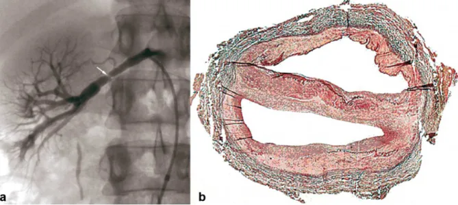
1) The intimal type corresponds to fibrous circumferential intimal thickening with proliferation of subendothelial connective tissue. The internal elastic lamina is always preserved, and the media and adventitia are normal. This form is mainly found in children, rarely in adults (5%). It appears as focal truncal stenosis (figure 1).
Figure 1. Example of intimal FMD lesions.
a- Renal angiogram showing short focal stenosis (white arrow) of the right renal artery, in favor of intimal FMD. b- Color histological slide of intimal FMD showing circumferential focal thickening. Media and adventitia are normal.
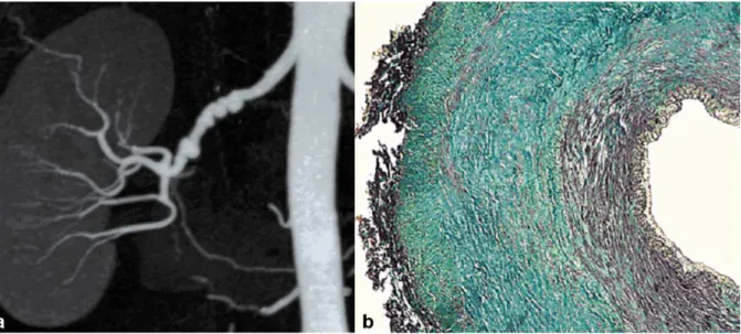
2) The medial type is the most frequent (60–70%) and corresponds to a rarefaction of smooth media muscle cells replaced by fibrosis. The intima, internal elastic lamina, and adventitia are normal. It appears as a succession of dilatations and multifocal stenoses with a characteristic string-of-beads aspect. It is mainly discovered in females aged 30–50 years (figure 2).
Figure 2. Example of medial FMD lesions.
a) Renal CTA with coronal plane MIP reconstruction, showing the typical “string of beads' aspect of the right renal artery, in favor of medial FMD.
b) Color histological slide of medial FMD showing extensive medial fibrosis. Intima and adventitia are normal.
3) The perimedial or subadventitial type (10–20% of cases) corresponds to excessive elastic tissue in the external area of the media. Its aspect is close to the medial type with fewer dilatations that do not exceed the diameter of the normal artery. Stenosis often appears tubular (figure 3).
Figure 3. Example of perimedial FMD lesions.
a) Aorta angiogram showing a long stenosis (black arrow) of the right renal artery, favor of perimedial FMD. b) Color histological slide of perimedial FMD showing extensive perimedial fibrosis.
This classification is not frequently used in daily practice because several types may co-exist in the same patient and/or the same artery. Today, pathological samples are analyzed in exceptional
17 stenosis or “string of beads” aspect), unifocal type (short stenosis less than 1 cm long), tubular type (stenosis more than 1 cm long) and mixed-type. The string-of-beads aspect is the most characteristic aspect of FMD, and indicates the presence of medial lesions [1]. The other angiographic aspects are less specific of a histological type.
In 2012, Savard et al. [24] proposed a simplified classification based only on two angiographic cervico encephalic subtypes of FMD : the multifocal type (presence of ≥2 stenoses on a given vessel
segment with or without the typical string-of-beads appearance) and the unifocal type (presence of a single focal or tubular stenosis) types. Interestingly, Savard et al. showed significant clinical
differences depending on the subtype such as: median age at diagnosis of FMD, hypertension, sex distribution, initial blood pressure, and current smoking.
b) Diagnostic tools
In case of renal impairment, Doppler ultrasound shows an accelerated blood flow in renal arteries with stenoses. It also allows to study the size of the kidneys, which is a good evaluation criterion for disease severity and follow up [19]. Magnetic resonance angiography (MRA) has a poorer spatial resolution than computed tomographic angiography (CTA) and is less sensitive for detecting accessory renal arteries [25], but remains a non-ionizing technique. CTA has the best
sensitivity/specificity ratio (1) and allows reconstruction of the renal arterial vasculature [25] but requires contrast media injection.
Although the diagnosis of cervicoencephalic FMD can be suspected with Doppler ultrasound in case of vascular loops, ectasia, intimal flap, or aliasing, it is most frequently established with cross sectional imaging, using either CTA of the supra-aortic trunks, MRA, and in some cases conventional
angiography. These imaging techniques present better sensitivity in detecting lesions of the middle and distal portions of the internal carotid and vertebral arteries at the level of the C1 and C2 vertebrae [1], which are the most frequently affected segments [3].
Despite the improvements in noninvasive screening techniques, negative results do not exclude the diagnosis of FMD. In case of high clinical suspicion, use of diagnostic arteriography [1; 9] or
c) Preferential locations, clinical manifestation and radiological diagnostic criteria
A-Renal arteries
The renal arteries are the most frequently affected location of FMD lesions. In 1997, in a series of 104 hypertensive patients with renal artery FMD, 80% were middle-aged females with preserved renal function. In 54% of the cases, the lesions were bilateral, and involved the branches in 42% of the cases. Multifocal string-of-beads lesions were found in 84% of the cases. The unifocal type most often affected young males, with tighter stenosis and more frequent downstream lesions [26].
FMD renal involvement does not necessarily lead to arterial hypertension, and progression toward kidney failure remains rare, even in cases of bilateral lesions [1; 12]. In case of stenosis in a patient with hypertension and renal FMD, severity of stenosis and it's relation to hypertension can only be asserted in case of asymmetrical kidney size. Severity of stenosis can also be assessed by measuring intra-renal arterial pressure compared to intra-aortic pressure. Thus, in case of a significant difference (> 20 mmHg), hypertension can be attributed to the FMD lesions and therefore lead to renal
angioplasty.
Even in cases of non-significant renal artery stenosis, the progression of the lesions ought to be monitored. In nearly one-third of cases, occurrence of either a new lesion, worsening stenosis, or an extending aneurysm can be visualized in these patients [23].
B-Cervicoencephalic arteries
The advent of multi-detector CT and the increase of awareness among physicians about
cervicoencephalic location of FMD have certainly participated in the raising of cervicoencephalic FMD prevalence. In 1982, Mettinger and Ericson reported, from an analysis of the literature about FMD, a prevalence of cervicoencephalic FMD lesion of 32% (in about 1,100 patients) [9]. In 2012, Olin et al. [6] reported a much higher prevalence of 74% (in 447 cases from the FMD registry from 9 states in the United States).
Although the most frequent symptoms of FMD are nonspecific (headache, dizziness, neck pain) [6], some clinical manifestations of cervicoencephalic FMD are more serious, such as transient ischemic attack (TIA), stroke, subarachnoid hemorrhage, and arterial dissection. FMD mainly affects the internal carotid artery in its extracranial part, but all territories can be affected [19] .
19 often occurs. FMD can also lead to subarachnoid hemorrhage (SAH) because of the occurrence of an intracranial dissection or a rupture of intracranial aneurysms (mainly due to the rupture of the internal elastic lamina in the medial types of FMD) [19].
C-Radiological diagnostic criteria
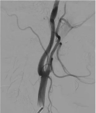
There are no formal radiological diagnostic criteria, but it is well established that the string-of-beads aspect found in renal or cervicoencephalic locations, on either arteriography, CTA, or MRA, is highly suggestive of FMD. Focal or tubular lesions on angiography allow to state the diagnosis when they are typical [27]. Another frequent finding suggestive of FMD diagnosis is the presence of a “web-like” defect at the origin of the internal carotid artery (figure 4) [3; 28].
Figure 4. Radiologic findings in a 39 year old female patient, presenting with non traumatic spontaneous subarachnoid hemorrhage. Carotid angiogram showing a “web-like” defect at the origin of the internal carotid
artery suggestive of FMD.
In such situations, lesions may be discrete at the cervical level and typical at the renal level. Thus, imaging of the renal arteries will help to establish the diagnosis. The opposite situation is also true and, it is recommended to perform imaging of the renal arteries in a patient presenting discrete but suggestive lesions in the cervicoencephalic arteries so as to confirm the diagnosis, and vice-versa (figures 5 and 6) [29].
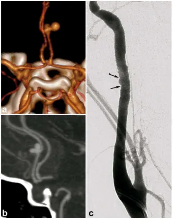
Intracranial FMD with a typical string-of-beads aspect (basilar artery, carotid, middle cerebral artery) is usually an intracranial extension of extracranial lesions [28].
Figure 5. Radiologic findings in a 68 year old female patient, who suffered from inferior myocardial infarct secondary to spontaneous dissection of the right coronary artery.
a) Renal coronal CTA MIP reconstruction, showing moderate involvement of the right renal artery (white arrows).
b) and c) 3D gadolinium enhanced MRA reconstructions, showing in the same patient typical 'string of beads aspect (white arrows) of the internal carotid artery, highly suggestive of FMD.
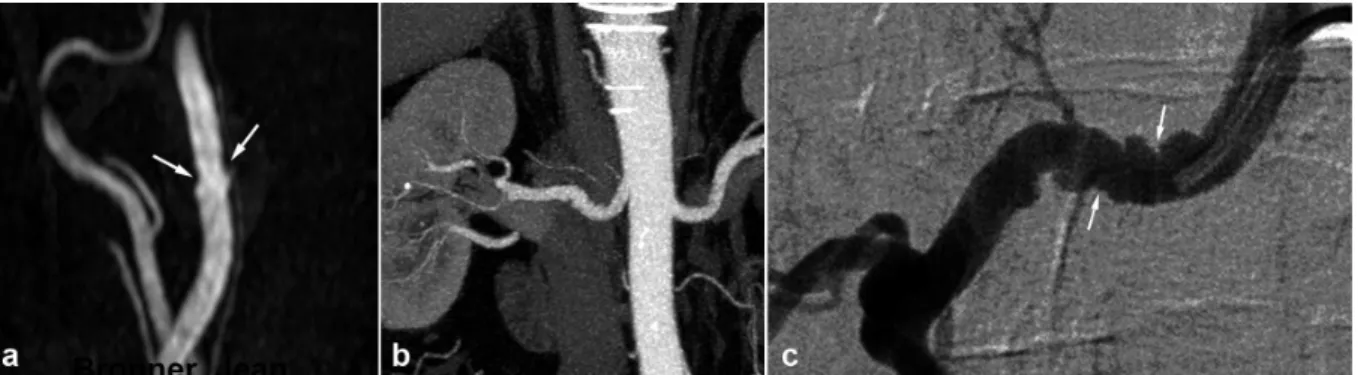
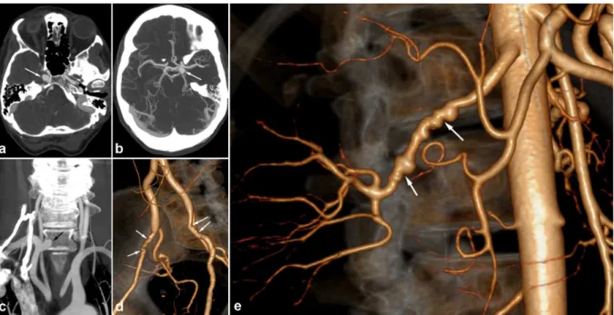
Figure 6. Radiologic findings in a 62 year old male patient with arterial hypertension.
a) Gadolinium enhanced MRA with MIP reconstruction showing typical but discrete involvement of the internal carotid artery (white arrows)
b) Renal CTA , coronal MIP reconstruction showing in the same patient a typical aspect of medial string-of-beads dysplasia of the right renal artery .
c) Right renal angiogram confirming the typical aspect of the right renal artery, in favor of FMD.
II) Less typical presentations (table 1)
a) Arterial dissection
According to Olin et al., out of 447 cases of FMD, 19.7% had an arterial dissection [6]. In 75% of cases, the dissection was located in the carotid artery, 22% in the renal artery, and 17% in a vertebral
21 Renal artery dissection may occur in 5–10%, especially in case of renal artery tubular stenosis [30]. It may be responsible of renal infarct by total occlusion or distal emboli, leading to sudden pain in the flank, hematuria, and/or rapidly progressive hypertension [1].
Extracranial dissection of the neck arteries, symptomatic or not, may occur as a complication of FMD lesions [4; 31]. However, it is not sufficient to establish a final diagnosis of FMD. In our experience, out of 64 patients with FMD, more than half of the cervicoencephalic artery dissections were discovered unexpectedly (unpublished data). Spontaneous dissection of a cervical artery is a frequent cause of stroke in young adults and may be associated with FMD in 15–20% of cases [32; 33]. For this reason, physicians should suggest FMD in patients with cervicoencephalic artery dissection, particularly if it is spontaneous, multifocal, or in case of atypical location. This situation should lead to further exploration of the renal arteries. The same approach could be proposed in case of pseudoaneurysm that may correspond to an aftermath of a dissection.
b) Aneurysms/vascular ectasia
Single or multiple aneurysms are frequent in FMD patients [5; 34]. Their prevalence reaches 17% in Olin et al.’s series [6]. Their preferential locations are the renal arteries (33%) where large aneurysms may develop, notably in medial FMD. Their rupture remains a rare complication. They may also be responsible for distal emboli or even arteriovenous fistula when rupturing in the renal vein [35]. The following most frequent location are carotid (21%), aortic (19.7%) and coeliac locations (15.8%). A strong association also exists between FMD and intracranial aneurysm, accounting for 11.8% of the aneurysms in the series reported by Olin et al. Several authors report a 22–51% prevalence of intracranial aneurysms in patients with FMD in the carotid and/or vertebral arteries [9; 17; 28; 36]. Nonetheless, these results come from a series of cerebral angiograms for the most part undertaken for hypertension. In 1998, in a meta-analysis of 18 studies including 615 FMD patients, Cloft et al. found a 24% prevalence of intracranial aneurysm [37]. However in the 212 patients without SAH, the prevalence was 7.3%, which remains higher than the prevalence expected in the general population, around 2.3% [38]. One may here suggest the diagnosis of FMD in patients with intracranial aneurysm and/or carotid ectasia, particularly in young females, especially in cases of associated personal or familial history of hypertension.
In some cases, these aneurysms have an intracavernous development and may be bilateral (figure 7). They therefore do lead to SAH but can be responsible for cavernous sinus syndrome. An exceptional
case of a carotid-cavernous fistula has even been described [34]. Loops and/or fusiform vascular ectasia in the subpetrous segment of the internal carotid arteries are also frequently found in FMD patients [29].
Figure 7. Radiologic findings in a 56 year old female patient presenting with non specific headaches.
a) and b) Cerebral CTA showing bilateral carotid aneurysms with intracavernous development (white arrows). c) Coronal CTA MIP reconstruction of the supra-aortic trunks, showing a right subclavicular aneurysm (black arrow).
d) and e) 3D volume rendering abdominal CTA showing typical medial FMD affecting the external iliac arteries (white arrows, figure d) ) and the right renal artery (white arrows, figure e)).
c) Unexplained SAH
SAH found in FMD patients is usually a consequence of subarachnoid intracranial aneurysm rupture (figures 8 and 9). [3]. SAH has also been described in patients with no visible aneurysm on
angiography but with ruptured of microaneurysms of the basilar artery at autopsy (figure 10) [39]. These SAH can also stem from intracranial dissection [29; 40]). The diagnosis is suggested on arteriography (figure 11). Intracranial dissection with SAH rebleeds usually, endovascular or
neurosurgical treatment could be proposed. In the case the arteriography at 3 months showed an ad integrum restitution of the artery.
Thus, the presence of nontraumatic SAH, especially in a young female patient, wherever it may be located and whatever its size, should bring up the diagnosis and discussion of cervicoencephalic
23
Figure 8. Radiologic findings in a 38 year old female patient, presenting with non traumatic spontaneous subarachnoid hemorrhage.
a) 3D volume rendering cerebral CTA showing a right pericallosal artery aneurysm. b) Cerebral CTA sagittal MIP reconstruction of the same aneurysm.
c) Therapeutic arteriography revealed right carotid artery typical medial FMD lesions (black arrows).
Figure 9. Radiologic findings in a 70 year old female patient, presenting a right hemisphere non traumatic SAH with tetraventricular inundation.
.a) Cerebral non enhanced CT showing SAH and intraventricular hemorrhage.
b) Cerebral CTA showing a dissecting aneurysm of the right antero-inferior cerebellar artery.
c) Angiogram of the right vertebral artery in arterial phase confirming the dissecting aneurysm of the right antero-inferior cerebellar artery (white arrow).
d) Right common carotid angiogram showing typical string of beads aspect, in favor of FMD.
e) Cerebral 3D volume rendering CTA of the dissecting aneurysm of the right antero-inferior cerebellar artery (white arrows).
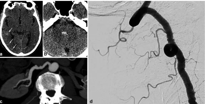
Figure 10. Radiologic findings in a 65 year old male patient with SAH of the posterior fossa. The CTA shows not particular signs. The arteriography found typical lesions of FMD in the right vertebral artery. The renal CT shows ectasia of the right renal artery termination.
a) and b) Cerebral non enhanced CT showing right temporo-occipital SAH (white arrows) associated with intraventricular hemorrhage (V4).
c) Right vertebral angiogram showing typical string of beads aspect.
d) Renal CTA , curved reconstructions of the right renal artery showing ectasia of the distal part of the right renal artery.
Figure 11. Radiologic findings in a 48 year old female, presenting with posterior headaches. The initial CT shows SAH of the posterior fossa. The angiography found dissection of the right superior cerebellar artery. Study of the supra-aortic arteries shows typical images of FMD. Ad integrum restitution at 3 months.
a) Cerebral non enhanced CT showing prepontine SAH (white arrow).
b) and c) Cerebral CTA and angiogram showing a dissection of the right superior cerebellar artery. d) Right carotid angiogram showing shows classical FMD imaging findings.
25
DIFFERENTIAL DIAGNOSIS
First of all, one must ensure that the images do not stem from vasospasm or stationary waves related to catheter use and/or the injection of a contrast agent. This is manifested by regular undulations with no significant stenosis, which are reversible after injection of a vasodilator and/or removal of the catheter.
Marfan or vascular Ehlers-Danlos syndrome should be discussed when presented with aneurysmal lesions and/or multiple dissections, particularly with a compatible morphotype or in a suggestive family context [1].
Vasculitis may mimic FMD lesions but, in most cases, presence of systemic inflammation provides the diagnosis [1].
Atherosclerotic lesions usually occur in a different setting. Most focal lesions are preferentially located at the ostium of the arteries or at a bifurcation. Most FMD patients are young and have few or none cardiovascular risk factors or aortic atheromatous plaques [1].
Finally, segmental arterial mediolysis (SAM) should be suggested in cases of an association of digestive tract artery lesions, notably the gastroduodenal artery. The similarity between the chronic vacular lesions of SAM and FMD lesions raises the question of a common pathophysiological stem of these lesions, one perhaps being a progressive form of the other [41; 42].
With typical string-of-beads lesions involving the renal and/or carotid arteries, possible differential diagnoses are limited.
CONCLUSION
FMD is a systemic nonatheromatous, noninflammatory disease that affects women more than men Renal and cervicoencephalic arteries are the most frequently affected sites. However, FMD may affect any artery. Radiologists play an important role in the diagnosis of FMD. A better knowledge of this disease is critical to shorten the delay between the first symptoms and the final diagnosis. It has been shown that an early diagnosis may improve prognosis and avoid complications. Common imaging findings of FMD, which mainly includes the usual 'string of beads aspect, are well known. However, it is important to recognize less common presentations of FMD, such as vascular loops, fusiform vascular ectasia, arterial dissection, aneurysm, and subarachnoid hemorrhage, to suggest diagnosis and to conduct further investigations.
27
References
1 Plouin PF, Perdu J, La Batide-Alanore A, Boutouyrie P, Gimenez-Roqueplo AP, Jeunemaitre X (2007) Fibromuscular dysplasia. Orphanet J Rare Dis, 2:28
2 Pasquini M, Trystram D, Oppenheim C, Ploin P, Touzé E (2011) Dysplasie fibromusculaire cervicale et intracrânienne. Press Med, 40:713-719.
3 Touze E, Oppenheim C, Trystram D, et al. (2010) Fibromuscular dysplasia of cervical and intracranial arteries. Int J Stroke, 5(4):296-305
4 Hugenholtz H, Pokrupa R, Montpetit VJ, Nelson R, Richard MT (1982) Spontaneous dissecting aneurysm of the extracranial vertebral artery. Neurosurgery, 10(1):96-100
5 Zimmerman R, Leeds NE, Naidich TP (1977) Carotid-cavernous fistula associated with intracranial fibromuscular dysplasia. Radiology, 122(3):725-726
6 Olin JW, Froehlich J, Gu X, et al. (2012) The United States Registry for Fibromuscular Dysplasia: results in the first 447 patients. Circulation, 125(25):3182-3190
7 Houser OW, Baker HL, Jr. (1968) Fibromuscular dysplasia and other uncommon diseases of the cervical carotid artery: angiographic aspects. Am J Roentgenol Radium Ther Nucl Med, 104(1):201-212
8 Manelfe C, Clarisse J, Fredy D, André J, Crouzet G (1974) Dysplasies fibromusculaires des artères cervico-céphaliques. J Neuroradiol., 1:149-231
9 Mettinger KL (1982) Fibromuscular dysplasia and the brain. II. Current concept of the disease. Stroke, 13(1):53-58
10 Blondin D, Lanzman R, Schellhammer F, et al. (2010) Fibromuscular dysplasia in living renal donors: still a challenge to computed tomographic angiography. Eur J Radiol, 75(1):67-71
11 Neymark E, LaBerge JM, Hirose R, et al. (2000) Arteriographic detection of renovascular disease in potential renal donors: incidence and effect on donor surgery. Radiology, 214(3):755-760
12 Cragg AH, Smith TP, Thompson BH, et al. (1989) Incidental fibromuscular dysplasia in potential renal donors: long-term clinical follow-up. Radiology, 172(1):145-147
13 Schievink WI, Bjornsson J (1996) Fibromuscular dysplasia of the internal carotid artery: a clinicopathological study. Clin Neuropathol, 15(1):2-6
14 Mettinger KL, Ericson K (1982) Fibromuscular dysplasia and the brain. I. Observations on angiographic, clinical and genetic characteristics. Stroke, 13(1):46-52
15 Begelman SM, Olin JW (2000) Fibromuscular dysplasia. Curr Opin Rheumatol, 12(1):41-47 16 Stanley JC, Fry WJ, Seeger JF, Hoffman GL, Gabrielsen TO (1974) Extracranial internal carotid and
vertebral artery fibrodysplasia. Arch Surg, 109(2):215-222
17 Stanley JC, Gewertz BL, Bove EL, Sottiurai V, Fry WJ (1975) Arterial fibrodysplasia. Histopathologic character and current etiologic concepts. Arch Surg, 110(5):561-566
18 Lüscher TF1, Lie JT, Stanson AW, Houser OW, Hollier LH, Sheps SG (1987) Arterial fibromuscular dysplasia. Mayo Clin Proc, 62(10):931-52
19 La Batide A, Perdu J, Ploin P (2007) Dysplasie fibromusculaire artérielle. Press Med, 36:1016-23. 20 Connett MC, Lansche JM (1965) Fibromuscular Hyperplasia of the Internal Carotid Artery: Report of a
Case. Ann Surg, 162:59-62
21 Harrison EG, Jr., McCormack LJ (1971) Pathologic classification of renal arterial disease in renovascular hypertension. Mayo Clin Proc, 46(3):161-167
22 Stanley J (1996) Renal artery fibrodysplasia In Renal Vascular Disease : in Renal Vascular Disease, : Novick A, Scoble J, Hamilton G. WB Saunders, London, 21–23.
23 Kincaid OW, Davis GD, Hallermann FJ, Hunt JC (1968) Fibromuscular dysplasia of the renal arteries. Arteriographic features, classification, and observations on natural history of the disease. Am J Roentgenol Radium Ther Nucl Med, 104(2):271-282
24 Savard S, Steichen O, Azarine A, Azizi M, Jeunemaitre X, Plouin PF (2012) Association between 2 angiographic subtypes of renal artery fibromuscular dysplasia and clinical characteristics. Circulation, 126(25):3062-3069
25 Vasbinder GB, Nelemans PJ, Kessels AG, et al. (2004) Accuracy of computed tomographic
angiography and magnetic resonance angiography for diagnosing renal artery stenosis. Ann Intern Med, 141(9):674-682; discussion 682
26 Pannier-Moreau I, Grimbert P, Fiquet-Kempf B, et al. (1997) Possible familial origin of multifocal renal artery fibromuscular dysplasia. J Hypertens, 15(12 Pt 2):1797-1801
27 HAS (2010) Protocole national de diagnostic et de soins : dysplasie fibromusculaire syptomatologique chez l’adulte In: G.d. l’HAS., (ed)^(eds),
28 Osborn AG, Anderson RE (1977) Angiographic spectrum of cervical and intracranial fibromuscular dysplasia. Stroke, 8(5):617-626
29 Varennes L, Tahon F, Grand S, et al. (2012) Aspect typique et variantes de la dysplasie fibro-musculaire des artères à destinée encéphalique. In: JFR, (ed)^(eds). JFR, Paris,
30 Lacombe M (2001) Isolated spontaneous dissection of the renal artery. J Vasc Surg, 33(2):385-391 31 Ringel SP, Harrison SH, Norenberg MD, Austin JH (1977) Fibromuscular dysplasia: multiple
"spontaneous" dissecting aneurysms of the major cervical arteries. Ann Neurol, 1(3):301-304 32 Schievink WI (2001) Spontaneous dissection of the carotid and vertebral arteries. N Engl J Med,
344(12):898-906
33 Debette S, Leys D (2009) Cervical-artery dissections: predisposing factors, diagnosis, and outcome. Lancet Neurol, 8(7):668-678
34 Bellot J, Gherardi R, Poirier J, Lacour P, Debrun G, Barbizet J (1985) Fibromuscular dysplasia of cervico-cephalic arteries with multiple dissections and a carotid-cavernous fistula. A pathological study. Stroke, 16(2):255-261
35 Goncharenko V, Gerlock AJ, Jr., Shaff MI, Hollifield JW (1981) Progression of renal artery fibromuscular dysplasia in 42 patients as seen on angiography. Radiology, 139(1):45-51
36 Mettinger KL, Soderstrom CE (1978) Pathogenetic profile of TIA before 55. A three-year investigation. J Neurol Sci, 36(3):341-348
37 Cloft HJ, Kallmes DF, Kallmes MH, Goldstein JH, Jensen ME, Dion JE (1998) Prevalence of cerebral aneurysms in patients with fibromuscular dysplasia: a reassessment. J Neurosurg, 88(3):436-440 38 Rinkel GJ, Djibuti M, Algra A, van Gijn J (1998) Prevalence and risk of rupture of intracranial
aneurysms: a systematic review. Stroke, 29(1):251-256
39 van de Nes JA, Bajanowski T, Trubner K (2007) Fibromuscular dysplasia of the basilar artery: an unusual case with medico-legal implications. Forensic Sci Int, 173(2-3):188-192
40 Andersen CA, Collins GJ, Jr., Rich NM, McDonald PT (1980) Spontaneous dissection of the internal carotid artery associated with fibromuscular dysplasia. Am Surg, 46(4):263-266
41 Gaud S, Cridlig J, Claudon M, Diarrassouba A, Kessler M, Frimat L (2010) [Segmental arterial mediolysis and renovascular hypertension]. Nephrol Ther, 6(7):597-601
29
31
En présence des Maîtres de cette Faculté, de mes chers condisciples et devant l’effigie D’HIPPOCRATE,
Je promets et je jure d’être fidèle aux lois de l’honneur et de la probité dans l’exercice de la Médecine.
Je donnerais mes soins gratuitement à l’indigent et n’exigerai jamais un salaire au-dessus de mon travail. Je ne participerai à aucun partage clandestin d’honoraires.
Admis dans l’intimité des maisons, mes yeux n’y verront pas ce qui s’y passe ; ma langue taira les secrets qui me seront confiés et mon état ne servira pas à corrompre les mœurs, ni à favoriser le crime.
Je ne permettrai pas que des considérations de religion, de nation, de race, de parti ou de classe sociale viennent s’interposer entre mon devoir et mon patient.
Je garderai le respect absolu de la vie humaine.
Même sous la menace, je n’admettrai pas de faire usage de mes connaissances médicales contre les lois de l’humanité.
Respectueux et reconnaissant envers mes Maîtres, je rendrai à leurs enfants l’instruction que j’ai reçue de leurs pères.
Que les hommes m’accordent leur estime si je suis fidèle à mes promesses. Que je sois couvert d’opprobre et méprisé de mes confrères si j’y manque.