HAL Id: inria-00147025
https://hal.inria.fr/inria-00147025
Submitted on 15 May 2007
HAL is a multi-disciplinary open access
archive for the deposit and dissemination of
sci-entific research documents, whether they are
pub-lished or not. The documents may come from
teaching and research institutions in France or
abroad, or from public or private research centers.
L’archive ouverte pluridisciplinaire HAL, est
destinée au dépôt et à la diffusion de documents
scientifiques de niveau recherche, publiés ou non,
émanant des établissements d’enseignement et de
recherche français ou étrangers, des laboratoires
publics ou privés.
A PROBABILISTIC OBJECTIVE FUNCTION FOR
3D RIGID REGISTRATION OF INTRAOPERATIVE
US AND PREOPERATIVE MR BRAIN IMAGES
Pierrick Coupé, Pierre Hellier, Xavier Morandi, Christian Barillot
To cite this version:
Pierrick Coupé, Pierre Hellier, Xavier Morandi, Christian Barillot. A PROBABILISTIC
OBJEC-TIVE FUNCTION FOR 3D RIGID REGISTRATION OF INTRAOPERAOBJEC-TIVE US AND
PREOP-ERATIVE MR BRAIN IMAGES. IEEE International Symposium on Biomedical Imaging: From
Nano to Macro, IEEE Signal Processing Society (SPS) and the IEEE Engineering in Medicine
and Biology Society (EMBS), Apr 2007, Washington DC, USA, United States.
pp.1320-1323,
A PROBABILISTIC OBJECTIVE FUNCTION FOR 3D RIGID REGISTRATION OF
INTRAOPERATIVE US AND PREOPERATIVE MR BRAIN IMAGES
Pierrick Coup´e
1,2,4
, Pierre Hellier
1,2,4, Xavier Morandi
1,2,3,4and Christian Barillot
1,2,4 1University of Rennes I, UMR CNRS 6074, IRISA, F-35042 Rennes, France
2
INRIA, VisAGeS U746 Unit/Project, F-35042 Rennes, France
3
CHU, University Hospital of Rennes, rue H. Le Guillou, F-35033 Rennes, France
4
INSERM, VisAGeS U746 Unit/Project, F-35042 Rennes, France
ABSTRACT
The registration of intraoperative Ultrasound (US) with preo-perative Magnetic Resonance (MR) images is a challenging problem due to the difference of information contained in each image modality. To overcome this difficulty, we intro-duce a new probabilistic function for similarity measurements based on the mean curvature of MR isophots and US hyper-chogenic structures. Experiments were carried out on 3 pa-tients and compared with three other registration approaches. The results show that the proposed method converges robustly compared to the standard registration techniques, with a com-putational time compatible with intraoperative use.
1. INTRODUCTION
The information and the artifacts present in US and MR images being of different nature, the registration of these two modalities is a difficult task. Registration approaches based on classical similarity measure such as Sum Square Difference (SSD), Mutual Information (MI) or Correlation Ratio (CR) are known to fail [1]. Previous works have studied three op-tions to register US and MR images : (a) the matching of ho-mologous features extracted from both images [2, 3, 4, 5, 6], (b) the preprocessing of the images to make US images and MR images more similar in order to use classical similarity measures [7, 8, 9] and (c) the iconic registration based on a specific similarity measure matching the US and MR image intensities [1].
Landmark-based registration represents the majority of the approaches in the context of US to MR registration.The mo-tivation is bound to the difficulty of finding a function mat-ching US image intensities with MR image intensities. Ba-sed on definition of region of interest in US and MR images, these methods register points, lines or surfaces. The main di-sadvantage of landmark-based registration is the sensitivity to the segmentation.
Intensity-based approach using histogram-based simila-rity measures tends to overcome the problem by preproces-sing the images in oder to register similar images. In [7] the
segmentation and the magnitude gradient of the MR image are used to simulate a pseudo-US, then the Correlation Coef-ficient (CC) is used as the similarity measure. In [8], the Nor-malized Mutual Information (NMI) is used to register the gra-dient magnitude images of both modalities. Intensity-based approaches use the MR image gradient in the registration pro-cedure because US images are considered as similar to gra-dient images.
To the best of our knowledge, Roche et al. [1] were the only to incorporate the transformation of MR image into pseudo-US in the similarity measure : the Bivariate Correlation Ratio (BCR). The registration procedure is split into two steps : (a) the estimation of the polynomial function matching the inten-sity and the gradient of the MRI with the US image and (b) the estimation of the transformation minimizing the BCR.
In the context of Image-Guided NeuroSurgery (IGNS), we propose a new objective function based on a probabilis-tic map of hyperechogenic structures composed of the mean curvature (Mlvv) of the preoperative MR image and the seg-mentation of the pathological tissues. The Mlvv operator was introduced to detect the sulci and the cerebral falx [10] which are the hyperechogenic structures of the brain in ultrasound images.
2. METHOD 2.1. Probabilistic objective function
Contrary to histogram-based approaches which match all the information in both images, the proposed approach consists in matching the informative features : the hyperechogenic struc-tures. The registration process is based on the estimation of the transformation ˆTmaximizing the conjoint probability for a voxel X = (x, y, z) to be included in hyperechogenic struc-ture in both modalities :
ˆ T = arg max T Z Ω p(X ∈ ΦU S, T (X) ∈ ΦM R) dX (1)
where p(X ∈ ΦU S)is the probability for X to be included
ΦM R)is the probability for X to be included in an
hyper-echogenic structure (in the sense of the ultrasound image) of the MR image. As the probabilities are independent, we can write : ˆ T = arg max T Z Ω p(X ∈ ΦU S).p(T (X) ∈ ΦM R) dX (2)
The maximization of Eq. 2 is achieved by a simplex procedure within a multiresolution scheme.
2.2. Construction of the probability maps
In order to construct the probability maps, we define a function f matching the intensity of the US image and the MR image with the probability for X to be included in an hyperechogenic structure :
p(X ∈ Φ) = f (u(X)) (3) where u : Ω 7−→ R is an image defined on Ω. For intraopera-tive US image U, the evaluation of f is done during surgery and is only a normalizing function :
p(X ∈ ΦU S) = U (X)/2n (4)
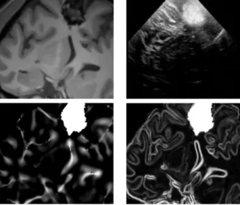
where n is the number of bits of the unsigned encoded image. For preoperative MR image V , the evaluation of f is done prior to surgery and is based both on the Mlvv operator and the manual segmentation of the pathological tissue performed by the neurosurgeon. The Mlvv operator is used instead of gradient operator because it is more specific than the gradient operator extracting relevant features that are present in both MR and US images (see comparison of Mlvv and gradient operator Fig. 1). Thus, f is defined as :
p(X ∈ ΦM R) = M lvv(V (X))/2n if X ∈ M 1 Ψ(X) if X ∈ M2 0 otherwise (5)
where M1represents the positive value of Mlvv map
com-puted on the brain tissue without the pathological tissue seg-mentation M2. The Mlvv operator is defined in 3D as :
M lvv(V (x, y, z)) = −2k ~wk1 2 h∂V(X) ∂x 2 “∂2V(X) ∂y2 + ∂2V(X) ∂z2 ” − 2 ∂V(X) ∂y ∂V(X) ∂z ∂2V(X) ∂y∂z + ∂V(X) ∂y 2“ ∂2V(X) ∂x2 + ∂2V(X) ∂z2 ” − 2 ∂V(X) ∂x ∂V(X) ∂z ∂2V(X) ∂x∂z + ∂V(X) ∂z 2“ ∂2V(X) ∂x2 + ∂2V(X) ∂y2 ” −2 ∂V(X) ∂x ∂V(X) ∂y ∂2V(X) ∂x∂y i (6) where k ~wk2 = ∂V(X) ∂x 2 + ∂V∂y(X)2+ ∂V∂z(X)2. Ψ(X) is the probability given to X in the segmentation of pathological tis-sue M2and Ψ() is used to incorporate a priori on pathology.
For hyperechogenic pathological tissue such as cavernoma or low-grade glioma Ψ(X) is high. For hyporechogenic patho-logical tissue such as necrosis or cyst Ψ(X) is low. In other
words, the Mlvv is computed from MR image on the extrac-ted brain by masking the pathological tissue. Then, only the positive values (i.e. the sulci and the falx) are kept. Finally, the Mlvv map is fused with the manual segmentation M2
(see Fig. 1).
2.3. Validation framework
Our approach was compared with three other approaches : (1) the Mutual information (MI), (2) the Normalized Mutual Information (NMI) on the gradient images of MR and US images as described in [8] and (3) the Correlation-Ratio on map extracted from MR (viewed as a “pseudo-US”) and the US image. The latter can be considered similar to the method described in [7]. The simplex was used as optimization pro-cedure within a multiresolution scheme for all the compared approaches.
3. MATERIAL 3.1. Data acquisition
T1 sense 3D sequences were used to acquired preopera-tive T1-weighted MR image on a 3T Philips Gyroscan scan-ner. The manual segmentation of the pathological tissue was performed before the surgery. After skull stripping from [11], the Mlvv was computed on the brain tissue denoised with the Non Local Means filter [12].
During the IGNS procedure, the US probe (Sonosite cra-nial 7 − 4MHz probe) was tracked by the Polaris cameras of the Stealth Station TREON (Medtronic Inc). The Sononav software installed on neuronavigator acquired the 2D B-scans and the probe positions. From the 2D B-scans and their posi-tions, a 3D volume was reconstructed with the Probe Trajec-tory method introduced in [13].
3.2. MR-US registration of the neuronavigation system
At the beginning of the neurosurgical procedure, the co-ordinate system of the preoperative MR image and the coor-dinate system of the intraoperative field are related by a rigid registration. Performed by the neuronavigator, this rigid re-gistration is based on surface matching between preoperative MR image and the position of points acquired on the patient’s head with the position pointer.
3.3. Pathology of the patients
In this study, hyperechogenic pathologies such as caver-noma (patient 1, see Fig. 2 and patient 2, see Fig. 3) and low-grade glioma (patient 3, see Fig. 4) have been chosen. For the experiments, we have tested the simplest model for Ψ(X). According to the neurosurgeron, the lesional tissue was consi-dered both homogenic and hyperechogenic; thus Ψ(X) was
Fig. 1. Top. Left : the denoising MR images. Right : the
cor-responding US images obtained with the registration perfor-med by the neuronavigation system. Bottom. Left : the pro-bability map based on Mlvv extracted from denoising MR image. Right : the same approach with gradient magnitude extracted from denoising MR image. Visually, the Mlvv is more specific to extract relevant structure of US image from MR image.
set to 1 (see Eq. 5). Example of intraoperative image and pro-bability map for patient 2 are presented in Fig. 1. For each pa-tient, two sequences were acquired before opening the dura. As it is generally admitted [8], we assumed that no brainshift occurs before opening of the dura.
4. RESULTS
Method Patient 1 Patient 2 Patient 3
MI - - - + -
-NMI on gradients - - -
-CR on maps - - + + -
-Our approach + + + + + +
Table 1. Results of the different MR/US registration methods.
For each patients two B-scans sequences were studied. The registration was classified as successful (+) if visually the re-gistration improves the overlay of the images given by neuro-navigator, or unsuccessful (-) in the other cases.
Tab. 1 shows the result of the registration procedures for the different similarity measures. These results underline that MI is not adapted for this kind of multimodal registration. Our implementation of the method proposed in [8] failed on all cases, maybe due to the acoustic shadows present in the data. The correlation ratio on maps converged when the
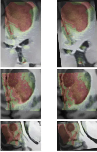
segmenta-Fig. 2. Patient 1. Left : registration given by the
neuronavi-gation system. Right : the result after correction with our re-gistration approach. For this case, even if the lesion was not entirely included in the US volume, the proposed registration procedure converged.
tion of the pathology is entirely included in the US volume. Nonetheless, for patient 1 and 3, only a part of the lesion inter-sects the US volume. In these cases, this approach failed. The results obtained with our methods are presented in Fig 2, 3 and 4. The proposed approach converged in all cases and was visually accurate. The registration procedures take less than two minutes on Pentium(R) M at 2GHz. A priori defining in the preoperative MR image the most probable locations of high intensity in the intraoperative US images seems to be an efficient way to register intraoperative US and preoperative MR brain images.
5. CONCLUSION
This paper presents a new framework for the 3D rigid re-gistration of US and MR brain images. This framework is based on the probabilistic objective function of the hyper-echogenic brain structures extracted from MR images. The experiments performed on 3 patients show that our approach converges robustly compared to methods such as the Mutual Information (MI), the Normalized MI on the gradient images or the Correlation Ratio on the US images and the pseudo-US created from MR images. The computational burden re-quired for our method is compatible with intraoperative use. The visually accuracy of our methods has been shown but its quantitative accuracy needs to be futher investigated.
Fig. 3. Patient 2. Left : registration given by the
neuronaviga-tion system. Right : results after correcneuronaviga-tion with our registra-tion approach. In this case, the acoustic shadow artifact was present on the US image. The signal below the lesion was to-tally dark. The proposed approach allowed to overcome these artifacts without specific detection of the shadows.
6. REFERENCES
[1] A. Roche, X. Pennec, G. Malandain, and N. Ayache, “Rigid registration of 3D ultrasound with MR images : a new approach combining intensity and gradient information,” IEEE TMI, vol. 20, no. 10, pp. 1038–1049, October 2001.
[2] N. Hata, M. Suzuki, T. Dohi, H. Iseki, K. Takakura, and D. Hashimoto, “Registration of ultrasound echography for intraoperative use : a newly developed multiproperty method,” in SPIE, 1994, vol. 2359, pp. 251– 259.
[3] D. G. Gobbi, R. M. Comeau, and T. M. Peters, “Ultrasound/MRI Over-lay with Image Warping for Neurosurgery,” in MICCAI. 2000, vol. 1935 of LNCS, pp. 106–114, Springer.
[4] A. P. King, J. M. Blackall, G. P. Penney, P. J. Edwards, D. L. G. Hill, and D. J. Hawkes, “Bayesian estimation of intra-operative deformation for image-guided surgery using 3-D ultrasound,” in MICCAI. 2000, vol. 1935 of LNCS, pp. 588–597, Springer.
[5] B. C. Porter, D. J. Rubens, J. G. Strang, J. Smith, S. Totterman, and K. J. Parker, “Three-dimensional registration and fusion of ultrasound and MRI using major vessels as fiducial markers,” IEEE TMI, vol. 20, no. 4, pp. 354–359, April 2001.
[6] I. Reinertsen, M. Descoteaux, S. Drouin, K. Siddiqi, and D. L. Collins, “Vessel driven correction of brain shift,” in MICCAI. 2004, vol. 3217 of LNCS, pp. 208–216, Springer.
[7] T. Arbel, X. Morandi, M. Comeau, and D. L. Collins, “Automatic non-linear mri-ultrasound registration for the correction of intra-operative brain deformations,” in MICCAI. 2001, vol. 2208 of LNCS, pp. 913– 922, Springer.
[8] M. M. J. Letteboer, M. A. Viergever, and W. J. Niessen, “Rigid regis-tration of 3D ultrasound data of brain tumours,” in CARS, 2003, pp. 433–439.
Fig. 4. Patient 3. Left : registration given by the
neuronaviga-tion system. Right : the result after correcneuronaviga-tion with our regis-tration approach.
[9] G.P. Penney, J.M. Blackall, M.S. Hamady, T. Sabharwal, A. Adam, and D.J. Hawkes, “Registration of freehand 3d ultrasound and magnetic resonance liver images,” Medical Image Analysis, vol. 8, pp. 81–91, 2004.
[10] G. Le Goualher, C. Barillot, and Y. Bizais, “Three-dimensional seg-mentation and representation of cortical sulci using active ribbons,” Int.
J. of Pattern Recognition and Artificial Intelligence, vol. 11, no. 8, pp.
1295–1315, 1997.
[11] J.-F. Mangin, O. Coulon, and V. Frouin, “Robust brain segmentation using histogram scale-space analysis and mathematical morphology,” in MICCAI. 1998, vol. 1496 of LNCS, pp. 1230–1241, Springer Verlag. [12] P. Coup´e, P. Yger, and C. Barillot, “Fast Non Local Means Denoising for 3D MR Images,” in MICCAI. 2006, vol. 4191 of LNCS, pp. 33–40, Springer.
[13] P. Coup´e, P. Hellier, N. Azzabou, and C. Barillot, “3D Freehand Ultra-sound Reconstruction based on Probe Trajectory,” in MICCAI. 2005, vol. 3749 of LNCS, pp. 597–604, Springer.