UNIVERSITE CATHOLIQUE DE LOUVAIN UNIVERSITE DE MONTREAL BELGIUM CANADA
EXPLORING THE NEURAL ENTRAINMENT TO MUSICAL RHYTHMS AND METER:
A STEADY-‐STATE EVOKED POTENTIAL APPROACH
Sylvie NOZARADAN, MD Promotors : Dr. André Mouraux (UCL)
Dr. Isabelle Peretz (UdeM)
Dissertation submitted for the degree of Doctor of Philosophy, Faculty of Medicine March 2013
Ces années de thèse font sans doute partie de mes meilleurs moments de vie. Elles ont été pour moi l’occasion de découvrir une passion pour la recherche, et de combiner mes centres d’intérêts, a priori éloignés. Mais cela n’aurait pas été possible sans l’encadrement optimal dont j’ai bénéficié durant ces années
Tout d’abord, c’est toujours une rare chance de croiser sur sa route un mentors. Je mesure la chance que j’ai eue d’en recontrer dans le cadre de ma thèse. Mes remerciements vont donc en premier à André, mon superviseur à l’UCL. Etre sa première doctorante est un grand honneur, dont j’espère avoir été à la hauteur. Je n’exagère pas en affirmant qu’il a été, et restera sûrement, un modèle, tant au niveau de ses très nombreux talents dans chaque étape du travail de chercheur qu’au niveau de ses qualités humaines. Associée dans cette supervision en équipe, je dois beaucoup à Isabelle, mon superviseur à Montréal, de m’avoir permis de découvrir au Brams, déjà avant la fin de mes études de médecine, la richesse de cette thématique de recherche. Je la remercie chaleureusement pour son soutien, ses encouragements et sa confiance depuis le début.
Je remercie vivement les membres de mon comité d’accompagnement et du jury, Pascal Kienlen-‐ Campard, Philippe Lefèvre, Marcus Missal, Alexandre Zénon, Charles Schroeder et Marc Schoenwiesner, pour leur lecture attentive de la thèse, ainsi que pour leurs commentaires et conseils tout au long du parcours. Je remercie aussi Etienne, Julie et Alex, ainsi que leur équipe, pour leur écoute et commentaires lors des journal clubs auxquels j’ai pu presenter mes travaux dès le début de la thèse, ainsi que leur bons conseils.
Je ne garderais sûrement pas un aussi bon souvenir de ces années si je n’avais eu la chance de réaliser cette thèse dans une si bonne ambiance. Je remercie donc les collègues de Nocions (dont Elisabeth, la plus agréable collègue de bureau qui soit!), et de COSY, dont le sourire et la bonne humeur sont autant de cadeaux que j’ai reçus chaque jour et qui vont me manquer. Que de bons souvenirs des conférences SfN, des verres du vendredi, de la campagne de vol… Merci également d’avoir été les meilleurs sujets d’expérience qui soient. Un grand merci aussi à l’équipe administrative et technique de COSY pour leur soutien et leur efficacité. J’ai eu la chance de trouver du côté Brams une ambiance tout aussi chaleureuse, qui aura rendu Montréal particulièrement difficile à quitter chaque fois. J’espère avoir encore de nombreuses occasions d’y séjourner et de collaborer avec le Brams.
Merci aussi à Vincent (aussi un de mes sujets d’expérience favoris!), Arnould, Leon van Noorden et le groupe Rhythm de Bruxelles, ainsi qu’aux chercheurs de la communauté Rhythm (RPPW et PoRT) pour ces échanges inspirants dans une ambiance amicale. La tonalité de ces rencontres est à cultiver, et a déjà débouché sur de belles amitiés. A propos d’amitié, je remercie bien sûr aussi Jessica Phillips-‐Silver et Coralie de Hemptinne, qui toutes deux ont fait preuve de patience pour m’accompagner dans mes premiers pas au labo, au Brams et à l’UCL. Je remercie aussi chaleureusement Marc Crommelinck, éternel créateur d’enthousiasme, qui m’a mis entre les mains, dès la 2ème candi, des lectures de neuroscience passionnantes (que je ne finis pas d’essayer de comprendre, alias Edelman!), et Bruno, qui après Marc a essayé de me les faire comprendre à son tour. Son enthousiasme ne cesse de m’inspirer depuis.
Ces réflexions sur la musique ne me sont pas tombées du ciel. Je remercie donc très chaleureusement ici mes mentors du côté musique, Françoise, Burkard, Jean-‐Claude, Santi et Caroline, qui continuent de m’inspirer un modèle de rigueur au travail, de curiosité et de créativité, qui s’applique si bien tant au métier d’artiste qu’à celui de chercheur.
Et bien sûr, un énorme merci à mon copain et ma famille de m’avoir supporté (au sens français et anglais du terme!) dans ce cheminement, et pour toute la force qu’ils me donnent chaque jour.
SUMMARY ... 9
FOREWORD...11
I. ENTRAINMENT IN NEURAL SYSTEMS ...13
I.1. ENDOGENOUS OSCILLATIONS ... 13
I.1.1. Spontaneous neural oscillations... 14
I.1.2. Frequency tuning function... 16
I.1.3. Synchronization. ... 17
I.1.4. A link between synchronization of oscillatory activities and brain function... 17
I.2. ENTRAINMENT TO OSCILLATORY INPUTS... 20
I.2.1. Synchronous oscillation to sound envelope in the auditory system ... 20
I.2.1.1. What is sound envelope? ... 20
I.2.1.2. Frequency decomposition of the sound... 21
I.2.1.3. Temporal coding versus rate coding. ... 23
I.2.2. The steady-‐state evoked potential approach ... 27
I.2.2.1. Nature of the steady-‐state evoked potentials... 28
I.2.2.2. Frequency tagging. ... 29
I.2.2.3. Methodological considerations... 30
II. ENTRAINMENT TO MUSICAL RHYTHMS ...35
II.1. BEAT IN MUSIC: A UNIVERSAL HUMAN ABILITY?... 35
II.1.1. Are beat and meter induced in all musical styles? ... 35
II.1.2. Language and music rhythms... 37
II.1.2.1. Rhythm in speech... 37
II.1.2.2. Music mimicking speech rhythms and vice versa... 38
II.1.3. Cultural differences ... 39
II.1.3.1. The Groove as a cultural specificity... 40
II.1.3.2. Meter, binary bias and integer ratios across cultures... 40
II.1.4. Human development ... 48
II.1.4.1. Production... 48
II.1.4.2. Perception. ... 49
II.1.4.3. Enculturation... 52
II.1.5.1. Innateness. ... 55
II.1.5.2. Domain-‐specificity and inter-‐individual differences... 55
II.1.5.3. Human specificity. ... 58
II.2. SENSORIMOTOR COUPLING ... 64
II.2.1. Production of periodic signals ... 64
II.2.2. Perception of periodic signals... 67
II.2.2.1. Models and empirical evidence on time processing. ... 67
II.2.2.2. Neural correlates of time processing. ... 69
II.2.2.3. Rhythm processing models. ... 74
II.2.2.4. Beat induction. ... 81
II.2.2.5. Neural correlates of rhythm processing... 86
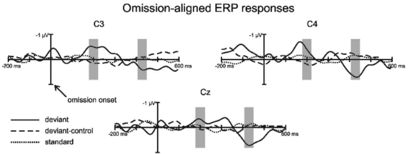
II.2.2.6. Tagging the neural entrainment to beat and meter: our novel approach ... 103
II.2.2.6.1. Study 1: TAGGING THE NEURONAL ENTRAINMENT TO BEAT AND METER (Sylvie Nozaradan, Isabelle Peretz, Marcus Missal, André Mouraux)... 103
II.2.2.6.2. Study 2: SELECTIVE NEURONAL ENTRAINMENT TO THE BEAT AND METER EMBEDDED IN A MUSICAL RHYTHM (Sylvie Nozaradan, Isabelle Peretz, André Mouraux) ... 124
II.2.3. Coupling of sensorimotor periodic signals... 156
II.2.3.1. Characteristics of sensorimotor synchronization to the beat. ... 156
II.2.3.2. A frequency tuning function for beat perception and synchronization. ... 160
II.2.3.3. Neural correlates of sensorimotor synchronization... 170
II.2.3.4. Study 3: CAPTURING WITH EEG THE NEURONAL ENTRAINMENT AND COUPLING UNDERLYING SENSORIMOTOR SYNCHRONIZATION TO THE BEAT (Sylvie Nozaradan, Younes Zerouali, Isabelle Peretz, André Mouraux) ... 179
II.2.3.5. Perception leading to movement... 212
II.2.3.6. Movement influencing perception... 214
II.2.3.7. Auditory prominence for sensorimotor coupling... 216
II.2.3.8. Study 4: STEADY-‐STATE EVOKED POTENTIALS AS AN INDEX OF MULTISENSORY TEMPORAL BINDING (Sylvie Nozaradan, Isabelle Peretz, André Mouraux) ... 220
III. DISCUSSION AND PERSPECTIVES ...245
III.1. STUDY 1: TAGGING THE NEURONAL ENTRAINMENT TO BEAT AND METER ... 245
III.1.1. Making a bridge between beat and meter-‐related SS-‐EPs, transient ERPs and ongoing oscillatory activities. ... 246
III.1.2. Some remarks on the frequency domain analysis of SS-‐EPs... 248
III.1.3. Musicians versus nonmusicians... 248
III.1.4. Beat and meter-‐related SS-‐EPs and head movement artifacts... 249
III.2. STUDY 2: SELECTIVE NEURONAL ENTRAINMENT TO THE BEAT AND METER EMBEDDED IN A MUSICAL RHYTHM ... 250
III.2.1. The neural sources of beat and meter-‐related SS-‐EPs... 251
III.2.2. Similarities between pitch and meter processing... 253
III.2.3. Retrieving time resolution and phase from SS-‐EPs... 254
III.3. STUDY 3: CAPTURING WITH EEG THE NEURONAL ENTRAINMENT AND COUPLING UNDERLYING SENSORIMOTOR SYNCHRONIZATION TO THE BEAT ... 256
III.4. STUDY 4: STEADY-‐STATE EVOKED POTENTIALS AS AN INDEX OF MULTISENSORY TEMPORAL BINDING .. 257
III.4.1. Cross-‐frequency modulation studied using frequency tagging. ... 258
III.4.2. Is periodicity special?... 258
IV. GLOSSARY...261
IV.1. ENTRAINMENT IN PHYSICS, BIOLOGICAL SYSTEMS AND NEUROSCIENCE... 261
IV.2. MUSICAL TERMS... 265
SUMMARY
The ability to perceive a regular beat in music and synchronize to it is a widespread human skill. Fundamental to musical behavior, beat and meter refer to the perception of periodicities while listening to musical rhythms, and usually involve spontaneous entrainment to move on these periodicities. However, the neural mechanisms underlying entrainment to beat and meter in Humans remain unclear. The present work tests a novel experimental approach, inspired by the steady-‐state evoked potential method, to explore the neural dynamics supporting the perception of rhythmic inputs. Using human electroencephalography (EEG), neural responses to beat and meter were recorded in various contexts: (1) mental imagery of meter, (2) spontaneous induction of a beat from rhythmic patterns, (3) multisensory integration, and (4) sensorimotor synchronization. Our results support the view that entrainment and resonance phenomena subtend the processing of musical rhythms in the human brain. Furthermore, our results suggest that this novel approach could help investigating the link between the phenomenology of musical beat and meter and neurophysiological evidence of a bias towards periodicities arising under certain circumstances in the nervous system. Hence, entrainment to music provides an original framework to explore general entrainment phenomena occurring at various levels, from the inter-‐neural to the inter-‐individual level.
FOREWORD
One of the richest features of music is its temporal structure. In particular, the beat, which usually refers to the perception of periodicities while listening to music, can be considered as a cornerstone of music and dance behaviors. Even when the music is not strictly periodic, humans perceive periodicities and are spontaneously entrained to move on these periodicities. Moreover, the beat can be grouped or subdivided in meters, which correspond to harmonics or subharmonics of the beat frequency (as in a waltz, which is a three-‐beats meter) (see also Glossary).
Getting entrained to music is an extremely common human activity, shared by humans of all cultures. It is a highly complex activity, which involves auditory (and also visual, proprioceptive and vestibular) perception, attentional capacities, as well as motor synchronization, performance and coordination. Hence, it is not surprising that a large network of brain structures is involved in entrainment to music, and that there is a growing interest in understanding the functional and neural mechanisms of the entrainment to music.
One of the major goals of this dissertation was to narrow the gap between scientific studies on neural entrainment on the one hand and entrainment to musical rhythms on the other hand. In both, entrainment processes and biases towards periodicity have been described as fundamental functional characteristics. Considering this, we tested whether periodicities induced by musical rhythms could entrain neural activities at frequencies corresponding to these periodicities.
In the present work, we have used the electroencephalogram (EEG), a technique particularly well suited to study a system that changes dynamically over short periods of times. We
developed an original EEG approach to capture the processing of beat and meter periodicities. This approach is based on the long-‐standing observation that when the brain is stimulated periodically, it synchronizes its activity to the inputs and produces periodic output (Lunel & Van der Tweel, 1965 ; Regan, 1966). This neural activity can be captured objectively in the form of a steady-‐state evoked potential (SS-‐EP) identified by analyzing the EEG in the frequency domain.
Our experiments show the interest of this approach to study various aspects of beat perception in normal individuals: elicited by mental imagery paced onto periodic sounds (Nozaradan et al., 2011), emerging spontaneously when listening to rhythmic patterns (Nozaradan et al., 2012), elicited by sensorimotor synchronization to the beat (Nozaradan et al., in revision), and finally, elicited by simultaneous auditory and visual beats which were temporally congruent or not (Nozaradan et al., 2012).
Several terms, either from the neural oscillation or the musical rhythm literatures, are recurrent in the present work. For a definition of these terms, as well as a description of some important concepts related to these terms, the reader is referred to the section Glossary.
The present work is attached with several media files. These are the stimuli of Studies 1 to 4, and also audio tracks which illustrate some of the musical aspects addressed in the theoretical parts.
I. ENTRAINMENT IN NEURAL SYSTEMS
Part I of the present thesis reviews the evidence supporting the view that our neural system is biased towards periodicity, under certain circumstances at least, and can act in some contexts as multiple coupled oscillators. This question is of particular interest in regard to research on rhythm, pulse and meter perception. Indeed, in order to explain the underlying mechanisms that lead to this ubiquitous human ability, a theoretical model of resonance for pulse and meter (see Section II.2.2.3.3.) has proposed to link the phenomenology of pulse and meter with the concepts of neural oscillation (Large and Kolen, 1994; Large, 2008). The basic idea of this model is that some neural oscillations, possibly dispersed across cortical and subcortical areas and spanning a range of natural frequencies for beat and meter induction in music, entrain to the rhythm of the auditory sequence.
Following a review of the possible tendencies towards periodicity and entrainment in the activity of neurons (Section I.1), we will review the evidence of neural entrainment in the particular case of synchronization to oscillatory inputs (Section I.2). We will then discuss the neurophysiological evidence for entrainment in the auditory system (Section I.2.1), whose stimulation forms can be seen as oscillatory in nature. Finally, we will focus on SS-‐EPs (Section I.2.2), an electrophysiological method making a specific use of periodic repeated stimulation to tag brain activity and which inspired the present experimental work.
I.1. ENDOGENOUS OSCILLATIONS
There is a large amount of evidence for rhythmic neural activities. To characterize these dynamic patterns, the term “oscillation” was first mentioned by Hans Berger in 1929, to describe cyclical fluctuations of the electrical currents of the human scalp at approximately
10 Hz. This electrical activity was enhanced when participants closed their eyes and constituted the first description of the alpha band, an ongoing neural activity between 8 and 12 Hz typically enhanced when the eyes are closed (see e.g., Klimesch, 1999, for a review). Since this seminal observation, numerous studies have explored the relationship between dynamic patterns recorded with EEG or other techniques, and behavioral states.
The human brain, with its numerous connections between areas, displays low-‐frequency and fast rhythmic patterns grouped within complex wave-‐sequences (Steriade, 2006). Some of these oscillations are due to intrinsic neuronal properties, while others arise from the large interconnections of neurons across distant brain areas. From this perspective, the mechanisms underlying oscillatory activities, synchronization across neurons and the emergence of a frequency tuning function within one neuronal population may be interpreted as different aspects of a common phenomenon.
I.1.1. Spontaneous neural oscillations. Two mechanisms at least have been proposed to explain the oscillatory behavior of neuronal discharge: (1) the mutual interconnection between an excitatory neuron and an inhibitory interneuron, or between two inhibitory interneurons, and (2) the pacemaker neuron.
The hypothesis of an oscillatory activity emerging from a mutual interconnection of at least two cells including an inhibitory neuron was proposed for the first time to account for the fast oscillations (between 6 and 10 Hz) observed in the rat hippocampus (Wang and Buzsaki, 1996). Such network models of oscillatory activity were further studied in the context of central pattern generators. In many animal species, functional units of a few cells, located in the spinal cord, have been shown to generate continuous periodic activity responsible for automatic movements such as locomotion in many animal species (Marder and Bucher, 2001). In these network models, two neurons reciprocally inhibit each other. When isolated,
these neurons do not fire in repetitive bursts. However, when they are coupled, they produce alternating patterns of activity (Fig. I.1.1.). The transition between activated and inhibited states occurs via various mechanisms. For instance, if the neuron shows spike-‐ frequency adaptation, the active neuron may slow down or stop firing, thus releasing the other neuron from inhibition. Alternatively, the inhibited neuron may escape from inhibition due to its intrinsic membrane properties and, in turn, activate or inhibit the first inhibiting neuron. This postinhibitory rebound has been shown to be crucial for the timing of firing of the central pattern generator unit (Marder and Bucher, 2001; Calabrese, 1998). By extension, similar mechanisms have been described to explain the oscillatory activity of thalamic neurons as well as neurons in the globus pallidus for instance (Bevan et al., 2002), based on the interplay between low-‐threshold excitatory calcium current and burst of GABA-‐ mediated inhibition.
According to the second kind of mechanism, some neurons are intrinsically rhythmic, and fire either endogenously or in response, for instance, to neuromodulatory substances such as neurotransmitters. Examples of such neurons have been observed in the inferior olive. Neurons of this structure exhibit sustained oscillatory activity that are generally observed between 4 and 10 Hz and are explained by the interplay between various ionic currents and their particular dynamics across the membrane (Bal and McCormick, 1997). Hence, when they receive stimulation, the dynamic of their responses lie within a narrow frequency range that coincides with their natural frequency of resonance, such that the transient response to the transient input takes the form of a transient oscillation. Neurons that are strongly oscillatory can provide important timing inputs for neuronal networks, by driving neurons that are not themselves intrinsically rhythmic (Fig. I.1.1.). However, they are more difficult to entrain or reset, except within a small frequency range.
Figure I.1.1. From Marder and Bucher (2001). Upper panel. Rhythmic network based on the coupling between a pacemaker neuron (in red) and a non pacemaker neuron. Bottom panel. Rhythmic network based on the reciprocal inhibition between two non rhythmic neurons.
I.1.2. Frequency tuning function. The frequency range in which sustained oscillatory activities are observed is determined by structural aspects, acting as bandpass filters, at the level of the single neuron and the network.
Low-‐pass and high-‐pass filtering is mainly constituted by timing constraints due to the conductance (which can be defined as the ease at which an electric current crosses the membrane) and capacitance (which can be defined as the ability of the neuron to store an electric charge) of the neuronal membrane (Hutcheon and Yarom, 2000). The combination between low-‐pass and high-‐pass filtering properties determines the frequency tuning function of individual neurons. If the dynamic activity is limited to a narrow frequency range, it results in an almost periodic activity, as found in pacemaker neurons for instance.
Moreover, this leads to cases where neurons would show a quasi-‐periodic discharge in response to non-‐periodic input such as white noise (Joris et al., 2004).
In addition, the frequency tuning function of neural oscillatory behaviors can also result from the physical architecture of neuronal networks and the limited speed of neuronal communication due to axon conduction and synaptic transmission (Buzsaki and Draguhn, 2004). That is, the size of the synchronous group also influences the period of oscillation. Higher frequency oscillations can involve a small neuronal space, whereas very large networks are only able to synchronize to slow oscillations. Hence, the frequency tuning function of the network is determined by both the properties of the individual neuron, and the properties of its interconnections.
I.1.3. Synchronization. It is generally assumed that integration of information requires synchrony, or coincidence, of convergent inputs (Buzsaki and Draguhn, 2004). Synchrony is defined as the simultaneous occurrence of activity in two or more cells (see the Glossary for more details on the concept of synchrony). Oscillation-‐based synchrony is thought to be the most efficient physical mechanism for temporal coordination (Pikowski et al., 2001). Oscillatory synchronization, as a synonym with entrainment, can be achieved through networks that include pacemaker or inhibitory processes (Fig. I.1.1.), thus emerging from the synaptic connections and their intrinsic properties.
I.1.4. A link between synchronization of oscillatory activities and brain function. Given the diversity of the voltage-‐dependent channels and the intrinsic properties of the cellular membrane within the whole brain, it is likely that every neuron has a resonance curve and the potential to exhibit oscillatory activity under certain circumstances. Whether resonance and oscillatory synchrony in neurons are simply epiphenomena or whether they are used to integrate and communicate information is still debated (Hutcheon and Yarom, 2000).
However, several mechanisms can be proposed to explain the advantages for neural systems to act as coupled oscillators.
First, oscillatory behaviors in neuronal groups may influence the response chronometry of the oscillating neurons, because their excitability becomes phase dependent (Llinas, 1988, Hutcheon and Yarom, 2000; Engel et al., 2001). The oscillatory fluctuation of the membrane potential of a given neuron creates predictable time windows during which the neuron is more likely to respond to external input. If the input occurs at an inappropriate time according to the excitatory phase, the neuronal response is dampened and/or delayed. Second, oscillatory synchronization may act as a filter and amplificator of the inputs. The amplification can be explained by the beacon effect. At equal input strength of each upstream neuron, the impact of the inputs ensemble is greater on the target cell when the inputs are synchronous. The filtering of the inputs is achieved based on the excitatory phase of the target neuron, but also based on the frequency of the stimulation. The inputs are thus selected when they fall within the frequency preference of the neuron, according to its intrinsic resonant oscillatory features or the resonant properties of the network to which the neuron is interconnected. This would thus determine the “sampling rate” of the neural network for a given input (see also Section I.2.2.1.).
Third, oscillatory synchronization could serve as a mean to bind cell assemblies. This is based on the assumption that information in the brain is processed, transferred and stored by flexible cell assemblies. These assemblies are defined as neuronal groups that are transiently synchronized (Edelman, 1978; 1989). Indeed, the binding of the neurons may depend on the coupling strength, itself influenced by the distribution of the resonant frequencies of the individual neurons of the group. As long as the frequencies of the coupled oscillators remain similar, synchrony can be sustained even with very weak synaptic links (Buzsaki and
Draguhn, 2004; Engel et al., 2001; Varela et al., 2001). This flexibility based on oscillatory synchronization is hypothesized to play a role in learning. In the rat hippocampus, a structure thought to play a crucial role in memory, brief pulse trains delivered at the peak of the neuronal oscillations induce long-‐term potentiation, whereas the same train applied out-‐ of-‐phase weaken the previously strengthened inputs (Csicsvari et al., 2003).
I.2. ENTRAINMENT TO OSCILLATORY INPUTS
In the previous section, we briefly reviewed the neurophysiological bases of the natural propensity of neurons to generate oscillatory activity, and the possible role of this oscillatory behavior in brain function. The present section addresses the question of how neurons behave in contact to external inputs that are themselves oscillatory (auditory stimuli). Finally, we will examine how such repeated stimuli can be used to “tag” neural processes using electrophysiology (steady-‐state evoked potential approach).
I.2.1. Synchronous oscillation to sound envelope in the auditory system
I.2.1.1. What is sound envelope? Acoustic stimuli contain multiple temporal dimensions. They can be summarized in at least two components, the “fine structure” and the “envelope”, which are usual terms to describe waveforms in physics. In acoustic, the fine structure is determined by the fast pressure variations corresponding to the spectral content of the sound. The processing of fine structure is involved in pitch perception, which can be defined as the perceptual phenomenon of sounds organized within a scale from low to high tones (Schnupp et al., 2010). The fine structure is itself modulated in amplitude, and the dynamic of this amplitude modulation constitutes the sound envelope. In humans, amplitude modulations produce various hearing sensations depending on the modulation frequency. Rhythms and fluttering, as well as most amplitude modulation frequencies found in ordinary speech for instance, correspond to envelope frequencies up to 20 Hz whereas roughness and pitch correspond to amplitude modulation frequencies above 20 Hz. As we will see, it has been proposed that this perceptual boundary may be related to a change in the coding form of these sound inputs (Eggermont, 2001).
Whether envelope processing is embedded in the auditory system is an important question, regarding the ecological prominence of low frequency sound modulations. Indeed, low-‐ frequency amplitude modulations (beneath 20 Hz) are prominent in acoustic natural environments (Nelken, 1999), and contain essential information, particularly for vocalization. For example, they have been shown to be necessary and almost sufficient for speech intelligibility (Shannon, 1995). This was evidenced by comparing speech intelligibility when manipulating the speech signal either by blurring the frequency content of the signal corresponding to the fine structure while keeping intact the frequency content up to 20 Hz, or by doing the inverse manipulation. Regarding the topic of the present thesis, the dynamics of amplitude modulation of the sound specifically beneath 5 Hz, constitutes a crucial, although not unique, cue for beat and meter perception. Hence, in the four experiments reported in the present work, the auditory stimuli have been designed such as to induce the beat and meter exclusively based on the dynamics of amplitude modulation of a pure tone.
I.2.1.2. Frequency decomposition of the sound. It is generally assumed that sounds are processed in the nervous system by cells responding to a specific frequency band. Already within the cochlea, these groups of cells are functional units defined by the strong correlation in the firing of individual cells in response to a preferred frequency band of stimulation. For this reason, these neuronal groups are often compared to mechanical filters, or a filterbank organized in a tonotopic map. This array of band-‐pass filters is assumed to decompose the sound input into several frequency bands according to the banwidth of frequency range to which each neuronal group responds preferentially (i.e., according to their frequency tuning function bandwidth). However, while a periodotopic organization of the cell groups encoding sound envelope has been widely observed in subcortical structures,
whether such functional arrangement exists at the level of the cortex is still debated. For example, the bandwidth of preferred frequency range appears to vary considerably across cortical neurons in the auditory cortex (Joris et al., 2004).
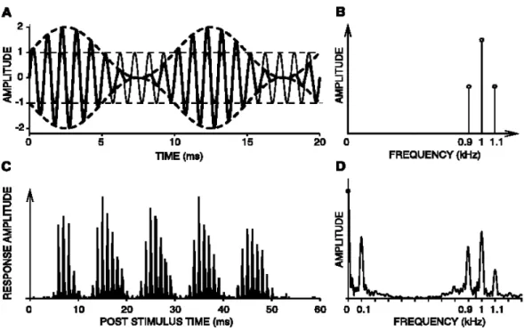
Importantly, groups of cells manifest nonlinear behaviors in the decomposition of the sound input that are crucial for sound envelope processing. Particularly, the cochlea exhibits patterns of responses similar to a demodulation function, which can be observed by comparing the spectrum of responses recorded in the cochlea to the spectrum of the eliciting stimulus, as represented in Figure I.2.1.2.
Figure I.2.1.2. From Joris et al. (2004). A. Superimposed waveforms of an unmodulated 1000 Hz tone (thin line) and the same tone sinusoidally amplitude modulated (thick line) at 100% depth with a frequency of 100 Hz. The dashed lines correspond to the sound envelope. B. Spectrum of the sound in A. C. Average nerve fiber response (poststimulus time histogram). D. Spectrum of the response. The activity at the carrier frequency and at sidebands indicates that there is a phase-‐locking to the fine structure of the sound. However, an additional component at the modulation frequency (0.1 kHz) emerges, compared to the sound spectrum.
I.2.1.3. Temporal coding versus rate coding. The first neural representation of the sound, and the sound envelope, is in the cochlea, as the first relay of the ascending auditory pathway (Fig. I.2.1.3.). Subsequently, this representation is conveyed by multiple, parallel pathways constituted by the auditory nerve to the cochlear nucleus. These parallel pathways, hypothesized to convey a detailed representation of the stimulus, diverge or converge further toward the inferior colliculus and finally the cortex (Fig. I.2.1.3.).
Figure I.2.1.3. Auditory pathway, from cochlea to cortex (copyright Allyn and Bacon, 2005).
Already at the level of the cochlea, auditory cells respond to sound envelope, as well as to the fine structure of sounds, in the form of firing patterns phase-‐locked to the temporal structure of the input. This temporal coding of information, referred to as envelope-‐locking in the context of sound envelope processing, contrasts with another form of encoding found extensively in the nervous system, based on the average firing rate. Referred to as the
average number of spikes per unit time, the rate coding assumes that the firing rate increases with increasing stimulus intensity or change in one stimulus feature.
Along the ascending auditory pathway, the sound envelope information is transmitted from the cochlea to the brainstem, through a principle of synchronization, or phase-‐locking, via the auditory nerve (Fig. I.2.1.3.). The temporal coding of sound envelope information is widely maintained across the subcortical structures, while expressed differently within each structure regarding the bandwidth of frequency tuning function of the cells groups, the possible gain modulations, etc. The temporal coding is then transformed, at least in part, in an average rate coding in structures as the superior olivary complex of the brainstem and in higher relays of the auditory pathway. On the whole, responses to amplitude modulation become more phasic than sustained at these stages. Sustained responses are transformed into on/off patterns of response that are hypothesized to serve several functions such as improving the signal-‐to-‐noise ratio under naturalistic listening or detecting changes in the stimulus content (Eggermont, 2001).
The transformation from a temporal coding to an average rate coding is assumed to occur due to the intrinsic and network properties of neurons that are no longer able to produce sustained frequency following. From this loss of synchrony with the stimulus dynamic, the transition to an average rate coding is performed for instance by a process of convergence of inputs from neurons differing in their response dynamics. The target neuron produces an output only if the timing and nature of the diverse combined inputs are such that the target neuron is depolarized strongly and quickly enough to reach threshold (Eggermont, 2001). This process thus requires coincident input from a relatively large number of input neurons. The diversity of the dynamic of the inputs makes this coincidence occurring transiently.
Importantly, numerous studies have brought evidence for a loss of sound envelope locking for modulation frequencies above 100 Hz in cortical areas (Eggermont, 2001). Indeed, as auditory information reaches progressively higher levels of processing, higher frequencies are progressively more represented using rate coding. However, frequencies below 100 Hz are still represented by means of temporal coding. The substantial lowpass filtering of the temporal coding of sound envelope is due, at least in part, to the fact that from the first to the last relays of the ascending auditory pathway, the neurons respond by temporal coding with a certain amount of temporal jitter. This jitter might be transmitted, and possibly amplified along the auditory pathway, making the transduction of high frequencies with sufficient temporal accuracy very limited.
The fact that, unlike the processing of tone, envelope processing at low frequencies remains largely represented as temporal coding at the level of the cortex constitutes an important argument in favor of the SS-‐EP approach to study rhythm processing. Indeed, the SS-‐EP approach might have been less adequate if the frequency range for rhythm perception was rate-‐encoded at the level of the cortex.
Nevertheless, as we will see, the perception of musical rhythm and meter do not only rely on the information conveyed by amplitude modulation, but also exploits harmonic structure, timbre modulations or even endogenous imagery of a temporal structure that can be imposed onto the sound (see Section II.2.2.4.1. for more details on the generation of metrical accents). In theory, one could hypothesize that these numerous features, processed by independent neurons, would be integrated within a unified representation corresponding to the percept of beat and meter. Such across-‐feature interactions may be hypothesized to emerge when the sound envelope and the other features set up widespread synchrony at
low frequencies across cortical neurons, thus adjusting to each other by synchrony of the periodic modulation of their responsiveness (Eggermont, 2001).
I.2.2. The steady-‐state evoked potential approach
The previous sections have briefly reviewed the evidence corroborating the view that neurons could show a natural propensity to generate oscillatory activity, and to synchronize to external inputs that are themselves oscillatory (auditory stimuli). Here, we examine how stimuli made oscillatory for experimental purpose can be used to “tag” neural processes using electrophysiology (steady-‐state evoked potential approach).
A large number of investigators have used non-‐invasive EEG techniques to study how the human brain processes external or endogenous inputs. The EEG signals recorded on the scalp lack spatial resolution. Indeed, the potentials measured at a given scalp position are not systematically determined by the activity of the cortical region located immediately underneath the electrode (Nunez and Srinivasan, 2005). However, it offers the advantage of measuring neural activity at the millisecond time scale.
The majority of studies have relied on the recording of event-‐related brain potentials (ERPs), i.e., changes in the ongoing electrical brain activity time-‐locked to a transient event, like the sudden onset of a sensory stimulus. In 1966, Regan introduced the approach of “steady-‐ state visual evoked potentials” (SSVEP) as an alternative to characterize stimulus-‐evoked activity in the ongoing EEG. Unlike conventional transient ERPs, which Regan described as “the response to a kick in the system”, SSVEPs reflect a sustained cortical response induced by the long-‐lasting periodic repetition of a feature in the input stimulation, described by Regan as “the response to a gentle shake of the system at a fixed repetition rate“ (Regan, 1989). Regan named this response “steady-‐state" because it remains virtually
constant in phase over time. Such responses have been described not only in the visual domain, but also in the auditory (Galambos et al., 1981), and somatosensory domains (Galambos, 1982). They will be referred in the present work as SS-‐EPs.
One of the advantages of the SS-‐EP approach is the prior knowledge of the frequency at which the neural response to the repeated stimulation should appear in the EEG, thus making the technique more objective. Moreover, since the response is expected to be concentrated within a very narrow frequency band, the technique has a very high signal-‐to-‐ noise ratio (Regan, 1989).
I.2.2.1. Nature of the steady-‐state evoked potentials. SS-‐EPs are thought to result from an entrainment or resonance of a population of neurons responding to the stimulus at the frequency of stimulation (Vialatte et al., 2010). An alternative to this view is that SS-‐EPs result from the linear superposition of independent transient responses elicited by the fast repetition of the stimulus (Regan, 1989; Capilla et al., 2011). Thus, how these activities emerge within the human EEG and their relationship with transient ERP and ongoing oscillatory activities remains a matter of debate.
Irrespective of the outcome of this debate, the SS-‐EP phenomenon appears to be caused by an increase in the neuronal response synchronization as a result of the presentation of a repetitive external input whose temporal presentation rate is close to the temporal activation cycle of a neuronal group. When a given input is repeated at fixed time intervals, it may force the group to respond at a certain rate, biasing the propagation of excitatory and inhibitory postsynaptic potentials. If the inter-‐stimulus interval coincides with the neuronal activation cycle, then a higher amount of neurons are available to respond to the input and can synchronize their response properly. This would result in an amplification of the output
along the targeted neural network, causing a noticeable increase in the amplitude of the signal registered on the scalp (Buszaki, 2006).
I.2.2.2. Frequency tagging. Another advantage of the SS-‐EP approach allows tagging responses to multiple inputs, based on their respective frequencies, and these inputs can even overlap each other spatiotemporally. Indeed, following Regan and Heron (1969), several studies have shown that different stimulation frequencies can be used to “tag” different inputs presented concurrently and, thereby, isolate the neural activity related specifically to the processing of each stream of stimulation (e.g., Morgan et al., 1996; Toffanin et al., 2009; Giani et al., 2012). For example, simultaneously presenting one stimulus modulated at frequency F1 and another stimulus modulated at frequency F2 elicits two distinct peaks in the EEG spectrum, at frequencies F1 and F2 (e.g., Chen et al., 2003). Based on this principle, the frequency-‐tagging method offers great advantage in studying for instance the processing of multisensory inputs, by disentangling the processing of each sensory input based on their respective frequencies of response (see Study 4 of the present thesis). Moreover, as movement-‐related SS-‐EPs can also be elicited by periodic hand movements (Gerloff et al., 1998), the frequency-‐tagging method may be suitable to study sensorimotor synchronization to a periodic input (see Study 3).
Importantly, when stimulating with two or more frequencies, peaks of activities can also emerge in the EEG spectrum at frequencies different from the frequencies of stimulation and their harmonics. These additional responses can appear at frequencies corresponding to the sum or difference of the stimulation frequencies (and/or their harmonics) (see also the Glossary). These crossmodulation products are hypothesized to result from the non-‐linear convergence or integration of the two input signals (Giani et al., 2012; Regan, 1989; Sutoyo and Srinivasan, 2009). The reader is referred to the Glossary for a more detailed description
of these concepts. The concept of crossmodulation is also addressed in Study 3 (Section II.2.3.4).
I.2.2.3. Methodological considerations. Several parameters of the periodic stimulation can be varied to test the response of the neural system experimentally.
I.2.2.3.1. Modulation waveforms. A large range of modulation waveforms, from pure sinusoids to square waves, can be used to elicit SS-‐EPs. The consistency of the neural response may be hypothesized to depend directly on the stimulation waveform. Indeed, if the neural system responds to the stimulation train based on the detection of periodic contrast changes in the stimulation, the neural assemblies would not respond in the case where the stimulus modulation is too slow to be perceived as a change in the input. In contrast, the abrupt contrast generated by a square waveform of stimulation should elicit more consistent neural responses at every stimulus with less jitter along time, compared to a sinusoidal waveform of stimulation.
However, a pure sinusoid waveform of stimulation could offer several advantages. First, in line with the view that SS-‐EPs constitute neural responses whose nature is distinct from transient ERPs, it can be hypothesized that a square wave train of stimulation elicits a larger amount of transient responses than a sinusoidal stimulation. This has been shown by comparing for instance the neural responses to a train of auditory clicks with a train of sinusoidal amplitude modulations of a sound (Draganova et al., 2002). Second, when stimulating with a pure sinusoid, the response is expected to result theoretically in a unique frequency in the EEG spectrum, corresponding to the frequency of the stimulation (Victor and Zemon, 1984). In reality, it is not systematically the case, as distinct frequency components at higher harmonics appear concomitantly in the EEG spectrum. Therefore, the appearance of harmonics in the EEG spectrum can be considered as the product of non-‐