Université de Montréal
EFFECT OF LEVODOPA ON CSTRIATAL AND C ORTICO-CEREBELLAR CIRCUITS IN PARKINSON’S DISEASE
par
KRISTINA MARTINU
DÉPARTEMENT DES SCIENCES BIOMÉDICALES FACULTÉ DE MÉDECINE
THÈSE PRÉSENTÉE À LA FACULTÉ DE MÉDECINE EN VUE DE L’OBTENTION DU GRADE PH.D.
EN SCIENCES BIOMÉDICALES
SEPTEMBRE 2012
Université de Montréal
Faculté des études supérieures et postdoctorales
Cette thèse intitulée:
EFFECT OF LEVODOPA ON STRIATAL AND CORTICO-CEREBELLAR CIRCUITS IN PARKINSON’S DISEASE
Présentée par : KRISTINA MARTINU
a été évaluée par un jury composé des personnes suivantes :
Dr. Pierre Rainville, président-rapporteur Dr. Oury Monchi, directeur de recherche
Dr. Richard Hoge, membre du jury Dr. Martin McKeown, examinateur externe Dre. Bernadette Ska, représentante du doyen de la FES
iii
Résumé
La maladie de Parkinson (MP) est la deuxième maladie neurodégénérative la plus commune. Les symptômes principalement observés chez les patients atteints de la MP sont la rigidité, les tremblements, la bradykinésie et une instabilité posturale. Leur sévérité est souvent asymétrique. La cause principale de ces symptômes moteurs est la dégénérescence du circuit dopaminergique nigro-striatal qui mène à un débalancement d’activité du circuit cortico-striatal. Ce débalancement de circuits est le point essentiel de cette thèse. Dans les protocoles de recherche décrits ici, des patients atteints de la MP (avant et après une dose de levodopa) et des participants contrôles sains ont effectué des mouvements auto-initiés ou en réponse à des stimulis externes pendant que l’on mesurait leur activité cérébrale en imagerie par résonance magnétique fonctionnelle (IRMf). Dans cette thèse, nous abordons et mettons en évidence quatre (4) points principaux.
En première partie (chapitre 2), nous présentons un recensement de la littérature sur les cicruits cortico-striataux et cortico-cérébelleux dans la MP. En utilisant des méthodes de neuroimagerie, des changements d’activité cérébrale et cérébelleuse ont été observés chez les patients atteints de la MP comparés aux participants sains. Même si les augmentations d’activité du cervelet ont souvent été attribuées à des mécanismes compensatoires, nos résultats suggèrent qu’elles sont plus probablement liées aux changements
pathophysiologiques de la MP et à la perturbation du circuit cortico-cérébelleux. En général, nous suggérons (1) que le circuit cortico-cérébelleux est perturbé chez les patients atteints de la MP, et que les changements d’activité du cervelet sont liés à la pathophysiologie de la MP plutôt qu’à des mécanismes compensatoires.
En deuxième partie (chapitre 3), nous discutons des effets de la levodopa sur les hausses et baisses d’activité observés chez les patients atteints de la MP, ainsi que sur l’activité du putamen pendant les mouvements d’origine interne et externe. De nombreuses études en neuroimagerie ont montré une baisse d’activité (hypo-activité) préfrontale liée à la déplétion de dopamine. En revanche, l’utilisation de tâches cognitives a montré des
augmentations d’activité (hyper-activité) corticale chez les patients atteints de la MP
hyper-activités des régions préfrontales dépendent de l’implication du striatum. Dans cette thèse nous suggérons de plus (2) que la levodopa ne rétablit pas ces hyper-activations, mais plutôt qu’elles sont liées à la perturbation du circuit méso-cortical, et aussi possiblement associées à l’administration de médication dopaminergique à long terme. Nous montrons aussi (3) que la levodopa a un effet non-spécifique à la tâche sur l’activité du circuit cortico-striatal moteur, et qu’elle n’a pas d’effet sur l’activité du circuit cortico-striatal cognitif.
Nous montrons enfin (chapitre 4) que la levodopa a un effet asymétrique sur les mouvements de la main droite et gauche. À peu près 50% des patients atteints de la MP démontrent une asymétrie des symptômes moteurs, et ceci persiste à travers la durée de la maladie. Nos résultats suggèrent (4) que la levodopa pourrait avoir un plus grand effet sur les patrons d’activations des mouvements de la main la plus affectée.
Mots clefs: Maladie de Parkinson, levodopa, circuit striatal, circuit cortico-cerebelleux, mouvements d’origine interne, mouvements d’origine externe, IRMf
v
Abstract
Parkinson’s disease (PD) is the second most common neurodegenerative disease, mainly manifested by tremor, rigidity, bradykinesia and postural instability, and often an asymmetry of symptom severity of the left and right sides of the body. The depletion of dopamine of the nigrostriatal pathway is the primary cause of the motor symptoms observed in patients with PD, leading to an imbalance in basal-ganglia prefrontal circuits. In the protocols described here, patients with PD before and after levodopa administration and healthy participants performed self-initiated (SI) and externally triggered (ET) movements with the left and right hand during functional magnetic resonance imaging (fMRI). In the chapters of this thesis, we argue and provide evidence for four main points.
The first portion (chapter 2) provides a literature review on cortico-striatal and cortico-cerebellar circuit disruption in PD. Using neuroimaging techniques, changes in cerebral and cerebellar activity have been observed in patients with PD compared with healthy participants. Although increases in activity in the cerebellum have often been interpreted as compensatory mechanisms, we provide evidence that they are more likely to be related to pathophysiological changes of the disease, and the disruption of the cortico-cerebellar circuit. In general, we argue (1) is that activity in the cerebellum is linked to the pathophysiology of PD.
In the second section (chapter 3) we discuss the effect of levodopa on the patterns of cortical hypo- and hyper-activity in PD, as well as the activity of the putamen in SI and ET movements. Many studies have shown cortical hypo-activity in relation to nigrostriatal dopamine depletion. In contrast, some cognitive studies have also identified increases in cortical activity in patients with PD as compared with healthy control participants. We have previously suggested that cortical hypo- and hyper-activations depend on striatal
recruitment. In this thesis, we further show that hyper-activations in the prefrontal cortex are not reestablished with levodopa administration. We suggest (2) that they are rather
associated with mesocortical dopamine circuit dysfunction, and perhaps linked with long-term dopaminergic medication administration. Furthermore, we show (3) that levodopa has
a non-task specific effect on the motor cortico-striatal loop, but does not affect the cognitive cortico-striatal circuit.
Finally (chapter 4), we show that the effect of levodopa on movements of the left and right hands is not symmetrical. Previous studies have shown that in about 50% of patients, one side of the body is more severely affected, and this asymmetry persists throughout the duration of the disease. Our results suggest (4) that levodopa may have stronger effects on the cerebral hemodynamic patterns related to the movements of the more affected hand than on those of the less affected hand.
Keywords: Parkinson’s disease, levodopa, cortico-striatal, cortico-cerebellar, self-initiated, externally triggered, fMRI
vii
Table of Contents
Résumé ... iii
Abstract ... v
List of figures ... ix
List of tables ... xi
Abbreviations ... xiii Acknowledgments ... xiv Chapter 1: Introduction ... 1 1.1 Parkinson’s disease ... 1 1.1.1 Shaking palsy ... 1 1.1.2 PD characteristics ... 3 1.2 Levodopa ... 6
1.3 Basal ganglia and cerebellum in PD ... 7
1.3.1 Anatomy ... 7
1.3.2 Functional roles of the striatum and cerebellum ... 14
1.4 Aims of this thesis ... 17
1.5 Magnetic resonance imaging ... 18
1.5.1 What is MRI? ... 18
1.5.2 History ... 19
1.5.3 Magnets and coils ... 20
1.5.4 Hemodynamics ... 21
1.5.5 Experimental design ... 22
Chapter 2: Pathophysiology versus compensation ... 24
Chapter 3: Striatal activity and cortical hyper-‐activity in PD ... 50
Chapter 4: Levodopa, disease asymmetry and hand proficiency ... 75
Chapter 5: Discussion ... 90
Appendix I ... xvii Appendix II ... xxvi Appendix III ... xlvi
ix
List of figures
Figure 1: Illustration of PD ... 3
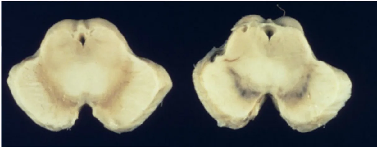
Figure 2: Midbrain showing loss of melanin-containing nerve cells of the substantia nigra in PD compared to healthy controls ... 4
Figure 3: Lewy bodies the cerebral cortex ... 4
Figure 4: UK clinical diagnostic criteria for PD ... 5
Figure 5: Fluorodopa PET in a healthy control and a PD patient ... 6
Figure 6: Dopamine, metabolism and drug treatment ... 7
Figure 7: Anatomy of the basal ganglia ... 8
Figure 8: Dopaminergic projections showing the nigro-striatal, limbic and meso-cortical pathways ... 9
Figure 9: The five ganglia-thalamocortical circuits as described by Alexander et al. ... 11
Figure 10: Cellular layers of the cerebellum ... 13
Figure 11: Schematic of cortical hypo- and hyper-activity in PD ... 15
Figure 12: Spatial and temporal resolution of neuroscience techniques ... 19
Figure 13: Model of the hemodynamic response ... 22
Figure 14: Schematic of a blocked design, with alternating conditions A and B ... 23
Figure 15: Schematic of cortico-basal ganglia circuits in healthy individuals ... 27
Figure 16: Schematic of the cortico-cerebellar circuit ... 29
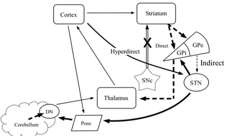
Figure 17: Schematic of the interaction between cortico-striatal and cortico-striatal circuits in PD ... 33
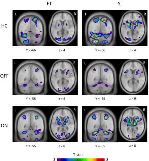
Figure 18: Activation peaks during ET and SI movements in healthy controls and PD patients before and after levodopa ... 43
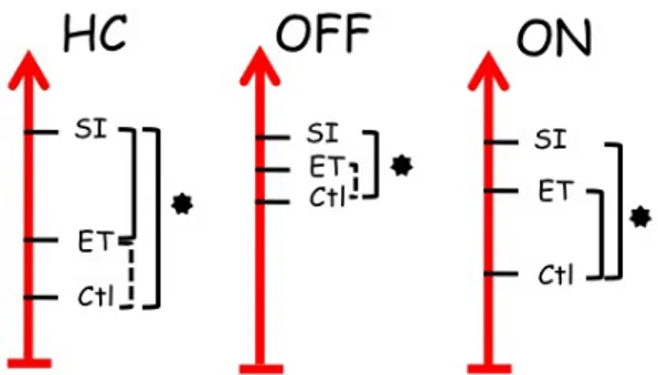
Figure 19: Diagram of putamen activity during control, ET and SI movements in HC, ON and OFF ... 62
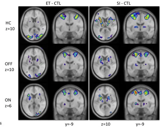
Figure 20: Location of peaks in the ET versus control and SI versus control for the three groups of participants ... 63
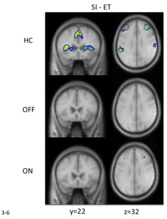
Figure 21: Location of peaks associated with the cognitive portion of SI movements ... 65
Figure 22: Peaks at the intersection of the VLPFC and the insula in healthy controls and patients ... 67
Figure 23: Location of peaks for SI – CTL and ET – CTL movements for sessions ON vs. OFF ... 83 Figure 24: fMRI BOLD activity correlating with performance in patients OFF medication
performing SI vs. CTL movements ... 92 Figure 25: Location of the putamen peaks in the various subtractions. ... xxxiii Figure 26: Location of the STN peaks for the left and right hand. ... xxxiv Figure 27: Location of the dorsolateral PFC and caudate nucleus peaks for the left and right
xi
List of tables
Table I - Demographics of patients (A) and healthy controls (B) ... 69
Table II - Activity peaks associated with ET movements, compared with the control condition ... 71
Table III - Activity peaks associated with SI movements, compared with the control condition ... 72
Table IV - Activity peaks associated with SI movements, compared with ET movements . 73 Table V - Inter-group comparisons for the ET versus control, SI versus control, and SI versus ET contrasts between healthy participants, patients on and patients off medication ... 74
Table VI: Demographics for the twelve patients with PD ... 87
Table VII: Mean reaction times (SD) and percent errors for SI, ET and CTL movements for left and right hand movements of patients OFF and ON medication ... 88
Table VIII: Activation peaks between patients ON and OFF performing SI compared with CTL movements ... 88
Table IX: Activation peaks between patients ON and OFF performing ET compared with CTL movements ... 89
Table X: Activation peaks for patients OFF performing SI versus CTL movements ... xix
Table XI: Activation peaks for patients OFF performing ET versus CTL movements ... xxi
Table XII: Activation peaks for patients ON performing SI versus CTL movements ... xxii
Table XIII: Activation peaks for patients ON performing ET versus CTL movements ... xxiv Table XIV: Activity peaks associated with SI movements, compared with the control
condition ... xlii Table XV: Activity peaks associated with ET movements, compared with the control
condition ... xliii Table XVI: Activity peaks associated with SI movements, compared with ET movements
... xliii Table XVII: Activity peaks associated with SI movements, comparing one hand with the
Table XVIII: Activity peaks associated with ET movements, comparing one hand with the other ... xliv Table XIX: Activity peaks associated with control movements, comparing one hand with
the other ... xlv Table XX: Summary of the major results: (a) left and right hand activations separately, (b)
comparing right and left hand activations ... xlv Table XXI: Sum of left and right side symptoms for individual patients ON and OFF
xiii
Abbreviations
aPFC: anterior prefrontal cortex ASL: arterial spin labeling
BDI-II: Beck’s depression inventory II CBF: cerebral blood flow
COMT: Catechol-O-methyl transferase CTL: control
DBS: deep brain stimulation
DLPFC: dorsolateral prefrontal cortex DTI: diffusion tensor imaging
EC: externally cued
ECu: externally cued-urgent ET: externally triggered FDG: Fludeoxyglucose (18F)
fMRI: functional magnetic resonance imaging
GP: globus pallidus
GPe: external segment of the GP GPi: internal segment of the GP HC: healthy controls
IFG: inferior frontal gyrus
LID: levodopa-induced dyskinesia LTD: long-term depression
MAO-B: monoamine oxidase B MCST: Montreal Card Sorting Task MoCA: Montreal cognitive assessment MPTP: 1-methyl-4-phenyl-1,2,3,6-tetrahydropyridine
MR: magnetic resonance
MRI: magnetic resonance imaging MSN: medium spiny neurons PD: Parkinson’s disease PDRP: PD-related pattern
PDTP: PD tremor-related pattern PET: positron emission tomography PFC: prefrontal cortex
PMC: premotor cortex
PPC: posterior parietal cortex pPFC: posterior prefrontal cortex rCBF: regional cerebral blood flow ROI: region of interest
rTMS: repetitive transcranial magnetic stimulation
SI: self-initiated
SMA: supplementary motor area SNc: substantia nigra pars compacta SNr: substantia nigra pars reticulata STG: superior temporal gyrus STN: subthalamic nucleus TBS: theta-burst stimulation
TMS: transcranial magnetic stimulation UPDRS: unified PD rating scale
VLPFC: ventrolateral prefrontal cortex VTA: ventral tegmental area
Acknowledgments
The last five years at Université de Montréal have been an absolute pleasure for me, and this is mainly due to the friends and colleagues I have had the opportunity to meet.
First of all, I want to thank my supervisor, Oury Monchi. Even though we first met during an infusion of radioactive isotopes into my bloodstream, the next few years haven’t been quite as unpleasant. I have kept many memorable moments from the years of training and collaboration. You have taught me tremendous amounts in scientific reasoning and criticism, but also valuable lessons in social interactions – or may I call it self-control? And for whatever reason, you have always had a knack for reading my thoughts before I even said anything. As a result, I think that my brain has started taking the shape of a pecan…
Thank you also to Oury’s lab (a.k.a. PCAN), past and present, which has not only been extremely helpful through the years, but also very fun to be part of. Thanks to Clotilde, Christophe, Jean-Sebastien, Atsuko, both Claudines, France, Thomas, Laura and Cécile. Although we clearly haven’t gotten together as many times as we said and hoped we would, I have truly appreciated all the scientific discussions, gossiping and practical jokes. Maybe with a delay on some of the practical jokes…
I must also thank Dr. Antonio Strafella and Dr. Julien Doyon for their help, guidance and advice throughout the years as members of my committee. It has been an absolute pleasure collaborating with you both, and I hope that more collaboration is to come. Thanks also to Mathieu for his help and patience on so many superscript issues, and Carollyn and André for all their help during scanning. I am proud to have been part of the UNF team – and perhaps will be again one day.
There are many people that have crossed my path during my work at the CRIUGM, and that I’ve had pleasure chatting, discussing and working with. I would rather avoid a list of names in fear of forgetting someone… thanks to all of you, you know who you are. A special thanks to Stuart and Brad for their help with my writing and for putting up with me, both at work and while curling.
I also want to send a special thanks to my family for all their help, support, and
inspiration through my years at school, which have finally come to an end. Thank you to my husband for his help in every aspect of my work, from listening to me ramble, both happy
xv
and frustrated, to building the response boxes I used in my experiments. I can’t forget my son Thomas; I’m so lucky to have such an easy-going child that has allowed me to finish my thesis without any delays. And also my second son Lukas, on the way; he has given me just the right amount of time to close this chapter before coming to this world.
This thesis has led to the following publications:
- Peer reviewed journals:
Martinu, K., Monchi, O. (In press) Cortico-basal ganglia and cortico-cerebellar circuits in Parkinson’s disease: pathophysiology or compensation? Behavioral Neuroscience Martinu, K., Degroot, C., Madjar, C., Strafella, A.P., Monchi, O. (2012) Levodopa
influences striatal activity but does not affect cortical hyper-activity in Parkinson's disease. Eur J Neurosci
Monchi, O., Martinu, K., Strafella, A. (2010) The contribution of neuroimaging in the study of cognitive deficits in Parkinson’s disease. Review. Clin EEG Neurosci 41(2): 76-81 Francois-Brosseau, F-E., Martinu, K., Strafella, A., Simard, F., Monchi, O. (2009) Basal
ganglia and frontal involvement in self-generated and externally-triggered finger movements in the dominant and non-dominant hand. Eur J Neurosci, 29(6): 1277-86
C’est le temps que tu as perdu pour ta rose qui fait ta rose si importante. – Antoine de Saint Exupéry, Le Petit Prince
1.1 Parkinson’s disease
1.1.1 Shaking palsy
The first detailed description of what is known today as Parkinson’s disease (PD) comes from 6 cases observed by James Parkinson, published in 1817. In his depiction of what he termed the ‘shaking palsy’, or paralysis agitans, the disease begins slowly, such that it is difficult for the patient to pinpoint its precise beginning. The patient first perceives a slight form of weakness, with a tendency to shake, most often in one of the hands and arms, followed by a change in posture. Fatigue and agitation slowly spreads to the lower limbs.
“At this period the patient experiences much inconvenience, which unhappily is found daily to increase. The submission of the limbs to the directions of the will can hardly ever be obtained in the performance of the most ordinary offices of life. The fingers cannot be disposed of in the proposed directions, and applied with certainty to any proposed point. As time and the disease proceed, difficulties increase: writing can now be hardly at all accomplished; and reading, from the tremulous motion, is accomplished with some difficulty. Whilst at meals the fork not being duly directed frequently fails to raise the morsel from the plate: which, when seized, is with much difficulty conveyed to the mouth. At this period the patient seldom experiences a suspension of the agitation of his limbs” (Parkinson, 2002).
In the following stages of the disease, walking becomes increasingly difficult, sleep
becomes troubled as tremor causes the patient to awaken, speech becomes unintelligible and feeding one’s self becomes virtually impossible.
“As the debility increases and the influence of the will over the muscles fades away, the tremulous agitation becomes more vehement. It now seldom leaves him for a moment; but even when exhausted nature seizes a small portion of sleep, the motion becomes so violent as not only to shake the bed-hangings, but even the floor and sashes of the room. The chin is now almost immoveably bent down upon the sternum. The slops with which he is attempted to be fed, with the saliva, are continually trickling from the mouth. The power of articulation is lost. The urine and faeces are passed involuntarily; and at the last, constant sleepiness, with slight delirium, and other marks of extreme exhaustion, announce the wished-for release” (Parkinson, 2002).
There were several accounts of what could be interpreted as PD from Egyptian papyrus, Leonardo da Vinci, Johannes Babtiste Sagar and Rembrandt (Lees, 2007). Shortly after James Parkinson’s essay, Prussian diplomat Wilhelm von Humboldt (1767 – 1835) described the symptoms of his disease in a written correspondence with a friend:
“Trembling of the hands … occurs only when both or one of them are inactive; now only the left one is trembling but not the right one that I am using to write – really odd to see … every line is starting with best intentions in large letters only to end … in barely legible small ones – in ageing one comes back to childhood's writing, because indeed childlike are these large [Latin] letters without connecting parts.” (Horowski, 2000)
Despite this, Wilhelm von Humboldt associated his symptoms of tremor, rigidity and bradykinesia to common consequences of ageing (Horowski, 2000).
It is only in the 1860’s that this ‘shaking palsy’ was further characterized by French neurologists Trousseau, Charcot and Vulpian at the Salpêtrières in Paris. Charcot in
particular recognized bradykinesia, posture (Figure 1) and gait as important signs of the disease, but also noted that dementia, depression, affective disorders and hallucinations ensued, notes that were largely ignored until the late 20th century. He subsequently rejected
3
the term ‘shaking palsy’ and attributed Parkinson’s name to the disease (Playfer & Hindle, 2008). It took another hundred years for researchers to establish dopamine depletion as the source of Parkinson’s disease (PD) (Ehringer & Hornykiewicz, 1960), after which levodopa became the first neurotransmitter replacement treatment (Birkmayer & Hornykiewicz, 1961). Further knowledge of mechanisms behind PD stems from the discovery of 1-methyl-4-phenyl-1,2,3,6-tetrahydropyridine (MPTP), a by-product of meperidine synthesis. In 1976, 23-year old chemistry graduate Barry Kidston self-injected himself with a concoction of meperidine and MPTP developing parkinsonism within three days. He displayed
dopaminergic neuron degeneration in the substantia nigra at his autopsy, 18 months later. The contamination of this illicit drug in northern California led to numerous additional cases of persistent parkinsonism in young drug abusers in 1982 (Langston et al.,1983), and spurred research with what became the animal model of PD.
Figure 1: Illustration of PD by William Richard Gowers (1886) 1.1.2 PD characteristics
The diagnosis of PD is not without downfalls. In a sample of 100 patients clinically diagnosed with PD, only 76 were found to have been correctly diagnosed post-mortem (Hughes et al., 1992). Most common misdiagnoses were attributed to multiple system atrophy, progressive supranuclear palsy, Alzheimer's disease, and cerebrovascular pathology. The typical neuropathological signs of PD are a loss of at least 50% of the melanin-containing nerve cells of the substantia nigra and a depletion of tyrosine
hydroxylase (Figure 2), the rate-limiting step in catecholamine synthesis (dopamine, epinephrine and norepinephrine). It is this neuronal loss that results in the dopamine depletion in PD. Another characteristic of PD is the presence of Lewy bodies (Figure 3), primarily consisting of alpha-synuclein agglomerations (Spillantini et al., 1997), in some of the remaining nerve cells (Perkin, 2008). According to Parkinson Society Canada, the prevalence of PD in Canada is estimated between 100 and 200 / 100,000, with an incidence rate of 10 to 20 / 100,000 each year; 85% of patients are over the age of 65.
Figure 2: Midbrain showing loss of melanin-containing nerve cells of the substantia nigra in PD (left) compared to healthy controls (right) (pathology.mc.duke.edu)
Figure 3: Lewy bodies the cerebral cortex (Love, 2005)
Symptoms of PD typically include tremor, rigidity, bradykinesia and postural disturbances. Bradykinesia mainly presents itself by the difficulty in performing tasks such
5
as lifting a fork or dressing, reduction in size of handwriting, reduced stride length and a stooped posture. In addition, patients have great difficulty maintaining their posture when pushed forwards or backwards. According to the United Kingdom PD society brain bank, the clinical criteria for a diagnosis of PD include bradykinesia, and either rigidity, tremor or postural instability (Hughes et al., 1992). Exclusion criteria consist of neurological
conditions or drug-induced symptoms (Figure 4). Further criteria, such as unilateral onset and response to levodopa, can support the diagnosis of PD. The unilateral onset is
maintained throughout the disease as symptom asymmetry; the side of the body first affected remains more severely affected throughout the duration of PD. This intriguing aspect will be the main focus of chapter 4, where we discuss the possibility that the effect of levodopa may be linked to disease asymmetry.
Patients also often show changes in brain imaging scans. In particular, 6-[18 F]-fluorodopa (a radioactively labeled dopamine precursor) positron emission tomography (PET) scans can show reduced isotope uptake in the putamen (Figure 5), particularly in the hemisphere that is more affected. As we will see in chapter 3, the blood oxygenation level dependent (BOLD) activity of the putamen and the cortical regions it communicates with are substantially affected in PD.
Figure 5: Fluorodopa PET in a healthy control and a PD patient (Longo et al., 2011)
1.2 Levodopa
Several options exist for the treatment of PD, through the alteration of the different metabolic steps of dopamine synthesis, release and reuptake (Figure 6). Levodopa (a.k.a. L-dopa), the cornerstone of PD treatment, enhances dopaminergic activity by providing more dopamine precursor. The effect of dopamine can also be enhanced by dopamine agonists acting on receptors (dopamine agonists), or by inhibiting dopamine reuptake (COMT inhibitors).
7
Figure 6: Dopamine, metabolism and drug treatment
COMT = Catechol-O-transferase; DA = dopamine; MAO = monoamine oxidase. Figure adapted from www.medscape.org
Levodopa, or L-3,4-dihydroxyphenylalanine, is one of the most common treatments for PD. Unlike dopamine, it crosses the blood brain barrier, increasing the concentration of
dopamine. Because dopamine receptors also exist in the periphery, however, the administration of levodopa has many adverse effects such as nausea, hypotension,
gastrointestinal complications, hair loss and sleep disturbance. It may also be the source of additional cell death and, as discussed in chapter 2, lead to levodopa-induced dyskinesias (LIDs).
1.3 Basal ganglia and cerebellum in PD
1.3.1 Anatomy
Given the importance of nigrostriatal degeneration in the pathophysiology of PD, describing basal ganglia anatomy and the dopaminergic pathways is crucial. Also, understanding the organization of the cortico-striatal circuits is fundamental for the remaining chapters. Finally, as will be discussed in chapter 2, the cerebellum plays an important role in the
pathophysiology of PD. We will therefore also introduce the cerebellum and cortico-cerebellar connections.
1.3.1.1 Basal ganglia
The basal ganglia are composed of the caudate nucleus, putamen, nucleus accumbens, subthalamic nucleus (STN), globus pallidus (GP) and substantia nigra, tightly
interconnected regions that process information from all cortical regions. The caudate nucleus and putamen, forming the striatum, are deep grey matter nuclei embedded within the c-shaped lateral ventricles (Figure 7). The putamen and the adjacent GP (a.k.a. the lentiform complex) with its internal and external segments (GPi and GPe, respectively) are anterior to the thalamus, separated by the posterior arm of the internal capsule. The
lentiform nucleus is covered laterally by the external capsule, claustrum, extreme capsule and insula. Anterior to the putamen, and joined to it at its most inferior point forming the nucleus accumbens, the caudate nucleus runs superiorly around the putamen, separated by the anterior arm of the internal capsule and forming the floor of the lateral ventricle. Inferior to the thalamus, as its name implies, lies the STN, just superior to the substantia nigra located in the midbrain.
Figure 7: Anatomy of the basal ganglia (Kandel et al., 2013)
9
1.3.1.2 Dopamine, dopamine receptors and dopaminergic pathways
Dopamine is a catecholamine neurotransmitter, synthesized from the amino acid tyrosine. Tyrosine hydroxylase first converts tyrosine to l-3,4-dihydroxyphenylalanine (L-dopa); this is the rate-limiting step of dopamine synthesis. The second step is the
decarboxylation of L-dopa to dopamine by the enzyme aromatic L-amino acid
decarboxylase (Vallone et al., 2000). Dopamine is produced in the cell bodies of the ventral tegmental area (VTA) and the substantia nigra, whose axons project to different regions of the brain, forming several dopaminergic pathways. The three major projections are the nigrostriatal, meso-cortical and meso-limbic pathways (Figure 8).
Figure 8: Dopaminergic projections showing the nigro-striatal, limbic and meso-cortical pathways (Chinta & Andersen, 2005)
The nigro-striatal pathway runs from the substantia nigra pars compacta (SNc) to the dorsal striatum (putamen and caudate nucleus). It is strongly involved in movement, and its degeneration is the primary source of PD symptoms. The meso-limbic pathway runs from the VTA to the ventral striatum (nucleus accumbens), the olfactory tubercle and other parts of the limbic system, and is mainly involved in motivated behavior. The meso-cortical pathway projects from the VTA to the frontal cortex, and is involved in memory and learning (Le Moal & Simon, 1991).
There are five dopamine G-coupled protein receptors, generally classified as either D1-like or D2-like. This classification stems from their effect on the production of cyclic adenosine monophosphate (cAMP) through the stimulation or inhibition of the adenylyl
cyclase protein (Stoof & Kebabian, 1981). The D1-like subfamily (D1 and D5), found exclusively post-synaptically, stimulate cAMP production, whereas the D2-like subfamily (D2, D3 and D4), expressed both post- and pre-synaptically, lead to an inhibition of adenlylyl cyclase and a decrease in cAMP production (for a review, see Beaulieu & Gainetdinov, 2011). D1 receptors are primarily found in the nigrostriatal, mesolimbic and mesocortical regions (striatum, nucleus accumbens, substantia nigra, olfactory bulb, amygdala and frontal cortex), as well as in the hippocampus, cerebellum, thalamic and hypothalamic areas. D2 receptor density is highest in the striatum, nucleus accumbens, olfactory tubercle, and also in the substantia nigra, VTA, hypothalamus, cortical areas, septum, amygdala and hippocampus. Segregation of D1 and D2 receptors has been found within the basal ganglia, such that the medium spiny neurons (MSNs) that project to different regions will selectively express one or the other. In particular, MSNs that project to the GPi and substantia nigra pars reticulata (SNr) express the D1 receptor, whereas a different group of MSNs that project to the GPe selectively express the D2 dopamine receptor. There is, however, a small portion (5-15%) of MSNs that express both D1 and D2 receptors in the dorsal striatum. D1 and D2 receptors are estimated to compose the majority of dopamine receptors within the striatum (Levey et al., 1993); D3, D4 and D5 receptors are expressed at much lower levels in several cortical and subcortical regions. All receptors are also expressed in the periphery, such as in the kidneys, adrenal glands, gastrointestinal tract, blood vessels and heart (Beaulieu & Gainetdinov, 2011).
1.3.1.3 Ganglia-‐thalamocortical circuits
The basal ganglia and cortex are linked through a series of ganglia-thalamocortical circuits, referred to in this thesis as cortico-striatal loops. Five parallel circuits have been described by Alexander et al., namely the “motor”, “oculomotor”, “dorsolateral prefrontal”, “lateral orbitofrontal” and “anterior cingulate” loops. Each one of these loops consists of non-overlapping regions of the striatum, GP, substantia nigra, thalamus and cortex (Figure 9). These circuits provide a topographical projection of information from functionally related cortical areas through the intermediate structures before being projected back to the cortex (Alexander et al., 1986). While the topography in these circuits is predominant, links
11
exist between these circuits at the cortical, striatal as well as thalamic levels. Furthermore, as discussed in the next chapter, there are important connections between the core of these circuits, in the thalamus, and the cerebellum. It must be noted, however, that the series of connections and funneling of information through these regions is part of the classical Albin-DeLong model (Albin et al., 1989; DeLong et al., 1990), and more complex models of basal ganglia function have been suggested (Bar-Gad & Bergman, 2001; Lanciego et al., 2012).
Figure 9: The five ganglia-thalamocortical circuits as described by Alexander et al., 1986
The two circuits of particular interest for this thesis are the motor and “dorsolateral prefrontal”, or cognitive, cortico-striatal circuits. The cognitive cortico-striatal loop consists of projections from the dorsolateral prefrontal cortex (DLPFC) and portions of the parietal cortex to the head of the caudate nucleus. From the latter, projections are sent to the dorsomedial one-third of the GP and rostral SNr, and finally to the thalamus before
projecting back to the DLPFC. The disruption of the cognitive cortico-striatal circuit leads to specific cognitive disabilities, and is thought to play a key role in the cognitive deficits sometimes observed in PD.
The motor cortico-striatal circuit is a closed loop of topographically organized projections between the motor cortex, premotor cortex (PMC) and supplementary motor area (SMA), putamen, GP, SNr, STN and thalamus. The motor loop further consists of direct, indirect and hyperdirect pathways. The direct pathway relays projections from the putamen to the GPi and then the thalamus. The indirect pathway consists of projections from the putamen first to the GPe, then the STN, and finally back to the GPi before relaying to the thalamus. Through excitatory and inhibitory connections of these two pathways, driven by the differential effects of D1 and D2 receptors, the direct pathway disinhibits thalamic activity, whereas the indirect pathway increases the inhibition of the thalamus. The balance between these two systems, described in detail in chapter 2, plays an important role in the symptomatology of PD.
1.3.1.4 The cerebellum and the cortico-‐cerebellar circuit
While cortico-striatal dysfunction is important in PD, cortico-cerebellar changes have also been reported. The nature and origins of these cortico-cerebellar alterations are still under debate and will be the focus of a large proportion of this thesis (chapter 2).
The cerebellum consists of tightly packed sulci and gyri of a very regular cell composition, and several deep nuclei. The grey matter of the cerebellum is formed of three cell layers (Figure 10); the granular cell layer holds all the granule cells, interneurons and Golgi cells, the thin Purkinje cell layer contains the Purkinje cell bodies, and, finally, the molecular layer comprises of the thick Purkinje cell dendritic trees, parallel fibers, interneurons, stellate cells and basket cells.
13
Figure 10: Cellular layers of the cerebellum (Kandel et al., 2013)
The cortico-cerebellar circuit consists of connections from the cerebral cortex to the cerebellar cortex through a series of brainstem nuclei, and feedback connections from the cerebellar cortex to the cerebral cortex through thalamic nuclei. Input to the cerebellum is carried out by two types of cells, the mossy fibers that bring cortical information from the pons (corticopontine and pontocerebellar projections), and climbing fibers that bring cortical information from the red nucleus and inferior olive. Purkinje cells from the cerebellar cortex then send their output to the deep nuclei of the cerebellum, which then
project through the red nucleus to thalamic nuclei, and finally back to the cortical region where information originated from, forming a closed loop. The cortico-cerebellar circuit and its connections with the cortico-striatal circuit are described in detail in chapter 2.
1.3.2 Functional roles of the striatum and cerebellum 1.3.2.1 The dorsal striatum and cortical activity
The dorsal striatum is an essential component of the cortico-striatal pathways
(Alexander et al., 1986). Using the Wisconsin Card Sorting Task (WCST) and the Montreal Card Sorting Task (MCST), Monchi et al. (2001, 2004, 2006, 2007) have been able to dissociate the roles of the components of the dorsal striatum, in particular those of the caudate nucleus and the putamen. The caudate nucleus has been shown to be involved different aspects of cognition such as the planning of a novel action (Monchi et al., 2006; Owen et al., 1996). In contrast, the putamen has been shown to be involved in the execution of novel actions. As discussed in the next two chapters, the recruitment of the caudate
nucleus and the putamen lead to differences in cortical activity observed in patients with PD. More specifically, patients with PD don’t simply have a hypoactive cortex, as the original model by Albin, Young & Penney suggests (1989); they display increased cortical activity as compared with healthy control participants in different cognitive and motor tasks. We define hypo-activations in patients with PD as a decrease in cortical activity compared with the activity of the same region in healthy participants during the same task or contrast, and hyper-activations as increases in activity in cortical regions as compared with those same regions in healthy participants. Based on results using the WCST, MCST, and self-initiated (SI) and externally triggered (ET) movements (Monchi et al., 2004, 2007; Martinu et al., 2012), we have suggested that the hypo-and hyper-activity patterns observed in PD are related to striatal requirement in the task at hand (Monchi et al., 2010). Moreover, in a task where healthy controls specifically recruit the striatum, patients with PD will show hypo-activity of prefrontal regions. However, in tasks where healthy controls do not specifically require basal ganglia activity, patients with PD will show hyper-activity of prefrontal and parietal regions (Figure 11).
15
Figure 11: Schematic of cortical hypo- and hyper-activity in PD
Our laboratory had previously shown that while levodopa had a significant effect on the structures involved in the motor cortico-striatal loop, it did not seem to affect activity of the cognitive cortico-striatal loop during the WCST (Jubault et al., 2009). We wanted to extend this concept to a motor task as well (chapter 3). But more specifically, we wanted to compare patients with PD to control participants to determine the effect of levodopa on the hypo- and hyper-activation patterns in PD. Does levodopa restore these hyper-activations to normal, or are they linked to the pathophysiology of PD and/or the prolonged use of
dopaminergic medications? 1.3.2.2 Hand dominance
According to lesion studies of right-handed subjects performed between the 1920’s and 1980’s, it seemed that the left hemisphere (considered to be dominant) played an important role in ipsilateral hand control (Mattay et al., 1998). A PET study with hand movements interpreted that the increase in left-hemispheric activity may be either due to a differential organization between the two hemispheres, or to the fact that more effort for right-handed subjects was necessary for movements of the left hand (Halsey et al., 1979). Kawashima et al. (1993) concluded that the left non-dominant hand recruited ipsilateral motor areas, a sign of functional asymmetry. However, Mattay et al. (1998) argued that the dominant hand movements used in these studies were over-learned sequences, or
automatized, and so required less ‘conscious effort’ to perform. The non-dominant hand, therefore, may require more resources to perform the same movements, and thus lead to the recruitment of ipsilateral regions. In their study, Mattay et al. (1998) compared a simple task performed by the left (non-dominant) hand to a more complex task performed by the right (dominant) hand, and found that subjects exhibited similar ipsilateral cortical activations in both tasks. The authors speculated, therefore, that ipsilateral activations in motor tasks represent the degree to which motor movements are automatic, rather than
differential organization for dominant and non-dominant hand control between the two hemispheres.
We have recently performed a functional magnetic resonance imaging (fMRI) study with healthy young adults while carrying out three conditions of a finger-moving task: a self-initiated (SI) button-press sequence, a computer-generated sequence, and a button-press repetition condition (François-Brosseau et al., 2009; this study is directly relevant to this thesis as we have applied the same protocol to the studies we describe, and is therefore included as an appendix for easy reference). We showed that the three tasks increasingly recruit the putamen, the repetition condition requiring the putamen the least, followed by the externally triggered (ET) condition, and the SI sequence generation requiring the putamen the most. In this particular protocol, the putamen was more involved in the
generation of SI movements than in ET ones, and there is increasing recruitment of cortical motor regions with the three tasks in the same order. The main finding of this study was that when comparing the dominant and non-dominant hands, task demand for striatal activity was higher when participants used the non-dominant hand. We wanted to use this same protocol with patients with PD to determine, first of all, whether they showed the same patterns of activity and whether levodopa reestablished this discrepancy, but mainly whether disease asymmetry could invert this effect. This would mean that if patients were more severely affected on the right side of their body, would they display opposite results? As discussed in chapter 4, however, this hypothesis turned out to be difficult to test
considering the recruitment restraints we faced. 1.3.2.3 The cerebellum
For many decades, the cerebellum was considered to be involved in motor functions. Although Charcot had been adamant about its role in cognitive functions, this aspect has greatly been ignored. In the 1930's, Abbie (1934) observed that there were degenerated regions of the pons after major lesions to the so-called association cortices. These
association areas are now known to be linked with the lateral hemispheres of the posterior lobe of the cerebellum through the pons and the thalamus (Schmahmann & Caplan, 2006). Additionally, Bard (1928) and Zanchetti & Zoccolini (1954) described animals that
17
developed sham rage after lesions and stimulations of the cerebellum. Even though early work hinted at its involvement in aspects such as emotional control and cognitive processes like executive functions and linguistic processing (Schmahmann & Caplan, 2006), it is only quite recently that experiments specifically designed to understand this involvement were developed. It is important to consider the cerebellum's implication in a combination of motor and cognitive tasks, such as the cognitive manipulation of motor sequences; a recent study showed that participants who perform worse at motor imagery compensated with the cortico-cerebellar network (Guillot et al., 2008). Taniwaki et al. (2006) showed that in contrast to the involvement of the striatal loop in SI movements, the
cortico-cerebellar loop is more involved in ET movements. Blouin et al. (2004), however, observed with PET that the cerebellum is involved in synchronized SI movements. As discussed in further detail in the following chapter, the involvement of the cerebellum in SI and ET movements is strongly dependent on the task used, partly relating to the type of planning involved in the individual movements. Not only do our results suggest that the cerebellum is preferentially involved in SI movements, we also find that levodopa has a significant effect on cerebellar activity. These results, discussed in chapter 2, support our hypothesis that levodopa increases activity in the cerebellum through connections between the cortico-striatal and cortico-cerebellar pathways, and suggest that it is this increase that eventually leads to the pathophysiological involvement of the cerebellum in symptoms such as LIDs.
1.4 Aims of this thesis
This general aim of this thesis is to understand the effect of levodopa on the neural processes as measured by BOLD fMRI underlying SI and ET movements of the left and right hands in patients with PD. The next section (chapter 2), recently accepted in Journal of Behavioral Neuroscience, reviews the current literature concerning cortico-striatal and cortico-cerebellar circuits in PD. In this section we will discuss the compensatory and pathophysiological involvement of these two circuits in PD, and the role that levodopa has to play in LIDs, a common side-effect. The following section (chapter 3) is a research article published in European Journal of Neuroscience on the effect of levodopa on cortico-striatal circuits in PD. There we demonstrate that cortico-striatal activity related to SI and ET
movements in PD is reduced compared with healthy controls, and that patients show hyper-activations linked to mesocortical dopamine pathway dysfunction. Finally, in the research article (chapter 4) that will be submitted to Movement Disorders shortly we describe the differential effect of levodopa on left and right hand movements in PD. More specifically, we suggest that levodopa leads to significant differences in cortico-striatal regions when patients use their left hand and not their right hand, implying that levodopa selectively acts on more affected / non-dominant hand movements.
1.5 Magnetic resonance imaging
1.5.1 What is MRI?
Magnetic resonance imaging (MRI) is an imaging technique that allows the
visualization of internal structures. Using a powerful magnet, often in the order of 1.5 to 7 Tesla (the earth’s magnetic strength is 0.00005 Tesla), and the magnetic properties of atomic nuclei in organ tissue and blood, MRI permits the imaging of the anatomical structures of body parts and brain function. The smallest unit of an MR image is called the voxel, i.e. the volumetric pixel, usually 1mm3 for an anatomical image. This is an extremely detailed spatial resolution when compared to other neuroimaging techniques such as
electroencephalography (EEG) or PET (Figure 12). The temporal resolution of MRI is <1s (fMRI) to minutes (MRI). Although it is far from the temporal precision of EEG, MRI provides an adequate balance of spatial and temporal resolutions for a wide range of physiological and pathological studies.
19
Figure 12: Spatial and temporal resolution of neuroscience techniques (Ward, 2006) 1.5.2 History
The development of the MRI technique was made possible by Wolfgang Pauli’s observation, in 1924, that atomic nuclei (e.g. hydrogen) spin at specific frequencies and have a magnetic moment. When placed into a surrounding magnetic field, atomic nuclei align themselves with the field, a phenomenon referred to as relaxation time. As Isidor Rabi demonstrated in 1937, if a surrounding magnetic field oscillates at the same frequency as the atomic nucleus, the latter would absorb energy from the field, just like pushing a
pendulum at the correct moment. This phenomenon was named magnetic resonance, and the frequency that has the most effect on the atomic nuclei in question is referred to as the resonant frequency. Just as the swing of a pendulum is highly dependent on gravity, the resonant frequency is highly dependent on the static magnetic field. In 1946, Edward Purcell and Felix Bloch independently developed experiments that tested the resonant frequency of solid matter (wax paper) and water. Bloch’s experiment in Physical Review involved a transmitter coil and a detector coil that recorded the energy, or nuclear resonance, absorbed by the sample of water. Nuclear magnetic resonance (NMR) is still the basis of all
MRI acquisitions today. The term ‘nuclear’ was eventually dropped due to its negative connotations with health, and magnetic resonance imaging was adopted in its stead.
After the observation that water atomic nuclei had different relaxation times depending on their surrounding tissue, the medical application of NMR was clear. If the measure of resonant frequencies of atomic nuclei could be transformed into images, one could potentially distinguish between different types of tissue. The simple detection of emitted energy from atomic nuclei from an oscillating magnetic field, however, lacked spatial information. In 1972, Paul Lauterbur suggested that if the strength of the static magnetic field varied across space, the resonant frequency detected could give information about its location. In 1976, using an electromagnetic pulse and rapidly changing field gradients, Peter Mansfield developed the echo-planar imaging used today to record images in a fraction of a second, making the acquisition of images of humans possible.
1.5.3 Magnets and coils
The MR scanner consists of several main components. The first is a wire wrapped in tight loops (a solenoid), forming the principal magnet (Huettel, 2004). The electrical current runs through this solenoid generating the static magnetic field, B0. The second important
component is the radiofrequency antenna, placed directly around the item being scanned, is composed of two electromagnetic coils: a transmitter and a receiver. This antenna generates the electromagnetic field at the atomic nuclei’s resonant frequency, and records the energy released (in the radiofrequency portion of the electromagnetic spectrum). Contrary to the static magnetic field, these coils are turned on and off during image acquisition. When placed in a static magnetic field, atomic nuclei align themselves with the field in what is referred to as relaxation time. The radiofrequency coils send pulses that disturb this
equilibrium, exciting the atomic nuclei. Following the pulse, it is the release of the absorbed energy and the return to baseline, or relaxation, that defines the magnetic resonance (MR) signal.
The third important components of the MR scanner are the gradient coils. These are three sets of coils that will cause a transient gradient in the magnetic field in the x, y, and z
21
directions, one after the other. The controlled changes in magnetic field give the MR signal spatial information, in that different locations have a different contribution to the MR signal. An MR signal that codes spatial information about the resonating atomic nuclei permits the reconstruction of spatial frequency data into image-space data. Through mathematical manipulations (an inverse Fourier transformation), one can generate 2D images from the recorded MR signal. A slice-by-slice acquisition of 2D images can finally be reconstructed into a 3D volume encompassing the entire structure being scanned, e.g. a participant’s head. 1.5.4 Hemodynamics
As different brain regions become involved in specific tasks, the energy consumed and therefore the demand for oxygen increases. The imaging of brain function is based on blood flow and the magnetic properties of hemoglobin. More specifically, water molecules in oxygenated hemoglobin have no magnetic moment, but deoxygenated hemoglobin has unpaired electrons and a significant magnetic moment; it is paramagnetic. In MR sequences sensitive to the changes in spin caused by deoxygenated blood, there will be a drop in MR signal. When measuring the BOLD response, however, what we observe is an initial dip followed by a strong increase in MR signal (Figure 13). One interpretation of this
phenomenon is that when a region becomes more active and begins consuming oxygen, an excess of oxygenated blood flushes the region and displaces the deoxygenated hemoglobin, causing the rise in MR signal.
Figure 13: Model of the hemodynamic response
Figure adapted from BOLD imaging, Radiopaedia, BOLD imaging, www.radiopaedia.org It is important to realize, when performing MRI experiments, that the changes in signal observed on a functional acquisition are a physiological phenomenon that correlates with changes in blood flow, which correlates with energy consumption of the underlying neurons, and which itself correlates with neuronal activity. It is not a direct measure of cellular activity; therefore interpretations of underlying cognitive processes have to be made with this in mind. The hemodynamic response is also very slow. The peak of the response occurs 4 to 6 seconds after the stimulus and the consequent neural response, and only returns to baseline after 12 to 14 seconds. This has important implications for the design of fMRI experiments.
1.5.5 Experimental design
In order to answer a scientific question, a proper experimental design needs to be put in place. Specific hypotheses require specific dependent and independent variables.
Additionally, in order to be able to make interpretations on neural processes involved in specific tasks, the experimental condition has to be compared to an adequate control, so that the comparison between the two can most accurately test the hypothesis in question. An fMRI protocol consisting of a motor or cognitive task will therefore usually consist of a timeframe with at least two alternating conditions. Subsequent analyses are performed to
23
correlate the hemodynamic activity (i.e. the dependent variable) involved in each condition, and the difference in the dependent variable between the experimental condition and the control condition provide an answer to the question as precisely as possible. Because the hemodynamic response is slow, researchers first developed the blocked design, which separates the different conditions in the distinct sections of an extended period of time (Figure 14), ranging easily from 15 to 30 seconds:
Figure 14: Schematic of a blocked design, with alternating conditions A and B The blocked design paradigm is a powerful method that involves a sustained
hemodynamic response, giving maximal amplitude of the BOLD response. Some questions cannot be answered by using the blocked design. For example, comparing correct and incorrect responses, or measuring the response associated with an “oddball” task involves events of a very short duration. Event-related paradigms are more appropriate for such experiments. They involve brief stimulus presentations presented randomly throughout the duration of the scan, with an inter-stimulus interval between each. Although this method does not allow the hemodynamic response to reach its maximum amplitude, it is based on the assumption that short stimuli will evoke a transient change in neural activity. In a situation where participants need to make responses to the events, this method allows the researcher to remove events with incorrect responses, or to make a comparison between them. The choice of experimental design is crucial to the questions that stem from the research hypothesis. One must ascertain that there are no confounding variables that would correlate with the variables of interest.
Chapter 2: Pathophysiology versus compensation
The first article presented in this thesis is a review that has recently been accepted for publication in a special issue on controversies in PD in Behavioral Neuroscience. Our preliminary data indicated that in patients with PD, activity in the cerebellum was increased after levodopa administration. Some studies have argued for the potential compensatory role of the cerebellum in PD pathology (Glickstein & Stein, 1991; Palmer et al., 2009a). If levodopa re-established cerebral activity to patterns observed in healthy controls, it should cause a reduction in activity instead. We wanted to examine more closely the role of the cerebellum in PD. In reviewing the literature we realized that there were two possible explanations for cerebellar changes in activity in PD, which are not necessarily mutually exclusive. We have found more convincing evidence, however, that changes in activity in the cerebellum are more closely linked with pathophysiology than compensatory
mechanisms, and that many studies suggesting that the cerebellum is involved in
compensation are inconclusive. In the following chapter we introduce in greater detail the cortico-striatal and cortico-cerebellar circuits, and provide evidence that the cerebellum may also be strongly affected by PD pathology and/or treatment. We also discuss the effect of levodopa on cerebellar activity, supported by our own results.
Cortico-striatal and cortico-cerebellar circuits in Parkinson’s disease: pathophysiology or compensation?
Martinu, K. & Monchi, O.
Accepted for publication in Behavioral Neuroscience in September 2012
Abstract
The basal ganglia and the cerebellum are anatomically and functionally linked to the cerebral cortex through a series of well-established circuits. The disruption of dopaminergic projections in PD leads to an imbalance within these circuits, leading to motor and cognitive symptoms. The cortico-cerebellar network has often been viewed as a compensatory
25
network, helping the dysfunction of the cortico-striatal circuits in PD. However, evidence for this compensatory role is scarce; most changes in cerebellar activity could equally be attributed to pathophysiological changes underlying PD. This paper will review the anatomy, interaction and function of the cortico-striatal and cortico-cerebellar circuits, the
pathophysiological, metabolic and functional changes observed in PD, as well as the effect of levodopa and deep brain stimulation (DBS) on these changes. We will use this
framework to discuss the pathophysiological and compensatory mechanisms behind cortico-striatal and cortico-cerebellar circuit activity in PD.
Keywords: Parkinson’s disease, Cerebellum, Striatum, Compensation, Levodopa
Introduction
PD is a debilitating neurodegenerative illness associated with the loss of dopaminergic neurons of the substantia nigra. Patients classically suffer from motor
symptoms such as tremor, rigidity and bradykinesia, although cognitive deficits in executive functioning, memory, language, and visuo-spatial processing are also pervasive (Taylor & Saint-Cyr, 1995). The cortex, thalamus, basal ganglia and cerebellum form a series of anatomically and functionally segregated circuits sub-serving a multitude of cognitive and motor functions. The disruption of these circuits through the degeneration of dopaminergic neurons in the substantia nigra leads to widespread changes in brain activity and
connectivity. It is not yet known whether these extensive neural changes are strictly the result of PD pathophysiology or, alternatively, are manifestations of compensatory
mechanisms in response to the disease. It has been suggested that the recruitment of cortico-cerebellar networks is one possible compensatory mechanism for the generation of
movement in PD (Rascol et al., 1997, Palmer et al., 2009a), such as SI and ET movements. However, many of the changes in the cortico-cerebellar circuits may be the result of
disruptions caused by PD or by the prolonged used of dopaminergic medication. In this review we will suggest that the pathophysiology behind changes in cerebral and cerebellar activity cannot be ignored, and that future research will be necessary to disentangle these
two alternative hypotheses. To this end, we will first describe the anatomy and function of the cortico-basal ganglia and cortico-cerebellar circuits, as well as the pathophysiological, metabolic and functional changes in these circuits as a result of the disease. We will then discuss the effect of levodopa and its side effects in the treatment of PD, DBS, and provide suggestions for future research that may help distinguish between compensatory and pathophysiological mechanisms.
Cortico-basal ganglia circuits
Anatomical connections
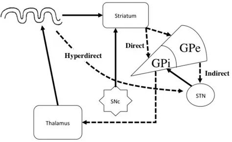
It is well established that motor, sensory and association areas of the cortex are extensively connected with specific subdivisions of the basal ganglia to form a series of ‘basal ganglia-thalamocortical’ circuits. Several distinct circuits have been described, including the motor, oculomotor, limbic and associative circuits (Alexander et al., 1986). These functionally and anatomically segregated pathways mainly relay information from functionally related cortical regions, the striatum, pallidum and substantia nigra and the thalamus. Understanding the connections within and between these circuits is crucial for procedures such as DBS, as an intervention in one area will have specific effects across a wide range of areas (Wichmann & DeLong, 2011). In the motor basal
ganglia-thalamocortical circuit, somatotopically organized information from the somatosensory, motor, premotor and supplementary motor cortices is projected through the putamen, STN, GPi, GPe and SNr to the ventrolateral nucleus of the thalamus. The thalamus finally projects back to the cortex, forming a closed loop of tightly interconnected regions
The motor cortico-striatal loop can further be divided into ‘direct’ and ‘indirect’ pathways, by which competing processes between the putamen, GP, STN and SNr
determine overall thalamic activity (Alexander et al., 1990). Specifically, the direct pathway connects the striatum to the GPi/SNr by a single inhibitory projection. By contrast, the indirect pathway connects the striatum to the GPi via inhibitory projections to the GPe and the STN and ultimately excitatory connections to the GPi/SNr. An overall output is finally sent from the GPi/SNr to the thalamus; the direct pathway causes the striatum to disinhibit
27
the thalamus, whereas the indirect pathway causes the striatum to inhibit thalamic activity (Figure 15). The signals from the direct and indirect pathways create a balance of opposing contributions, allowing movement to be regulated via thalamocortical connections.
However, these pathways are not entirely independent as evidence suggests that there are synaptic connections between the direct and indirect motor cortico-striatal pathways (Yung et al., 1996).
Figure 15: Schematic of cortico-basal ganglia circuits in healthy individuals
Solid lines represent excitatory connections, dashed lines represent inhibitory connections. There is also evidence for additional connections directly from the cortex to the STN (Monakow et al., 1978), referred to as the ‘hyperdirect’ pathway. One possibility would be that signals to the thalamus are first modulated by the inhibitory hyperdirect pathway, followed by the excitatory direct pathway, and finally by the inhibitory indirect pathway (Nambu et al., 2002). The STN, reflecting the organization of the basal ganglia into motor and associative and limbic portions, functions as a major relay station and modulator in the processing of cortico-striatal information (Hamani et al., 2004).
Neuromodulators
Dopamine is a prominent neurotransmitter in the basal ganglia, and, among other functions, plays a major role in movement through the cortico-striatal pathway. Although
many different neurotransmitters are implicated in brain circuitry and PD, this review will focus on dopamine in particular. Dopaminergic projections are sent from the substantia nigra to the striatum, forming the nigrostriatal pathway. Additional dopaminergic
projections run from the VTA to the nucleus accumbens (mesolimbic pathway) or frontal cortex (mesocortical pathway). In the context of the motor cortico-striatal circuit, dopamine has a contrasting effect on the direct and indirect pathways through a differential effect on D1 and D2 receptors (Gerfen et al., 1990; DeLong & Wichmann, 2007). Specifically, dopamine has a net inhibitory effect on the indirect pathway and a net excitatory effect on the direct pathway. The end result is that dopamine effectively favours the direct pathway, and its depletion or excess creates an imbalance in the two circuits, affecting activity in most cortical regions through the different cortico-striatal loops. This ultimately leads to the movement-related difficulties observed in different patient populations.
Cortico-cerebellar circuit
Anatomical connections
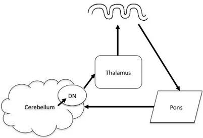
The cortico-cerebellar circuit is similarly organized into functionally segregated pathways that connect regions of the cerebellar cortex with the cerebral cortex. Lateral portions of the cerebellar cortex send projections, via the dentate nucleus, to the thalamus, which in turn projects to specific cortical areas (Figure 16). Retrograde transneuronal transport methods using neurotropic viruses have shown that these cortical areas include the motor, premotor, oculomotor, prefrontal cortex (PFC) and posterior parietal cortex (PPC) with minimal overlap between different termination sites (Clower et al., 2001; Middleton & Strick, 2001; Strick et al., 2009). Projections from the cortex back to the lateral cerebellum pass either through the pons or the red nucleus and inferior olive (Leiner et al., 1989). Furthermore, the segregation of connections to the cerebral cortex is maintained in the cerebellar cortex (Kelly & Strick, 2003), such that the separate compartments of the cerebellum form closed anatomical loops with the specific cortical region they send projections to and receive input from (Strick et al., 2009).
29
Figure 16: Schematic of the cortico-cerebellar circuit Wave: cortex; DN: dentate nucleus.
The cerebellar cortex is organized into very regular molecular, Purkinje and granular cell layers, suggesting that the type of information processing in the cerebellar cortex is mainly related to its associations with different cortical regions, rather than local circuitry (Ramnani, 2006). The dentate nucleus seems to consist of distinct sections that process motor and non-motor information (Dum & Strick, 2003), with the non-motor portion of the dentate nucleus substantially larger than the motor section (Matano, 2001). In fact, two main circuits have been described, notably the ‘motor’ loop that projects from the motor and PMC to the dorsal part of the dentate nucleus, and the ‘prefrontal’ loop that connects Brodmann area 9/46 and the ventral dentate nucleus (Glickstein et al., 1985; Orioli & Strick, 1989; Schmahmann & Pandya, 1995; Kelly & Strick, 2003). This segregation of motor and non-motor connections from the dentate nucleus is maintained in the cerebellar cortex, with separate locations being connected to areas such as the primary motor cortex and area 46 (Strick et al., 2009). Diffusion tensor imaging (DTI) examining the distribution of fibers in the cerebellar peduncle in humans and macaque monkeys in vivo has revealed that the majority of fibers in the macaque consist of motor fibers, whereas humans have a much