Université de Montréal
The Association between Caries and
Periodontal Diseases
Par
Arezou Roufegari Nejad
Département de santé buccale Faculté de médecine dentaire
Mémoire présenté à la Faculté des études supérieures et postdoctorales en vue de l’obtention du grade de
Maîtrise ès Sciences (M.Sc.) en sciences bucco-dentaires
Mars 2012
Université de Montréal Faculté des études supérieures
Ce mémoire intitulé:
The Association between Caries and Periodontal Diseases
Présenté par : Arezou Roufegari Nejad
A été évalué par un jury composé des personnes suivantes :
Dr Daniel Fortin, président-rapporteur Dr Robert Durand, directeur de recherche Dre Elham Emami, co-directrice de recherche Dr Bryan S. Michalowicz, examinateur externe
Résumé
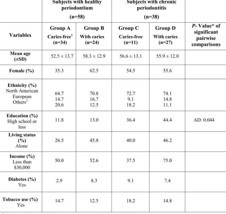
Objectifs: Le but de cette étude clinique était de comparer un groupe d’adultes ayant un parodonte sain avec un groupe d’adultes atteints de parodontite chronique en terme de risque carieux et mesures cliniques et microbiologiques de la carie.
Méthodes: Quatre-vingt-seize individus ont été divisés en deux groupes en fonction de leur état de santé parodontal et ont été appariés pour l'âge, le sexe et l'origine ethnique. Trente-huit sujets étaient atteints de parodontite chronique définie comme ayant au moins quatre dents avec ≥ 1 site avec une profondeur de sondage ≥ 4 mm et une perte d'attache clinique ≥ 2 mm, et 58 sujets présentaient un parodonte sain. Par la suite, les groupes ont été subdivisés en deux groupes en fonction de leur statut carieux : les participants ayant au moins une lésion carieuse non traitée sur une surface dentaire et ceux n’ayant pas de lésion carieuse non traitée. Les données ont été recueillies par le biais d’un questionnaire, un examen clinique et des échantillons de plaque supra- et sous-gingivale. L’évaluation de la charge buccale de Streptococcus mutans et de six agents pathogènes parodontaux a été réalisée par la technique d'amplification de la réaction en chaine de la polymérase (PCR). Les données ont été analysées à l'aide d’analyses statistiques descriptives et bivariées.
Résultats: Les individus atteints de parodontite chronique étaient 3,5 fois plus susceptibles d'avoir des caries que les individus en bonne santé (OR 3,5 ; IC: 1,5 - 8,3 ; P = 0,006). Les sujets à la fois atteints de parodontite chronique et de caries dentaires ont eu un niveau d’éducation significativement plus faible que les sujets ayant un parodonte sain et sans caries dentaires (OR 6,0 ; IC: 1,7 à 21,7 ; P = 0,04). La proportion de sujets ayant une charge buccale élevée de Porphyromonas gingivalis (P. g.) et
Treponema denticola (T. d.) était significativement plus élevée chez les patients atteints de parodontite
chronique et de carie que chez les patients sains présentant des caries (P. g.: OR 8,6 ; IC: 2,4 - 30,3 ; P = 0,004 et T. d.: OR 10,0 ; CI: 2,6 - 38.1 ; P = 0,003).
Conclusions: Les résultats de cette étude suggèrent que, chez les sujets adultes atteints de la parodontite chronique, la fréquence des caries est plus élevée que chez les sujets ayant un parodonte sain. De plus, le faible niveau d'éducation influence négativement le statut parodontal des individus.
Mots-clés: Maladie parodontale, caries, Streptococcus mutans, Porphyromonas gingivalis, Treponema
Abstract
Aim: The aim of this clinical study was to compare adults with a healthy periodontium and those with chronic periodontitis, in terms of caries’ risk and caries’ clinical and microbiological measures.
Methods: Ninety-six healthy adults were divided into chronic periodontitis (n= 38) and healthy periodontium (n=58) based on their periodontal status, and matched for age, gender, and ethnic background. Chronic periodontitis was defined as having at least four teeth with ≥1 site with a pocket depth ≥4 mm and clinical attachment loss ≥2 mm. Each group were subsequently subdivided in 2 groups according to their caries status: participants having at least one untreated decayed surface and those with no untreated caries. Data were collected by means of self-administrated questionnaire, clinical examination, and supra- and subgingival plaque sampling. Assessments of oral levels of
Streptococcus mutans and six periodontal pathogens were conducted by PCR amplification techniques.
Data were analyzed using descriptive and bivariate statistical tests.
Results: Individuals with chronic periodontitis were 3.5 times more likely to have caries than healthy individuals (OR 3.5; CI: 1.5 – 8.3; P = 0.006). Subjects with both chronic periodontitis and dental caries had a significantly lower level of education than periodontally healthy subjects without dental caries (OR 6.0; CI: 1.7 – 21.7; P = 0.04). A significant higher proportion of subjects with high oral levels of
Porphyromonas gingivalis (P. g.) and Treponema denticola (T. d.) was found among subjects with
chronic periodontitis and untreated caries compared to periodontally healthy subjects with untreated caries (P. g.: OR 8.6; CI: 2.4 – 30.3; P = 0.004 and T. d.: OR 10.0; CI: 2.6 – 38.1; P = 0.003).
Conclusion: The results from this study suggest that, adults with chronic periodontitis are more prone to caries disease than those adults with a healthy periodontium. Furthermore, low educational level could have a negative impact on the periodontal status of individuals.
Keywords: periodontal disease, caries, Streptococcus mutans, Porphyromonas gingivalis, Treponema
Table of Contents
Résumé ... iii
Abstract ... v
Table of Contents ... vi
List of Tables ... viii
List of Figures ... ix
List of Acronyms and Abbreviations ... x
Acknowledgments ... xii
CHAPTER ONE ... 1
1: Introduction ... 2
1.1: Periodontal Disease ... 3
1.1.1: Definition and Classification ... 3
1.1.2: Epidemiology ... 6
1.1.3: Etiology ... 6
1.1.4: Pathologic Pathways ... 8
1.1.5: Risk Factors ... 9
1.1.6: Diagnosis... 12
1.1.7: Prevention and Treatment ... 13
1.2: Dental Caries ... 14
1.2.1: Definition and Epidemiology ... 14
1.2.2: Stages of Disease ... 15
1.2.3: Etiology ... 16
1.2.4: Pathologic Pathways ... 19
1.2.5: Risk Factors ... 21
1.2.6: Diagnosis... 24
1.2.7: Prevention and Treatment ... 26
1.3: Current Knowledge on the Association between Caries and Periodontal Diseases ... 28
1.3.1: In Vitro Studies ... 28
1.3.2: Human Studies ... 28
2: Material and Methods ... 32
2.1: Problem Statement ... 32
2.2: Hypotheses ... 32
2.3: Research Aims ... 33
2.4: Study Design and Study Population ... 33
2.5: Data collection: ... 35
2.5.1: Medical and Socio-demographic Questionnaires ... 35
2.5.2: Clinical Examination ... 35 2.5.3: Microbiological Investigation ... 36 2.6: Statistical Analysis ... 41 2.7: Bioethical Considerations ... 42 CHAPTER THREE... 43 3: Results ... 44 3.1: Manuscript ... 44 CHAPTER FOUR ... 64 4: Discussion ... 65 5: Conclusion ... 73 BIBLIOGRAPHY ... 74 APPENDICES ... 95
Appendix I: Consent form for participants with periodontal disease ... 96
Appendix II: Consent for healthy participants ... 102
Appendix III : Socio-demographic questionnaire ... 108
Appendix IV: Medical questionnaire ... 109
Appendix V: Caries record form ... 110
Appendix VI: Periodontal record form ... 111
List of Tables
Chapter 1
Table 1.1: Abbreviated Version of the 1999 AAP Classification of Periodontal Disease and Conditions
Chapter 2
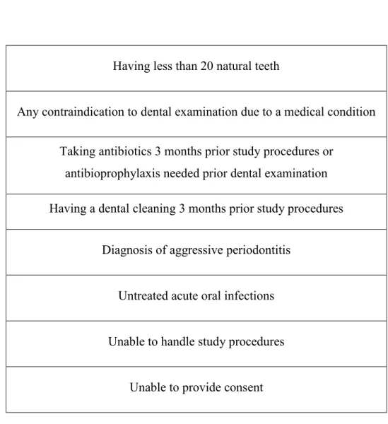
Table 2.1: Exclusion Criteria
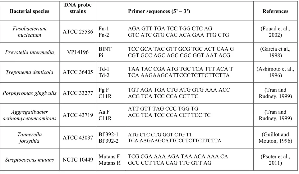
Table 2.2: Primer Sequences for Selected Bacterial Species
Chapter 3
Table 3.1: Socio-demographic Characteristics by Subgroups Table 3.2: Clinical Parameters by Subgroups
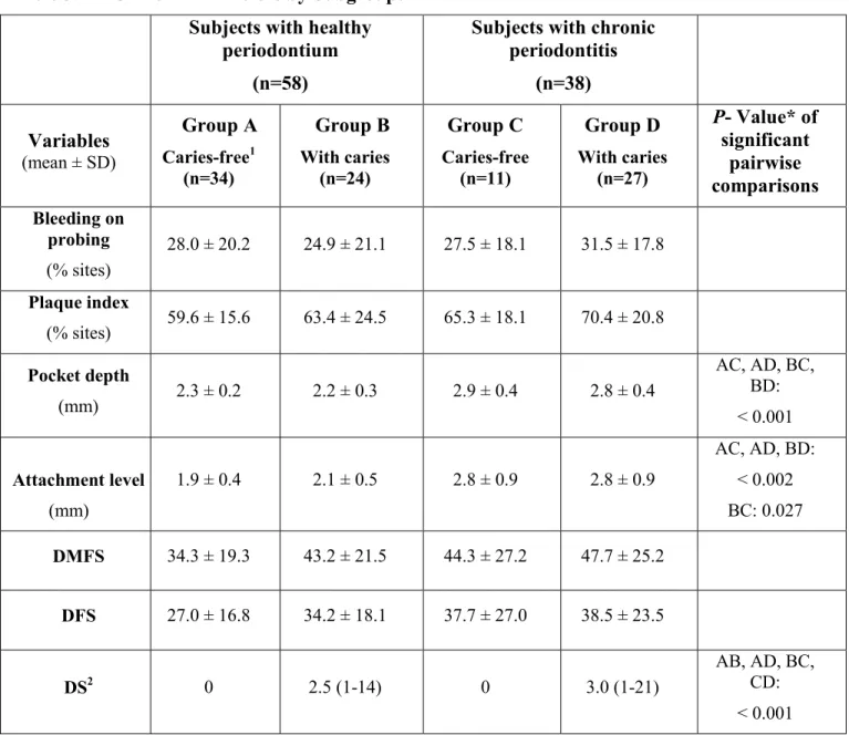
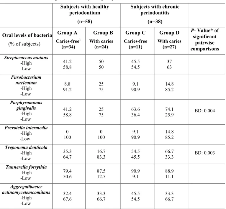
Table 3.3: Microbiological Profiles by Subgroups
List of Figures
Chapter 1
Figure 1.1: Conceptualizing the Caries Process
Figure 1.2: Main Etiological Risk Factors of Dental Caries in Caries Development Figure 1.3: Risk Factors of Dental Caries
Chapter 2
Figure 2.1: Serial Dilutions for PCR Semi-quantitative Assessments
Chapter 3
Figure 3.1: Percentage of Study Population with Caries According to Their Periodontal Status Figure 3.2: Example of a PCR Semi-quantitative Assessments for P. gingivalis
List of Acronyms and Abbreviations
° Degree mm Millimeter mM Millimolar μl Microliter mg Milligram g GramDMFS Decayed, Missing and Filled tooth surfaces index DMFT Decayed, Missing and Filled teeth index
BOP Bleeding on probing PD Pocket depth
PI Plaque index
CAL Clinical attachment loss
P. g. Porphyromonas gingivalis T. d. Treponema denticola P. i. Prevotella intermedia A. a. Aggregatibacter actinomycetemcomitans S. m. Streptococcus mutans T.f. Tannerella forsythia CI Confidence interval OR Odds ratio SD Standard deviation P P value
PCR Polymerase chain reaction
dNTP Deoxyribonucleotide triphosphate AAP American Academy of Periodontology CHMS Canadian Health Measure System OD Optical density
Acknowledgments
~
I would like to give my sincere appreciation and deep gratitude to my supervisor, Dr. Robert Durand, for his exceptional support, encouragement and guidance from the very early stages of this thesis. Most importantly, he gave me never-ending encouragement and support in many ways. I am indebted to him more than he knows.~
I gratefully acknowledge my co-supervisor, Dr. Elham Emami, for her valuable advice and continuous support throughout my thesis. Her patience alone with her quest for excellence is incredible and makes her an exceptional factor. I am really grateful for all her support and excellent supervision.~
I am grateful to Dr. Fatiha Chandad from the Université Laval for sharing her knowledge and all experience and training during my project. She has taught me so much and offered me tremendous help.~
I would like to thank Mr. Jabrane Azelmat from the Université Laval for his assistance with the microbiological assessment at the laboratory.~
I would like to thank everyone at the laboratory of Dr. Jean Barbeau for providing a familiar and friendly working environment, especially Dr. Jean Barbeau and Ms. Annie Leduc for their advice and assistance. Without their help and support, I would never been here in the position to continue and finish laboratory procedures of my research.~
I wish to sincerely thank Mrs. Chantal Morand for categorizing and scheduling study subjects.~
I would like to thank Mr. Pierre Rompré for his assistance with statistical analysis.~
I would like to thank my brother and colleague Ali, his wife Nahal and their lovely daughter Rejina, for their understanding, endless patience and encouragement when it was most needed during the past two years.~
Lastly, I am deeply and forever indebted to my parents for their never-ending love, support, and encouragement throughout my entire life and for providing me high-quality education and enthusiasm for graduate studies. They are my source of strength and without their never-ending support, this thesis would not have been possible.CHAPTER ONE
Introduction
and
Literature Review
1: Introduction
Poor oral health is an important determinant of morbidity and mortality in the population at large (World Health Organization., 1987; Appollonio et al., 1997; Shimazaki et al., 2001). There is also growing evidence of the association between oral and general health. Periodontal diseases have been shown to be implicated in the causal pathway of diabetes, coronary heart disease, and premature birth, among other conditions (Chambrone et al., 2011; Lakschevitz et al., 2011; Manjunath et al., 2011). Unfortunately, oral diseases such as dental caries and periodontal diseases are among the most prevalent chronic diseases in Canada and worldwide (Ong, 1998; Beltran-Aguilar and Beltran-Neira, 2004; Sanz and van Winkelhoff, 2011).
A recent Canadian Health Measures Survey (CHMS) found that 57% of children aged 6–11 years, 59% of 12–19-year-olds, 96% of adults, and 21% of dentate Canadian adults have moderate to severe periodontal disease (Health Canada, 2010). Furthermore, 96% of adults reported history of dental caries (Health Canada, 2010). Epidemiologic data from the province of Québec demonstrated that a majority of adult Quebecers suffer from periodontal disease and almost half of their tooth surfaces are affected by caries (Brodeur et al., 2000; Brodeur et al., 2001).
These data indicate that the Canadian oral healthcare system faces major challenges. In 2009, Canada spent 12.8 billion dollars on oral health care (Health Canada, 2010). Thus, there is a need to identify risk factors of poor oral health that could be improved by preventive measures. However, clinical studies examining the interaction of oral diseases, their common ethological pathogens, risk factors, and microbiological interactions are scarce and need to be addressed.
This research project investigating the association between dental caries and periodontal diseases in Quebec adults was therefore conducted to provide new evidence to further our understanding of whether exposure to periodontal pathogens will influence the susceptibility of individuals to dental caries. This chapter consists of a review of the literature offering background knowledge on this topic.
1.1: Periodontal Disease
1.1.1: Definition and Classification
Periodontal disease is defined as “an inflammatory disease of the supporting tissues of the teeth caused by specific microorganisms or groups of specific microorganisms, resulting in progressive destruction of the periodontal ligament, alveolar bone loss, pocket formation, and gingival recession” (Novak, 2012).
The classification of periodontal diseases has been modified over the years. In 1989, the “World Workshop on Clinical Periodontics” proposed classification of different forms of periodontal disease including early-onset periodontitis, adult periodontitis, necrotizing ulcerative periodontitis, refractory periodontitis, and periodontitis associated with systemic diseases (Nevins et al., 1989; Novak, 2012). Ten years later, a different classification of periodontal diseases was reported by the “International Workshop for a Classification of Periodontal Diseases and Conditions” and has been endorsed by the American Academy of Periodontology (International Workshop for a Classification of Periodontal Diseases and Conditions, 1999) (Table 1).
The most prevalent form of periodontal disease is chronic periodontitis, formerly known as adult periodontitis. The term “chronic” was selected because it was less limited in being neither specific nor age-dependent (International Workshop for a Classification of Periodontal Diseases and Conditions, 1999; Armitage, 1999). The 1999 International Workshop defined chronic periodontal disease as “an infectious disease resulting in inflammation within the supporting structures of teeth, and progressive attachment and bone loss. It is characterized by pocket formation and ⁄ or gingival recession. Its onset may be at any age but is most commonly detected in adults. The prevalence and the severity of the disease increase with age. It may affect a variable number of teeth and has variable rates of progression.”(Armitage, 1999). Moreover, the amount of destruction is in line with the presence of local factors, and signs of inflammation are variable according to the patient’s plaque control (International Workshop for a Classification of Periodontal Diseases and Conditions, 1999).
Depending on the number of sites involved, chronic periodontitis can be either localized or generalized. Periodontitis is classified as localized periodontitis when 30% or less of sites are affected, and as generalized periodontitis if more than 30% of sites are affected (International Workshop for a Classification of Periodontal Diseases and Conditions, 1999).
Table 1.1: Abbreviated Version of the 1999 AAP Classification of Periodontal Diseases and Conditions
Gingival Disease
Dental Plaque-Induced Gingival Diseases Non-Plaque-Induced Gingival Diseases Chronic Periodontitis
Localized
Generalized (More than 30% of sites are involved) Aggressive Periodontitis
Localized
Generalized (Interproximal attachment loss affecting at least three permanent teeth other than first molars and incisors)
Periodontitis as a Manifestation of Systemic Diseases Associated with Hematological Disorders
Associated with Genetic Disorders Not Otherwise Specified
Necrotizing Periodontal Diseases Necrotizing Ulcerative Gingivitis Necrotizing Ulcerative Periodontitis Abscesses of the Periodontium
Gingival Abscess Periodontal Abscess Pericoronal Abscess
Developmental or Acquired Deformities and Conditions
Localized Tooth-related Factors that Modify or Predispose to Plaque-induced Gingival Disease/Periodontitis
Mucogingival Deformities and Conditions Around Teeth
1.1.2: Epidemiology
Periodontal disease can affect up to 90% of the population, depending on race, geographic location, and diagnostic threshold (Pihlstrom et al., 2005). Mild to moderate chronic periodontitis is the most common form of periodontitis worldwide, with prevalence ranging from 13% to 57% depending on oral hygiene and socio-economic status (Rylev and Kilian, 2008). According to the 2007-2009 Canadian Health Measure Center survey (CHMS), 32.3% of Canadian adults showed signs of gingivitis in one or more locations in the mouth (Health Canada, 2010). In addition, 21% of adults Canadian demonstrated a moderate or severe form of periodontal disease (Health Canada, 2010). Moreover, 21.1% of Canadians had lost 4 mm or more of attachment and 6.0% had lost 6 mm or more (Health Canada, 2010). In addition, periodontitis is one of the major causes of tooth loss among the Quebec population and about half of 35-to-44-year-old Quebecers have at least one tooth with a periodontal pocket of 4 to 5 mm and one person out of five has at least one periodontal pocket >6 mm (Brodeur et al., 2001). Moreover, it was estimated that just 5.2% of Quebec adults do not need any periodontal therapy (Brodeur et al., 2001).
1.1.3: Etiology
The nature and the quantity of the oral microflora differ between sites within a patient and among patients (Moore et al., 1984). The oral biofilm contains mainly microbes and host proteins that start to adhere to teeth within a few minutes following dental oral hygiene procedures. Equal proportions of gram-positive cocci, especially streptococcus spp and actinomyces sp., dominate in the healthy gingival sulcus (Abiko et al., 2010; Darveau, 2010). Maturing plaque contributes to the development of facultative anaerobic microorganisms, spirochaetes, and motile rods. With increased disease severity, the strict proportions of anaerobic, gram-negative, and motile organisms increase significantly (Zambon, 1996; Sakamoto et al., 2005).
Transition from gingivitis to periodontitis is not well understood but it may be explained by the implication of host defense mechanisms, additional microbial species, and the invasion of certain
species into the periodontal tissue (Modeer and Wondimu, 2000). Investigators have underlined the fact that there are more than 500 species of microorganisms, such as gram-positive and gram-negative bacteria, yeast, protozoa, and viruses inhabiting the oral cavity, and their interactions within the biofilm are yet to be understood (Moore and Moore, 1994; Socransky and Haffajee, 1994; Paster et al., 2001). Anaerobic gram-negative rods and spirochetes are among the mostprevalent bacteria in the periodontal lesion (Holt and Ebersole, 2005; Zijnge et al., 2012). The predominant species include Porphyromonas
gingivalis, Prevotella intermedia, Tannerella forsythia formerly known as Bacteroides forsythus, Aggregatibacter actinomycetemcomitans, Veillonella parvula, Wollinella recta, Eikenella corrodens, Treponema denticola, Peptostreptococcus micros, Prevotella melaninogenica, Fusobacterium nucleatum, Campylobacter rectus, Selenomonas sputigena, andStreptococcus intermedius (Lovegrove,
2004; Holt and Ebersole, 2005; Zijnge et al., 2012). Most of these bacteria are highly motile and proteolytic. Some studies indicated that among these bacteria, A. actinomycetemcomitans, P. gingivalis, and T. forsythia are highly related to destruction of the periodontal tissues (Sanz et al.,
2004). On the other hand, colonization of these bacteria has been reported to vary among populations with different race/ethnicity or geographic origins (Sanz et al., 2000; Haffajee et al., 2004; Lopez et al., 2004). For instance, Sanz et al. in 2000 studied subgingival plaque samples of people living in Spain and the Netherlands and matched the groups for age, gender, and periodontal disease variables (Sanz et al., 2000). They observed that samples of Spaniards exhibited high oral levels of P. gingivalis and low levels of A. actinomycetemcomitans, while Dutch samples demonstrated high prevalence of A.
actinomycetemcomitans and P. micros. Griffen et al. studied patients of The Ohio State University
College of Dentistry, and showed that there was a difference in the level of P. gingivalis in healthy periodontium depending on ethnic background, being positive in 22% of whites, 53% of African-Americans, and 60% of Asian-Americans (Griffen et al., 1998). A. actinomycetemcomitans is mostly
associated with aggressive periodontitis in young adolescents (Henderson et al., 2010). P. gingivalis is a gram-negative black-pigmenting anaerobic bacterium, playing a key role in the initiation and progresssion of chronic periodontal disease (Loesche, 1999; Grenier and La, 2011). Furthermore, a cross-sectional study on 1,300 individuals indicated that P. gingivalis and Tannerella forsythia are associated with attachment loss or alveolar bone loss (Grossi et al., 1994; Grossi et al., 1995). In 1998, Socransky and colleagues using DNA probes identified that T. forsythia, P. gingivalis, and T. denticola were associated with increasing pocket depth and bleeding on probing (Socransky et al., 1998). A variety of other local factors including oral hygiene, pH, temperature, availability of nutrients and
water, anatomy of oral structures, salivary flow, and use of chemotherapeutic agents have been also reported to influence the growth of oral microorganisms (Marcotte and Lavoie, 1998; Burt, 2005; Pihlstrom et al., 2005).
1.1.4: Pathologic Pathways
Microorganisms inhabiting the oral biofilm, such as Gram anaerobes, can release endotoxins that penetrate periodontal tissues and trigger a host immune response. This response can result in tissue destruction either directly, by the action of enzymes and endotoxins, or indirectly by stimulating an inflammatory reaction within the host tissues through different pathways (Somma et al., 2010). Tissue response to bacterial antigens can be both protective and destructive. For example, bacterial toxins can stimulate the immune system to overproduce cytokines. Although inflammatory mediators such as cytokines are usually beneficial for tissue healing, in high quantity they can result in inflammation and tissue damage. The immune response to bacteria results in stimulating the periodontal tissue host cells to release enzymes such as matrix metalloproteinase that breaks down the connective tissue, leading ultimately to tooth loss. These enzymes and inflammatory mediators can not only affect the tissues of the oral cavity, but also can potentially damage other body organs by entering into the vascular system (Pender et al., 1997; Graves and Cochran, 2003).
Chronic periodontitis often results in gingival recession, which exposes the root surface to the aerobic flora of the mouth. The saliva buffering action can neutralize the pH level on the root surface and change the environmental condition into one that enables the growth of cariogenic bacterial species on the root surface (Saotome et al., 2006). This phenomenon can explain the potential risk of root caries in patients affected with chronic periodontitis, since the root surface is more susceptible to caries than the enamel of the tooth crown.
1.1.5: Risk Factors
Risk factor is defined as “an environmental exposure, aspect of behavior or an inherent characteristic which is associated with a disease” (Last and Association internationale d'épidémiologie, 2001). Different biological, behavioral, and systemic conditions have been introduced as potential risk factors for periodontal disease. These include age, gender, race/ethnicity, genetics, socioeconomic status, smoking, diabetes, obesity, HIV infection, osteoporosis, and psychological factors (Borrell and Papapanou, 2005).
Age
Although severe periodontitis can also be found in young adults, the severity and prevalence of periodontal disease increases with age (Burt, 1992; Borrell and Papapanou, 2005; Burt, 2005; Pihlstrom et al., 2005). It is hypothesized that degenerative systemic changes as a result of age may increase the susceptibility of elders to this chronic disease. The level of attachment loss increases with age, while pocket depth variation is minimal over the years (Albandar, 2002). Attachment loss and bone loss in elders might be caused by prolonged exposure to other risk factors during the individual’s life. Conversely, studies have demonstrated that elders following a specific preventive program can have minimal attachment loss (Persson et al., 1998).
Gender
Although, there is no inherent difference between men and women in susceptibility to periodontal disease, this disease is more prevalent in men. This might be explained by the fact that women pay usually more attention to their oral hygiene and use dental services more often (Albandar, 2002; Burt and Eklund, 2005). Therefore, gender differences observed in the prevalence and severity of periodontal diseases can be a result of a higher frequency of preventive methods rather than genetics.
Race/Ethnicity
National surveys in the United States have demonstrated that ethnicity is an important risk factor for periodontal disease (Albandar et al., 1997; Albandar et al., 1999). Periodontal disease has been seen to
vary significantly among different populations as a result of different levels of exposure to various etiological and risk factors of periodontitis in addition to differences in genetic profiles (Vo and Park, 2008; Sanders Thompson et al., 2009). Numerous studies have reported a higher prevalence of periodontal diseases in African-Americans and a similar prevalence in Mexicans and non-Hispanic white people (Borrell et al., 2002; Hyman and Reid, 2003). Similarly, Borrell et al found Black individuals to demonstrate twice as often periodontal disease versus Caucasian subjects (Borrell et al., 2005). Moreover, specific periodontal pathogens are seen in certain racial and ethnic populations. For example, Aggregatibacter actinomycetemcomitans serotype c colonizes more often Asian populations while the JP2 clone of A. actinomycetemcomitans periodontitis causes aggressive periodontitis in adolescents of the Mediterranean and Western parts of Africa (Haubek et al., 2001; Kim et al., 2009).
Genetic factors
Genetic factors play a significant etiologic role in about half of the subjects affected by periodontal diseases (Michalowicz, 1994). Studies on twins have demonstrated that clinical measures of periodontitis such as probing pocket depth and attachment loss can be affected by genetic factors. The family history of aggressive periodontitis indicates the genetic involvement of this category of periodontal disease (Loos et al., 2005; Frydman and Simonian, 2011). Although the most important modifier of periodontal phenotypes is genetic determination, the role of single nucleotide polymorphisms is ambiguous (Hart and Kornman, 1997; Schenkein, 2002).
Socio-economic status
Lower socio-economic status does not alone result in increased prevalence of periodontal diseases when adjusted for other factors such as smoking and poor oral hygiene (Novak, 2012). However, periodontal disease is more prevalent in low socio-economic populations (Lopez et al., 2001). This might be due to reduced accessibility to oral health facilities.
Smoking
The prevalence of periodontal disease, gingival recession, and attachment loss is higher in smokers than in non-smokers (Barbour et al., 1997; Albandar et al., 2000), and there is a strong association between smoking habit and both prevalence and severity of periodontal disease (Albandar et al., 2000; Bergstrom et al., 2000). Longitudinal clinical studies have reported that the influence of smoking has a
greater impact on the healing process of periodontal disease than on disease progression (Kinane and Radvar, 1997; Faddy et al., 2000; Papantonopoulos, 2004; Rieder et al., 2004).
Diabetes mellitus
Over the last decades, diabetes mellitus has been considered as one of the major risk factors for periodontitis (Grossi and Genco, 1998; Lalla et al., 2008). In fact, a two-way relationship between diabetes mellitus and periodontal diseases has been reported (Soskolne and Klinger, 2001). Subjects with diabetes mellitus have higher prevalence and severity of periodontal disease (Taylor et al., 1998; Lalla et al., 2008). Moreover, individuals with controlled diabetes have exhibited good response to treatment of periodontitis compared to poorly controlled diabetes subjects (Tervonen et al., 1991; Christgau et al., 1998; Mealey and Oates, 2006).
Obesity
In clinical studies, a positive relationship between obesity (body mass index [BMI] ≥ 30) and periodontitis has been reported (Saito et al., 2001; Al-Zahrani et al., 2003; Wood et al., 2003). Ithas been found that overweight subjects with insulin resistance have 1.5 times more periodontitis than individuals with high BMI and low insulin resistance (Borrell and Papapanou, 2005). Moreover, Al-Zahrani and co-workers reported a significant association between high BMI, waist-to-hip ratio, and periodontal disease in young adults (Al-Zahrani et al., 2003).
Osteopenia /Osteoporosis
Some studies reported that women with low bone mineral density have more clinical attachment loss, gingival recession, and gingival inflammation (Mohammad et al., 1997; Tezal et al., 2000; Inagaki and Noguchi, 2002). According to these studies, it has been hypothesized that low bone density in osteoporosis in combination with abnormal hormone function and genetic factors may affect the host immune system and increase susceptibility to periodontal diseases (Wactawski-Wende, 2001).
HIV disease
Patients with HIV have shown higher prevalence and severity of periodontitis (Winkler and Murray, 1987). However, in some studies, the progression of periodontal disease was not different between HIV-positive and HIV-negative groups (Robinson et al., 2000; Hofer et al., 2002). This might be
explained by the fact that some HIV-positive individuals might be under good control, with their immune system being virtually unchanged compared to healthy individuals.
Psychosocial factors
The mechanism of the potential impact of psychosocial factors in periodontal health is complex and not well understood. It has been suggested that psychosocial stress results in a change of behavior, i.e. smoking or poor oral hygiene (Genco et al., 1999), which in turn may lead to periodontitis. In a study of 1,426 subjects, it was reported that under a similar situation of financial stress, adults with poor coping behavior had more susceptibility to severe periodontitis than subjects with good coping strategies (Genco et al., 1999).
1.1.6: Diagnosis
A complete periodontal examination plays an important role in the detection and treatment of periodontal disease. This examination usually includes demographics, medical and dental history, radiographic findings, and clinical observations. The first step in detection of gingivitis is the observation of the shape, colour, and texture of gingival tissues. Common clinical signs of gingivitis include redness, swelling, and bleeding on probing (Armitage, 2004b). Redness and swelling are often seen together on the gingival margin. Bleeding on gentle probing results from minute blood ulceration and fragility of inflamed periodontal tissues and blood vessels (Armitage, 2004a). Specific periodontal measures include recording of pocket depth, clinical attachment level (Ramfjord, 1967), and plaque index (O'Leary et al., 1972) and are often taken at four to six sites around every tooth. Pocket depth is the distance between the tip of the gingival margin to the base of gingival crevice. The clinical attachment level and radiographic crestal bone levels are known to be indicators of past disease activity and damage caused by periodontitis. Clinical attachment level is the distance from the cement-enamel junction (CEJ) to the base of crevice measured with a periodontal probe (Ramfjord, 1967). Although using fewer sites in epidemiology surveys can be useful to estimate disease severity, it tends to underestimate prevalence of the disease (Ainamo et al., 1982; Stoltenberg et al., 1993).
1.1.7: Prevention and Treatment
Prevention of periodontal disease is based on the control of risk factors (Pihlstrom, 2001). When oral hygiene procedures are withdrawn, the biofilm begins to form a layer on the tooth in a 24-hour period and can result in gingivitis within 10 to 12 days (Loe et al., 1965). Tooth cleaning can reverse gingivitis into a healthy condition within one week (Loe et al., 1965; Sambunjak et al., 2011). Some studies have demonstrated that good oral hygiene, tooth brushing, flossing, and regular dental cleaning can prevent periodontal diseases and inhibit further clinical attachment loss (Loos et al., 1988; Brothwell et al., 1998; Berchier et al., 2008). A clinical study has found that using a toothbrush twice a day could reduce gingival bleeding by 35%, and when combined with flossing techniques at home resulted in an overall 67% reduction (Ahovuo-Saloranta et al., 2004). In addition, daily supervised tooth brushing in child populations can prevent gingivitis (Honkala et al., 1986). Importantly, tooth brushing and flossing have more effect on gingivitis than tooth brushing alone (Sambunjak et al., 2011).
Some publications have recommended for some patients to use chemotherapeutic agents daily as oral hygiene adjuncts in order to increase the efficacy of their oral hygiene methods. An oral rinse containing chlorhexidine 0.12%, an anti-bacterial agent, and fluoride-containing mouthrinses have been found to be effective to reduce supra-gingival plaque and gingivitis (Westfelt et al., 1983).
Initial therapy usually involves removing the supra- and subgingival biofilm and calculus around all accessible tooth surfaces through scaling and root planing (Armitage, 2004a). This procedure is efficient at reducing gingival inflammation and at preventing establishment and progression of periodontal diseases (Pihlstrom et al., 2005). However, most pathogens are not removed completely and remain in periodontal tissues and on the tooth surface due to anatomical structures, poor host defense, or bacterial invasion of soft tissues. Systemic antibiotic therapy can help the host defense system to control and remove the infection in certain refractory cases. However, using antibiotics has some disadvantages such as adverse drug reactions and increasing microorganism drug-resistance (Slots and Rams, 1990; Slots and Ting, 2002; Seiler and Herold, 2005). When patients do not respond to initial therapy, surgical therapy can also be effective in treating chronic periodontal disease (Heitz-Mayfield et al., 2002). Periodic periodontal maintenance must be part of a comprehensive therapeutic approach in order to maintain stability of the periodontium.
1.2: Dental Caries
1.2.1: Definition and Epidemiology
Dental caries is a result of imbalance of the indigenous bacteria that accumulate on the tooth surface, and occurs when demineralization of the hard tissue and destruction of the organic matter of the tooth is initiated by acid production of cariogenic bacteria (Kutsch and Young, 2011).
It is difficult to estimate the global prevalence and distribution of dental caries since the diagnosis criteria differ among studies (Selwitz et al., 2007). Despite the decrease in severity and prevalence of dental caries in developed countries, it is still one of the most common diseases especially in adults aged 35 and over (Brodeur et al., 2000). The 2007-2009 Canadian Health Measures Survey studied 5,586 Canadian people and reported that 96% of adults had one or more decayed, missing, or filled teeth (Health Canada, 2010). However, dentate adults had fewer teeth with untreated decay but had more teeth extracted and teeth filled. Also, Canadian males had more untreated tooth caries than females. Moreover, individuals with low income level have a higher amount of dental caries compared with people with higher income (Touger-Decker and van Loveren, 2003; Beltran-Aguilar et al., 2005; Health Canada, 2010). The National Health and Nutrition Examination Surveys (NHANES III) conducted in 1988–1994 and 1999–2004 in the United States reported tooth decay in 91% of dentate adults aged ≥ 20 years, 86.8% of dentate persons aged 20–39 years, 95.1% of people aged 40–59 years, and 93.1% of individuals aged more than 60 years (Beltran-Aguilar et al., 2005). In addition, dentate non-Hispanic white adults aged ≥ 20 years had higher coronal caries than non-Hispanic Black and Mexican-American individuals. Moreover, Kidd et al. found that English elderly individuals who live in their own homes had less root caries than those living in nursing homes (Kidd et al., 2000).
It was reported that generally women have demonstrated a higher level of dental caries than men (Lukacs, 2010). Three factors are related to the higher rate of caries among women (Lund, 2009):
- Flow rate of saliva in women is less than in men and results in reducing the removal of food debris from teeth.
- Food craving and alteration of immune response during pregnancy.
1.2.2: Stages of Disease
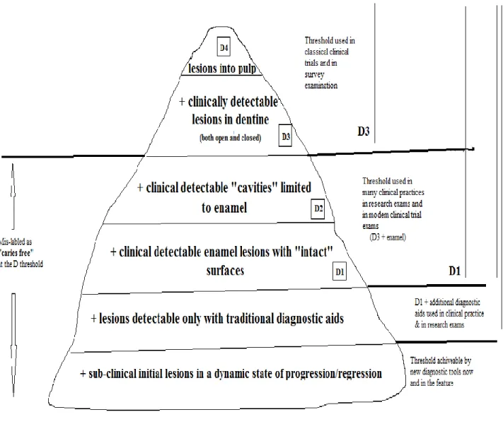
In 2002, the International Consensus Workshop on Dental Caries Clinical Trials developed a caries classification system divided into six clinical stages (Pitts, 2004; Shivakumar et al., 2009). Diagnostic threshold is a term that explains the cut-off level used to describe which lesions are “caries” and which are classified as “sound” (Pitts, 2004). This can be shown in the form of an “iceberg” of disease progression in which only the tip represents clinically detectable lesions (Figure 1.1).
The first stage is a sub-clinical lesion in a dynamic state of demineralization and remineralization with initial colonization of bacteria on the teeth. In the second stage, when the demineralization predominates, enamel progressively breaks down, a process that can be detected only with fiber-optic transillumination or bitewing radiographs. It has been many years since non-cavitated (or pre-cavitated) enamel lesions are detected and measured (Marthaler, 1984; Neilson and Pitts, 1991; Ismail, 1997; Pitts, 1997b; a; 2001). In the third stage, direct visualization after drying can detect non-cavitated
enamel lesions clinically (white and brown spots) (D1). Sound enamel is translucent and microporous.
After repeated demineralization challenges, the first sign of caries is a change in translucency and light reflection after drying for a short time. Ekstrand et al. reported the histological depth of caries according to their severity (Ekstrand et al., 1995). They indicated that white spots with air-drying are limited to the outer half of the enamel. However, the depth of white spot lesions that are obvious without air-drying is located between the inner half of the enamel and the outer third of the dentin. In the fourth stage, acid starts to weaken and dissolve parts of the enamel and sub-surface enamel is dissolved away. The surface collapses and a cavity appears that can be detected clinically with an explorer or direct visualization and is limited to the enamel (D2). When the enamel breaks down
without visible dentin, caries extend to the middle third of the dentin and in case of cavities with visible dentin, decay extends to the inner third of the dentin (Ekstrand et al., 1995). In the fifth stage, caries extend to the underlying dentin in the absence of therapy (D3). In the sixth and final stage, the bacteria
Figure 1.1: Conceptualizing the Caries Process (Pitts, 2001)
1.2.3: Etiology
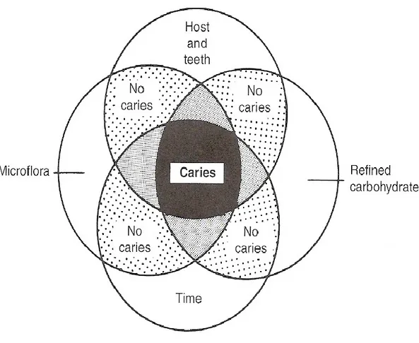
Nowadays, most experts in cariology agree that caries is an infection that can be transmitted from one person to another and that its initiation and progression are influenced by several factors (Clarkson, 1999). Initially, it was proposed that the three main etiological factors for dental caries were the host,
including the hard tooth surfaces and saliva, carbohydrate substrates, and bacteria such as mutans
streptococci and lactobacilli species (Keyes, 1960). When these three factors were present in the oral
cavity, dental caries could occur. It was later found that the frequency with which teeth are exposed to the cariogenic environment influences caries development (Newbrun, 1989). Therefore, time became the fourth primary etiological factor that was added to the initial three essential factors (Figure 1.2). Preventive methods such as topical fluoride applications, oral hygiene instructions, and dietary counseling were then developed and showed clinical efficacy against caries. Consequently, Fejerskov and Manji added more factors implicated in the caries process to create a more comprehensive risk assessment model (Figure 1.3) (Fejerskov and Kidd, 2008).
The oral cavity contains a vast array of bacteria but only a few of them are believed to be cariogenic (Teanpaisan et al., 2007). Lactobacilli and mutans streptococci species have a mutual synergy and are positively correlated with dental caries (Loesche, 1986; Berkowitz, 2003; Beighton, 2005). Two strains of mutans streptococci, S. mutans and S. sobrinus, play an important role in the etiology of dental caries (Bowden, 1990). Many studies have demonstrated the role of S. mutans in the initiation of dental caries while lactobacilli are known to play a role in the progression of carious lesions (Tanzer et al., 2001; Nishikawara et al., 2006). Higher amounts of S. mutans are seen in enamel caries compared to root caries (Brown et al., 1986). A recent study found a high prevalence of lactobacilli in root caries in elderly people, confirming earlier results (Preza et al., 2009). Many theories have been postulated to explain dental caries. As early as 1890, Miller brought forward the “Acid-parasitic Theory” of dental caries. He described oral microorganisms as capable of initiating caries by producing acid from dietary carbohydrates. These acid products lead to the loss of the mineral composition of the tooth (Miller, 1890). The “Proteolytic Theory” was then brought forward by Gottlieb in 1944 (Gottlieb, 1944). He theorized that initiation of caries was due to proteolytic enzymes of bacteria targeting protein elements of the tooth, such as lamellae and rod sheaths, and destroying the matrix of enamel by dissolving the enamel apatite crystals. Decades later, Schatz and Martin reported the proteolysis chelation mechanism (Schatz and Martin, 1962). This theory supported the simultaneous microbial degradation of the organic components known as proteolysis, and the remineralization of the tooth by the process known as chelation. The current etiologic concept of dental caries focusing on the effect of cariogenic bacteria-producing organic acid in fermentation of carbohydrates was first discovered and developed in the 1970s (Loesche, 1976). The main organic acids in the presence of fermentable carbohydrates are lactic, formic, and acetic acids. These acids contribute to decrease the pH of the oral biofilm located on the
tooth surface, causing surface demineralization and providing an advantageous environment for
Streptococcus mutans. These bacteria are responsible for the cariogenicity of the dental plaque by
expressing wide range of virulence factors.
Figure 1.3: Risk Factors of Dental Caries (Fejerskov and Kidd, 2008)
1.2.4: Pathologic Pathways
The main pathogenic characteristics of mutans streptococci and lactobacilli are adhesion, acid production, and acid tolerance (Marsh, 2009).S. mutans is a facultative anaerobic bacterium producing
lactic acid, surface antigen I/II, and a water-insoluble glucan (Hamada and Slade, 1980; Takahashi and Nyvad, 2011). Its adherence to the tooth surface is formed mainly by proteins such as glucane-binding that is triggered by the enzymatic actions of glucose transferases and water-insoluble glucans (WIGs) (Loesche, 1986; Simon, 2007). WIGs play an important role in developing initial caries through the
adhesion and aggregation processes (Mattos-Graner et al., 2000). This protein modifies physio-chemical properties of dental plaque and increases porosity of the dental plaque matrix in order to make it more cariogenic (Zero et al., 1992; Cury et al., 1997; Napimoga et al., 2004). Also, WIG decreases the calcium and phosphate concentrations in the saliva, decreasing the saliva buffer action (Napimoga et al., 2004). In addition, colonization of mutans streptococci on the tooth surface is mediated via both sucrose-independent and sucrose-dependent adhesion metabolisms. The attachment process is initiated by sucrose-independent adhesion to salivary components within the acquired enamel pellicle, while sucrose-dependent adhesion may be primarily responsible for establishing permanent colonization (Loesche, 1986). The surface protein P1, also known as antigen I/II (ca. 185 kDa), is the surface adhesion protein that is released by S. mutans and other streptococci and influences the sucrose-independent adhesion metabolism (Lee, 1992; Whittaker et al., 1996). Consequently, these microbial acid-induced adaptation and selection processes change the demineralization/mineralization balance and shift it toward mineral loss (Takahashi and Nyvad, 2011). On the other hand, S. mutans can resist other types of bacteria and thrive at low pH levels and in high glucose concentrations (Loesche, 1986). This can result in the maintenance of an acidic environment and demineralization of the tooth surface, resulting in caries.
When the lesion progresses to the stage of cavitation, the organisms penetrate into the enamel crystals and underlying dentine. In the advanced clinical stage of carious lesions, mutans streptococci do not have ability to survive at pH levels below 5.0, and lactobacilli, as a secondary cariogenic bacteria, can be found in higher concentrations (Loesche, 2007). Lactobacillus is a gram-positive facultative
anaerobic bacteria which is rod-shaped and non-spore forming (Falsen et al., 1999). It derives all its energy from the metabolism of glucans into acid fermentation and can also tolerate a low pH environment (Marcotte and Lavoie, 1998). Specific mechanisms by which lactobacilli can adhere to the tooth surface are not well understood. Recognition of collagen, a major component of dentine, might be a mechanism used by these bacteria to adhere to root surfaces as well as caries (McGrady et al., 1995; Featherstone, 2000; Love and Jenkinson, 2002).
1.2.5: Risk Factors
Inadequate Salivary flow and composition
Saliva contributes to maintain balanced oral flora that promote health and integrity of tooth surfaces (Hicks et al., 2003; Garcia-Godoy and Hicks, 2008; Kutsch and Young, 2011). Saliva protects teeth against caries with mechanisms including bacterial clearance, direct antibacterial activity, buffering, and remineralization. The buffering capacity and the ability of saliva to lubricate the oral cavity and wash away microorganisms and food debris from the tissues and teeth, help in balancing the demineralization/remineralization process on the tooth surface (Dowd, 1999; Stookey, 2008; Hara and Zero, 2010; Paice et al., 2011). For example, a reduction in the salivary flow causes an inadequate saliva buffering action, resulting in a decrease of the oral pH. Therefore, the host inability to counterbalance the acidic environment willcreate an ideal condition for the establishment of cariogenic bacteria (Simmonds et al., 2000). Moreover, saliva has a significant antibacterial activity through its enzymes that can contribute to tooth health (Spielmann and Wong, 2011). Some medications, medical conditions, and radiotherapy to the head and neck region can cause a reduction of saliva flow and dry mouth (xerostomia) resulting in more caries (Spielmann and Wong, 2011).
Oral levels of cariogenic bacteria
Tooth caries are caused by an imbalance of indigenous bacteria and can be transmitted as early as the infant stage (Caufield and Griffen, 2000; Harris et al., 2004). Many studies have demonstrated the role of S. mutans in initiating dental caries, while lactobacilli are known to have a role in the progression of carious lesions (Tanzer et al., 2001; Nishikawara et al., 2006). Teanpaisan et al. studied the acquisition of mutans streptococci and lactobacilli species in relation to dental caries. They used saliva of 3 to 24-month-old Thai children as a medium for colonization by mutans streptococci and lactobacilli (Teanpaisan et al., 2007). Subjects who had a significant early colonization by mutans streptococci and
lactobacilli showed a higher number of dental caries than whose saliva exhibited later colonization.
The authors found a significant association between high mutans streptococci and lactobacilli salivary levels and tooth caries. The level of cariogenic bacteria differs among individuals of different age
groups. For example, level of lactobacilli in saliva increases with age, particularly in individuals aged 70 years and older, while the prevalence of mutans streptococci in saliva or plaque is not related to age (Percival et al., 1991).
Diet
Sugar is a major component of our daily diet. It has a fundamental role in caries development since it is used directly by cariogenic bacteria (Mobley et al., 2009). Sugar and fermentable carbohydrates are metabolized into acids by cariogenic bacteria, resulting in low pH of the oral environment and enhancing growth of acidogenic and aciduric bacteria such as mutans streptococci. The initial colonization of mutans streptococci on the tooth surface is facilitated by sucrose. In contrast, a diet with lower intake of sugar and more calcium-rich nutrients can favor remineralization and prevent caries (Touger-Decker and van Loveren, 2003; Makinen, 2010). However, the frequency of fermentable carbohydrate intake seems to be correlated to caries more importantly than the total amount of sugar in the diet (Paes Leme et al., 2006). The 1945–1953 Vipeholm study is one of the largest clinical studies investigating the relationship between the amount of sugar intake and frequency of dental caries (Gustafsson et al., 1954). The authors concluded that consumption of sugar during meals and between meals increases the risk of dental caries. In summary, dietary factors that are related to the incidence of dental caries are:
Amount of fermentable carbohydrate consumption; Concentration of sugar in food;
The duration teeth are exposed to carbohydrates; Frequency of meals and snacks;
Sequence of food consumption.
Poor Oral hygiene
On the basis of existing literature, oral hygiene status appears to be an important factor in caries incidence (Andlaw, 1978; Bellini et al., 1981; Selwitz et al., 2007). The European Workshop on Mechanical Plaque Control approved the following theory in 1998: “Forty years of experimental research, clinical trials and demonstration projects in different geographical and social settings have
confirmed that effective removal of dental plaque is essential to dental and periodontal health throughout life” (Lang et al., 1998).
The use of fluoride toothpaste and toothbrush is the most common form of oral hygiene practiced by individuals (Selwitz et al., 2007). Mechanical oral hygiene measures such as tooth brushing and dental floss can remove the oral biofilm and food debris from the smooth and interdental surfaces of teeth (Fejerskov and Kidd, 2008). Also, toothpaste provides an ideal vehicle for the carriage of agents like fluoride for the prevention of dental caries. Papas and coworkers demonstrated that the toothbrush had the beneficial effect of improving the oral hygiene of patients with medically induced xerostomia (Papas et al., 2007). Moreover, Bellini et al. indicated that the quality of tooth cleaning was more effective than its frequency (Bellini et al., 1981).
Fluoride Exposure
Dental caries have decreased in prevalence in developed countries over the years. This decline can be explained in part due to the widespread use of fluoride, both topically and in public water supplies (Buzalaf et al., 2011). Therefore, insufficient systemic or topical fluoride exposure can enhance the prevalence of dental caries (Selwitz et al., 2007).
Smoking
Tobacco use, notably cigarette and pipe smoking, is considered a risk factor for dental caries (Hirsch et al., 1991; Jette et al., 1993; Axelsson et al., 1998). It causes bad breath, tooth discoloration, accumulation of plaque and tartar, and compromises salivary flow (Lie et al., 2001; Rooban et al., 2011). The oral levels of lactobacilli and Streptococcus mutans were shown to increase in smokers, thereby explaining the association between smoking and dental caries (Sakki and Knuuttila, 1996).
Social factors
Dental caries are significantly related to low income, low level of education, and lack of dental insurance coverage (Burt et al., 1999). Therefore, dental health education through school systems and
oral health professionals, as well as an increase in public awareness of oral health through advertisements, could explain the decrease in the prevalence of dental caries worldwide.
Medications, systemic diseases, and physical/mental disabilities
Some medications and systemic diseases can lead to xerostomia and ultimately, dental caries due to the reduced buffering action and quantity of saliva. For example, medications such as antihistamines and antidepressants, patients receiving radiotherapy to the head and neck, and some medical conditions like diabetes mellitus, diabetes insipidus, Parkinson’s disease, and sarcoidosis as well as autoimmune diseases like HIV/AIDS and Sjogren’s syndrome can cause xerostomia (Ship, 2003; Scully and Felix, 2005; Kielbassa et al., 2006; Neville, 2007; Seo et al., 2009; Johnson, 2010). In addition, a wide variety of physical and mental disabilities can result in a reduced frequency and quality of oral hygiene measures, exposing individuals to caries.
Local and iatrogenic factors
Presence of gingival recessions exposing root surfaces that are less resistant to caries (Selwitz et al., 2007), use of orthodontic appliances (Ogaard et al., 1988; van der Veen et al., 2010), poorly designed or ill-fitting fixed or removable prostheses (Featherstone et al., 2011), and defectuous restorations (Kidd et al., 2000; Aleksejūnienė et al., 2009) can result in an increase in the amount of dental caries.
1.2.6: Diagnosis
The detection and classification of dental caries is a complex task in clinical research (Ricketts et al., 1997). Investigators have studied the use of several procedures to detect dental caries (Stookey and Gonzalez-Cabezas, 2001). Different technologies are used depending on the severity, depth, and location of the carious lesion. The subclinical carious lesion in a dynamic state of demineralization and remineralization (first stage) can be detected with quantitative light-induced fluorescence (QLF) technology, near-infrared fluorescence (DIAGNOdent®, KaVo Dental GmbH, Biberach, Germany), electronic caries monitor, and digital radiography. QLF is a diagnostic tool for assessment of dental
caries lesions, dental plaque, and bacterial activity. It is also used to detect parameters like percentage of mineralization lost as well as lesion depth and size. However, it seems to be limited to a lesion depth of about 400 microns and cannot detect occlusal or secondary caries (Amaechi and Higham, 2002; Heinrich-Weltzien et al., 2005). DIAGNOdent® is an examination tool for detecting early and very small caries on occlusal surfaces (Kuhnisch et al., 2007). However, the instrument’s sensivity can be reduced by factors like presence of plaque, staining and dehydration of teeth (Lussi et al., 1999).Both methods have the same merit in quantification of smooth surface caries, although research shows that QLF can analyze changes in mineral content with better precision (Shi et al., 2001). Non-cavitated carious lesions associated with enamel breakdown (second stage) are detected only by fiber-optic transillumination technology or bitewing radiographs. Proximal lesions were diagnosed by fiber-optic transillumination two times more than was the case for clinical examination. Digital fiber-optic transillumination can diagnose early caries and even lesions that cannot be detected with radiography by using safe white light or standard light. In proximal surfaces, light is shone from one surface and captured from the opposite surface by the camera. In occlusal surfaces, the mouthpiece illuminates the tooth from both facial and lingual surfaces and images from the top of the tooth. The images are sent to the computer and analyzed with algorithms (Schneiderman et al., 1997). White carious lesions (third stage) with appearance of sound surfaces are visually detected only when forced air is blown on the suspected surfaces. The aim is to eliminate moisture and change the properties of the unmineralized enamel surface. A dental explorer is used for the detection of cavitated dental caries (fourth stage) involving the enamel of the occlusal, facial, and lingual surfaces.
In 1968, Radike reported clinical criteria for detection of frank carious lesions (Ismail, 2004). These criteria rely on visual detection, the use of gentle exploring with a dull-ended explorer for detection of caries, or radiographic images. Frank lesions are usually easily detected and the cavity corresponds to either a discontinuity of the enamel surface caused by loss of tooth surface or a cavitation resulting from the caries process. However, lesions that are not cavitated are more difficult to detect. These non-cavitated lesions are considered caries if the explorer “catches” or resists upon removal of the explorer after its insertion into a pit or fissure with a moderate to firm pressure or if a surface loses the translucency of its enamel adjacent to a pit compared to the surrounding tooth structure. They are also considered caries if the surfaces are etched or if there are white spots showing subsurface demineralization when an explorer or blown air is used. For detection of interproximal caries, the procedure for detection includes a combination of visual-tactile methods, radiographs, and
transillumination. A bitewing radiograph is recommended to detect any break in continuity of enamel surfaces caused by caries. Transillumination is mostly used for anterior teeth and will reveal a loss of translucency on calculus-free and stain-free proximal surfaces when caries are present. Root caries are mostly seen in the maxillary arch and on facial surfaces of exposed roots of mandibular teeth and can be easily detected most of the time with a visual and tactile examination (Katz et al., 1982). They are encountered in the presence of gingival recessions that leave the roots exposed to the cariogenic flora. NIDCR criteria for root caries are lesions in root surfaces that are yellow/orange, tan, or light brown (National Institute of Dental Research (U.S.). Epidemiology and Oral Disease Prevention Program., 1987). These lesions show softness to an explorer tip.
1.2.7: Prevention and Treatment
Daily removal of plaque by tooth brushing, fluoride toothpaste and dental flossing are among the most efficient ways to remove plaque and prevent caries on the tooth surfaces (Hamilton, 1977). Flossing helps in removing the debris and oral biofilm from the interproximal surfaces (Bellini et al., 1981; Roberson et al., 2006; Selwitz et al., 2007). Dietary changes such as substitution of sugar-free foods for snacks are effective at diminishing caries prevalence (Touger-Decker and van Loveren, 2003; Makinen, 2010). Reducing sugar consumption and careful oral hygiene can therefore reduce the incidence of dental caries. Another preventive method against caries is the use of pit and fissure sealants in children and adolescents aged less than 20 years (Simonsen and Neal, 2011). It aims at preventing the colonization of cariogenic bacteria into pits and fissures, areas that are hard to reach with daily oral hygiene measures, with a durable restorative material.
There are many caries prevention programs like fluoridation of water, salt, mouthrinses, and intake of fluoride tablets in schools (Selwitz et al., 2007). Fluoride can bind to hydroxyapatite crystals of the enamel (Robinson, 2009; Denbesten et al., 2011) and increase the resistance of tooth structure to demineralization. Therefore, it has a significant protective effect against bacterial carbohydrate metabolism and even reduces bacterial-driven acid formation (Balzar Ekenback et al., 2001). Fluoride can stimulate remineralization in small carious lesions but in deeper ones, the caries will have to be removed and restored to prevent the progression of caries into the tooth structure. It is generally recommended to provide topical fluoride like fluoride varnishes and gels to high-risk populations like
infants and patients with reduced salivary gland functions in a professional setting (Roberson et al., 2006; Tubert-Jeannin et al., 2011). Varnishes and gels provide high uptake of fluoride ion into enamel and have shown clinical efficacy (Lee et al., 2010).
The individual risk factors and the extent of caries dictate the treatment regimen. For small lesions, topical fluoride is often effective at promoting tooth remineralization and treating caries. However, in cavitated advanced lesions, the missing tooth structure is replaced by a restoration. For many years, dentists have use mechanical removal and tooth reconstruction with restorative materials to prevent and treat caries, and to restore the tooth structure, function, and aesthetic. When caries are too extensive and the pulp of the tooth is infected, a root canal therapy may be advised to prevent further infections. If there is not enough tooth structure remaining, replacing the tooth structure with a restoration may not be possible and dental extraction might be the only option (Roberson et al., 2006). Periodic recalls based on individual risk factors and including a dental examination, radiographs, and oral prophylaxis are recommended to monitor the patient’s caries status and detect early carious lesions. Therefore, a caries management program combining restorative therapy with a preventive approach and a comprehensive maintenance phase will reduce caries incidence.
1.3: Current Knowledge on the Association between
Caries and Periodontal Diseases
1.3.1: In Vitro Studies
The potential association between periodontal pathogens and cariogenic bacteria has been studied using different in vitro models. Four strains of periodontopathogens, F. nucleatum (F. n.), A.
actinomycetemcomitans (A. a.), P. gingivalis (P. g.), Prevotella intermedia (P. i.) and four strains of
cariogenic bacteria, S. mutans (S. m.), S. sanguis (S. s.), A. viscosus (A. v.), and L. acidophilus (L. a.) were used in an in vitro study (Rao and De-Yi, 2005). The authors found that the co-aggregations of periodontopathogens and cariogenic bacteria were specific. Fine and colleagues studied the relationship between A. actinomycetemcomitans and cariogenic bacteria growth in an in vitro model using an agar diffusion method. The agar diffusion assay showed that A. actinomycetemcomitans had an inhibitory effect on cariogenic bacteria. The authors found that the saliva from A. actinomycetemcomitans-positive subjects killed S. mutans. (Fine et al., 2007). Another in vitro study of the effect of oral streptococci on anaerobic bacteria such as P. intermedia, F. nucleatum, Veillonella, P. gingivalis, and
P. micros showed that these anaerobic bacteria were inhibited by certain strains of streptococci,
particularly S. mutans and S. salivarius, as a result of their lactic acid production(Doran et al., 2004). Alakomi et al. observed that lactic acid produced by lactobacilli can permeate the outer membrane of gram-negative bacteria and induce the inactivation of periodontopathogens (Alakomi et al., 2000). These studies suggest that there are interactions between cariogenic bacteria and periodontal pathogens.
1.3.2: Human Studies
Findings of human studies on the association between caries and periodontal diseases are controversial. Several clinical studies did not find any relationship, either positive or negative, between dental caries and periodontal diseases (Miller and Seidler, 1940; Brucker, 1943; Massler et al., 1952; Kinane et al.,