B
OVINE VIRAL DIARRHOEA VIRUS INDUCES APOPTOSIS IN BLOOD
MONONUCLEAR CELLS BY A MECHANISM LARGELY DEPENDENT ON
MONOCYTES
Michel Lambot,1 Emmanuel Hanon,1 Corine Lecomte,1 Claude Hamers,1 Jean-Jacques Letesson2
and Paul-Pierre Pastoret1
1 Department of Immunology/Vaccinology, Faculty of Veterinary Medicine, University of Liège, B43b,
B-4000 Liège, Belgium
2 Department of Immunology, Faculties Notre-Dame de la Paix, Namur, Belgium
KEYWORDS: mots-clés ABSTRACT
The induction of apoptosis by bovine viral diarrhoea virus (BVDV) was examined in bovine peripheral blood mononuclear cells (PBMC) incubated with an antigenically homologous pair of non-cytopathic and cytopathic BVDV. Our results show that the cytopathic biotype, in contrast to the non-cytopathic counterpart, induces apoptosis in PBMC. Flow cytometry analysis of cells undergoing apoptosis revealed that: (1) monocytes constitute the major cell population undergoing apoptosis; (2) cytopathic virus also induces apoptosis in BoCD4 , BoCD8 and BoWC1 T cells in whole PBMC cultures but not in purified T cell suspensions; (3) the degree of apoptosis of BoCD4 and BoCD8 T cells incubated with the cytopathic virus was significantly enhanced by the presence of monocytes. Taken together, these results suggest that bovine monocytes play an important role in apoptosis induced by cytopathic BVDV.
Bovine viral diarrhoea virus (BVDV), a positive-stranded and enveloped RNA virus, shares the genus Pestivirus, family Flaviviridae, with ovine border disease virus and classical swine fever virus (Wengler, 1991). Field BVDV isolates can be divided into two biotypes, non-cytopathic and cytopathic, according to their pathogenicity in cultured cells. An important aspect of BVDV infection is an apparent affinity of the virus for cells of the immune system. Animals acutely infected with BVDV undergo a transient alteration of the absolute number of circulating leukocytes (Ellis et al., 1988 ; Bolin et al., 1985 ; Howard et al., 1992) while exhibiting BVDV antigens in multiple lymphoid organs (Marshall et al., 1996 ; Bolin et al., 1985 ; Bezek et al., 1994). Reports of several investigations, done both in vivo and in vitro, also indicate multiple abnormalities of the immune response to BVDV infection (reviewed by Potgieter, 1995). The mechanism that leads to these functional alterations in the infected host and the kind of interaction that takes place between the virus and the immunocompetent cells remains unresolved.
In a variety of viral infections, there is increasing evidence that changes observed in lymphoid cell functions are directly associated with the induction of apoptosis or programmed cell death (Razvi & Welsh, 1995). For BVDV, the occurrence of apoptosis has recently been reported with a few cytopathic strains in long-term cultures, including kidney and testis cells (Zhang et al., 1996 ; Hoff & Donis, 1997) and bone marrow-derived macrophages (Adler et al., 1997). The purpose of the present study was to examine the induction of apoptotic cell death by BVDV in peripheral blood mononuclear cells (PBMC). We addressed the question whether the non-cytopathic/ cytopathic biotypes of the same BVDV antigenic pair were able to induce apoptosis in bovine PBMC and if so, to identify the cell population(s) involved. The PBMC were isolated from blood of normal healthy cattle by cen trifugation on Ficoll–Hypaque (Pharmacia). The cells (2x10 /ml) were cultured with the virus inoculum at an m.o.i. of 6·3 TCID50 per cell in complete RPMI 1640
medium as described previously (Lambot et al., 1997). A homologous pair of non-cytopathic and cytopathic strains, Pe515, obtained from a field case of mucosal disease (Brownlie et al., 1984), was used in this study. Viruses were multiplied onto calf testis (CT) cell cultures as described previously (Lambot et al., 1997). The supernatant from a non-infected CT cell preparation was used as control medium. After culture for 3 days, the PBMC were processed for in situ detection of apoptosis as described by Hanon et al. (1997). In a series of preliminary experiments, we observed that only the cytopathic biotype of strain Pe515 induced a significant degree of apoptosis in the PBMC (data not shown). No apoptosis over background levels was observed for blood cells cultured in the presence of the noncytopathic counterpart.
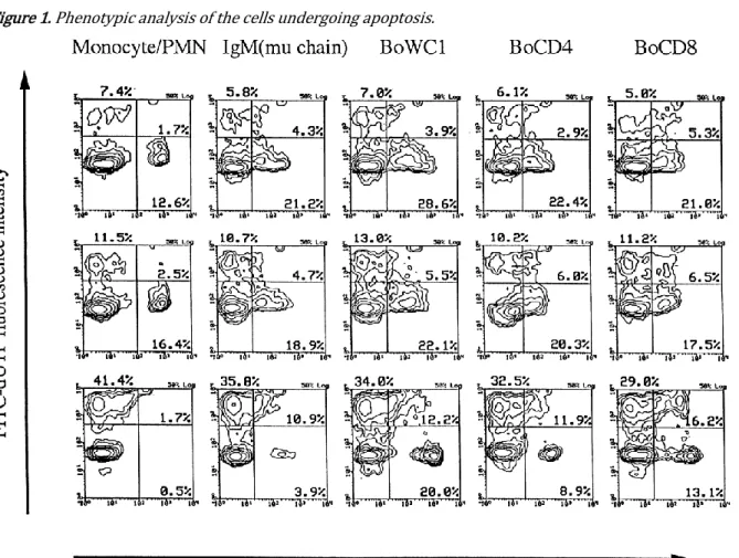
To determine which cell type(s) undergoes apoptosis, we performed double labelling experiments to detect con-comitantly cell phenotype and DNA fragmentation. The procedure followed is described by Dequiedt et al. (1997). The following monoclonal antibodies (MAbs) directed against specific surface markers were used for characterization of peripheral blood populations: IL-A30 against IgM (Naessens et al., 1988); IL-A24 against monocytes and polymorphonuclear cells (Ellis et al., 1988); CC8 against BoCD4 (Bensaid & Hadam, 1991); CC63 against BoCD8 (McHugh & Sopp, 1991); IL-A29 against BoWC1 (Davis et al., 1996). Fig. 1 shows that 38 % of the total BoWC1+ T cell population was apoptotic when PBMC were cultured with
cytopathic virus. The total percentage of apoptotic cells rose to 55–57 % in the BoCD4 and BoCD8 T cell populations. In contrast, we were unable to evaluate B cells and monocytes for apoptosis due to loss of specific surface markers after exposure to the cytopathic virus.
Therefore, to determine whether the cytopathic virus induced apoptosis in monocytes and B cells, we examined the extent of apoptosis in a cell population enriched from PBMC prior to culture in the presence of virus. The enrichment procedure for different cell populations was carried out by negative selection with paramagnetic beads (Miltenyi et al., 1990). MAbs 1H4 (Letesson et al., 1985), 11F5 (Morrison & Davis, 1991) and 2G1 (Letesson & Delcommenne, 1993), which are, respectively, specific for B cells, BoWC1+ T cells and monocytes of PBMC, were
included in the MAb solution used to enrich in T cells. MAbs 1H8, which defines T cells (Letesson & Bensaid, 1991), 11F5 and 2G1 were used to enrich in B cells. For the monocyteenriched population, MAbs 1H8, 11F5 and 1H4 were used. A BoWC1+ T-cell-enriched population was
achieved by using a mixture of MAbs 1H8, 1H4 and 2G1. Flow cytometry analysis clearly showed that a high degree of apoptosis was induced by the cytopathic virus in the monocyte-enriched population (Fig. 2). In assays with enriched B cells, the levels of apoptotic cells were only marginally elevated by the cytopathic virus in comparison with the controls. With the T cell-enriched population, the percentage of cells undergoing apoptosis induced by the cytopathic virus was not consistently higher than that observed with the cells cultured alone or with non-cytopathic virus. Importantly, this latter finding suggests that, upon enrichment, T cells become resistant to apoptosis induced by the cytopathic virus. To determine whether the presence of monocytes is required to trigger apoptosis in T cells, we performed mixing experiments in which monocytes were added, at different concentrations, to non-adherent cells isolated from PBMC by a plasma/gelatin method (Goddeeris et al., 1986). We showed that the percentages of BoCD4 and BoCD8 T cells undergoing apoptosis in cytopathic virus-containing medium were consistently greater in the presence of autologous monocytes than those observed without addition of monocytes (Fig. 3). Within the BoCD8 population, the effect of the monocytes appears to be proportion dependent, since the maximal level of apoptosis was observed at a proportion of 1 : 5 monocytes. In contrast, at the highest proportion of monocytes, the degree of apoptosis of BoCD4 T cells was 2 fold greater than that observed in non-adherent cells cultured alone. To determine whether BVDV gene expression was required for induction of apoptosis, the virus was first inactivated by a psoralen–UV treatment, as described by Hanon et al. (1997), prior to being added to cell cultures. The finding that inactivation of the virus affects its ability to induce apoptosis in PBMC (data not shown) raised the possibility that the differences observed between the biotypes for apoptosis may be explained by differences in virus load between cell populations. To examine this hypothesis, we carried out semi-quantitative infectivity studies on positively selected single cell populations. After a 24 h incubation period with each biotype of BVDV Pe515, cell populations were sorted from PBMC cultures by a FACStar equipped with the Automatic Cell Deposition Unit (ACDU). Multiple samples (n = 12) containing defined cell numbers (101 , 102 , 103 and 104 sorted cells per well) of each population were loaded onto CT
cell cultures. After two consecutive passages, an indirect immunofluorescence assay was performed to detect BVDV (Mignon et al., 1992). The percentage of cells containing BVDV antigen in the prescribed populations was calculated according to Bertram et al. (1995). BVDV was detected only in the monocytes and not in the three other populations (T, B, BoWC1+ T
cells). The rates of BVDV infectivity in monocytes have been found to be surprisingly low, ranging from 0·01 to 0·2 %. However, we found no significant difference between biotypes in the percentage of these cells positive for viral antigen.
In conclusion, three interesting points emerge from our results. First, we show that only the cytopathic biotype was able to kill cells by apoptosis in PBMC cultures while the homologous non-cytopathic virus could not. The observations by Zhang et al. (1996) and Adler et al. (1997) that other cytopathic strains were able to trigger this process in cell lines prompted us to suggest that apoptosis might be a general mechanism associated only with cytopathic BVDV strains and not with non-cytopathic ones.
Second, we report that cells undergoing apoptosis were predominantly found in the monocyte population of PBMC. However, the level of apoptosis is out of proportion to the number of cells containing BVDV antigens (ACDU results). These weak rates of infectivity suggest that the induction of apoptotic death by the cytopathic virus is not a direct consequence of the productive infection in the blood cells. This contrasts with reports from others laboratories (Zhang et al., 1996 ; Adler et al., 1997 ; Hoff & Donis, 1997). They have demonstrated apoptotic cells productively infected with BVDV. The contrasting results could be based on the use, by different laboratories, of cell types with different metabolic activity [MDBK for Zhang et al. (1996) and Hoff & Donis (1997) and bone marrow-derived macrophages for Adler et al. (1997)]. Here, we reported experiments addressing the apoptotic effect of BVDV on PBMC. Although further experiments on BVDV genome transcription and viral expression in blood cells are necessary to determine the exact type of infection involved in these cells, we hypothesize that the mechanism(s) by which cytopathic virus triggers apoptosis in monocytes could involve the expression of specific BVDV proteins rather than virus replication in infected cells. In this regard, the viral protein NS3 is a likely candidate because it is a nonstructural protein restricted to cytopathic viruses (Wiskerchen & Collett, 1991). The fact that NS3 has proteinase activities is intriguing, since apoptosis may be triggered by certain proteinases in a variety of cell types (Steller, 1995). At this time, no homology has, however, been identified between the NS3 protein and such apoptosis-inducing proteases. The other possibility for the role of NS3 in apoptosis could be its interaction with cellular factor(s). Recent evidence that infection with cytopathic BVDV activates cellular proteases of the ICE family supports this contention (Hoff & Donis, 1997). Third, the monocytes contribute to the induction of apoptosis of BoCD4+ and BoCD8 T
cells. This apoptosis-inducing function of monocytes might be mediated either by or by membrane-associated molecule(s) expressed after infection with cytopathic virus. Moreover, we cannot rule out an indirect effect of monocytes on T cell death as a withdrawal of protective factors due to a functional impairment of monocytes in the presence of cytopathic virus. The indirect pathway hypothesis would be strengthened by the fact that the cytopathic biotype, and not the homologous non-cytopathic virus, induces secretion of factor(s) from bone marrow-derived macrophages which are able to prime uninfected macrophages for activation-induced apoptosis (Adler et al., 1997). The nature of this factor(s) remained to be elucidated, although it was tentatively characterized as type I interferon because of its physico-chemical properties. In the case of monocytes from blood, M. Campos & H. Bielefeldt Ohmann (unpublished data, 1988) have reported that BVDV elicits production of TNF in culture supernatant. This is particularly interesting since the recent study of Zheng et al. (1995) found that anti-TNF-α antibodies can block the apoptotic process induced in mouse T cells. However, a significant down-regulation of TNF-α production in bovine bone marrow-derived macrophages subsequent to BVDV infection has recently been demonstrated (Adler et al., 1996). Therefore, studies on the factor(s) that drive lymphocyte apoptosis upon exposure to virus need to be done. The identification of these
signals may not only lead to a better understanding of the cell-death mechanism induced by cytopathic BVDV in vitro, but also shed light on the role of this biotype in severe manifestations of BVDV infection such as the so-called mucosal disease, which is typically correlated with the appearance of a cytopathic virus in animals persistently infected with a non-cytopathic BVDV (Brownlie, 1991).
Figure 1. Phenotypic analysis of the cells undergoing apoptosis.
Légende de la figure. PBMC were cultured with control medium (a), or with non-cytopathic (b) or homologous cytopathic (c) BVDV Pe515 for 3 days. Cells were stained first by indirect immunofluorescence with MAbs A24, IL-A30, IL-A29, CC8 and CC63 for detection of cell surface antigens and second by 3' end labelling with FITC–dUTP using the TUNEL method for detection of apoptosis. The gates that delimited the histograms are defined according to double staining of PBMC with control medium (c). Ten thousand events were collected from each sample, using a gate set to exclude debris. Results represent data obtained for each cell surface marker used within one experiment.
Figure 2. Percentages of cells undergoing apoptosis in cell cultures enriched into different populations from PBMC.
Légende de la figure. Cells were enriched from PBMCs by negative selection using paramagnetic beads and incubated for 3 days with control medium (•), or with non-cytopathic (IZI) or cytopathic ([]) BVDV. At the end of the culture period, cells were stained using the TUNEL method and analysed for percentage of apoptotic cells by flow cytometry. The purity of each enriched population is shown in parentheses. Results represent the percentage of apoptotic cells in one representative experiment for each enriched cell type.
Figure 3. Dose-dependent effect of monocytes on induction by cytopathic BVDV of apoptosis in T cell-enriched cultures.
Légende de la figure. Non-adherent cells were cocultured for 3 days with increasing amounts (indicated as total cell percentage of culture) of monocytes in control medium (•) or in medium containing the non-cytopathic (IZI) or cytopathic ([]) virus. The non-adherent cell population was derived from PBMC by the plasma-coated gelatine method. The monocyte population was enriched from PBMC by negative selection with paramagnetic beads. At the end of the culture period, cells were stained by a double-labelling procedure using MAbs CC63 (a) and CC8 (b) for detection of BoCD8+ and BoCD4+ celles, as described for Fig. 1. Percentages of apoptotic cells denote the number of
We would like to thank Professor J. Brownlie for providing the Pe515 strains, Drs P. Hubert and J. Lyaku for critical reading of this manuscript and Mrs M. Loncar for technical assistance. This work was supported by a grant from the Ministry of Middle Classes and of Agriculture (convention 5711A).
References
Adler, H., Jungi, T. W., Pfister, H., Strasser, M., Sileghem, M. & Peterhans, E. (1996). Cytokine regulation by virus infection: bovine viral diarrhea virus, a flavivirus, downregulates production of tumor necrosis factor alpha in macrophages in vitro. Journal of Virology 70, 2650–2653.
Adler, B., Adler, H., Pfister, H., Jungi, T. W. & Peterhans, E. (1997). Macrophages infected with cytopathic bovine viral diarrhea virus release a factor(s) capable of priming uninfected macrophages for activation induced apoptosis. Journal of Virology 71, 3255–3258.
Bensaid, A. & Hadam, M. (1991). Bovine CD4 (BoCD4). Veterinary Immunology and Immunopathology 27, 51–54.
Bertram, S., Hufert, F. T., Neumann-Haefelin, D. & von Laer, D. (1995). Detection of DNA in single cells using an automated cell deposition unit and PCR. BioTechniques 19, 616–620
Bezek, D. M., Grohn, Y. T. & Dubovi, E. J. (1994). Effect of acute infection with noncytopathic or cytopathic bovine viral diarrhea virus soluble factor(s) released into the microenvironment Bolin, S. R., McClurkin, A. W., Cutlip, R. C. & Coria, M. F. (1985). Response of cattle persistently infected with bovine viral diarrhea virus to vaccination for bovine viral diarrhea and to subsequent challenge exposure with cytopathic bovine viral diarrhea virus. American Journal of Veterinary Research 46, 2467–2470.
Brownlie, J. (1991). The pathway for bovine virus diarrhoea virus biotypes in the pathogenesis of disease. Archives of Virology Supplementum 3, 79–96.
Brownlie, J., Clarke, M. C. & Howard, C. J. (1984). Experimental production of fatal mucosal disease in cattle. Veterinary Record 114, 535–536.
Davis, W. C., Brown, W. C., Hamilton, M. J., Wyatt, C. R., Orden, J. A., Khalid, A. M. & Naessens, J. (1996). Analysis of monoclonal antibodies specific for the gd TcR. Veterinary Immunology and Immunopathology 52, 275–283.
Dequiedt, F., Hanon, E., Kerkhofs, P., Pastoret, P.-P., Portetelle, D., Burny, A., Kettmann, R. & Willems, L. (1997). Both wild-type and strongly attenuated bovine leukemia viruses protect peripheral blood mononuclear cells from apoptosis. Journal of Virology 71, 630–639.
Ellis, J. A., Davis, W. C., McHugh, N. D., Emery, D. L., Kaushal, A. & Morrison, W. I. (1988). Differentiation antigens on mononuclear phagocytes identified by monoclonal antibodies. Veterinary Immunology and Immunopathology 19, 325–332.
Goddeeris, B. M., Baldwin, C. L., Ole-Moiyoi, O. & Morrison, W. I. (1986). Improved methods for purification and depletion of monocytes from bovine peripheral blood mononuclear cells. Journal of Immunological Methods 89, 165–173.
Hanon, E., Hoornaert, S., Dequiedt, F., Vanderplasschen, A., Lyaku, J., Willems, L. & Pastoret, P.-P. (1997). Bovine herpesvirus 1-induced apoptosis occurs at the G0/G1 phase of the cell cycle. Virology 232, 351– 359.
Hoff, H. S. & Donis, R. O. (1997). Induction of apoptosis and cleavage of poly(ADP-ribose) polymerase by cytopathic bovine viral diarrhea virus infection. Virus Research 49, 101–113.
Howard, C. J., Clarke, M. C., Sopp, P. & Brownlie, J. (1992). Immunity to BVDV in calves: the role of different T-cell subpopulations analysed by specific depletion in vivo with monoclonal antibodies. Veterinary Immunology and Immunopathology 32, 303–314.
Lambot, M., Douart, A., Joris, E., Letesson, J.-J. & Pastoret, P.-P. (1997). Characterization of the immune response of cattle against non-cytopathic and cytopathic biotypes of bovine viral diarrhoea virus. Journal of General Virology 78, 1041–1047.
Letesson, J. J. & Bensaid, A. (1991). Bovine CD6 (BoCD6). Veterinary Immunology and Immunopathology 27, 61–64.
Letesson, J. J. & Delcommenne, M. (1993). Production of a monoclonal antibody to the light chain of the bovine b2-integrin family (BoCD18). Veterinary Immunology and Immunopathology 39, 103–108.
Letesson, J. J., Lostrie, N. & Depelchin, A. (1985). Production d’anticorps monoclonaux spécifiques d’isotypes d’immunoglobulines bovines. Annales de Médecine Vétérinaire 129, 131–141.
McHugh, N. D. & Sopp, P. (1991). Bovine CD8. Veterinary Immunology and Immunopathology 27, 65–70. Marshall, D. J., Moxley, R. A. & Kelling, C. L. (1996). Distribution of virus and viral antigen in specific pathogen-free calves following inoculation with noncytopathic bovine viral diarrhea virus. Veterinary Pathology 33, 311–318.
Mignon, B., Waxweiler, S., Thiry, E., Boulanger, D., Dubuisson, J. & Pastoret, P.-P. (1992). Epidemiological evaluation of a monoclonal ELISA detecting bovine viral diarrhoea pestivirus antigens in field blood samples of persistently infected cattle. Journal of Virological Methods 40, 85–94.
Miltenyi, S., Muller, W., Weichel, W. & Radbruch, A. (1990). High gradient magnetic cell separation with magnetic activated cell sorting (MACS). Cytometry 11, 231–238.
Morrison, W. I. & Davis, W. C. (1991). Differentiation antigens expressed predominantly on CD4-CD8-T lymphocytes (WC1, WC2). Veterinary Immunology and Immunopathology 27, 71–76.
Naessens, J., Newson, J., Williams, D. J. L. & Lutje, V. (1988). Identification of isotypes and allotypes of bovine immunoglobulin M with monoclonal antibodies. Immunology 63, 569–574.
Potgieter, L. N. D. (1995). Immunology of bovine viral diarrhea virus. In Bovine Viral Diarrhea Virus. Edited by J. C. Baker & H. Houe. Veterinary Clinics North America 11, 501–520.
Razvi, E. S. & Welsh, R. M. (1995). Apoptosis in viral infections. Advances in Virus Research 45, 1–60. Steller, H. (1995). Mechanisms and genes of cellular suicide. Science 267, 1445–1449.
Wengler, G. (1991). Family Flaviviridae. Classification and Nomenclature of Viruses. Fifth Report of the International Committee on Taxonomy of Viruses. Edited by R. I. B. Francki, C. M. Fauquet, D. L. Knudson & F. Brown. Archives of Virology Supplementum 2, 223–233.
Wiskerchen, M. & Collett, M. S. (1991). Pestivirus gene expression: protein p80 of bovine viral diarhhea virus is a proteinase involved in polyprotein processing. Virology 184, 341–350.
Zhang, G., Aldridge, S., Clarke, M. C. & McCauley, J. W. (1996). Cell death induced by cytopathic bovine viral diarrhoea virus is mediated by apoptosis. Journal of General Virology 77, 1677–1681.
Zheng, L., Fisher, G., Miller, R. E., Peschon, J., Lynch, D. H. & Lenardo, M. J. (1995). Induction of apoptosis in mature T cells by tumor necrosis factor. Nature 377, 348–351.