Topical ascorbic acid on photoaged skin. Clinical, topographical and
ultrastructural evaluation: double-blind study vs. placebo
Philippe G. Humbert1, Marek Haftek2, Pierre Creidi1, Charles Lapière , Betty Nusgens3, Alain Richard4, Daniel
Schmitt , Andre Rougier4 and Hassan Zahouani5
1Department of Dermatology, Hospital Saint Jacques, University of Franche-Comté, Besançon, France, 2INSERM U.346/CNRS, 'Peau humaine et Immunité', E. Herriot Hospital, Lyon, France,
3Laboratory of Connective Tissues Biology, Tour de Pathology, University of Liège, Sart Tilman, Belgium, 4La Roche Posay Pharmaceutical Laboratories, Asnières, France,
5The Laboratoire de Tribologie et Dynamique des Systèmes, UMR CNRS 5513, Ecole Centrale de Lyon, Ecully, France
Abstract
Vitamin C is known for its antioxidant potential and activity in the collagen biosynthetic pathway.
Photoprotective properties of topically applied vitamin C have also been demonstrated, placing this molecule as a potential candidate for use in the prevention and treatment of skin ageing.
A topically applied cream containing 5% vitamin C and its excipient were tested on healthy female volunteers presenting with photoaged skin on their low-neck and arms in view to evaluate efficacy and safety of such treatment. A double-blind, randomized trial was performed over a 6-month period, comparing the action of the vitamin C cream vs. excipient on photoaged skin. Clinical assessments included evaluation at the beginning and after 3 and 6 months of daily treatment. They were performed by the investigator and compared with the volunteer self assessment. Skin relief parameters were determined on silicone rubber replicas performed at the same time-points. Cutaneous biopsies were obtained at the end of the trial and investigated using
immunohistochemistry and electron microscopy. Clinical examination by a dermatologist as well as self-assessment by the volunteers disclosed a significant improvement, in terms of the 'global score', on the vitamin C-treated side compared with the control. A highly significant increase in the density of skin microrelief and a decrease of the deep furrows were demonstrated. Ultrastructural evidence of the elastic tissue repair was also obtained and well corroborated the favorable results of the clinical and skin surface examinations.
Topical application of 5% vitamin C cream was an effective and well-tolerated treatment. It led to a clinically apparent improvement of the photodamaged skin and induced modifications of skin relief and ultrastructure, suggesting a positive influence of topical vitamin C on parameters characteristic for sun-induced skin ageing.
Keywords: vitamin C ; photoaged skin ; skin relief ; clinical assessment ; elastic fibres ; ultrastructure
Introduction
Various types of connective tissue, especially well-exposed skin, undergo notable modifications during the life span of an individual. This fact had led to the hypothesis that cells in the extracellular matrix, as well as the extracellular matrix itself, are involved in the ageing process. Ultraviolet (UV) radiation is thought to penetrate deep enough into the skin and to induce adverse reactions in the epidermis and superficial dermis, leading to photodamage. Cumulation of clinical, topographical and histologic changes in chronically sun-exposed skin characterizes the process of photoageing. From the clinical point of view, photoaged skin is characterized by wrinkles, laxity, irregular pigmentation with apparition of brown spots and a leathery appearance (1). Prevailing long-term histologic and ultrastructural alterations include fragmentation and homogeneization of elastic fibers in the reticular dermis, as well as pigment cell redistribution and dysfunctions (2). The UV-induced lesions are, in part, the result of generated reactive oxygen species in the target tissue (3-5) and should be, to some degree, prevented by antioxidants (6-8).
Vitamin C (L-ascorbic acid) is known for its antioxidant potential (6). It has been also shown to regulate collagen production, as it stimulates type I procollagen synthesis in cultured human skin fibroblasts (9,10). On the basis of in vitro and in vivo studies, it has been postulated that vitamin C could be used topically for prevention and correction of skin ageing. Indeed, vitamin C levels in the skin can be severely depleted after UV irradiation and, on the other hand, topical vitamin C has been shown to reduce chronic UVB damage to mouse skin (11). In humans, a 10% L-ascorbic acid solution applied on the volar forearm increases the minimal erythema dose induced by UVB irradiation (12). Moreover, the molecule seems to function also as a sunscreen when applied topically (13). Taking into account such a promising profile, we found it interesting to evaluate experimentally the impact of topical vitamin C on photoaged skin.
To be biologically active, vitamin C must remain in a free form and be stable. Accordingly, a formulation fulfilling these two important criteria was tested in our double-blind study vs. placebo.
Patients and Methods
A double-blind randomized trial was performed to evaluate the clinical effects and the modifications of skin relief and structure over a 6-month period of use of a cream containing 5% vitamin C (Active C®, Laboratories
La Roche Posay, 92407 Courbevoie Cedex, France) on photoaged skin. It was conducted in agreement with the 'Declaration of Helsinski' and approved by the Ethics Committee of the University Hospital St Jacques in Besançon, France.
Patients
Twenty healthy female volunteers having signed their informed consent, aged 51-59 years (mean ± SD = 55.3 ± 2.8 years), were chosen for this study. Subjects with a known allergy to any of the ingredients of the cream, or those having used topical applications of any cream on the region of interest (RI) during the preceding month were not included. No topical application of any other product onto the RI was permitted. The other exclusion criteria were: acute or chronic illness likely to necessitate a treatment with corticoids; topical vitamin A acid or alpha-hydroxy-acid treatment during the month before the beginning of the study; and disorders resulting from excessive alcohol or toxic substances consumption.
The probands presented with signs of skin photoageing on the skin regions selected for treatment and evaluation, i.e. the low-neck and dorsal aspects of both forearms. The left or right half of the upper chest and the
corresponding forearm were randomly assigned to once a day application of a fingertip unit of either 5% vitamin C cream or excipient alone. Each patient received a randomized pair of identical-appearing tubes, color coded and labeled right or left. Among these 20 patients, 10 accepted to be biopsied on both forearms at the end of the trial, after 6 months of treatment.
Clinical assessments
Assessment by the investigator
Before and after 3 and 6 months of treatment, clinical evaluation of physical properties was performed by the investigator on each side of the low-neck. Hydration was graded: 0, normal; 1, dry or 2, very dry. Roughness, smoothness, laxity, fine wrinkles and coarse wrinkles were graded: 0, absent; 1, mild; 2, moderate or 3, severe. Brown spots were graded: 0, absent; 1, mild; 2, marked. Suppleness of the skin was graded: 0, marked; 1, moderate; 2, mild; 3, absent. Glare was graded: 1, high; 2, normal; 3, lustreless; 4, dull. On entering the trial and at each return visit, the investigator assessed also overall skin sensitivity to the treatment at every RI.
Desquamation, erythema, stinging or burning sensations were graded: 0, absent; 1, mild; 2, moderate or 3, severe. Afterwards, a global clinical score was calculated. It represented the sum of the six following items: hydration, roughness, laxity, suppleness, fine wrinkles, and coarse wrinkles. The maximal score, corresponding to the most pronounced alterations, was therefore 17.
The analyzed population was the population in intention-to-treat. Quantitative variables were described by the mean, the standard error of mean, the median, the minimum and the maximum.
For analysis of the main criterion (global score), the means of both groups were compared with a rank test on paired series. The evolution of the global score during the trial was evaluated by variance analysis with two factors (subject, time) on each area of treatment and on the difference between the vitamin C and the placebo areas. For analysis of both investigator and volunteer judgements, the percentages were compared between each
group using the χ2-test and Fischer's test. The Ryan-Einot-Gabriel-Welsch test was performed in the case of a
significant time effect (variance analysis on the ranks) (SAS® software, Domaine de Grégy, BP 5, 77166
Grégy-Sur-Yerres, France). Volunteer self assessment
The volunteers were asked to score from 0 (unsatisfactory) to 10 (very satisfactory), on an analogic scale, the state and evolution of each of the following items: hydration, fine wrinkles, coarse wrinkles, firmness, glare, withering, tonicity, suppleness, pigmented spots, roughness, comfort, smoothness, imperfections, smoothing (= sleekness), dryness, erythema. The results were recorded for each coded and randomly assigned RI.
Skin relief measurements
Skin surfaces were molded using a silicone rubber (Silflo®, Flexico, Davis Healthcare Services Ltd, UK).
Adhesive rings were used to delineate sampling sites and keep track of specimen orientation. Thus, replicas of the RI were performed at baseline and after 3 and 6 months of treatment with either 5% vitamin C cream or the placebo, according to the technique described by Grove et al. (14), Corcuff et al. (15), and Creidi et al. (16). The negative replicas of the cutaneous relief were analyzed by a defocussing laser probe with an optical measuring head, which is a servomotor operating as a transmitter-receiver. A laser diode (transmitter) sends out a beam of light to the surface and the reflected signal is sent back to a set of four photodiodes (receiver). A servomotor drives a lens in order to obtain a maximal reflected light intensity on the photodiodes. The diameter of the spot on a flat surface is 1 µm; this diameter varies when it is projected onto a rough surface. Variation of the diameter of the spot in the course of the measurement triggers an automatic localization control by vertical shifting of the lens. The lens is part of a vertical movement system which records the localization height that forms the
topographical signal. The resulting skin surface image was digitally analyzed with Toposurf® software (Imagerie
Digitale Ltd, Lyon, France), and numeric values were plotted to create profiles reflecting surface features at these specific locations.
This approach allowed us to classify skin furrows of the micro-relief according to their depth, width and
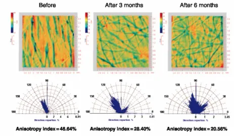
orientation. The furrows could be classified into three main categories, i.e. microrelief (0-10 µm depth), medium (10-20 µm) and deep furrows (>20 µm). The anisotropic index was defined as follows: the orientation
distribution of the furrows is quantified as a compass rose, the density in a triangular part corresponds to the number of motifs of the furrows that have this orientation. A completely anisotropic surface gives rise to a direction rose concentred into only one angular sector. Conversely, a perfectly isotropic surface leads to a circular direction rose.
So if N is the number of angular sectors, inspected between 0 and π, an anisotropy index (A.I) can be defined as:
where Ri is the rose value corresponding to angular sector i, and
where S/N should be the Ri value for all i in the case of a perfectly isotropic surface and the factor 1/2 comes
from the fact that an Ri value greater than S/N must be exactly compensated by lower values.
The analyses were carried out in a double-blind fashion without knowledge of the assigned treatment. Changes from baseline of wrinkle and roughness features were evaluated by the paired Wilcoxon test. The quantitative variables were described as mean, standard error of mean, median, minimum and maximum. The qualitative variables were described as percentages and evaluated using the χ2-test.
Skin biopsies
Three-millimeter punch biopsies were taken from the extensory side of the patients' forearms in 10 individuals who had agreed to participate in the study. The sides were assigned to treatment with the active principle and its placebo in the same manner as for the low-neck. The biopsies were divided in half, each proceeded for
investigations with light or electron microscopy. Light microscopy
Skin fragments were fixed in Baker's solution (buffered formalin) for 3h and routinely embedded in paraffin. Five-micrometer sections were dewaxed and stained with hematoxylin-eosin-saphran, orcein, as well as immuno-labelled using monoclonal antibodies to vimentin (VIM 3B4; Progen, Heidelberg, Germany) and to collagen IV (CIV 22: Dako, Glostrup, Denmark); both revealed with streptavidin-biotin-alkaline phosphatase (LSAB kit; Dako). Visualization of collagen IV required pretreatment of sections with 0.1% trypsin (15min, 37°C; Merck, Darmstadt, Germany).
Electron microscopy
Small skin fragments were fixed in 2% glutaraldehyde in cacodylate buffer for 6h, postfixed in 1% osmium tetroxide, dehydrated in ethanol and embedded in Epon. Ultrathin sections were counterstained with lead citrate and uranyl acetate.
Analysis of the morphological results
Light and electron microscopy findings were first compared within pairs of biopsies obtained from the same patient in a blind manner. The striking differences were evaluated semi-quantitatively, taking into account the epidermal compartment, the papillary dermis and the underlying reticular dermis. For semi-quantitative analysis at the EM level, several ultra-thin sections from at least three different blocks of each biopsy were carefully examined at low- and high-power magnification. Because the most pronounces differences were observed in the papillary dermis and concerned elastic fibers, the whole length of this compartment was analyzed at an
intermediate magnification of × 6000 on all sections available. Normally looking elastic fibers composed of an electron-dense fibrillin network embedded in electron-lucent elastin were classified as 'composite'. Their frequency between the fragmented structures was estimated as: sporadic (+), few (++), often (+++), or plenty (++++). After decoding of the treated and placebo sides, the results were analyzed within the whole group of patients.
Results
One patient dropped out just after the beginning of the trial. The remaining 19 patients were evaluated at 3 and 6 months. Clinical examination by the dermatologist as well as self assessment by the volunteers disclosed an improvement in terms of the 'global score'. Global score consisted of the sum of six items (hydration, roughness, laxity, suppleness, fine wrinkles, coarse wrinkles). Statistical evaluation of the values from the treated side vs. placebo was performed using a rank test on paired series This score evolved in a significant manner from 6.7 ± 1.6 to 5.0 ± 1.0, and 4.4 + 1.0, at baseline, at 3 and 6 months, respectively, on the treated area (P < 0.05). On the control area, the score was 6.7 ± 1.6, 5.7 ± 1.5 and 5.3 ± 0.7, at baseline, 3 months and 6 months, respectively (P > 0.05). According to dermatologist assessment, variance analysis showed a statistically significant improvement of hydration, small wrinkles, wrinkles, glare, and brown spots items in each group, throughout the trial.
Furthermore, roughness, suppleness, small wrinkle scores significantly improved in the vitamin C group. Self assessment by the volunteers disclosed a statistically significant and beneficial evolution of most of these clinical items (hydration, firmness, glare, tonicity, suppleness, brown spots, roughness imperfections, smoothing and dryness) on the treated area during the 6-month duration of the trial. Our results concerning the analysis of the skin replicas showed that compared with the placebo there exists a highly significant increase in the density of skin microrelief (P > 0.01) as well as a decrease of deep furrows at RI treated with the vitamin C cream over a 6-month period (P > 0.05) (Figs 1 and 2). Ra (which is the arithmetic mean of roughness) and Rz (which is the peak-to-valley mean of roughness) (17) values are given in (Table 1).
Tissue damage typical for chronic sun exposure could be observed in all studied biopsies
In the epidermis, melanocytes were irregularly distributed and often clustered. These vimentin-expressing cells were mostly encountered in the basal epidermal layer and, in some patients, displayed an extensive network
of dendrites reaching up to the granular layer. Transfer of the pigment to keratinocytes, visualized ultrastructurally, was most frequently enhanced, as judged by a higher melanin content in the neighboring epithelial cells compared with melanocytes. The observed changes were basically patient-dependent and not related to the presence or absence of the vitamin C treatment.
Figure 1. This histogram shows the evolution in percentage of the furrow density (number of furrows with a given depth) as compared with the baseline before treatment (= 0). The microrelief (0-10 µm depth) and medium (10-20 µm) furrows have been gathered in order to compare them with deeper furrows (> 20 µm). At 6 months, the differences in density of the furrows were significantly different between the vitamin C group and the placebo group, χ2-test: P < 0.01. Density of the skin furrows
disclosed a continuous improvement throughout the trial, showing the reappearance of small furrows (< 20 µm) and the disappearance of deeper furrows in the vitamin C treated group.
Figure 2. Example showing the reappearance of the isotropic morphology of skin relief and thus the decrease of the anisotropy index.
Table 1. Ra (the arithmetic mean of roughness) and Rz (the peak-to-valley mean of roughness) values
Ra T0 T3 T6 Vitamin C 8.16 µm 8.15 µm 6.98 µm Placebo 7.73 µm 7.56 µm 7.13 µm Rz T0 T3 T6 Vitamin C 61.74 µm 62.88 µm 53.59 µm Placebo 60 µm 59.22 µm 55.34 µm
In the dermis, a variable degree of alteration of the elastic tissue could be observed, ranging from absence of the oxytalan fibers in the papillary region to pronounced coalescence of the fibers in the reticular part. Major
differences related to the application of the active compound were noticed in the ultra structure of superficial dermis. On the untreated side, there were few elastic fibers; mostly fragmented electron-dense cores in the papillary and down to the upper reticular dermis. By contrast, biopsies of the treated skin revealed several 'composite' elastic fibers comprised of electron-dense bundles embedded in a relatively electron-lucid substance (Fig. 3). These were largely reduced or even absent from the papillary level in the control, placebo-treated biopsies. Inversely, the remnants of partially degraded fibers were still encountered in the superficial dermis after treatment. Fibers of the composite type predominated also in the lower parts of the reticular dermis of all
patients, independently of the vitamin C application. Once again, the degree of the photo-damage varied from patient to patient, but the overall tendency was towards re-appearance of 'composite' elastic structures in the upper dermis under influence of the active principle (Table 2).
Vimentin labelling alone did not allow for distinction between various types of dermal cells. The hematoxylin-eosin-stained serial sections were carefully examined for the presence of rare focci of perivascular or dermal infiltrate to exclude such areas from evaluation. The remaining individual, spindle-shaped, vimentin-positive cells were regarded as being likely of fibrocyte type. These latter cells tended to be more numerous in the papillary dermis on the vitamin C-treated side. However, a semi-quantitative examination of the EM samples did not confirm the increase of fibrocyte number after topical ascorbic acid application. The cells with fibrocyte morphology were more voluminous and their cytoplasm was richer in mitochondria and endoplasmic reticulum, which might have contributed to their better visualization in light microscopy in the vitamin C-treated biopsies. There were no clear-cut changes in the expression or distribution of the dermal collagen network. However, type I collagen bundles appeared less unidirectional and more evenly distributed on the side treated with ascorbic acid. The dermal-epidermal junction was not altered. Continuous staining for type IV collagen was present and ultrastructural examination did not reveal any basement membrane multiplication or rupture.
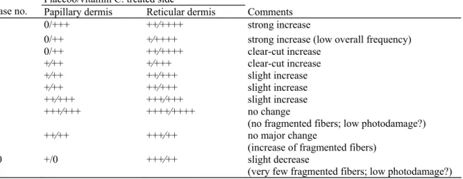
Table 2. Distribution of normally looking «composite» elastic fibers in the superficial dermis of photoaged skin
Placebo/vitamin C: treated side
Case no. Papillary dermis Reticular dermis Comments
7 0/+++ ++⁄++++ strong increase
3 0/++ +⁄++++ strong increase (low overall frequency)
1 0/++ ++⁄++++ clear-cut increase 9 +⁄++ +⁄+++ clear-cut increase 5 +⁄++ ++⁄+++ slight increase 8 +⁄++ ++⁄+++ slight increase 4 ++⁄+++ +++⁄+++ slight increase 6 +++⁄+++ ++++⁄++++ no change
(no fragmented fibers; low photodamage?)
2 ++⁄++ +++⁄++ no major change
(increase of fragmented fibers)
10 +/0 +++⁄++ slight decrease
(very few fragmented fibers; low photodamage?)
0, absent; +, sporadic; ++, few; +++, often; ++++, plenty.
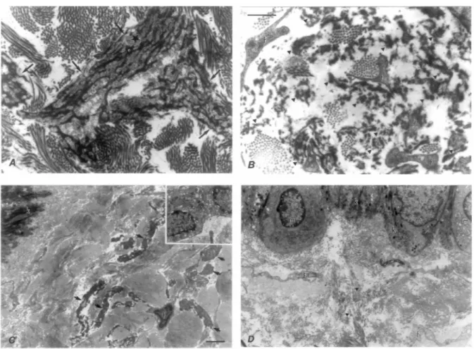
Figure 3. Representative pictures illustrating the ultrastructural changes observed in chronically photodamaged skin treated with (a,c) topical vitamin C and (b,d) its placebo. The most striking sign of long-term photodamage is the presence of electron-dense fragments of disrupted elastic fibers (arrow heads) in the reticular dermis. In (b), such debris are visualized between (f) fine fibrocyte extensions and sparse collagen bundles. Normal-looking 'composite' elastic fibers in (a) show an electron-dense microfibrillar scaffold (white asterisk) embedded in an amorphous substance of a lighter shade (black asterisk). Such fibers become more numerous in the papillary dermis after the treatment with ascorbic acid. They are surrounded by a well-organized multidirectional collagen network, visible also in (c). Metabolically active fibrocytes are often encountered in vitamin C-treated dermis (inset). Composite elastic fibers are indicated by arrows; k = basal keratinocytes. In contrast, fine, elongated portions of fibrocytes in the placebo-treated papillary dermis, (d), are surrounded by fiber-free spaces and dispersed collagen bundles. The epidermis contains pigment-free melanocytes (m) among melanin-abundant basal keratinocytes. Original magnification in (a,b): × 10000; in (c,d): × 4000. Bars: 1 µm in (a,b); 2 µm in (c,d).
Discussion
The results of this clinical trial confirm that topical application of 5% vitamin C over a period of 6 months significantly improves the clinical appearance of photodamaged skin when compared with vehicle alone. The dermatologic examination, which was performed in a double-blind manner, allowed to distinguish differences in the aspect of the skin between the areas treated with vitamin C cream and placebo. Significant reduction in small and coarse wrinkles, assessed by both an investigating dermatologist and a volunteer, and improvement in the overall aspect of the skin assessed by the volunteers, especially concerning firmness, smoothness, and dryness, were observed after 6 months of daily treatment. Significant favorable modifications of skin relief were induced, leading to the reappearance of an isotropic surface pattern. Optical profilometry of a skin replica was used to quantify these changes. It is a valid and objective measure of skin topography (14,18-23). The 3D relief parameters according to the Industrial Standards known from surface engineering (17) have often been used in the field of cosmetic dermatology for the quantitative analysis of human skin relief. The skin surface presents a number of depressions that are roughly rectilinear and can be classified according to depth and the magnification required to describe them. The most visible features are wrinkles, which vary in depth from 100 µm to several millimeters according to age and environmental factors. Optical-skin imaging discloses a particular network of features: parallel and criss-cross furrows, forming rectangles, squares, lozenges, trapezoids, and triangles. Primary lines are wide and 20-100 µm deep according to site and age. They cross to form
primary lines and form diagonals within the shapes made by the latter. This study showed two particular changes concerning skin relief: reappearance of small furrows (which are typical of young people) and a decrease of deeper furrows (which characterize aged skin). Such evolution of skin relief was already noticed 3 months after the beginning of treatment. Thus, clinical and topographical assessment supported the view that the observed changes in skin appearance well represented an improvement. In a study including 19 volunteers aged between 36 and 72 years, Traikovich (24) demonstrated a statistically significant improvement in at least one skin relief parameter, and good clinical results when testing a formulation containing ascorbic acid, tyrosine and zinc on the face during a 3-month period of treatment.
Indeed, vitamin C displays biochemical properties on collagen synthesis and collagen synthesis regulation in vitro (25-28) and in vivo (29). The results of the morphological evaluation also suggest that vitamin C induces the synthesis or improves the assembly of the various components of the elastic fibers, rather than exerting inhibitory action on elastases. Indeed, fragmented and incomplete elastic fiber structures were observed in the papillary and reticular dermis on both the treated and control sides, suggesting that the damage occurred before the vitamin C application. Increased proportion of composite fibers on the treated side in seven out 10 patients is in favor of a vitamin C-induced neo-synthesis or tissue organization. Parallel studies measuring the expression of the genes of gelatinases A and B, as well as their inhibitors, did not show a significant difference between the treated and control sites (29).
In any case, the quantities of gelatinases synthesized in the skin of our patients was low enough to prevent any degradation of the basement membrane's collagen IV, which is also these enzymes' substrate, and thus even on the side not treated with vitamin C. To limit potential sampling artifacts afferent to the EM approach, several ultra-fine tissue sections have been examined thoroughly in a 'blind' manner, and the results were analyzed only after decoding of the placebo and vitamin C-treated sides. For more precision, we evaluated separately the papillary dermis, where the initial destructive changes to elastic fibers were most pronounced, and the reticular dermis, where the elastic tissue damage was less evident. Although statistical analysis of our findings has been postponed because of a highly intricated structure of the dermal elements, the semi-quantitative analysis based on the presence of various proportions of normal-looking 'composite' fibers could be performed. We observed a correlation between the increased frequency of 'composite' fibers and the exposure to vitamin C in two-thirds of the patients, the proportion suggesting that the finding was not fortuitous. The remaining three patients, showing no increase of the pool of 'composite' fibers after the vitamin C treatment, may possibly illustrate the potential difficulties related to the EM-based semi-quantitative evaluation. However, in two of these patients, the
frequency of appearance of the fragmented elastic fibers was low on the control side, which may be indicative of a relatively mild initial photodamage to the elastic tissue and partially explain the lack of a marked difference after treatment. Our observations suggest that a prolonged topical treatment with ascorbic acid, applied in an appropriate vehicle, may result in the activation of a dermal synthesis of elastic fibers. Such an induction should be potentially beneficial for restoration of elasticity of the photodamaged skin. Improvement of the skin relief parameters observed in our patients seems to corroborate this possibility.
The proposal of topical application of vitamin C on photoaged skin relies on different experiments which tend to demonstrate its beneficial interest. Blood levels of ascorbate after application on skin exposed to UV light were reduced by 66% compared with the situation when the same product was applied on non-irradiated skin (6). Topical vitamin C has also UV photoprotective properties (8,12). In animals, a 5% solution of ascorbate applied 2h before UVB and UVA exposure reduced UVB-induced skin wrinkling (11). Measurement of serum levels of L-ascorbate after topical application demonstrated transcutaneous absorption of the vitamin C (7,8). These levels are assumed to be much higher than that obtained with oral supplementation (6).
Ascorbic acid is an alpha-kelactone with biochemically effective reducing properties. In tissues, it acts as a free radical scavenger. It may also function as an enzymatic cofactor and thus participate in hydroxylation of procollagen, a process involved in maturation of collagen fibers. Moreover, vitamin C influences the production of collagen by post-translational and transcriptional mechanisms (30,31). L-ascorbic acid is known to be necessary for prolyl hydroxylase and lysyl hydroxylase, enzymes essential for the stabilizing and crosslinking of collagen molecules, respectively. These reactions increase the tissue strength and may also influence the skin relief. Another possible action of topically applied vitamin C may be the scavenging and quenching of free radicals, leading to regeneration of local vitamin E levels (32) and prevention of further photoinduced damage. Aged skin compared with younger skin shows a decreased baseline production of collagen (33). In vitro studies showed that ascorbic acid, when added to fibroblast cultures from elderly donors, stimulated cell proliferation (34).
Wrinkles, roughness, laxity, brown spots and elastosis belong to clinical signs related to photo-ageing (1). Fitzpatrick and Rostan (35) have recently demonstrated a decrease of photoaging scores of the cheek and the peri-oral area in a double-blind study including 10 patients, with a formulation containing 10% vitamin C. As the amount of vitamin C available in the skin after oral ingestion are limited by regulatory mechanisms, topical delivery of vitamin C becomes attractive.
Conclusion
We conclude that topical application of 5% vitamin C is an effective and well-tolerated treatment. Topical vitamin C leads to a clinically apparent improvement in the appearance of photodamaged skin. It also induces skin relief modifications with disappearance of the anisotropic pattern on the surface and recovery of small furrows. Moreover, a prolonged topical treatment with ascorbic acid, applied in an appropriate vehicle, may result in activation of a dermal synthesis of elastic fibers.
References
1. Gilchrest B A. Skin aging and photoaging: an overview. J Am Acad Dermatol 1989: 21: 610-613.
2. Braverman I M, Fonferko E. Studies in cutaneous aging. I. The elastic fiber network. J Invest Dermatol 1982: 78: 434-443. 3. Abbey M, Nestel P J, Baghurst P A. Antioxidant vitamins and low-density lipoprotein oxidation. Am J Clin Nutr 1993: 58: 525-532. 4. Ranodive N S, Menon I A. Role of reactive oxygen species and free radicals from melanins in photoinduced cutaneous inflammations. Pathol Immunopathol Res 1986: 5: 118-139.
5. Black H S. Potential involvement of free radical reactions in ultraviolet light-mediated cutaneous damage. Photochem Photobiol 1987: 46: 213-221.
6. Colven R M, Pinnell S R. Topical vitamin C in aging. Clin Dermatol 1996: 14: 227-234.
7. Darr D, Combs S, Dunston S, Manning T, Pinnell S. Topical vitamin C protects porcine skin from ultraviolet radiation-induced damage. Br J Dermatol 1992: 127: 247-253.
8. Darr D, Dunston S, Faust H, Pinnell S. Effectiveness of antioxidants (vitamin C and E) with and without sunscreens as topical photoprotectants. Acta Derm Venereol (Stockh) 1996: 76: 264-268.
9. Dumas M, Chaudagne C, Bonte F, Meybeck A. Age-related response of human dermal fibroblasts to 1-ascorbic acid, study of type I and III collagen synthesis. C R Acad Sci III- 1996: 319: 1127-1132.
10. Chung Jh, Youn Sh, Kwon O S, Cho Kh, Youn J I, Eun H C. Regulations of collagen synthesis by ascorbic acid, transforming growth factor-beta and interferon-gamma in human dermal fibroblasts cultured in three-dimensional collagen gel are photoaging and aging independent. J Dermatol Sci 1997: 15: 188-200.
11. Bisset D L, Chatterjee R, Hannon D P. Photoprotective effect of super-oxide scavenging antioxidants against ultra-violet radiation-induced chronic skin damage in the hairless mouse. Photodermatol Photoimmunol Photomed 1990: 7: 56-62.
12. Murray J, Darr D, Reich J, Pinnell S. Topical vitamin C treatment reduces ultraviolet B radiation-induced erythema in human skin. J Invest Dermatol 1991: 96: 587.
13. Pinnel S R, Madey D L. The benefits of topical vitamin C (1-ascorbic acid) for skin care and UV protection. J Appl Cosmetol 1999: 18: 126-134.
14. Grove J L, Grove M J, Leyden J J et al. Skin replica analysis of photodamaged skin after therapy with tretinoin emollient cream. J Am Acad Dermatol 1991: 25 (1): 231-237.
15. Corcuff P, Chatenay F, Leveque J L. A fully automated system to study skin surface patterns. Int J Cosmet Sci 1984: 6: 167-176. 16. Creidi P, Vienne M P, Ochonisky S, et al. Profilometric evaluation of photodamage after topical retinaldehyde and retinoic acid treatment. J Am Acad Dermatol 1998: 39: 960-965.
17. Zahouani H. Spectral and 3D motifs identification of anisotropic topographical components. Analysis and filtering of anisotropic patterns by mophological rose approach. Int J Mach Tools Manufact 1998: 38: 615-623.
18. Grove L, Grove M J, Leyden J L. Optical profilometry: an objective method for quantification of facial wrinkles. J Am Acad Dermatol 1989: 21 (2): 631-637.
19. Mignot J, Zahouani H, Rondot D, Nardin Ph. Morphological study of human skin topography. Int J Bioeng Skin 1987: 3: 177-196. 20. Agache P, Mignot J, Makki S. Microtopography of the skin and aging. In: Kligman A M, Takase Y, eds. Cutaneous Aging. Tokyo: University of Tokyo Press, 1988: 475-499.
21. Cook Th, Craft T J, Brunelle R L, Norris F, Griffin W A. Quantification of the skin's topography by skin profilometry. Int J Cosmet Sci 1982: 4: 195-205.
22. Makki S, Agache P, Mignot J, Zahouani H. Statistical analysis and three dimensional representation of the human skin surface. J Soc Cosmet Chem 1984: 35: 311-325.
23. Makki S, Barbenel J C, Agache P. A quantitative method for assessment of the microtopography of human skin. Acta Derm Venereol (Stockh) 1979: 59: 285-291.
24. Traikovich S S. Use of topical ascorbic acid and its effects on photodamaged skin topography. Arch Otolaryngol Head Neck Surg 1999: 125: 1091-1098.
25. Murad S, Grove D, Lindberg K A, Reynolds G, Sivarajah A, Pinnell S R. Regulation of collagen synthesis by ascorbic acid. Proc Natl Acad Sci U S A 1981: 78: 2879-2882.
26. Murad S, Sivarajah A, Pinnell S R. Prolyl and lysyl hydroxylase activities of human skin fibroblasts. effect of donor age and ascorbate. J Invest Dermatol 1980: 75: 404-407.
27. Murad S, Sivarajah A, Pinnell S R. Regulation of prolyl and lysyl hydroxylase activities in cultured human skin fibroblasts by ascorbic acid. Biochem Biophys Res Commun 1981: 101: 868-875.
28. Murad S, Tajima S, Johnson G R, Sivarajah S, Pinnell S R. Collagen synthesis in cultured human skin fibroblasts: effect of ascorbic acid and its analogs. J Invest Dermatol 1983: 81: 158-162.
29. Nusgens B, Humbert P, Rougier A et al. Topically applied vitamin C enhances the mRNA level of col-lagens I and III, their processing enzymes and TIMP1 in the human dermis,. J Invest Dermatol 2001: 116: 853-859.
30. Padh H. Cellular functions of ascorbic acid. Biochem Cell Biol 1990: 68: 1166-1173.
31. Pinnell S R, Murad S, Darr D. Induction of collagen synthesis by ascorbic acid: a possible mechanism. Arch Dermatol 1987: 123: 1684-1686.
32. Beyer R E. The role of ascorbate in antioxidant protection of biomembranes. interaction with vitamin E and coenzyme Q. Arch Bioeng Biomed 1994: 26: 349-358.
33. Mays P K, McAnulty R J, Campa J S, Laurent G J. Age-related changes in collagen synthesis and degradation in rat tissues. Biochem J 1991: 276: 307-313.
34. Phillips C L, Combs S B, Pinnell S R. Effects of ascorbic acid on proliferation and collagen synthesis in relation to the donor age of human dermal fibroblasts. J Invest Dermatol 1994: 103: 228-232.
35. Fitzpatrick R E, Rostan E F. Double-blind, half-face study comparing topical vitamin C and vehicle for rejuvenation of photodamage. Dermatol Surg 2002: 28: 231-236.