HAL Id: hal-01626728
https://hal.archives-ouvertes.fr/hal-01626728
Submitted on 31 Oct 2017HAL is a multi-disciplinary open access archive for the deposit and dissemination of sci-entific research documents, whether they are pub-lished or not. The documents may come from teaching and research institutions in France or abroad, or from public or private research centers.
L’archive ouverte pluridisciplinaire HAL, est destinée au dépôt et à la diffusion de documents scientifiques de niveau recherche, publiés ou non, émanant des établissements d’enseignement et de recherche français ou étrangers, des laboratoires publics ou privés.
Critical Overview of the Use of Ru(II) Polypyridyl
Complexes as Photosensitizers in One-Photon and
Two-Photon Photodynamic Therapy
Franz Heinemann, Johannes Karges, Gilles Gasser
To cite this version:
Franz Heinemann, Johannes Karges, Gilles Gasser. Critical Overview of the Use of Ru(II) Polypyridyl Complexes as Photosensitizers in One-Photon and Two-Photon Photodynamic Ther-apy. Accounts of Chemical Research, American Chemical Society, 2017, 50 (11), pp.2727-2736. �10.1021/acs.accounts.7b00180�. �hal-01626728�
1
Critical Overview of the Use of Ru(II) Polypyridyl Complexes
as Photosensitizers in One-Photon and Two-Photon
Photodynamic Therapy
Franz Heinemanna,#, Johannes Kargesa,#, and Gilles Gassera,*
a
Chimie ParisTech, PSL Research University, Laboratory for Inorganic Chemical Biology, F-75005 Paris, France.
#
these authors have contributed equally to the work
* Email: gilles.gasser@chimie-paristech.fr; www.gassergroup.com; Tel. +33 1 44 27 56 02.
ORCID-ID:
Franz Heinemann: 0000-0001-9590-443X Johannes Karges: 0000-0001-5258-0260 Gilles Gasser: 0000-0002-4244-5097
2 Conspectus
Photodynamic Therapy (PDT) is an emerging technique to treat certain types of cancer, bacterial/fungal/viral infections and skin diseases. In the past years, different research groups developed new ruthenium-containing photosensitizers (PSs) with tuned photophysical and biological properties to better fit the requirements of PDT. In this account, we report on and discuss the latest results in this research area, emphasizing particularly our own research. For example, inspired by the DNA intercalating complex [Ru(bpy)2(dppz)]2+ (bpy =
2,2’-bipyridine; dppz = (dipyrido[3,2-a:2’,3’-c]phenazine), a series of ruthenium complexes bearing differently functionalized dppz ligands were synthesized to target DNA. The introduction of the substituents on the dppz ligand did not reduce much the complexes’ affinity to DNA but highly affected their cellular uptake. The most effective complex in this series, [Ru(bpy)2(dppz-7-OMe)]2+, showed IC50 values in the low micro-molar range against
several types of cancer cells upon irradiation and, importantly, a high phototoxic index (PI) of >150. This value is comparable or even better than several PSs used in clinics under comparable experimental conditions. This compound was found to localize in the nucleus and to induce DNA damage in HeLa cells upon light irradiation. Interestingly, cells in the mitotic
3
phase were found to be more affected and to have a different mechanism of cell death (apoptosis) upon light irradiation than those in the interphase (paraptosis). To take advantage of that, the PS was combined with a cell cycle inhibitor to synchronize cells in the mitotic phase, further improving the photo-toxicity by a factor of 3.6. In addition, our group could recently demonstrate that [Ru(bphen)2(benzene-1,2-dislufinate)] (bphen =
4,7-Diphenyl-1,10-phenanthroline) localizes in mitochondria and has an IC50 value of 0.62 µM with a PI of over
80 in HeLa cells upon light irradiation at 420 nm. Interestingly, this complex was also found to efficiently kill gram-positive S. aureus under light irradiation. Antimicrobial PDT (aPDT) is another field of research where Ru(II) polypyridyl complexes can play an interesting role to fight antibiotics resistance. [Ru(dqpCO2Me)(ptpy)]2+ (dqpCO2Me =
4-methylcarboxy-2,6-di(quinolin-8-yl)pyridine), ptpy = 4′-phenyl-2,2′:6′,2′′-terpyridine) is additionally efficient against gram-negative E. coli. The efficacy of positively charged Ru(II) PSs is related to their affinity to the negatively charged membrane of gram-negative bacteria.
A drawback of many Ru(II) polypyridyl PSs is their low absorption in the biological optical window (600-900 nm) where light penetration depth into tissue is the highest. The lowest energy transition in the UV/Vis spectra of Ru(II) polypyridyl complexes is usually a metal-to-ligand charge-transfer band. To shift the absorption into this range, tuning of the metal-to-ligand system, e.g. by extending π-systems, has been described in the literature. Another approach to make excitation in the optical biological window possible is Two-Photon Absorption (2PA). High photon density is needed and usually confocal laser beams are used for excitation. In collaboration with the Chao group, a series of homoleptic Ru(II) complexes bearing tertiary alkyl ammonium substituted bipyridine ligands with two photon cross sections between 185-250 GM at around 800 nm was tested in vitro. They showed IC50 values in the micromolar
range and PIs between 103 and 313. The highly positive-charged complexes were found to enter the cell via endocytosis and to target lysosomes. Studies on 3D tumor cell spheroids, a
4
model closer to real tumors than commonly used 2D cell monolayers, were also performed. It could be demonstrated that 2P-PDT treatment with 800 nm laser irradiation was significantly more effective than that with 450 nm laser irradiation.
5 INTRODUCTION
Photodynamic Therapy (PDT) is a medical technique, which is approved in several countries, to treat various types of cancer (i.e. lung, bladder, oesophageal and brain cancer) but also bacterial, fungal or viral infections as well as different skin diseases. The use of PDT is a two-stage process (Figure 1). At first, a (preferably nontoxic) photosensitizer (PS) is administered into the patient either locally or systemically. Upon irradiation at a specific wavelength in the absorption window, the PS is excited to the singlet state S1. Since this excited state is
relatively unstable, the PS can emit the excess of energy by fluorescence or a vibronic radiationless relaxation. Alternatively, the PS can undergo an intersystem crossing (ISC) process to the more stable excited triplet state T1. This excited triplet state T1 can then decay
to the ground state by a vibronic radiationless relaxation, produce phosphorescence or influence the biological environment by two different pathways called Type I or Type II.1–3
6
In a Type I mechanism, an electron or proton is transferred from/to the PS to/from the biological environment to create a radical anion and its corresponding cation (Figure 2). These are able to generate reactive oxygen species (ROS), which can trigger cell death. Beside direct reactions of the PS with substrates, PS∙- is able to generate a superoxide anion radical (O2∙-) by oxidative quenching. The latter can further react and create hydrogen
peroxide by a disproportion/one-electron transfer reduction. Hydrogen peroxide can further react through a Haber-Weiss Reaction or a Fenton Reaction to create the powerful hydroxyl radical. The highly reactive hydroxyl radical is able to cause oxidative damage, which were, for example, thoroughly investigated for fatty acids or other lipids. Additionally, the hydroxyl radical can initiate a cascade of other free radicals, which can oxidise several substrates and hence create cellular damage.3-5
On the opposite, during a Type II mechanism, the excited 3PS state transfers its energy to molecular oxygen (3O2). During this process, singlet oxygen (1O2) with a very short lifetime
in an estimated range of <0.04-3 μs in biological environment is formed.2 Based on its short lifetime and high reactivity, the radius of action of 1O2 is in the order of 0.01-0.155 μm.6
Therefore, it reacts extremely quickly with its close biological surroundings. Most of the PSs applied in the clinics are working through a Type II mechanism although the recently approved Palladium-containing PS TOOKAD-soluble 1 (Figure 3) was found to work mainly through a Type I mechanism.7 Worthy of note, a recent study still showed the necessity of oxygen for the treatment with TOOKAD-soluble to avoid the non-radiative decay of the PS to the ground state corresponding with heat development.8
7
Figure 2. Schematic representation of favorable photochemical reactions during PDT. Sub = biological Substrate, O2∙- = superoxide anion, SOD = superoxide dismutase.
The majority of the currently approved PSs are based on a tetrapyrrolic scaffold (i.e. porphyrin, chlorine). However, these compounds have several drawbacks, namely 1) poor water solubility; 2) tedious synthesis; 3) absorption (e.g. Photofrin 2; λ = 630 nm) in the spectral range of the biological environment (i.e. skin, fat, blood); 4) low cancer selectivity; 5) photobleaching and 6) slow clearance from the body causing photosensitivity.2
Next to organic porphyrins, tetrapyrrolic structures containing metals like Sn(IV) (Purlytin 3), Pd(II) (TOOKAD soluble 1) and Lu(III) (Lutex 4) have already been approved or are currently being intensively investigated as PDT PSs (Figure 2).2,9 Of special interest is TOOKAD soluble 1 with high absorption at 763 nm which is in the biological optical window of PDT (600-900 nm). For the PDT treatment with TOOKAD soluble 1 at 763 nm the light penetration depth into tissue is 2-3 times higher than with Photofrin 2.2,10
Beside metal-containing porphyrin structures, other types of metal complexes containing Pt(II), Pt(IV), Os(II), Re(I), Ir(III) have been or are currently investigated. However, the most
8
studied metal in PDT has been undoubtedly Ru(II), and more specifically Ru(II) polypyridyl complexes.11-16 The latter have a large Stokes shift, implying only minimal interference between excitation and emission. Additionally, these complexes have usually a high water solubility compared with porphyrins or phthalocyanines as well as a high 1O2 production rate,
which is essential as a Type II PDT PS. Importantly, Ru(II) polypyridyl complexes have generally a high chemical and photochemical stability and are little or even not photobleaching in contrast to porphyrin derivatives. Of note, the introduction of Ru(II) moieties to porphyrins like in 5 can improve the PDT characteristics (water-solubility, cellular uptake, 2PA-properties) compared to the porphyrin itself.1,17
Importantly in the context of this Account, the compound of McFarland et al. TLD-1433 6 has very recently entered into clinical trials as a novel PDT PS for the treatment of non-muscle invasive bladder cancer.18 Using 6, a strong photodynamic effect in vitro and in vivo was observed.19 It was demonstrated that binding 6 with transferrin enhances the physical and biological properties of the PS. Upregulation of the transferrin receptor is characteristic for many cancer types, particularly for bladder cancer.20 Therefore, the mixture of 6 and transferrin generates a suitable way of transportation of the PS to the tumor tissue. Additionally, an improved PDT efficacy could be demonstrated based on an about 15 % lowered photobleaching effect and a decreased dark toxicity during in vivo studies than 6 alone. A highly increased extinction coefficient makes the excitation at 850 nm possible allowing for a deeper tissue penetration.21 In this Account, we report on and discuss the latest results in this research area, emphasizing particularly our own research.
9 Figure 3. PSs investigated for PDT treatment.
10
Figure 4. Structures of Ruthenium complexes investigated for 1P-PDT and aPDT by our group (8-19, 21) and other groups (20, 22).
DNA targeting Photosensitizers
Effective targets for PSs are cell compartments, which are important for cell survival (i.e mitochondria, lysosomes, membranes or the nucleus). Transcription and replication of DNA
11
is crucial for cell proliferation.1 Many cytostatic cancer drugs exert their effect by targeting DNA. Because cancer cells divide much faster than healthy cells, they are more affected by DNA damage.22 DNA has been previously shown to be an effective target in PDT. Attachment of a nuclear targeting peptide to an organic PS led to a 2000-fold increase in phototoxicity compared to the unmodified PS.23 Additionally, a variety of Ru-based DNA photo-cleavers are known in the literature that can damage DNA by oxidative mechanisms.24 Inspired by this, we investigated a series of dppz-containing Ru(II) complexes to target DNA (dppz = dipyrido[3,2-a:2’,3’-c]phenazine) (Figure 4). 7 was first investigated as a DNA probe using the so-called light-switch effect by the Barton group.25
In the investigated series, 8-17, the ancillary ligands were bpy (bipyridine) or phen (phenanthroline) and the dppz ligand was substituted with different functional groups.23,26,27 Experiments confirmed the ability of 8-13 to generate 1O2 with quantum yields between
55-94 % in acetonitrile and 1-6 % in Phosphate-Buffered Saline (PBS). This example clearly confirms that 1O2 quantum yields are highly depended on the used solvent (e.g. quenching
effects). Biological studies revealed striking differences in their phototoxicity on HeLa cells. The major reason for that was the cellular uptake. 8 and 9 showed around 3-9 times higher cellular uptake compared to 10-14. This is especially striking since the compounds have a similar lipophilicity. Although less lipophilic, the uptake of 9 (1.76 nmol per mg of protein, 4 h, 20 µM) was much higher than the one of 8 (1.08 nmol per mg of protein, 4 h, 20 µM) in HeLa cells. Significantly lower uptake and a reverse situation was observed for MRC-5 cells with an uptake of 0.76 nmol per mg of protein for 8 and 0.18 nmol per mg of protein for 9. Thus, other factors than lipophilicity must play an important role for the uptake of these compounds. 8 and 9 were found to be the most phototoxic compounds (420 nm, 9.27 J cm-2) with phototoxic indices (PIs) of >150 and 42, respectively at low micro-molar concentrations on HeLa cells (PI = IC50(dark)/IC50(light)). This is significantly higher compared to Photofrin (PI
12
>4.3) and Hypericin (PI >12), two known PDT agents, under similar conditions (5 Jcm-2, 1000 W halogen lamp). High DNA affinity, DNA photo-cleavage ability and nuclear localization of 8 and 9 suggest DNA involvement in the mode of action.24 For 9, this was further confirmed by an observed DNA damage response of the treated cells caused by oxidative DNA damage, including double strand breaks and guanine oxidation. Furthermore, cell cycle dependence for the mechanism of cell death was observed leading to paraptosis or apoptosis (Figure 5). The latter was observed for mitotic cells which were also much more sensitive to PDT treatment. The combination of a cell cycle inhibitor to synchronize cells in the mitotic phase and 9 resulted in a 3.6 times lower PS dose needed. This combinational approach bears significant potential to improve the outcomes of PDT treatments.23
Figure 5: Cell cycle-dependent sensitivity and mechanism of cell death for in vitro PDT (HeLa cells) with 9.
Antimicrobial Photodynamic Therapy
Resistance of bacteria to antibiotics has become an urgent problem and new treatments to fight these infections are needed.28 Antimicrobial PDT (aPDT) is one promising way for that purpose.28,29 It is also referred to as Photodynamic Inactivation (PDI) and is intended to treat also infections with fungi, yeasts or viruses besides bacteria.29 In our group, the aPDT properties of complexes 18 and 19 were investigated.28 Biological studies were undertaken on two bacterial strains, namely Gram(+) Staphylococcus aureus (S. aureus) and Gram(-)
13
Escherichia coli (E. coli). Both compounds were found to reduce the viability of S. aureus
over 6 log10 (99.9999 %) under 420 nm light irradiation (8 J cm-2). While 18 exerted no
phototoxicity on Gram(-) E. coli, 19 could reduce the viability over 4 log10 (99.99 %). This is
remarkable since aPDT of Gram(-) bacteria with organic PSs has been challenging.29 The good efficacy of different Ru(II) polypyridyl complexes, among them 7 and 20, against Gram(-) bacteria was also reported by other groups.30 It was linked to electrostatic interaction of the positively charged complexes with the negatively charged outer membrane of Gram(-) bacteria and intercalation of the dppn ligand of 20 into the membrane.30,31 Worthy of note, McFarland et al. tested several Ru(II) polypyridyl PS for aPDT showing the potential of the oxygen-independent, type I, Ru(II)-based PS 6 against Methicillin resistant Staphylococcus
aureus under low oxygen tension.32
Tuning the Absorption Properties of 1P-Photosensitizers
The majority of Ru(II) polypyridyl PSs, among others those reported by our group (8-19), suffer from the fact that they lack significant absorption in the biological optical window (600-900 nm) and have therefore low tissue penetration depth (Figure 6a). This limits their potential to topical or very localized internal applications. Regarding the UV/Vis absorption properties of Ru(II) polypyridyl complexes, the lowest energy absorption band is usually of the metal-to-ligand charge transfer (MLCT) type. It typically lies in the region of 400-500 nm. In the case of the prototype [Ru(bpy)3]2+, the MLCT transition occurs at 450 nm and can be
seen as an excitation of a metals d-electron (HOMO) to the π*-orbital of the bpy-ligand (LUMO) (Figure 6b).33
14
Figure 6: (a) Light penetration depth into skin. The optical biological window between 600-900 nm is mainly limited by haemoglobin and melanin absorbing at the lower as well as water and fat at its upper end.34 (b) Simplified MO scheme of the frontier orbitals for octahedral d6 complexes like [Ru(bpy)3]2+.
To increase the absorption wavelength, the HOMO-LUMO energy gap must be reduced. This is possible by destabilization of the metals t2g-orbitals (HOMO) or by ligands possessing
low-lying π*-orbitals (LUMO). Increasing electron density at the metal center by electron donating ligands leads to destabilization of the LUMO.35 Originally 18 was designed in this way inspired by a Ru(II) complex bearing a benzene dithiol (bdt) donor ligand with an absorption maximum at 622 nm. Unfortunately, in our case, the sulfur atoms of the bdt ligand were oxidized under air contact resulting in the disulfinato complex 18. The desired red-shift in absorption was not achieved. On the contrary, a blue-shift was observed (λmax =
396-440 nm) compared to [Ru(bpy)3]2+. However, besides the aPDT activity discussed above, 18
showed very promising results on HeLa cells under 420 nm irradiation (6.95 J cm-2) at remarkable low concentrations of 0.62 µM with a PI of 80. The subcellular localization indicated the mitochondria as possible target of 18.28
M - t2g M - eg L - π* L - π M L C T L C filled orbitals empty orbitals (a) (b)
15
Lower π*-orbitals of the ligand are reached with electron-withdrawing groups like the carboxyl group attached to the bpy or phen ligand for example. This approach was already used in the context of dye-sensitized solar cells to achieve red light-absorbing Ru(II) polypyridyl complexes.35 Also, ligands with enlarged π-systems possess low lying π*-orbitals and could be introduced into complexes to lower the absorption energy.36 It is of interest that some of these complexes have ligand centred (LC) π-π* triplet states at lower energy level than the 3MLCT. Importantly, these 3LC states can participate in type II and oxygen-independent type I reactions.19 Hypoxic tumors are especially challenging since they can contribute to the development of resistances against radio- and chemotherapy, rendering them therefore difficult to treat37 The oxygen dependence of approved type II PSs is one of the main factors limiting the outcomes of PDT. The ability to challenge this problem with oxygen independent type I PS was demonstrated in vitro in a malignant melanoma model, a hypoxic and difficult to treat skin cancer.38
The lifetime of emissive 3MLCT states is usually shorter compared to those of 3LC states. The metal’s orbital contribution to the 3
MLCT excited state promotes ISC back to the singlet ground state because of the spin orbit coupling. If the 3MLCT and 3LC lie close together, equilibration between them can prolong the emission lifetime.33 Longer triplet state lifetimes can increase 1O2 generation even under low oxygen tension.19,38
In collaboration with the Würthner group, we investigated complex 21 with an extended π-system as a potential PDT PS. 21 possesses an azabenz-annulated perylene bisimide (ab-PBI) ligand and bpy as ancillary ligands. Perylene bisimides are electron poor and withdrawing aromatic molecules.39,40 A large red-shift for the MLCT band of 21 was observed compared to [Ru(bpy)3]2+. A broad absorption band lied at 520 nm with absorption
up to 600 nm. TD-DFT calculations suggested that besides the MLCT also intra-ligand charge transfer (ILCT) of the ab-PBI ligand contributed to this absorption and also to the T1 state.39
16
A prolonged luminescence lifetime of 4.2 µs (CH2Cl2, room temperature) was observed
compared to [Ru(bpy)3]2+ (τ = 488 ns, CH2Cl2, 25 °C)33. Therefore, the 1O2 production was
tested with higher wavelength excitation at 575 nm. Unfortunately, 1O2 formation could not
be detected at this wavelength, contrary to that observed when excited at 420 nm in acetonitrile and PBS. Since the complex was toxic in the dark, only low PI values of 2 were determined for different cancerous and non-cancerous cell lines under 420 nm irradiation.40 Worthy of note, the group of McFarland systematically investigated Ru(II) complexes with attached aromatic chromophores that possess low lying triplet intra-ligand (3IL) states for PDT. With these so-called dyads, compared to [Ru(bpy)3]2+ extremely long triplet state
lifetimes up to hundreds of microseconds could be reached.38 These long lived 3IL states of dyad PS like 6 and 20 are highly photosensitizing. This allows for excitation and 1O2
generation in the biological optical window although only weak absorption (<100 M-1 cm-1) is observed in that region.19
Very recently, Wang et al. reported a complex 22 with a merocyanine substituted oxyquinolate ligand that showed a strong absorption maximum at 650 nm (ε = 17300 M-1
cm-1).30 Oxyquinolate ligands are so-called non-innocent ligands. Non-innocent ligands are characterized by a large π-overlap with the metals orbitals. This results in a large hybrid metal-ligand HOMO with an increased energy and so a diminished HOMO-LUMO gap for ruthenium complexes with oxyquinolate ligands.42 When irradiated in the biological optical window at 635 nm (1.5 mW cm-2, 20 min), 22 showed good phototoxicity against E. coli. Low activity against HeLa cells was rationalized with a low cellular uptake.31
17
2-Photon absorbing Photosensitizers
Currently approved PSs are excited in the spectral range from 375-763 nm.2,9,10 Due to absorption and light scattering effects of the biological tissue, the light penetration depth into the tissue is low at these wavelengths (Figures 6a, 7). As a result, these PSs cannot be used to treat deep tumors.43
Figure 7. Average penetration depth for a procaine brain.43,44
A strong red-shift of the PS’s excitation wavelength can be achieved with a chromophore, which is able to perform a simultaneous absorption of two photons, a so-called two-photon absorption (2PA) process (Figure 8). In addition to a highly increased penetration depth, a 2PA process has an enhanced spatial resolution due to its dependence to the square of the luminescence intensity, whereas a one-photon process linearly depends on the intensity. This explains why 2PA processes mostly work with intense laser beams or focused laser pulses with a high photon density. Additionally, based on the use of a low energy near-infrared irradiation, reduced photodamages of the PS have been observed.43
18
Figure 8. Comparison of a Single Photon Excitation (a) and a simultaneous Two-Photon Excitation (b).
Although 2P-PDT is currently not employed in the clinics, the use of this technique has been demonstrated in in vitro studies on cervical cancer HeLa cells, HeLa multicellular tumor spheroids (MCTS)17,45 as well as, more importantly, in in vivo studies.46 One of the factors influencing the success of this technique is the ability of the PS to absorb two photons simultaneously. This ability is quantified by the so-called two-photon cross-section σ2, which
is expressed in Goeppert-Mayer (GM) with 1 GM = 10-50 cm4 s.molecule-1. The great majority of investigated PSs in 2P-PDT are porphyrins and other tetrapyrrolic derivatives, which show in principle good physical and biological properties. However, they have relatively low σ2 values. For example, Photofrin in methanol has a σ2 value of 10 GM at
800 nm.47
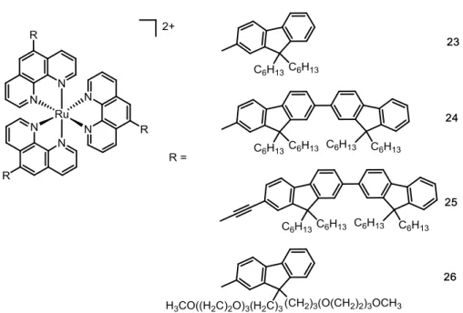
The group of Lemercier et al. was, to the best of our knowledge, the first to demonstrate the use of Ru(II) polypyridyl complexes in 2P-PDT. These researchers demonstrated that the substitution of a Ru(II)-1,10-phenanthroline complex with oligofluorene groups in position 5 (Figure 9) was able to enhance the ability of the complex 23 to absorb two photons simultaneously as well as to increase luminescence lifetime (τ = 1700 ns, σ2 = 40 GM, λ =
19
890 ns, σ2 = 20 GM, λ = 750 nm in acetonitrile). It was demonstrated by additional studies
that the photophysical properties of 23 can be further increased by the incorporation of a second 5-oligofluorene group 24 (τ = 2500 ns, σ2 = 90 GM, λ = 750 nm in acetonitrile).
Furthermore, these researchers investigated the connection of the oligofluorene substituent to the Ru(II) phenanthroline complex through a triple bond (complex 25). As a result, a drastic increase of the σ2 value (τ = 708 ns, σ2 = 225 GM, λ = 750 nm in dichloromethane) was
demonstrated, which facilitates a 2P process.48 In view of applications as a PDT PS, the solubility of the compound 25 was enhanced by incorporation of a poly-ethoxy group in the 5-oligofluorene group. In vitro experiments on F98 glioma cells demonstrated that cell death was triggered upon laser irradiation (λ = 740 nm, P = 30 mW, tirradiation = 5 min, tobserved PDT effect
= 15 min) when the poly-ethoxy substituted 5-oligofluorene-1,10-phenanthroline Ru(II) complex 26 was incubated.49 These compounds are referred, in this Account, as the first generation of Ru(II) polypyridyl 2P-PSs.
Figure 9. Chemical structures of the first investigated Ru(II) phenanthroline complexes as 2P-PSs.
20
As discussed above, the type of the substituents on a Ru(II) polypyridyl complex has a significant influence on its photophysical and biological properties. However, an important factor is also the biodistribution of the PS inside a cell. Ru(II) polypyridyl complexes which are able to specifically target a cellular organelle are called from here on second generation of Ru(II) polypyridyl 2P-PSs. One potential target of a PDT PS is mitochondria which function as the apoptosis center of the cell. It has already been demonstrated that apoptosis of cells can be triggered effectively in the mitochondria by production of oxidative stress. Additionally, studies have shown that mitochondria targeting anticancer agents, which are able to disturb the redox balance in the cell, are able to trigger the mitochondrial-dependent cell death signaling pathway and therefore overcome different cancer resistances.50 The Chao research group described a series of Ru(II) polypyridyl complexes 27-28 (Figure 10) that were able to selectively target mitochondria.45 Nevertheless, based on the function of mitochondria as the cellular energy machinery, the localization of the PS there can possibly result in a high dark cytotoxicity.51 Our group then designed, in cooperation with the Chao group highly charged, lysosome targeting, tertiary alkyl ammonium substituted Ru(II) polypyridyl complexes 29-31 (Figure 10). Since the lysosomes are known to be the waste disposal center by digesting biomolecules both from outside and inside the cell though different kinds of enzymes, it is an ideal localization for PDT purposes. Interestingly, due to the introduction of highly positively charged groups, the water solubility of the complexes has been increased. The Ru(II) polypyridyl complexes 29-31 showed a similar one-photon absorption profile (i.e. metal-to-ligand-charge transfer band from about 400-500 nm) and τ values (810-900 ns in methanol) than other similar Ru(II) polypyridyl complexes.45,48,49 However, very importantly, the 2PA profile of these compounds was enhanced by introducing tertiary alkyl ammonium groups. In the literature, it is known that nitrogen groups enhance the ability of a compound to absorb 2P simultaneously.52 In addition to that, σ2 values from 185 GM for 31 up to 250 GM for 29
21
(800 nm, in methanol) were determined. These values represent an important increase in comparison to clinically approved PSs like Photofrin (10 GM, 800 nm in methanol)47 but are still in the same range than other Ru(II) polypyridyl 2P-PSs.45,48,49 Importantly in view of applications in 2P-PDT, the compounds 29-31 are able to produce 1O2 with 1O2 quantum
yields of 92-99% in methanol and of 49-67% in D2O. However, crucially, high phototoxic
indices of 103 for 31 and even 313 for 29 were determined. As part of the biological investigations, the cellular uptake of the compounds in HeLa cells were studied. Complex 29 was found to internalize in cells through endocytosis. Once inside HeLa cells, complex 29 was found to localize in the lysosomes. Interestingly, upon light irradiation, cell shrinkage and generation of blebs as well as a change in the localization of the compound was observed. The localization of the compound 29 changed first from the lysosomes to the cytoplasm and, after an extended irradiation time, to the nucleus and nucleoli.17 Worthy of note, such a change in localization upon light irradiation has already been reported for other Ru(II) complexes.17
22
Figure 10. Chemical structures of investigated Ru(II) polypyridyl 2P-PSs.
Capitalizing on the results obtained with one-photon absorbing PSs (see above), we then investigated the potential of Ru(II) complexes with a doubly substituted dppz ligand 16-17 as 2P-PSs in cooperation with the Chao group. We could show that a small structural change, such as different substituents, i.e. a methoxy group in 16 and a hydroxyl group in 17, had a significant influence on the photophysical properties. For example, a σ2 value of 93 GM was
determined for 17 while an impressive value of 245 GM was obtained for 16 (λ = 800 nm, in methanol). 16 was found to have a higher PI value (11.7) than 17 (> 5.9) on 2D HeLa cell monolayers. Apart from monolayers, the biological effects on 3D HeLa MCTS upon 2P irradiation were tested. Interestingly, 16 was able to penetrate deep into the core of the MCTS (up to 300 m) with a PI value of 11, whereas 17 was mostly found at the outer surface making this compound uninteresting for (2P-)PDT. Overall, this study highlighted that a small change in the structure of a Ru(II) polypyridyl complex had a significant influence on its photophysical and biological properties (i.e. stability, cellular localization, anticancer activity).26
CONCLUSION
Ru(II)-based PSs have undoubtedly a great potential in PDT thanks to their tuneable photo-physical properties. As described herein in details, biological properties such as cellular uptake or subcellular distribution are strongly affected by small structural changes and many different cell compartments can be envisaged as targets for those compounds.
23
Recent efforts led to metal complexes with excitation wavelength in the biological optical window (600-900 nm) which are efficient against cancer cells or bacteria. Excitation in this spectral region can also be achieved with two-photon excitation and some Ru(II) polypyridyl complexes were found to be very promising 2P-PDT PSs. However, the lack of in vivo studies in this field of research does not allow yet to assess the full potential of such compounds. This important gap needs to be filled over the next years. At our end, we will focus our attention on the development and biological evaluation of novel PSs for excitation in the biological optical window and enhanced two-photon cross-sections in the case of 2P-PDT.
AUTHOR INFORMATION
Corresponding Author: Email: gilles.gasser@chimie-paristech.fr; www.gassergroup.com. Tel. +33 1 44 27 56 02.
Notes
The authors declare no competing financial interest.
Biographical Information
Franz Heinemann graduated from the University of Hamburg with a Master of Chemistry in 2016. During his studies, Franz performed internships at Chimie ParisTech and at the University of Zurich. He started in 2016 his PhD thesis in Gasser Group at Chimie ParisTech, Paris Sciences & Lettres (PSL) Research University working on the development of new Ru(II)-based PSs for PDT.
Johannes Karges studied Chemistry at the Philipps-University Marburg and the Imperial College London (Erasmus+). In 2015 he received a Deutschlandstipendium scholarship based
24
on his excellence during his studies. Johannes graduated in 2016 with a Master in Chemistry from the Philipps-University Marburg. He is currently working on his PhD thesis on the development of Ru(II)-based PSs for PDT in the Gasser Group at Chimie ParisTech, Paris Sciences & Lettres (PSL) Research University.
Gilles Gasser was born, raised and educated in Switzerland. Gilles started his independent research career at the University of Zurich, first as Swiss National Science Foundation (SNSF) Ambizione fellow (2010) and then as a non-tenure track SNSF Assistant Professor (2011). In 2016, Gilles moved to Chimie ParisTech, Paris Sciences & Lettres (PSL) Research University, to take a permanent position thanks, among others, to an ERC Consolidator Grant and a PSL Chair of Excellence Program Grant. Gilles’ research interests lie in the use of metal complexes in medicine and chemical biology.
ACKNOWLEDGMENTS
This work was financially supported by an ERC Consolidator Grant PhotoMedMet to G.G. (GA 681679) and has received support under the program «Investissements d’Avenir » launched by the French Government and implemented by the ANR with the reference ANR-10-IDEX-0001-02 PSL (G.G.). G.G. would like to sincerely thank his esteemed colleagues PD Dr. Stefano Ferrari (University of Zurich), Prof. Hui Chao (Sun Yat-Sen University), Prof. Frank Würthner (University of Würzburg), PD Dr. Tim Maisch (University Hospital Regensburg), Dr. Ilaria Ciofini and Prof. Carlo Adamo (both Chimie ParisTech, PSL Research University) for their precious collaboration towards the projects presented in this Account.
25 Refrences
(1) Mari, C.; Pierroz, V.; Ferrari, S.; Gasser, G. Combination of Ru(II) complexes and light: new frontiers in cancer therapy. Chem. Sci. 2015, 6, 2660–2686 and references therein.
(2) O’Connor, A. E.; Gallagher, W. M.; Byrne, A. T. Porphyrin and Nonporphyrin Photosensitizers in Oncology: Preclinical and Clinical Advances in Photodynamic Therapy. Photochem. Photobiol. 2009, 85 (5), 1053–1074 and references therein. (3) Plaetzer, K.; Krammer, B.; Berlanda, J.; Berr, F.; Kiesslich, T. Photophysics and
photochemistry of photodynamic therapy: fundamental aspects. Lasers Med. Sci. 2009,
24 (2), 259–268.
(4) Turrot R. Chemical Aspects of Photodynamic Therapy, Advanced Chemistry Texts - Gordon and Breach Science Publishers, Amsterdam, 2000 and references therein. (5) Josefsen, L. B.; Boyle R. W. Photodynamic Therapy and the Development of
Metal-Based Photosensitisers. Met. Metal-Based Drugs 2008, 1-24 and references therein.
(6) Ogilby, P. R. Singlet oxygen: there is indeed something new under the sun. Chem. Soc.
Rev. 2010, 39, 3181–3209 and references therein.
(7) Ashur, I.; Goldschmidt, R.; Pinkas, I.; Salomon, Y.; Szewczyk, G.; Sarna, T.; Scherz, A. Photocatalytic Generation of Oxygen Radicals by the Water-Soluble Bacteriochlorophyll Derivative WST1l, Noncovalently Bound to Serum Albumin. J.
Phys. Chem. A 2009, 113, 8027–8037.
(8) Kimm, S. Y.; Tarin, T. V; Monette, S.; Srimathveeravalli, G.; Gerber, D.; Durack, J. C.; Solomon, S. B.; Scardino, P. T.; Scherz, A.; Coleman, J. Nonthermal Ablation by Using Intravascular Oxygen Radical Generation with WST11: Dynamic Tissue Effects and Implications for Focal Therapy. Radiology 2016, 281, 141571.
(9) Dolmans, D. E. J. G. J.; Fukumura, D.; Jain, R. K. Photodynamic therapy for cancer.
Nat. Rev. Cancer 2003, 3, 380–387.
(10) Chen, Q.; Huang, Z.; Luck, D.; Beckers, J.; Brun, P.; Wilson, B. C.; Scherz, A.; Salomon, Y.; Hetzel, F. W. Preclinical studies in normal canine prostate of a novel palladium-bacteriopheophorbide (WST09) photosensitizer for photodynamic therapy of prostate cancers. Photochem. Photobiol. 2002, 76, 438–445.
(11) Knoll, J. D.; Turro, C. Control and utilization of ruthenium and rhodium metal complex excited states for photoactivated cancer therapy. Coord. Chem. Rev. 2015, 282–283, 110–126.
(12) Zeng L.; Pranav G.; Chen Y.; Wang E.; Liangnian J.; Chao H.; Chen Z.-S. The development of anticancer ruthenium(II) complexes: from single molecule compounds to nanomaterials Chem. Soc. Rev. 2017, DOI: 10.1039/C7CS00195A.
(13) Padilla R.; Maza W. A.; Dominijanni A. J.; Winkel B. S. J.; Morris A. J.; Brewer K. J. Pushing the limits of structurally-diverse light-harvesting Ru(II) metal-organic chromophores for photodynamic therapy J. Photochem. Photobiol. A, 322-323 67-75. (14) Bonnet, S. Shifting the Light Activation of Metallodrugs to the Red and Near-Infrared
Region in Anticancer Phototherapy. Comments Inorg. Chem. 2015, 35, 179-213.
(15) Higgins, S. L. H.; Brewer, K. J. Designing Red-Light-Activated Multifunctional Agents for the Photodynamic Therapy. Angew. Chem. Int. Ed. 2012, 51, 11420-11422. (16) Sharma, R.; Knoll, J. D.; Martin, P. D.; Podgorski, I.; Turro, C.; Kodanko, J. J.
Ruthenium Tris(2-pyridylmethyl)amine as an Effective Photocaging Group for Nitriles
Inorg. Chem. 2014, 53, 3272-3274.
(17) Huang, H.; Yu, B.; Zhang, P.; Huang, J.; Chen, Y.; Gasser, G.; Ji, L.; Chao, H. Highly Charged Ruthenium(II) Polypyridyl Complexes as Lysosome-Localized Photosensitizers for Two-Photon Photodynamic Therapy. Angew. Chem. Int. Ed. 2015,
26
54, 14049–14052 and references therein.
(18) http://www.theralase.com/pressrelease/health-canada-approves-clinical-trial-application-anti-cancer-drug. (Accessed 10.04.2017)
(19) Shi, G.; Monro, S.; Hennigar, R.; Colpitts, J.; Fong, J.; Kasimova, K.; Yin, H.; DeCoste, R.; Spencer, C.; Chamberlain, L.; Mandel, A.; Lilge, L.; McFarland, S. A. Ru(II) dyads derived from α-oligothiophenes: A new class of potent and versatile photosensitizers for PDT. Coord. Chem. Rev. 2015, 282–283, 127–138 and references therein.
(20) Tortorella, S.; Karagiannis, T. C. Transferrin Receptor-Mediated Endocytosis: A Useful Target for Cancer Therapy. J. Membr. Biol. 2014, 247, 291–307.
(21) Kaspler, P.; Lazic, S.; Forward, S.; Arenas, Y.; Mandel, A.; Lilge, L. A ruthenium(II) based photosensitizer and transferrin complexes enhance photo-physical properties, cell uptake, and photodynamic therapy safety and efficacy. Photochem. Photobiol. Sci. 2016, 15, 481–495.
(22) Hurley, L. H. DNA and Its Associated Processes as Targets for Cancer Therapy. Nat.
Rev. Cancer 2002, 2, 188–200.
(23) Pierroz, V.; Rubbiani, R.; Gentili, C.; Patra, M.; Mari, C.; Gasser, G.; Ferrari, S. Dual mode of cell death upon photo-irradiation of a RuII polypyridyl complex in interphase or mitosis. Chem. Sci. 2016, 7, 6115–6124.
(24) Mari, C.; Pierroz, V.; Rubbiani, R.; Patra, M.; Hess, J.; Spingler, B.; Oehninger, L.; Schur, J.; Ott, I.; Salassa, L.; Ferrari, S.; Gasser, G. DNA Intercalating RuII Polypyridyl Complexes as Effective Photosensitizers in Photodynamic Therapy. Chem. Eur. J. 2014, 20, 14421–14436 and references therein.
(25) Friedman, A. E.; Chambron, J. C.; Sauvage, J. P.; Turro, N. J.; Barton, J. K. Molecular “Light Switch” for DNA: Ru(bpy)2(dppz)2+. J. Am. Chem. Soc. 1990, 112, 4960–4962.
(26) Hess, J.; Huang, H.; Kaiser, A.; Pierroz, V.; Blacque, O.; Chao, H.; Gasser, G. Evaluation of the Medicinal Potential of Two Ruthenium(II) Polypyridine Complexes as One- and Two-Photon Photodynamic Therapy Photosensitizers. Chem. Eur. J. 2017,
23, 9888-9896.
(27) Mari, C.; Rubbiani, R.; Gasser, G. Biological evaluation of nitrile containing Ru(II) polypyridyl complexes as potential photodynamic therapy agents. Inorg. Chim. Acta 2017, 454, 21–26.
(28) Frei, A.; Rubbiani, R.; Tubafard, S.; Blacque, O.; Anstaett, P.; Felgenträger, A.; Maisch, T.; Spiccia, L.; Gasser, G. Synthesis, Characterization, and Biological Evaluation of New Ru(II) Polypyridyl Photosensitizers for Photodynamic Therapy. J.
Med. Chem. 2014, 57 (17), 7280–7292 and references therein.
(29) Hamblin, M. R.; Hasan, T. Photodynamic therapy: a new antimicrobial approach to infectious disease? Photochem. Photobiol. Sci. 2004, 3, 436–450.
(30) Wang, Y.; Zhou, Q.; Wang, Y.; Ren, J.; Zhao, H.; Wu, S.; Yang, J.; Zhen, J.; Luo, Y.; Wang, X.; Gu, Y. In Vitro Photodynamic Inactivation Effects of Ru(II) Complexes on Clinical Methicillin-resistant Staphylococcus Aureus Planktonic and Biofilm Cultures.
Photochem. Photobiol. 2015, 91, 124–133 and references therein.
(31) Zhang, Y.; Zhou, Q.; Tian, N.; Li, C.; Wang, X. Ru(II)-Complex-Based DNA Photocleaver Having Intense Absorption in the Phototherapeutic Window. Inorg.
Chem. 2017, 56, 1865–1873.
(32) Arenas, Y.; Monro, S.; Shi, G.; Mandel, A.; McFarland, S.; Lilge, L. Photodynamic inactivation of Staphylococcus aureus and methicillin-resistant Staphylococcus aureus with Ru(II)-based type I/type II photosensitizers. Photodiagnosis Photodyn. Ther. 2013, 10, 615–625.
27
and Photophysics of Coordination Compounds: Ruthenium. In Photochemistry and
Photophysics of Coordination Compounds I; Balzani, V., Campagna, S., Eds.;
Springer: Berlin, Heidelberg, 2007; pp 117–214.
(34) Barun, V. V.; Ivanov, A. P.; Volotovskaya, A. V.; Ulashchik, V. S. Absoprtion Spectra and Light Penetration depth of normal and pathologically altered human skin. J. Appl.
Spectrosc. 2007, 74, 430–439.
(35) Anderson, P. A.; Strouse, G. F.; Treadway, J. A.; Keene, F. R.; Meyer, T. J. Black MLCT Absorbers. Inorg. Chem. 1994, 33, 3863–3864.
(36) Chouai, A.; Wicke, S. E.; Turro, C.; Bacsa, J.; Dunbar, K. R.; Wang, D.; Thummel, R. P. Ruthenium(II) Complexes of 1,12-Diazaperylene and Their Interactions with DNA.
Inorg. Chem. 2005, 44, 5996–6003.
(37) Wilson, W. R.; Hay, M. P. Targeting hypoxia in cancer therapy. Nat Rev Cancer 2011,
11, 393–410.
(38) Lincoln, R.; Kohler, L.; Monro, S.; Yin, H.; Stephenson, M.; Zong, R.; Chouai, A.; Dorsey, C.; Hennigar, R.; Thummel, R. P.; McFarland, S. A. Exploitation of Long-Lived 3IL Excited States for Metal-Organic Photodynamic Therapy: Verification in a Metastatic Melanoma Model. J. Am. Chem. Soc. 2013, 135, 17161–17175 and references therein.
(39) Schulze, M.; Steffen, A.; Würthner, F. Near-IR Phosphorescent Ruthenium(II) and Iridium(III) Perylene Bisimide Metal Complexes. Angew. Chem. Int. Ed. 2015, 54, 1570–1573.
(40) Mari, C.; Huang, H.; Rubbiani, R.; Schulze, M.; Würthner, F.; Chao, H.; Gasser, G. Evaluation of Perylene Bisimide-Based RuII and IrIII Complexes as Photosensitizers for Photodynamic Therapy. Eur. J. Inorg. Chem. 2017, 12, 1745–1752.
(41) Caspar, J. V; Meyer, T. J. Photochemistry of Ru(bpy)32+. Solvent Effects. J. Am. Chem.
Soc. 1983, 105, 5583–5590.
(42) Ngo, K. T.; Lee, N. A.; Pinnace, S. D.; Szalda, D. J.; Weber, R. T.; Rochford, J. Probing the Noninnocent π-Bonding Influence of N-Carboxyamidoquinolate Ligands on the Light Harvesting and Redox Properties of Ruthenium Polypyridyl Complexes.
Inorg. Chem. 2016, 55, 2460–2472.
(43) Ogawa, K.; Kobuke, Y. Recent Advances in Two-Photon Photodynamic Therapy.
Anticancer. Agents Med. Chem. 2008, 8, 269–279.
(44) Wilson, B. C.; Jeeves, W. P.; Lowe, D. M. In Vivo and Post Morten Measurments of the Attenuation Spectra of Light in Mammalian Tissues. Photochem. Photobiol. 1985,
42, 153–162.
(45) Liu, J.; Chen, Y.; Li, G.; Zhang, P.; Jin, C.; Zeng, L.; Ji, L.; Chao, H. Ruthenium(II) polypyridyl complexes as mitochondria-targeted two-photon photodynamic anticancer agents. Biomaterials 2015, 56, 140–153.
(46) Zhang, T.; Lan, R.; Chan, C.-F.; Law, G.-L.; Wong, W.-K.; Wong, K.-L. In Vivo Selective Cancer-Tracking Gadolinium Eradicator as New-Generation Photodynamic Therapy Agent. Proc. Natl. Acad. Sci. USA 2014, 111, E5492–E5497.
(47) Karotki, A.; Khurana, M.; Lepock, J. R.; Wilson, B. C. Simultaneous Two-photon Excitation of Photofrin in Relation to Photodynamic Therapy. Photochem. Photobiol. 2006, 82, 443–452.
(48) Girardot, C.; Cao, B.; Mulatier, J. C.; Baldeck, P. L.; Chauvin, J.; Riehl, D.; Delaire, J. A.; Andraud, C.; Lemercier, G. Ruthenium(II) Complexes for Two-Photon Absorption-Based Optical Power Limiting. ChemPhysChem 2008, 9, 1531–1535 and references therein.
(49) Boca, S. C.; Four, M.; Bonne, A.; van der Sanden, B.; Astilean, S.; Baldeck, P. L.; Lemercier, G. An ethylene-glycol decorated ruthenium(II) complex for two-photon
28
photodynamic therapy. Chem. Commun. 2009, 30, 4590–4592.
(50) Hockenbery, D. M. Targeting Mitochondria for Cancer Therapy. Environ. Mol.
Mutagen. 2010, 51, 476–489.
(51) Dickerson, M.; Sun, Y.; Howerton, B.; Glazer, E. C. Modifying Charge and Hydrophilicity of Simple Ru(II) Polypyridyl Complexes Radically Alters Biological Activities: Old Complexes, Surprising New Tricks. Inorg. Chem. 2014, 53, 10370– 10377.
(52) Pawlicki, M.; Collins, H. A.; Denning, R. G.; Anderson, H. L. Two-Photon Absorption and the Design of Two-Photon Dyes. Angew. Chem. Int. Ed. 2009, 48, 3244–3266.