Université de Sherbrooke
Interactions Between Fibroblast Growth Factor 2 and Distinct Asthma Mediators
Enhance Bronchial Smooth Muscle Cell ProliferationPar Ynuk Bossé
Département de pédiatrie, Service d'immunologie/allergologie
Thèse présentée à la Faculté de médecine et sciences de la santé en vue de l'obtention du grade de
Philosophiae Doctor (Ph.D.) en immunologie
l+I
Library andArchives Canada Archives Canada Bibliothèque et Published Heritage
Branch Direction du Patrimoine de l'édition
395 Wellington Street Ottawa ON K1A ON4 Canada
395, rue Wellington Ottawa ON K1A ON4 Canada
NOTICE:
The author has granted a non-exclusive license allowing Library and Archives Canada to reproduce, publish, archive, preserve, conserve, communicate to the public by
telecommunication or on the Internet, loan, distribute and sell theses
worldwide, for commercial or non-commercial purposes, in microform, paper, electronic and/or any other formats.
The author retains copyright ownership and moral rights in this thesis. Neither the thesis nor substantial extracts from it may be printed or otherwise reproduced without the author's permission.
ln compliance with the Canadian Privacy Act some supporting forms may have been removed from this thesis.
While these forms may be included in the document page count,
their removal does not represent any loss of content from the thesis.
•
••
AVIS:
Your file Votre référence ISBN: 978-0-494-30943-8 Our file Notre référence ISBN: 978-0-494-30943-8
L'auteur a accordé une licence non exclusive permettant
à
la Bibliothèque et Archives Canada de reproduire, publier, archiver,sauvegarder, conserver, transmettre au public par télécommunication ou par l'Internet, prêter, distribuer et vendre des thèses partout dans le monde, à des fins commerciales ou autres, sur support microforme, papier, électronique et/ou autres formats.
L'auteur conserve la propriété du droit d'auteur et des droits moraux qui protège cette thèse. Ni la thèse ni des extraits substantiels de celle-ci ne doivent être imprimés ou autrement reproduits sans son autorisation.
Conformément
à
la loi canadienne sur la protection de la vie privée, quelques formulaires secondaires ont été enlevés de cette thèse. Bien que ces formulaires aient inclus dans la pagination, il n'y aura aucun contenu manquant.Interactions Between Fibroblast Growth Factor 2 and Distinct Asthma
Mediators Enhance Bronchial Smooth Muscle Cell Proliferation
Par Y nuk Bossé
Département de pédiatrie, Service d'immunologie/allergologie
Thèse présentée à la Faculté de médecine et sciences de la santé en vue de l'obtention du grade de Philosophiae Doctor (Ph.D.) en immunologie
Novembre 2006
Résumé
L'augmentation de la masse des muscles lisses autour des bronches et bronchioles est une caractéristique typique de l'asthme. L'hyperplasie des cellules musculaires lisses (CML) contribue de façon majoritaire à ce changement structural des voies
respiratoires. Plusieurs mediateurs retrouvés dans les poumons des asthmatiques sont susceptibles d'influencer de façon concertée ou de façon antagoniste la prolifération des CML. Les travaux de cette thèse ont pour objet de quantifier le rôle de certains facteurs de croissance, de certaines cytokines de type T H2 et des cystéinyl-leucotriènes (cys-LTs) dans la prolifération des CML bronchiques humaines in vitro en tenant compte de leurs séquences de surexpression dans les poumons des asthmatiques suite à une provocation allergique. Les résultats démontrent que le transforming growth
factor (TGF)f31, !'interleukine (IL)-4 et l'IL-13, trois médiateurs tardivement régulés à la hausse suivant une provocation allergique, ont aucun effet sur la prolifération des CML lorsqu'ils sont administrés seul. Par contre, un pré-traitement avec le fibrobast
growthfactor (FGF)2, un facteur rapidement libéré dans la lumière des bronches suite
à une provocation allergique, confère au TGFf31, ainsi qu'à l'IL-4 et l'IL-13 des effets mitogéniques. Dans tous les cas, les synergies semblent partiellement dépendantes d'une boucle autocrine de facteurs de croissance dans la famille du platelet-derived
growth factor (PDGF), où le FGF2 induit l'expression de la chaîne a du récepteur des PDGFs et le TGFf31, l'IL-4 et l'IL-13 induisent l'expression des ligands dépendants de cette chaîne pour signaler, soient le PDGF-AA et le PDGF-CC. Les cys-LTs, tant qu'à eux, n'ont aucun effet direct sur la prolifération des CML bronchiques avec ou sans pré-traitement avec le FGF2. Cependant, ils stimulent la production du TGFf31 par les cellules épithéliales. Dans des conditions in vivo, où les CML ont déjà été stimulées par le FGF2, les cys-LTs pourraient donc induire la prolifération par une boucle paracrine impliquant la production de TGFf31 par les cellules épitheliales. En somme, les résultats suggèrent que plusieurs facteurs surexprimés dans les poumons des asthmatiques peuvent collaborer pour induire la prolifération des CML. Le FGF2, entre autre, est un facteur essentiel, puisqu'en plus de son effet direct sur la
prolifération des CML, il confère un effect mitogénique au TGFf31, ainsi qu'aux cytokines de type T H2, IL-4 et IL-13. Étant donné que les séquences de stimulations utilisées dans ces travaux réflètent temporellement les évènements se produisant dans des conditions in vivo, les synergies prolifératives répertoriées dans cette thèse sont susceptibles de contribuer à l'hyperplasie des CML que l'on retrouve dans les voies respiratoires des sujets asthmatiques.
Mots clés: muscle lisse des voies respiratoires, mitogénèse, facteurs de croissance, cytokines, leucotrienes.
Interactions Between Fibroblast Growth Factor 2 and Distinct Asthma
Mediators Enhance Bronchial Smooth Muscle Cell Proliferation
Abstract
by
Ynuk Bossé
Département de pédiatrie, Service d'immunologie/allergologie Thèse présentée à la Faculté de médecine et sciences de la santé
en vue de l'obtention du grade de
Philosophiae Doctor (Ph.D.) en immunologie Novembre 2006
Increased bulk of smooth muscle mass arround the airways is a typical feature of asthma. Severa} mediators act in concert or antagonistically to regulate airway smooth muscle (ASM) cell proliferation. This thesis focuses on fibroblast growth factor (FGF)2 and transforming growth factor (TGF)~l, which are known to be sequentially upregulated in the lung following allergie challenge and have recently been shown to synergize together in ASM cell proliferation. Emphasis is put toward the conflicting studies documenting the mitogenic effect of TGF~ 1 in vitro and to its seemingly potent effect in vivo. Thereafter, different asthma mediators, such as IL-4 and IL-13, are introduced and how their mitogenic potential toward ASM cells could be altered by FGF2 is presented. Finally, how the controversial issue between in vitro and in vivo data regarding the mitogenic effect of leukotrienes could be reconciliated and how it could be related to FGF2 and TGF~l proliferative synergism is discussed. Key words : airway smooth muscle, mitogenesis, growth factors, cytokines, leukotrienes
TABLE OF CONTENTS
TABLE OF CONTENTS ... I
FIGURE AND TABLE LIST ... .111
LIST OF ABBREVIATION ... IV INTRODUCTION ... 1
GENERAL AND SPECIFIC OBJECTIVES ... 4
CHAPTER 1: TGF~l ... 10 1.1. TGFBl ... 10 1.2. TGFBl ACilVATION ... 10 1.3. TGFBl RECEPTORSANDSIGNALING ... 12 1.4. EXPRESSIONOFTGFBl IN ASTHMA ... 14 Temporal concems ... 15 Spatial concems ... 18
1.5. CELLULAR SOURCES OF TGFB 1 IN ASTHMA ... 30
Neutrophils ... 32
Eosinophils ... 3 3 Macrophages ... 36
Mast cells ... 37
Epithelium ... 38
1.6. CELLULAR SOURCES OFTGFBl IN OTHER TYPES OF INFLAMMATION ... 41
1.7. ACTIVE TGFB SIGNALING IN ASTHMA ... 42
1.8. SPECULATIVE ARGUMENT ... 47
1.9. TGFBl RECEPTORS IN ASTHMA ... 49
1.10. INTERIM CONCLUSIONS AND PERSPECTIVES ... 49
1.11. IN VIVO LINKS BETWEENTGFBl AND ASM CELLHYPERPLASIA ... 53
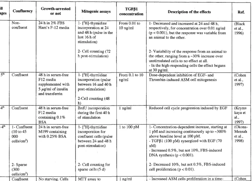
1.12. IN VITRO EFFECT OFTGFBl ON ASM CELLPROLIFERATION ... 60
CHAPTER 2: FGF2 ... 70
2.1. FGF FAMILY AND BIOLOGY ... 70
2.2. FGF2 STRUCTURE, SECRETION AND EXTRACELLULAR LOCALIZATION ... 70
2.3. FGFS RECEPTORS AND CO-RECEPTORS ... 73
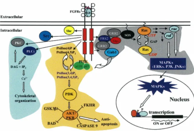
2.4. FGF RECEPTOR SIGNALING ... 75
2.5. INCREASED FGF2 EXPRESSION IN ASTHMA OR IN EXPERIMENTAL ANIMAL MODELS OF ASTHMA .. 77
2.6. RAPID RELEASE OF FGF2 FOLLOWING ALLERGEN CHALLENGE ... 79
Proteolysis-mediated FGF2 desequestration from ECM ... 79
Mast cell de granulation ... 80
Released following epithelium damage ... 82
2.7. FGF2 RECEPTORS ON ASM CELLS ... 87
2.8. IN VIVO LINKS BETWEEN FGF2 AND ASM CELL HYPERPLASIA ... 88
2.9. IN VITRO EFFECT OF FGF2 ON ASM CELL PROLIFERATION ... 88
CHAPTER 3: FGF2 AND TGF~l SYNERGISM IN HUMAN BRONCHIAL SMOOTH MUSCLE CELL PROLIFERATION ... 92
CHAPTER 4: SYNERGISM BETWEEN FGF AND TGF~ F AMIL Y MEMBERS ... 93
CHAPTER 5: IL-4 AND IL-13 ... 99
5.1. IL-4 AND IL-13 ANDTHEIR RECEPTORS ... 99
5.2 INVOLVEMENT OF IL-4 AND IL-13 IN THE DEVELOPMENT OF ASTHMA PATHOGENESIS ... 100
5.3 CELLULAR AND MOLECULAR MECHANISMS MEDIATING THE EFFECTOR FUNTIONS OF 4 AND IL-13 ... 103
5.4 INCREASED EXPRESSION OF IL-4 AND IL-13 IN ASTHMA AND EXPERIMENTAL ANIMAL MODELS OF
ASTHMA ... 106
5.5 EFFECTS OF IL-4 AND IL-13 ON ASM CELLS ... 108
CHAPTER 6: THE Tu2 CYTOKINES, IL-4 AND IL-13, ENHANCE HUMAN BRONCHIAL SMOOTH MUSCLE CELL PROLIFERATION ... 110
CHAPTER 7: LEUKOTRIENES ..•...•..•...•••...••...•. 111
7 .1. LEUKOTRIENES AND RECEPTORS ... 111
7 .2. INCREASED EXPRESSION OF CYS-LTS AND CYSL Tl IN ASTHMA AND EXPERIMENTAL ASTHMA ... 113
7.3. CYs-LTs IN ASTHMA PATHOGENESIS ... 114
7.4. CYSL Tl RECEPTOR TRANSDUCES THE ASTHMA-LIKE EFFECTS OF CYS-LTS ... 117
7.5. EFFECT OF LEUKOTRIENES ON ASM CELL MITOGENESIS IN VIVO VS IN VITRO ... 118
Effects ... 123
References ... 123
7.6. INDUCTION OFTGFf:H IN VITRO AND IN VIVO ... 123
CHAPTER 8: LEUKOTRIENE D4-INDUCED, EPITHELIAL CELL-DERIVED TRANSFORMING GROWTH FACTOR fH IN HUMAN BRONCHIAL SMOOTH MUSCLE CELL PROLIFERATION ... 125
CONCLUSION ... 126
THERAPEUTIC OPTIONS TO ALTER THESE PROLIFERATIVE SYNERGISMS ... 136
FINAL REMARKS AND PERSPECTIVES ... 142
ACKNOWLEDGMENTS ... 144
FIGURE AND TABLE LIST
Figure 1: TGFf31 signaling ...•...••...•••...••...•.•...•...•...•....•.•...•...•••...••.. 13
Table 1: Expression ofTGFf31 in human asthma •.•.••••••....•...•..•...•...•...••...•.•...•... 21
Table 2: Increased expression of TGFf31 in animal models of asthma ...•... 25
Table 3: Effect of TGFfH on ASM cell proliferation in vitro ... 67
Figure 2: FGF2 signaling ...•...•.•.••...•...•...•...•... 77
Table 4: Expression of FGF2 in human and in a non-human primate model of asthma ....•...•....•. 78
Figure 3: IL-4 and IL-13 receptors and signaling ... 101
Table 5: Cys-LTs on ASM cell mitogenesis ... 123
Figure 4: Sequence of upregulated mediators in asthma ...••...•...•...•.. 131
Figure 5: PDGF receptors •...•...••...•••...•.•...•...•...•...•.•... 132
Figure 6 : Variability in the therapeutic response of asthmatics to a leukotriene receptor antagonist ...•....•...•...•...•...••... 137
LIST OF ABBREVIATION
aa Amino acid
AA Arachidonic acid
Ab Antibody
AHR Airway hyperresponsiveness ALK Activin receptor-like kinase
AM Alveolar macrophages
AR-Smad Activin-receptor activated Smads ASM Airway smooth muscle
BALF Bronchoalveolar fluid
BOP Becl omethasone di pro pi onate BLT Leukotriene B4 receptor
CCIO Clara cell 10-KD prote in Co-Smad Co-mediator Smad cys-LTs Cysteinyl leukotrienes CysLTl Cys-LTs receptor 1
DG Diacylglycerol
Dox Doxycycline
ECM Extracellular matrix ECP Eosinophil cationic protein EGF Epidermal growth factor
FBS FEV1 FGF FGFR FHFs FLAP FRS2 GAG GGL y-GT GM-CSF GPCR Grb2 GSH GTPase HBGF HMW 5-HPETE HSPG IGF IGFR IGFBP IL
Fetal bovine serum
Forced expiratory volume in 1 sec Fibroblast growth factor
FGF receptor
FGF homologous factors
5-lipoxygenase activating protein FGF receptor substrate 2
Glycosaminoglycans y-glutamyl leukotrienase
gamma-gl utamyl transpeptidase
Granulocyte-macrophage colony-stimulating factor G protein-coupled receptor
Growth factor receptor-binding protein Gl uthathi one
Guanosine triphosphatase Heparin binding growth factor Higher molecular weight
5-hydroperoxyeicosatetraenoic acid Heparan sulfate proteoglycans Insulin-like growth factor IGF receptor
IGF binding protein Interleukin
IP JAK-STAT LAP LAR LBP LMW 5-LO LPA LPS LRPl LTBP LTC4S LTRA LTs MAPK MBP MMP MTl-MMP NLS OR OVA p7ÜS6K PDGF Inositol phosphate
Janus kinase-Signal transduction and activator of transcription latency-associated peptide
late asthmatic reaction LPS binding protein Low molecular weight 5-lipoxygenase
Lysophosphatidic acid Li popol ysaccharide
Low-density lipoprotein receptor-related protein 1 latency TGF~ binding protein
LTC4 synthase
Leukotriene receptor antagonist Leukotrienes
Mitogen-activated protein kinase Major basic protein
Matrix metalloproteinase Membrane type 1-MMP Nuclear localization sequence Odds ratio
Ovalbumin
70-KD kinase of S6 Ribosomal protein Platelet-derived growth factor
PDG FR
PBK
PKC
PLC
PPAR pSmad PTB RTK SAC SBE SH2 SNP Sos TGF j3 Tj3RTH
TNFa tPA uPA VEGF VSM PDGF receptor Phosphoinositide 3-kinase Protein kinase C Phospholipase CPeroxisome proliferator-activated receptor Phosphorylated Smad
Phosphotyrosine binding domain Receptor tyrosine kinase
Segmental allergen challenge Smad binding element Src homology 2 domain
Single nucleotide polymorphism Son of sevenless
Transforming growth factor beta TGF beta receptor
T helper lymphocyte
Tumor necrosis factor alpha Tissue plasminogen activator urokinase plasminogen activator Vascular endothelial growth factor Vascular smooth muscle
INTRODUCTION
Allergie asthma is cause by an immunological response toward innocuous inhaled enviranmental triggers and is characterized by recurrent breathing symptoms secondary to inflammation, airway hyperresponsiveness (AHR) and airway
obstruction. In 1922, Huber and Koesler (Huber and Koessler, 1922) described for the first time an increase in ASM tissue in the airways of asthmatics. Since then,
enlargement of peribranchial ASM tissue has become a histopathologic signature of asthma (Hirst et al., 2004). Other structural alterations of the lung, collectively called remodeling, also occur in asthma. It includes epithelial metaplasia, which is largely characterized by goblet cells hyperplasia, and hypertraphy of branchial glands, which both lead to mucus hypersecretion. Altered deposition of extracellular matrix (ECM) is also observed and mostly translates into subepithelial fibrasis, furthering the thickness of the airway wall (Pascual and Peters, 2005). However, due toits
contractile nature and its increased abundance in asthma, ASM tissue has alway been thought responsible for AHR. Current evidence for this contention includes: 1-Exagerated branchial responsiveness of asthmatic patients to non-inflammatory contractile agonists, such as cholinergie stimuli, in periods of asthma remission where inflammation and mucus hypersecretion are largely attenuated (Seow and Fredberg, 2001); 2- Mathematical modeling established in the early 1990's, which suggested that among all features of airway remodeling found in asthma, increased bulk of ASM tissue arround the branchi was likely the main contributor to AHR (reviewed in (Pare et al., 1997)); and 3- Branchial thermoplasty, which alters the structure and the
long term improvement in AHR in human asthmatics (Brown et al., 2005; Cox et al., 2006; Danek et al., 2004).
In the last few years, new aspects of ASM cell biology have gained interest of researchers working in the field of asthma. Information emanating from these studies suggests that ASM cells are not only a contractile unit, but are also able to express adhesion molecules for inflammatory cell recruitment and are potent secretagogues for cytokines, chemokines and ECM components (Joubert and Hamid, 2005). Hence, beyond their role in bronchoconstriction, ASM cells are now believed to play a significant role in the initiation and/or perpetuation of airway inflammation, as well as in the architectural changes that arise in asthmatic airways.
These new emerging concepts, together with the ability of ASM to contract in response to spasmogens, highlight the possible deleterious consequences of an
increased peribronchial ASM mass in the pathology of asthma. In the same direction, elucidation of the main factors involved in ASM cell hyperplasia may significantly enhance our understanding of asthma etiology and may, hopefully, culminate in the elaboration of improved therapeutics for the treatment of this prevalent disease.
This thesis aims to present the current evidence supporting the role of TGFf)l and FGF2 in ASM cell hyperplasia, to document the lack of data concerning the effect of IL-4 and IL-13 in this altered phenotype and to underscore recent developments made in this field that may shed light on some of the paradoxical results published so far
concerning the mitogenic effects of TGF~l and leukotrienes on ASM cells. New evidence is given that FGF2 may be the cornerstone of ASM cell hyperplasia owing to its ability to confer mitogenic potential to different asthma mediators.
GENERAL AND SPECIFIC OBJECTIVES
Chapter 1
General Objective: To review published studies supporting the involvement of TGFPl on ASM cell hyperplasia characterizing asthma.
Specific Objectives: The first objective of this chapter is to give a global overview of the TGFPl cytokine, its cell-surface receptors and the intracellular signaling pathways that are responsible for transducing its biological effects. The second objective is to compute the results of published studies regarding the expression of TGFP 1 in asthma or experimentally-induced asthma. Finally, the last objectives of this chapter are to discuss the current in vivo evidence supporting arole for TGFPl in ASM cell hyperplasia and the rather controversial state of the literature regarding the eff ect of TGFBl on ASM cell proliferation in vitro.
Chapter 2
General Objective: To review the current literature documenting the potential role of FGF2 on asthmatic ASM cell hyperplasia.
Specific Objectives: The first objective was to give an overall picture of the FGF2 growth factor and to present the different cell-surface receptors onto which this ligand can bind. The intracellular signal transduction pathways that ensure its biological effects are also briefly discussed. Then, the consitent results obtained by diff erent groups of investigators, revealing its increased expression in asthmatic airways, together with its unequivocal effect on ASM cell proliferation in vitro are reviewed.
However, its as yet unrecognized contribution to ASM tissue remodeling in vivo is also highlighted.
Chapter
3
General Objective: To document the mitogenic effect of TGFBl on primary human bronchial smooth muscle cells with or without a pre-treatment with FGF2.
Working hypothesis: FGF2 pre-treatment influences the mitogenic potential of TGFBl, which would help to explain the conflicting results obtained so far concerning the effect of TGFBl on ASM cell proliferation in vitro.
Specific Objectives: To determine the time- and concentration-dependent effect of TGFBl on ASM cell proliferation with or without a 24 hour pre-treatment with increasing concentrations of FGF2. The most important finding included in this chapter is a striking synergism between FGF2 and TGFBl on ASM cell proliferation. The next objectives then turn toward elucidating the operational mechanisms involved in this proliferative synergism.
Chapter4
General Objective: Discuss the results obtained in chapter 3 in relation to the
collective proliferative synergism of ASM cells that might occur in vivo between any member of the TGFBl and FGF2 families.
Specific Objectives: The first objective of this chapter was to identify ail other members of TGFBl and FGF2 families that have been shown to be upregulated in asthmatic airways. Since the different members of each of these families transduce their biological effects via similar signaling pathways, we postulate that they might
exert similar effects. If true, any member of one family potentially synergizes with any member of the other family in inducing ASM cell proliferation. This chapter underscores the potential contribution of other members of the TGFj31 and FGF2 families to the enlarged ASM tissue found in the bronchi and bronchioles of asthmatic patients. Overall, it proposes the hypothesis that ASM cell hyperplasia occuring in
vivo may not simply be the result of the individual proliferative synergism between FGF2 and TGFj31, but rather to the potential additive action of ail individual synergisms susceptible to occur between every upregulated members of the FGF2 family with any of the upregulated members of the TGFj31 family.
ChapterS
General Objective: To review the current state of the literarure in regard to the effect of IL-4 and IL-13 on ASM cell proliferation.
Specific Objectives: The first objective of this chapter was to introduce 4 and IL-13 cytokines in terms of their structures, their cognate cell-surface receptors and the intracellular signaling pathways that they use to transduce their biological effects. This chapter also aims to review the knowledge aquired so far concerning the role that each of these cytokines might have in asthma etiology and diathesis. lt is concluded that even if either IL-4 or IL-13 were shown to be required, and sometime sufficient, in the development of many pathognomonic features of asthma, their respective role in ASM cell hyperplasia is still unknown.
Chapter6
General Objective: To document the individual and the FGF2-combined effects of IL-4 and IL-13 on primary human bronchial smooth muscle cell proliferation.
Specific Objectives: To determine the concentration-dependent effect of 4 and IL-13 on ASM cell proliferation with or without a pre-treatment with increasing
concentrations of FGF2. Similar to what we observed with TGFj31 in chapter 3, the results suggest that both IL-4 and IL-13 have no mitogenic effect on their own, but synergize in a concentration-dependent manner with FGF2 to induce ASM cell proliferation. The next objectives were then to determine the operational mechanisms involved in this proliferative synergism.
Chapter7
General Objective: To review the current state of knowledge concerning the effect of cys-LTs on ASM cell hyperplasia.
Specific Objectives: The first objectives of this chapter are to give a general overview of cys-LTs structure, their synthesis and their cognate cell-surface receptors, and to briefly discuss their well-established increased expression in asthma. Another objective is to revise the conflicting results between in vitro studies, which suggest
very weak or no direct effect of cys-LTs on ASM cell proliferation, and the in vivo
studies, which consistently report a trophic effect of these mediators in ASM cell hyperplasia. Owing to the great reminiscence of this last observation with the effect of TGFj31 highlighted in chapter 1, the final objective of chapter 7 was to wrap up recent evidence suggesting that cys-LTs are potent inducers of TGFf31 in vivo, as well as in
Chapter8
General Objective: To document the potential paracrine influence of epithelial cell-derived, cys-LT-induced TGFf:H on the proliferation of primary human branchial smooth muscle cells.
Working hypothesis: Based on our previous results suggesting an important effect of TGF~l on ASM cell proliferation (chapter 3), we hypothesized that the mitogenic effect of cys-LTs reported in vivo, together with their lack of effect in vitro, may be related to their capacity to stimulate the production of this growth factor in airway epithelial cells, which will, in turn, act as a paracrine factor to induce ASM cell proliferation.
Specific Objectives: The initial objective of this chapter was to determine whether cys-LTs are capable to upregulate TGF~l expression in an epithelial cell line that overexpressed the high affinity receptor for leukotriene D4, CysLTl. Another major
concern was to determine whether this endogenously produced TGF~l is able to support ASM cell proliferation. This initial part of the work presented in chapter 8 reports that cys-LTs are potent inducers of TGF~l and that this cys-LT-induced TGF~l is able to support the proliferation of FGF2-pretreated ASM cells. It was thereof important to determine whether untransformed airway epithelial cells behaved in a similar manner. Hence, the hypothesis that CysLTl is expressed on airway epithelial cells and that epithelial cells have the capacity to respond to cys-LTs in a CysLTI specific manner in term of TGF~l production were tested. The cumulated data highlights the possibility that cys-LTs can induce TGF~l expression in airway epithelial cells. Altogether, these results shed light on the conflicting results reported
so far between in vivo and in vitro studies documenting the effect of cys-LTs on ASM cell proliferation, and suggest that the mitogenic effect observed in vivo may be related to a paracrine involvement of airway epithelial cell-derived ASM cell mitogens that are induced by cys-LTs.
1.1. TGFfJl
CHAPTER 1 TGFfU
TGFf31 was first isolated and characterized in platelets in 1983 (Assoian et al., 1983) and is now the prototypic member of a superfamily of cytokines, which actually counts 33 members in man (de Caestecker, 2004). TGFf31 is encoded by a 7-exon gene localizes on chromosome 19ql3 and several genetic studies have associated some of its common single nucleotide polymorphisms (SNP) with asthma phenotypes (Buckova et al., 2001; Hakonarson et al., 2001; Hobbs et al., 1998; Mak et al., 2006; Nagpal et al., 2005; Pulleyn et al., 2001). TGFf31 protein has a short half-life
(normally less than 3 min) in cell free systems (Wakefield et al., 1990). However, to overcome its lability, TGFf31 is usually secreted in a latent formas a 180-210-KD multi-protein complex containing the glycosylated 125-190-KD latency TGFf3 binding protein (LTBP), the 75-KD latency-associated peptide (LAP) and the 25-KD mature form of TGFf31 (Harpel et al., 1992).
1.2. TGFfJl activation
Activation of latent TGFf31 occurs through different mechanisms including: 1-proteolytic dissociation from LAP by the urokinase plasminogen activator
(uPA)/plasmin system (Khalil et al., 1996a; Lyons et al., 1988), or by other proteases such as metalloproteinase (MMP)-2 (McMahon et al., 2003), MMP-9 (Lee et al., 2001a; Yu and Stamenkovic, 2000) and the lysosomal serine protease cathepsin D (Lyons et al., 1988); 2- conformational alteration in its structure by thrombospondin
(Crawford et al., 1998) or integrin such as avj36 (Morris et al., 2003; Munger et al., 1999); 3- oxidation and nitrosylation (Barcellos-Hoff and Dix, 1996; Vodovotz et al., 1999); 4- removal of carbohydrate structure on LAP by glycosidases such as sialidase (Miyazono and Heldin, 1989); and 5- integrin avj38-mediated latent TGF(31
recruitment to the cell membrane for membrane type 1 (MTl)-MMP-dependent proteolytic activation (Fjellbirkeland et al., 2003). Extracellular regulation of TGFfH activation is also influence by different cell-surface molecules and ECM constituents that bind TGF(31 and ensure its activation in restricted localised compartment. For example, mannose 6-phosphate/insuline-like growth factor II (IGF-11) receptor
(Gleizes et al., 1997; Kovacina et al., 1989) as well as integrins a8(31 (Lu et al., 2002) and avj31 (Munger et al., 1998) bind latent forms of TGF()l and are thus believed to target the latent complex on the surface of cells for subsequent proteolytic activation with ensuing binding toits signaling receptor. TGF()l is also a heparin binding
growth factor (HBGF) (Miyazono et al., 1994a). Consequently, its binding availability for cell surface receptors is regulated extracellularly by heparan sulfate proteoglycans (HSPG). Whereas certain proteoglycans, such as betaglycan and endoglin (Cheifetz et al., 1992), facilitate TGF()l binding toits receptors; others, such as biglycan and decorin, sequester TGFj31 in the ECM (Redington et al., 1998). In addition, because of their ability to release TGF() 1 from pericellular stores, certain enzymes such as thrombin, neutrophil elastase or mast cell chymase may be essential in the process of TGFj31 activation, even if they cannot activate latent TGFj31 directly (Taipale et al., 1992; Taipale et al., 1995).
1.3. TGF{Jl receptors and signaling
Six receptors have been identified for TGFf31 (Huang and Huang, 2005), but the most studied are the 65-KD type 1 receptor (Tf3RI or ALK5), the 85-KD type II receptor (Tf3RII), the 280-KD type III receptor (Tf3RIII or betaglycan, a heparan
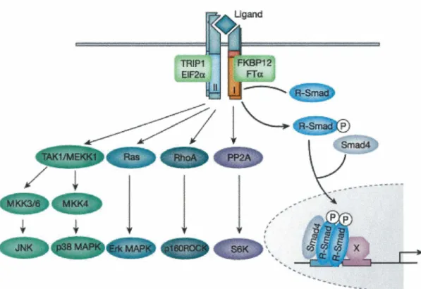
sulfate/chondroitin sulfate proteoglycan), and more recently the 504-KD Tf3R5, which is also known as the low-density lipoprotein receptor-related protein 1 (LRPl). The canonic mechanisms by which TGFf31 binds and activates its cognate cell surface receptors as well as the intracellular signaling pathways that transduce intracellularly the TGFf31 message from the cell membrane to the nucleus have been reviewed extensively (Shi and Massague, 2003). Briefly, TGFf31 initially binds to the single transmembrane, constitutively active, serine/threonine kinase Tf3RII homodimer. The formed complex subsequently recruits the single transmembrane, activable,
serine/threonine kinase Tf3RI homodimer, which is concomitantly activated by Tf3RII-mediated phosphorylation of several threonine and serine residues in its intracellular GS juxtamembrane domain. This phosphorylated GS domain then serves as a docking site for activin-receptor activated Smads (AR-Smads; namely Smad2 and Smad3), which are, in turn, phosphorylated by Tf3Rl. The phospho-AR-Smads (pSmad2 and pSmad3) then homo or hetero-oligomerize with each other and with at least one co-mediator Smad (Co-Smad; most often called Smad4) and the complex ultimately translocates to the nucleus where it binds Smad binding element (SBE)-containing promoters or interacts with other transcriptional partners to regulate gene expression. Apart from the Smad pathway, it is now clear that other intracellular signaling pathways such as mitogen-activated protein kinase (MAPK), the phosphoinositide
3-kinase (PI3K), the PP2A phosphatase-mediated p70s6K inactivation and the
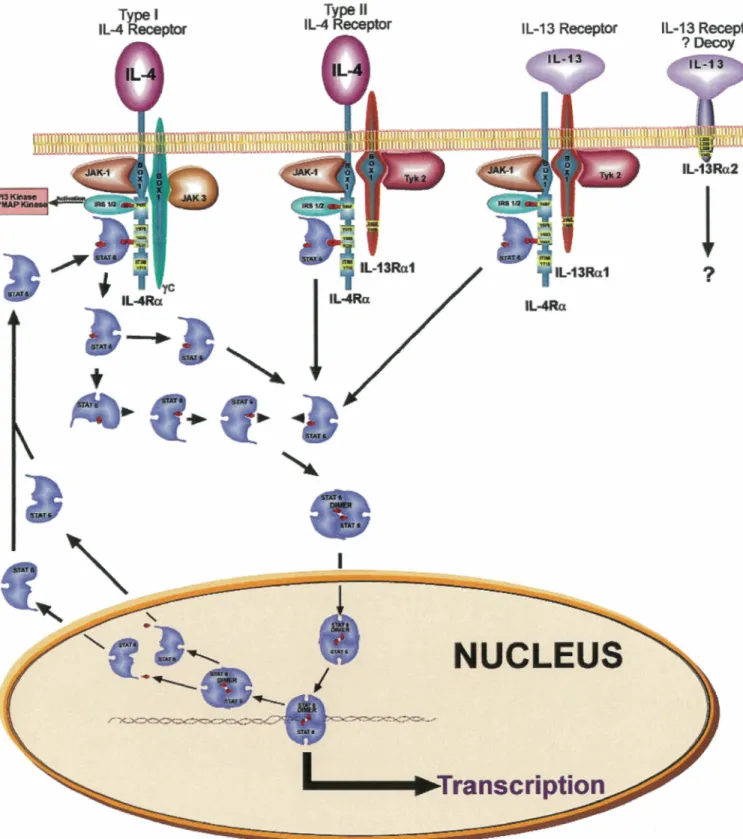
Rho-family of small guanosine triphosphatase (GTPase) pathways are activated by TGFj31 and transduce some of its biological activities (reviewed in (Derynck and Zhang, 2003) and (Moustakas and Heldin, 2005)) (Figure 1). In addition, Smads were shown to cross talk with other important signaling pathways such as Janus kinase-Signal transduction and activator of transcription (JAK-STAT) (Ulloa et al., 1999) and WNT (Nishita et al., 2000).
Figure 1: TGFfH signaling
1.4. Expression of TGF
fJJ
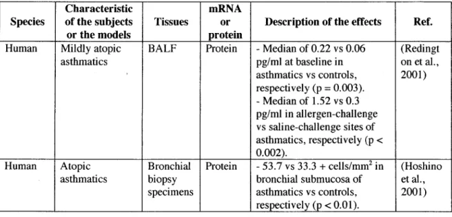
in asthmaExpression of TGFf3 l is altered in asthma and the current weight of evidence suggests that TGFf3 l is upregulated in human and animal asthmatic airways (summarized in Table 1 and 2). However, 5 studies performed with human tissues have shown no regulation of TGFf3 l expression in asthma. In contrast to their previous articles, in which they reported an increased expression of TGFf31 in branchoalveolar lavage fluid (BALF) before and after allergie challenge (Redington et al., 1997), Redington and coworkers (Redington et al., 1998) have demonstrated indistinguishable pattern of TGFf31 immunohistochemical staining between asthmatic and contrai subjects.
Aubert and coworkers (Aubert et al., 1994) had previously reported similar findings, but their results were contested since their contrai subjects were heavy smokers. The relative intensity of TGFf31 immunostaining in the branchial mucosa was also similar between asthmatics and healthy subjects in Hoshino and coworkers' study (Hoshino et al., 1998). More recently, Chu and coworkers (Chu et al., 2004) confirmed these results by documenting a lack of significant augmentation of TGFf3 l
immunoreactivity in asthmatic epithelium. Agreeing with these studies, Balzar and coworkers (Balzar et al., 2005) have shown no difference in the number of cells staining positive for TGFf31 in the submucosa of normal subjects and asthmatic patients suffering from different severity of the disease.
Reasons for these discrepancies are currently unknown. However, ail conflicting results came from studies measuring TGFf31 expression by immunohistochemical appraach, using tissue specimens obtained by branchial biopsies or lung resections.
Immunohistochemistry requires extensive tissue handling. Ali the steps before microscopie reading, including immediate precaution to preserve tissue integrity, reagent used for fixation or to embed the tissue, strength and specificity of the
detection and the staining antibodies, and bleaching of the fluorochrome or attenuated chemiluminescence signal occurring during the procedures could all lead to erroneous results and false interpretations. It is thus reasonable to surmise that the conflicting results concerning the increased expression of TGFPl in the airways of asthmatics may be the result of technical artefacts. However, alternative hypothesis may explain this conundrum.
TeIDporalconcerns
It is worth mentioning that collection of lung specimens off ers promiscuous
advantages for studying mRNA or protein expression at the tissue level. For example, staining of cross-sectional sections of these Jung specimens by immunohistochemical approach or by in situ hybridization brings ample information regarding the tissue or the cellular sources of TGFPl. Combined with laser microdissection, tissue specific expression of a particular gene can even be confirmed by more conventional
technique such as RT-PCR (Kelly et al., 2005). Unfortunately, limits of these
techniques are also prominent. As such, results obtained from these experiments must be interpreted with caution. Protein or mRNA detected in Jung specimens reflect their expression levels at a particular time point. Asthma is a waxing and waning disease, where a period of exacerbation is usually followed by a period of remission and where the severity of symptoms is temporally associated with the degree of airway
likely to be inducible and transient in nature. Correspondingly, TGF~l was shown to
be increased at 24 h, but not at 10 min, following segmental allergie challenge (SAC) and its concentration returned to baseline level after 1 week (Batra et al., 2004; Redington et al., 1997). Whether TGF~l expression starts to increase earlier is
unknown, but in animal models of acute or chronic antigen challenge, TGFf31 expression in BALF is still unaffected 6 h following the last allergen exposure (Kumar et al., 2004). In contrast to Batra and coworkers (Batra et al., 2004), Redington and coworkers (Redington et al., 1997) also reported a statistically significant increase in TGFf31 level in BALF of asthmatics at baseline compared to healthy controls (8 pg/ml vs 5.5 pg/ml), but whether this difference is physiologically relevant remains questionable. Tillie-Leblond and coworkers (Tillie-Leblond et al., 1999) have also reported no difference in the levels of latent and active form of TGF~l in BALF at baseline between mild asthmatics and healthy volunteers. In the same study, both the latent and active form of TGFf31 were significantly increased in patients suffering from status asthmaticus compared to healthy controls or to patients presenting similar severity of the disease, but distant from an acute exacerbation period. Nomura and coworkers (Nomura et al., 2002) have substantiated these results by examining longitudinal changes that occur in the lung fonction (forced expiratory volume in 1 sec,% of predicted (%FEV1)) and the percentage of TGFf31 positive cells in induced sputum samples of five asthmatic subjects. They demonstrated that during asthma exacerbation, %FEV1 decreased from 86.5 to 51.0% and that TGFf31 positive cells rose from 1.9 to 55.4% during the same time period. These results confirmed the inducible and transient upregulation of TGFf31 that have been
demonstrated by others in BALF following SAC (Batra et al., 2004; Redington et al., 1997). Based on results obtained with animal models of chronic allergen challenge-induced airway remodeling, it was also suggested that several allergen provocations must be required before the upregulation of TGF~l could be appreciated (Corbel et al., 2003).
These aformentioned findings suggest that the samples would need to be collected following bronchoprovocative challenge to observe the transient increase of TGF~ 1 expression by immunohistochemistry. In all studies documenting no regulation of TGF~l expression in asthma, lung specimens had been taken at baseline, i.e. in a remission period where no sign of exacerbation was present or without prior experimentally-induced bronchoprovocation. In Hoshino and coworkers' study (Hoshino et al., 1998) for example, asthmatics presented daily symptoms, but based on their attack score, the number and severity of symptoms were very low, suggesting that subjects were not in an exacerbation period when biopsies were taken. In the immunohistochemical study carried out by Redington and coworkers (Redington et al., 1998), asthmatic subjects were presented as mildly symptomatic. However, they were clinically stable despite being restricted from use of oral or inhaled
glucocorticoids for 4 weeks, indicating once again that no acute exacerbation was present at the time bronchial biopsies were taken. Hence, failure to demonstrate a significant upregulation may simply reflect the punctual expression of TGFf:H measured in the two extreme poles of a transient response. Taken together, these results implied that TGF~l is not necessarily overexpressed in asthmatics at baseline,
but it is inducible upon allergen challenge. Determining the sequence and the kinetics of TGF~l expression may be important to increase our understanding of the role of this cytokine in asthma.
Spatial concerns
Along with the transient nature of TGFf31 response following allergie challenge, failure to detect an increased expression of TGFf31 in certain immunohistochemical studies may be related to the airway compartment studied. Expression of TGFf3 was shown to be heterogenous within the same sample (Vignola et al., 1997) and its increased expression in asthma may occur exclusively in very localized
compartments. For instance, periglandular tissues or sites of epithelial desquamation were shown to stain strongly for this cytokine (Kokturk et al., 2003; Vignola et al., 1997). On the other hand, Magnan and coworkers (Magnan et al., 1997) have
demonstrated homogenous intensity of TGFf3 immunostaining in ciliated and mucous cells as well as in areas of epithelial impairment, such as sites of deciliated cells or desquamated regions. However, apart from a homogenous staining in the epithelium within each sample, Magnan and coworkers (Magnan et al., 1997) suggested an altered compartmentalization of TGFf3 expression in asthmatic airways. Whereas TGFf3 immunoreactivity was strong in the epithelium of control subjects, negative or faintly positive staining was observed in this particular compartment of asthmatics. In contrast, asthmatics expressed higher amounts of TGFf3 in the submucosa compared to healthy individuals. This epithelial to submucosal redistribution of TGFf3 was in accordance with an increased number of inflammatory cells staining positive for
TGFj3 in the submucosa of human asthmatics (Chakir et al., 2003; Chu et al., 2000; Flood-Page et al., 2003; Minshall et al., 1997; Ohno et al., 1996; Vignola et al., 1997).
Whatever the physiologie or pathophysiologic reason for this altered
compartmentalization, the same trend of TGFj31 relocalization was observed in murine models of allergie airway inflammation. In this regard, McMillan and coworkers (McMillan and Lloyd, 2004) have demonstrated that TGFj31 expression was confined to the bronchiolar and alveolar epitheliums in control animais and was relocalized to the submucosal compartment in association with inflammatory infiltrates after repeated allergen challenges of sensitized animais. In this particular mode!, even smooth muscle became positive for TGFj31 immunostaining during the chronic phase of allergen challenge. lnterestingly, this altered compartmentalisation also occurred in other types of airway inflammation, such as the one induced by prolonged (4 wk) lipopolysaccharide (LPS) exposure (Savov et al., 2002). Initially, TGFj31 expression was confined to the airway epithelium, but subsequent to LPS exposure, TGFj31 immunostaining was mainly localised in the subepithelial area (Savov et al., 2002). Hence, in addition to look at the right time, investigators
attempting to document an increased expression of TGFj31 in asthma need to look at the right place.
With the use of techniques permitting to appreciate the overall expression of TGFj31, such as in studies using BALF, serum or plasma, or with the use of animal models, which allow sufficient biologie materials to be homogenized, it is becoming clear that
TGFf:H is upregulated in asthma following allergie challenge. But once again, controversies are reported and are related to different peculiarities of the studied populations. For example, Joseph and coworkers (Joseph et al., 2003) have reported an increase of TGF~ 1 expression in the plasma of nonatopic, but not of atopic asthmatic patients. However, using atopic patients only, which were included based on skin prick test positivity and corroborating medical history of allergen-induced asthma, Karagiannidis and coworkers (Karagiannidis et al., 2006) reported a significant increase of TGF~l in the serum of asthmatics, attaining levels almost 7-fold higher than those measured in healthy controls. No clear explanation is currently ascribed to explain these contrasting results and the question of whether TGF~l expression is influence by the atopic phenotype will need further exploration.
Increased expression of TGF~l measured in the BALF must also be interpreted with caution. Epithelium desquamation is a characteristic feature of remodeled asthmatic airways. Epithelium denudation may give access to a certain amount of TGF~l, which is otherwise masked by an intact epithelium in non-asthmatic individuals. Thus, the increased expression of TGF~l observed in BALF of asthmatics following challenge may simply be related to an easiest accessibility to TGF~l stores cause by epithelium
desquamation. In support to this contention, increased concentration of TGF~l has been noted in BALF following a sham bronchoprovocation procedure, which is likely to be the result of epithelial damage (Redington et al., 1997). Moreover, positive correlation (r
=
0.89) has been reported in the same study between concentration ofThese results suggest that epithelium denudation renders a bulk of TGFj31 normally sequestered in the subepithelial layer, such as the one associated with the basal lamina (Aubert et al., 1994; Redington et al., 1998), collectable by bronchoalveolar lavage. Of major concern, degranulation products of eosinophils such as major basic protein (MBP) and eosinophil cationic protein (ECP) (Frigas et al., 1980; Robinson et al., 1992), as well as mast cell proteases, such as tryptase and chymase (Redington et al., 1995), are damaging for the airway epithelium. Increased expression of TGFj31 observed at 24 h, but not at 10 min, following SAC may consequently be caused by eosinophil-mediated epithelium desquamation, rather then a true de novo TGFj31 protein synthesis. Together, these observations raise doubts on the techniques
currently used to measure the expression of different mediators in the airways and for instance, to the increased expression of TG Fj31 in asthma.
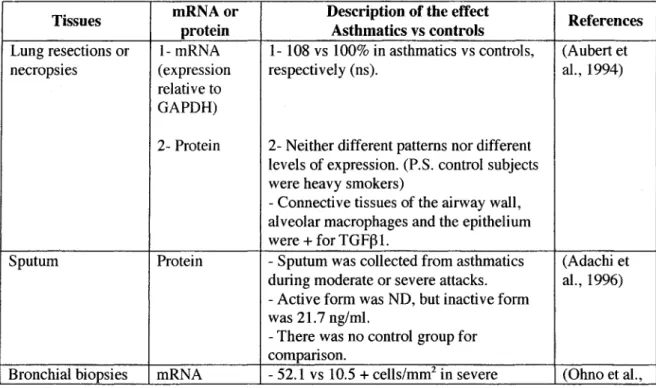
Table 1: Expression ofTGFfH in human asthma
Tissues mRNAor protein Description of the effect Asthmatics vs controls References Lung resections or 1-mRNA 1- 108 vs 100% in asthmatics vs controls, (Aubert et necropsies (expression respectively (ns). al., 1994)
relative to GAPDH)
2- Protein 2-Neither different patterns nor different levels of expression. (P.S. control subjects were heavy smokers)
- Connective tissues of the airway wall, alveolar macrophages and the epithelium were +for TGF~l.
Sputum Prote in - Sputum was collected from asthmatics (Adachi et during moderate or severe attacks. al., 1996)
- Active form was ND, but inactive form was 21.7 ng/ml.
- There was no control group for comparison.
asthmatics vs controls, respectively (p < 1996) 0.02), but was not elevated in mild
asthmatics (1.0 + cell/mm2)
- In asthmatics, + cells were eo.
Alveolar Protein - Median of 435 vs 210 ng/million cells in (Vignola et
macrophages asthmatics vs controls, respectively (p
=
al., 1996)0.005).
Bronchial biopsies Protein* - Altered compartmentalization: In (Magnan et
asthmatics, TGFBl intensity of staining al., 1997) decreased in the epithelium (p < 0.01) and
increased in the submucosa in association with an increase inflammatory cell infiltrate.
Bronchial biopsies Prote in* - Median of 4 vs 0 + cells/mm of BM in (Vignola et
the epithelium and 31.5 vs 0 + cells/mm2 al., 1997)
in the submucosa in controls vs asthmatics, respectively (p =::; 0.015).
BALF Protein - 8.0 vs 5.5 pg/ml in asthmatics vs (Redington
controls, respectively (p
=
0.027). et al., 1997)- lncreased expression following SAC: 31.3 vs 25.0 at 10 min (p
=
0.78) and 46.0 vs 21.5 pg/ml at 24 h (p=
0.017) post-allergen- vs saline-challenged sites, respectively.Bronchial biopsies 1-mRNA - 18.5, 10.8 and 7.8 vs 3.5 + cells/mm of (Minshall et
BM in severe, moderate and mild al., 1997)
asthmatics vs controls, respectively (p <
0.05 for severe and moderate asthmatics). 2- Protein - 18.8, 12.3 and 9.2 vs 5.2 + cells/mm of
BM in severe, moderate and mild asthmatics vs controls, respectively (p <
0.05 for all groups of asthmatics).
Bronchial biopsies Protein - 18 vs 16% relative intensity in the (Hoshino et
bronchial mucosa of asthmatics vs al., 1998)
controls (ns).
Bronchial biopsies Prote in - Neither different patterns nor different (Redington
levels of expression between asthmatics et al., 1998) and controls.
- Positive staining was observed in subepithelial connective tissues of the airway wall and in the bronchial epithelium.
Bronchial biopsies Protein* - 12.5 vs 6.6 + cells/mm2 in the (Chu et al.,
submucosa of asthmatics vs controls (ns, p 1998)
=
0.06).BALF Protein -Active TGFBl: median of -350 and 30
(Tillie-vs 60 pg/ml in status asthmaticus and Leblond et
stable asthmatic patients vs controls, al., 1999)
- Latent TGF~l: median of-900 and 75
vs 1 OO pg/ml in status asthmaticus and stable asthmatic patients vs controls, respectively (p < 0.05).
Bronchial biopsies Protein* -Medianof-41.4vs7.8, 11.1, 15,and8.l (Wenzel et
cell/mm2 in the submucosa of eo + severe al., 1999)
asthmatics vs controls, mild asthmatics, moderate asthmatics and eo - severe asthmatics, respectively (p = 0.0003). - TGFB + cells correlate with eosinophils, neutrophils and macrophages number (p < 0.0001).
BALF-enriched mRNA - Increased in mild atopic asthmatics vs (Prieto et al.,
alveolar controls (p < 0.005). 2000)
macrophages
1-B ronchial 1- Protein* 1- 108 vs 24 + cells/mm2 of airway (Chu et al.,
biopsies submucosa in asthmatics vs controls (p = 2000)
0.002).
2-Peripheral blood 2- Protein 2- 115 vs 46 pg/106 cells in asthmatics vs
neutrophils (spontaneous controls, respectively (p = 0.007).
release ex
vivo)
Airway epithelial Prote in -~4 vs 1.5 pg/ 104 ce lis in asthmatics vs (Hastie et
ce lis (ex vivo) controls (p = 0.032). al., 2002)
Cells in sputum Prote in - 23 out of 26 asthmatics demonstrated (Nomura et
samples TGF~ 1 + cells vs 0 out of 8 in normal al., 2002)
volunteers.
Bronchial biopsies Protein* -~11and6 vs< 1 + cells/mm2 of (Chakir et
bronchial submucosa, in severe-to- al., 2003)
moderate and mild asthmatics vs controls, respectively (p < 0.05).
- Immunoreactivity was mainly localized in inflammatory cells.
1- Bronchial 1-mRNA 1- Median of ~43 vs 5 + eo/mm2 before vs (Flood-Page
biopsies after treatment with mepolizumab in mild et al., 2003)
atopic asthmatics, respectively (p
=
0.04). - Median of ~20 vs 39 + eo/mm2 before vsafter placebo treatment, respectively.
2- BALF 2- Protein 2- Median of ~5 vs 4 pg/ml before vs after
treatment with mepolizumab in mild atopic asthmatics, respectively (p
=
0.05). - Median of ~4 vs 4 pg/ml before vs afterplacebo treatment, respectively.
- In both cases, there was no control group for comparison.
0.02), but was not elevated in intermittent asthmatics (2.7%) and was not affected in neither the epithelial nor the submucosal tissues.
Plasma Prote in - 2.5 vs 1.5 ng/ml in nonatopic asthmatics (Joseph et
vs controls, respectively (p
=
0.002), but al.,2003) was not different in atopic asthmatics ( 1.4ng/ml).
Bronchial biopsies Protein - Higher intensity of staining in the (Kokturk et
submucosa, but not in the epithelium, of al., 2003) asthmatics vs controls (p < 0.05).
- TGFf31 was mainly localized in association with connective tissue in all groups.
BALF Prote in - Identical at baseline ( ,,_,50 pg/ml) (Batra et al.,
- Significantly increased after SAC in 2004)
asthmatics only (""170 pg/ml) (p < 0.05).
Bronchial Protein - ( 1.0-2.4) vs (0.2-1.6)% of total epithelial (Chu et al.,
epithelium area (interquartile 25-75% range) in 2004)
asthmatic vs normal subjects, respectively (ns).
Bronchial biopsies 1-mRNA 1- no diff erence between mild and severe (Balzar et
(with or without persistent eosinophilia) al.,2005) asthmatics vs controls.
2- Protein* 2- The majority of subjects in every
groups expressed undetectable or very low number of+ cells in the submucosa.
1-Serum 1- Protein 1- 11.7, 14.5 and 36.8, vs 5.5 ng/ml in (Karagiannid
severe asthmatics with inhaled and is et al.,
systemic GCs, moderate asthmatics with 2006)
inhaled GCs and moderate asthmatics without any drug treatment vs healthy subjects, respectively (p ::5 0.001 for all
groups of asthmatics vs controls).
2-CD4+ T cells 2-mRNA 2- Decreased expression in moderate
asthmatics without any drug treatment compared to each of the 3 other groups (p
::5 0.01).
Exhaled breath Protein* - 1.69-fold increase in asthmatics (Matsunaga
condensate compared to healthy subjects (p < 0.01). et al., 2006)
*Antibody used did not discriminate between TGFj31, 2 or 3 or is not specified. Unless otherwise indicated, amounts of TGFf:H represent the mean values.
Abbreviation: +, positive; BALF, bronchoalveolar lavage fluid; BM, basement membrane; eo, eosinophils; GCs, glucocorticoids; ns; not statistically significant; SAC, segmental allergen challenge.
Table 2: lncreased expression of TGFfH in animal models of asthma
Species Models Tissues mRNAor Protein Extent of the increase References
Female OVA Whole-lung Active protein - Increased in fonction of allergen (Tanaka et BALB/c sensitization lavage concentration. From - 10, 60, 145 al., 2001) mi ce and and 190 pg/ml in saline, 0.01, 0.1
challenge and 1 % OVA, respectively (p < 0.01).
C57BU6 IL-13 1- Lung 1-mRNA 1- Increased expression in (Lee et al., mi ce transgenic homogenates transgenic mice vs littermate 200la)
controls.
2- Whole-lung 2- Protein 2- -294 vs 132, 1794 vs 59 and lavage 2610 vs 88 pg/ml in transgenic mice vs littermate controls at 1, 2 and 3 mo of age, respectively (p < 0.01).
Active protein - -2.4 to 4.3-fold increases in Iuciferase activity when CCI-64 cells stably transfected with a luciferase reporter gene driven by the PAI-1 promoter were stimulated with BALF of transgenic compared to littermate control mice (p < 0.001). 3- Lung sections 3-mRNAand 3- Expression was restricted to the
Protein airway epithelium and to some AM in littermate controls, but increased in AM and was appreciated in airway epithelium, type Il pneumocytes and occasionally in eosinophils of transgenic mice.
Male Endotoxin- Lung sections Prote in 1- 2.64 vs 0 relative intensity in air- (Savov et C3H/HeBF induced 1- (Staining vs LPS-exposed animais after 4 wk al., 2002) el mice asthma Subepithelium intensity was of LPS exposure (p < 0.005), but
are a graded from 0 this difference did not persist after to 3) a 4 wk recovery period.
2- Epithelium 2- 2.59 vs 0.83 relative intensity in air- vs LPS-exposed animais after 4 wk of recovery of the 4 wk LPS exposure (p < 0.005), but no difference (0.83 vs 1.14 in air- vs LPS-exposed animais) after the 4 wk of exposure.
Male BP2 OVA -BALFand Protein* - Increased expression in both of (Corbel et mi ce sensitization whole lung these Jung compartments 48 h after al., 2003)
and the Jast challenge of a 8 mo challenge protocol of bronchoprovocation,
but not after 48 h of a single challenge, compared to control mi ce.
Female OVA Whole-lung Active protein - 44.9, 41.8 and 583.6 pg/ml in (Masudaet WBB6F1, sensitization lavage control, sensitized only and al.,2003)
control, sensitized only and sensitized/challenged W/Wv, respectively (p < 0.001). - 102.4 vs 464.6 pg/ml in sensitized only vs sensitized/challenged Sl/Sld, respectively (p < 0.05).
Male Endotoxin- Whole-lung 1- Protein 1- -865 vs 58 pg/ml in C3HeB/FeJ (Brass et C3HeB/FeJ induced lavage vs C3H/HeJ after 5 d of LPS al., 2003) (LPS- asthma exposure (p < 0.05), but was not
sensible) different after 4 h of exposure or 96
and h after an 8 wk of exposure.
C3H/HeJ
(LPS- 2- Active 2- -111vs19 pg/ml in C3HeB/FeJ insensible) protein vs C3H/HeJ after 5 d of LPS mi ce exposure (p < 0.05), and -22 vs 0
pg/ml in C3HeB/FeJ vs C3H/HeJ after 4 h of LPS exposure (p < 0.05), but not different 96 h after a 8 wk of LPS exposure.
Female OVA Whole-lung Active protein - 47.5, 57.9 and 178.0 pg/ml in (Nagao et C57BL/6 sensitization lavage control, sensitized only and al., 2003)
and and sensitized/challenged C57BL/6,
congenic IP challenge respectively (p < 0.01). KO mice - 39.1, 44.3 and 342.5 pg/ml in
control, sensitized only and sensitized/challenged IP KO, respectively (p < 0.01).
Female OVA Whole-lung Active protein - 17.l and 265.3 pg/ml in sensitized (Komai et BALB/c, sensitization lavage only and sensitized/challenged al., 2003) BALB/cJ, and BALB/c, respectively (p < 0.01).
and challenge - 27.5, 32.5 and 333.8 pg/ml in
BALB/cJ control, sensitized only and
congenic sensitized/challenged BALB/cJ,
IL-4KO respectively (p < 0.01).
mi ce - 27.9, 29.5 and 81.6 pg/ml in
control, sensitized only and sensitized/challenged IL-4 KO, respectively (p < 0.01).
Female OVA 1- Lung sections 1- Protein 1- Altered compartmentalization of (Mc Millan BALB/c sensitization TGFfH immunoreactivity, from and Lloyd, mi ce and airway epithelium to submucosal 2004)
challenge compartment.
2- Lung 2- Active 2- -66, 180, 160 and 66 vs 50 homogenates protein pg/ml in 25, 35, 55 days challenged
and 80 days challenged, 1 month recovered vs control mice, respectively (p < 0.05 for 35 and 55 days challenged vs control mice).
Female OVA 1- Whole-lung Prote in 1- 300 vs 135, 356 vs 156 and 369 (Cho etal., BALB/c sensitization lavage fluid vs 146 pg/ml in sensitized mice 2004b) mi ce and challenged vs unchallenged for 1, 3
challenge and 6 mo, respectively (p = 0.03). - Reduction to 241 and 269 pg/ml with ISS treatment in sensitized mice challenged for 3 mo and 6 mo, respectively, (p = 0.05), but no reduction in the l mo challenged group.
2- Lung 2- 1946 vs 664 pg/mg of lung homogenates protein in challenged (3 mo) vs
unchallenged sensitized mice, respectively (p
=
0.02).- Reduction to 939 pg/mg of lung protein in challenged sensitized mice treated with ISS (P
=
0.05)C57BU6 OVA 1- Lung 1- Protein 1- 3740 vs -1855 pg/ml in (Cho et al., and sensitization homogenates challenged vs unchallenged 2004a) congenic and sensitized WT mice, respectively (p
IL-5 KO challenge < 0.01).
mi ce - 2170 vs -1455 pg/ml in
challenged vs unchallenged sensitized IL-5"1 mice (p < 0.05
when compared challenged mice with WT and IL-51 genotypes).
2- Lung sections 2- Protein* 2- 64.5 VS -5 TGFB +
(numberof cells/bronchus in challenged vs peri bronchial unchallenged sensitized WT mice, cells +for respectively (p < 0.001).
TGFB per - 22.6 vs -1 TGFB + cells/bronchus bronchiole of in challenged vs unchallenged 150-200 µm of sensitized IL-51· mice (p < 0.001
internai when compared challenged mice diameter) with WT and IL-5"1
· genotypes).
- Peribronchial cells + for TGFB were mainly eo (63%) and macrophages (35%), but increased expression of TGFB was also observed in the epithelium of both WT and IL-s-1· challenged mice.
Male Endotoxin- Whole-lung 1- Protein 1- 266. l, 173.8 and 43.2 vs nd (Brass et C57BU6 induced lavage fluid pg/ml in 4 wk exposed, 3 d al., 2004) and asthma recovered, 5 d and 4 h exposed vs
congenic control mice, respectively.
LBPKO - 115.7, 87.7 and 34.2 vs nd pg/ml
mi ce in 4 wk exposed, 3 d recovered, 5 d and 4 h exposed vs control LBP·1·
mice, respectively.
2-Active 2- 4.3, 58.3 and 6.5 vs nd pg/ml in protein 4 wk exposed, 3 d recovered, 5 d
and 4 h exposed vs control mice, respectively.
- nd, 23.5 and 5.4 vs nd pg/ml in 4 wk exposed, 3 d recovered, 5 d and 4 h exposed vs control LBP·' mice, respectively.
Female OVA 1- Lung sections la) Protein la)- Intense staining in airway (Kumar et BALB/c, sensitization (Ab used was epithelium of naïve mice al., 2004) congenic and acutely specific for - Median values of 3 vs 2 in the
IL-13·'· and challenge the active epithelium of chronically CD4+- or form, but the challenged (6 wk), sensitized WT depleted chronically latent form vs similarly treated IL-131 mice (p
mi ce challenge was revealed < 0.01).
following - Median values of 3 vs 3 in the proteinase K epithelium of chronically treatment) challenged (6 wk), sensitized WT
intensity was mice (ns). graded from 0
to 3)
lb) Active lb)- Median values of 2, 1.5 and 0 protein in the epithelium at 3, 6 and 24 h
following the last exposure, respectively, in chronically challenged, sensitized WT mice compared to 0, 0 and 0.5 in acutely challenged, non-sensitized and to l,
1.5 and 0.5 in acutely challenged, sensitized WT mice at the same time points.
- Median values of 2, 2 and 2 in the subepithelial zone at 3, 6 and 24 h following the last exposure, respectively, in chronically challenged, sensitized WT mice compared to 0, 0 and 0 in acutely challenged, non-sensitized and 2, 3 and 2 in acutely challenged, sensitized WT mice at the same time points.
- Median values of 3, 2, 2 and 1 vs 0 in the subepithelial zone of sensitized WT mice, challenged for 8, 6, 4 and 2 wk vs naïve mice, respectively (p < 0.05 for 8 and 6 wk compared to naïve).
- Median values of 2 vs 1 in the subepithelial zone of chronically challenged (6 wk), sensitized WT vs similarly treated IL-13·1
• mice (p
< 0.05).
- Median values of 2 vs 0 in the epithelium of chronically challenged (6 wk), sensitized WT vs similarly treated IL-13·1· mice 3 h after the last exposure (p < 0.01). - Median values of 2 vs 0.5 in the subepithelial zone of chronically challenged (6 wk), sensitized WT vs similarly treated CD4•-depleted mice (p < 0.01).
2- Whole-lung 2-Active 2- 11.8, 6.5, 4.3 and 8.0 VS 5.1,
lavage prote in ng/ml in non-sensitized acute, sensitized acute, non-sensitized chronic and sensitized chronic at 6 h following the last challenge vs naïve, respectively (ns).
- 80% ofTGF(:H in the BALF was in an active form.
Male OVA Whole-lung Active -300 vs 30 pg/ml in sensitized and (Peng et al., BALB/c sensitization lavage Protein* challenged vs shammed animais, 2005)
mi ce and (ns).
challenge
Female OVA 1- Whole lung mRNA 1- No difference between sensitized (Kelly et BALB/c sensitization mice challenired with OV A or al., 2005)
mi ce and saline at either 2 or 8 wk following challenge the Iast exposure.
2- 2- Increased at 2 wk, but not at 8 Microdissected: wk, following the last exposure in a) Branchial the branchial wall (p < 0.05). wall - When the epithelium and the b) Epithelium ASM cells of the branchial wall c)ASM were analysed separately, the
increase observed at 2 wk after the last exposure was restricted to the epithelium (p < 0.05).
BALB/c OVA Lung mRNA - Weak, but seemingly significant (Karagianni mi ce sensitization homogenates decrease at 2, but not 12 h, dis et al.,
and following two or four 1-h antigen 2006) challenge challenge in mice recovered from a
first period of challenge 228 days ago (p ,;; 0.0 l ).
BALB/c OVA Lung Prote in - -6000 vs 7250 and 5250 pg/rat (Kiwamoto T-cell sensitization homogenates Jung in WT vs GAT A and T-bet et al., 2006) GATA-3 or and transgenic miive mice, respectively,
T-bet challenge compared to 5750 vs 8375 and
transgenic 4425 in the same groups 1 d
mice following the last challenge on
sensitized mice (p < 0.05 for both transgenic mice vs WT after sensitization/ challenge).
Female Occupationa 1- Lung Protein 1- Increased expression 48 h after (Lee et al., BALB/c 1 asthma homogenates the last TDI inhalation. 2006a) mi ce (TDI- - PPARy agonists rasiglitazone and
induced) pioglitazone, as well as transferred of adenovirus gene vector expressing PPARy2 cDNA or BA Y 11-7085 partial! y prevent TDI-induced TGFfH.
2- Whole-lung 2- nd vs ~ 120 pg/ml 48 h after the
lavage last TOI inhalation in control vs experimental group, respectively (p <0.05).
- PPARy agonists rosiglitazone and pioglitazone, as well as transferred of adenovirus gene vector
expressing PPARy2 cDNA partially prevent TDI-induced TGF{31 (p < 0.05).
Female OVA 1- Whole-lung Protein 1- 236 vs 789 and 543 pg/rnl in (Lee et al., BALB/c sensitization lavage control vs sensitized and 2006c) mi ce and challenged mice treated or not with
challenge fluticasone (p < 0.05).
2- Lung sections 2- lncreased positive cells in the peribronchial region of control vs sensitized and challenged mice, which was reduced by fluticasone treatrnent.
Female OVA Whole-lung Protein -~650 vs 450 pg/ml in OVA- (Munitz et
BALB/c sensitization lavage challenged vs saline-challenged al., 2006) mi ce and sensitized mice, respectively.
challenge pratocol, up to ~900
pg/ml.
- Correlated with eo counts in the lavage (R2
=
0.89).- The increase was completely reversed with a bispecific antibody directed against CD300a that targeted CCR3+ cells (LCl) (p < 0.01).
Brown OVA - Lung sections - Pratein* - Staining intensity scores in airway (Mansoor et Norway rats sensitization (midlevel epithelium and ASM tissue were al., 2006)
and segmental significantly increased from isotype challenge branchi) control in sensitized/challenged
(4.71), but not in contrai rats (2.00).
- Addition of pirfenidone in sensitized/challenged animal's diet may slightly decreased staining intensitv ( 4.5).
Female OVA 1- Whole-lung - Pratein 1-~125 vs nd pg/ml in OVA- (Lee et al.,
C57BL/6 sensitization lavage (7 days challenged vs saline-challenged 2007) and after the last sensitized mice, respectively (p <
challenge challenge) 0.05).
- Reduced to ~60, 65 and 70 vs 126
or 128 pg/ml in Montelukast-, Pranlukast- and anti-IL-11 Ab- vs vehicle- or control Ab-treated animais, respectively (p < 0.05). 2- lncreased in OV A-challenged vs saline-challenged sensitized mice 2- Lung (p < 0.05).
homogenates (7 - Reduced in Montelukast-, days after the Pranlukast- and anti-IL-11 Ab- vs last challenge) vehicle- or contrai Ab-treated
animais (o < 0.05).
*Antibody used did not discriminate between TGF~l, 2 or 3 or is not specified. Unless otherwise indicated, amounts of TGF~l represent the mean values.
Abbreviation: AM, alveolar macrophages; d, day; eo, eosinophils; h, hour; IP, prostaglandin (PG)l2 receptor; KO, knockout; LBP, LPS binding protein; nd, none
detected; ns, not statistically significant; OVA, ovalbumin; PAI-1, plasminogen activator inhibitor-1; PPAR, peroxisome proliferator-activated receptor; TDI, toluene diisocyanate; wk, week; WT, wild type.
1.5. Cellular sources of TGF{Jl in asthma
Studies investigating the cellular source of TGF~l in asthma have also yielded inconsistent results. It is known that TGF~l is widely expressed throughout the body and every resident structural and immune cell in the lung, as well as every
express and secrete TGFf31. In the lung of non-asthmatic human or animal, airway epithelium seems to be the major site of TGFf31 expression (de Boer et al., 1998; Lee et al., 2001a; Magnan et al., 1994; Magnan et al., 1997; Pelton et al., 1991;
Warshamana et al., 2001; Yamauchi et al., 1988). However, other stroma! cells in the airways such as fibroblasts (Kelley et al., 1991a; Kelley et al., 1991b; Lee et al., 1995; Vignola et al., 1997), endothelial cells (Coker et al., 1996), vascular smooth muscle (VSM) cells (de Boer et al., 1998) and ASM cells (Berger et al., 2003; Black et al., 1996; Coutts et al., 2001; de Boer et al., 1998; Fukuda, 1993; Hamet et al., 1991; Kokturk et al., 2003; Lee et al., 2006b; Magnan et al., 1994; Majesky et al., 1991; McKay et al., 1998) are also potential sources of this cytokine, since they were all shown to express and produce detectable amount of TGFf31. Due toits affinity to certain components of the ECM, latent forms of TGFf31 in the lung have a tendency to accumulate in particular compartments of the airway wall. In fact, many
immunohistochemical studies have localized TGFf31 mainly in extracellular
compartments in association with connective tissues of the airway wall (Aubert et al., 1994; Kokturk et al., 2003; Redington et al., 1998). However, the cellular sources of ECM-sequestered TGFf31 are bard to infer. Contrasting these results, Magnan and coworkers (Magnan et al., 1997) were unable to identify TGFf3 expression in
extracellular space, but rather identified inflammatory cells infiltrating the submucosa and the epithelium as the major source of this cytokine. One could speculate that this controversy may be related to the use of a pan-TGFf3 antibody (Ab) in Magnan and cow orkers study, but the detecti on of all three forms of TG Ff3 instead of TG Ff31 onl y would be an additional reason to find TGFf3 in extracellular spaces.
Neutrophils
In asthma, the cellular origin of TGFj31 is less clear and numerous inflammatory cells as well as structural cells were shown to contribute. Blood- or airway-derived
neutrophils in normal and asthmatic individuals were shown to express TGFj3 (Chu et al., 2000; Grotendorst et al., 1989). Since, airway neutrophilia is particularly
prominent in nonatopic asthma (Amin et al., 2000) and in more severe forms of the disease (reviewed in (Ennis, 2003)), neutrophils could contribute significantly to the increased expression of TGFj3 in these types of asthma. In this regard, Chu and coworkers (Chu et al., 2000) have demonstrated that around 55% of TGFj3 positive cells in the submucosal compartment of asthmatics and normal controls were neutrophils (Chu et al., 2000). However, only a fraction of neutrophils expressed TGFj3 (29 and 20% in asthmatic and normal submucosa, respectively). In animais, increased TGFj31 expression in the subepithelial area following prolonged (4 weeks) exposure to LPS was also neutrophil-dependent, as neutropenic animais did not develop this altered expression of TGFj31 (Savov et al., 2002). Interestingly,
upregulation of TGFj31 expression in the airway epithelium observed after a recovery period of 4 weeks following exposure to LPS was also dependent on the presence of neutrophils during the LPS exposure period (Savov et al., 2002). Hence, it was concluded that neutrophils may be a direct source of TGFj31 following their mobilisation into the submucosa, but may also alter the subsequent expression of TGFj31 in other airway compartments, for instance the epithelium. In addition to the epithelium, Lee and coworkers (Lee et al., 2006b) have recently demonstrated that neutrophil elastase increased the expression of TGF(31 in ASM cells, suggesting once